
Innovative Solid-Phase Extraction Strategies for Improving the Advanced Chromatographic Determination of Drugs in Challenging Biological Samples

 ,
,  and
and 
Abstract
:1. Introduction
2. Materials and Methods
2.1. Eligibility Criteria
2.2. Search Strategy
3. Solid-Phase-Based Extraction Procedures
3.1. Application of Non-Stimuli-Responsive Adsorbents
3.1.1. Solid-Phase Extraction (SPE)
3.1.2. Dispersive Solid-Phase Extraction (d-SPE)
3.1.3. Solid-Phase Microextraction (SPME)
3.1.4. Molecularly Imprinted Polymers (MIPs)

| Analyte(s) | Matrices | Extraction Conditions | Analysis Conditions | Analysis Method | LOD (ng/mL) | Ref. |
|---|---|---|---|---|---|---|
| Dextromethorphan | Plasma | Sorbent: micropipette tip-based C18; Extraction Solvent: MeOH; Sample Volume: 100 μL | Column: Equity-5 fused silica capillary column (30 × 0.32, 0.5 μm); Column Temperature: 20 °C for 1 min; then at 20 °C/min to 270 °C; and finally increased at 30 °C/min to 300 °C; Carrier Gas: Helium; Flow rate: 2 mL/min | SPME- GC/MS | 1.25 | [32] |
| Beta-blocker drugs | Urine | Sorbent: MIP-SPE (Hydrophilic Co-Monomers: 2-hydroxyethyl methacrylate and glycerol dimethacrylate); Functional Monomer: Methacrylic acid; Crosslinker: Ethylene glycol dimethacrylate; Extraction Solvent: ACN | Column: Shim-Pack XR-ODS C18 (100 × 3 mm, 2.2 μm); MP: (0.01%) FA/MeOH (30:70 v/v) and NH4FA (pH 5.0); Detector: MS/MS with ESI-POS; Operating mode: MRM | MIPs-SPE- HPLC- MS/MS | 1.0 | [38] |
| Cocaine, its metabolites | Plasma | Sorbent: Synthesized MIPs using ethylene dimethacrylate (monomer); divinylbenzene-80 (cross-linker); Extraction Solvent: dichloromethane; 2-propanol, and ammonium hydroxide (76:20:4) | Column: Phenomenex Kinetex 5 C18 (100 × 2.1 mm, 5.0 μm); MP: (A)NH4OAc (2 mM)/MeOH; (B)NH4OAc (2 mM)/Water; Flow rate: 0.2 mL/min; Detector: ABSciex 3200 Q TRAP LC-MS/MS system with ESI; Operating mode: MRM | MIPs-µ-SPE- HPLC- MS/MS | 0.061–0.87 | [39] |
| Roxithromycin | Plasma | Sorbent: polyethylene tablet-shape (magnetic MIP-SPME); Extraction Solvent: MeOH/Acetic acid (5%); Sample Volume: 500 μL | Column: Symmetry C18 (150 × 4.6 mm × 5 µm); MP: Acetic acid (0.1%)/(10 mmol/L) NH4OAc and MeOH (30:70, v/v); Flow Rate: 1.0 mL/min, (30 °C); Injection Volume: 20 μL; Detector: MS/MS with ESI-POS; Operating mode: MRM | MMIP- SPME –HPLC- MS/MS | 3.8 | [40] |
| Methadone | Plasma | Sorbent: polyethylene tablet-shape (MIP-sol-gel); Extraction Solvent: MeOH; (5 mM) NH4FA (pH 4.0) (8:1:1, v/v/v); Sample Volume: 200 μL | Column: Zorbax Bonus-RP (100 × 2.1 mm, 3.5 µm); MP: (0.1%) FA in ACN/water (5:95) and (0.1%) FA in MeOH (45 °C); Detector: MS/MS with ESI-POS; Operating mode: MRM | MIPs-µ-SPE- HPLC- MS/MS | 1.0 | [41] |
| Amphetamine | Urine | Sorbent: MIP-gel form; Extraction Solvent: MeOH; Sample Volume: 1000 µL | Column: Zorbax Bonus-RP (100 × 2.1 mm, 3.5 μm); MP: (0.1 mol/L) NH4FA in Water and (0.1%) FA in MeOH; Column temperature: 30 °C; Detector: MS/MS with ESI-POS; Operating mode: MRM | MIPs-µ-SPE- HPLC- MS/MS | 1.0 | [42] |
| Cannabinoids | Urine | Sorbent: polypropylene porous membrane protected μ-SPE system; Extraction Solvent: Heptane/2-Propanol/Ammonium hydroxide (75:20:5 v/v/v) | Column: RP Kinetex C18(100 × 2.1 mm, 2.6 μm); MP: (0.1%) FA in water and (0.1%) FA in meOH; Flow Rate: 0.40 mL/min; Injection Volume: 20 μL; Detector: MS/MS with ESI-POS; Operating mode: MRM | MIPs-µ-SPE- UHPLC- MS/MS | 0.032–0.75 | [43] |
| Carbamazepine | Urine | Sorbent: MIPs (55 mg); Cartridge volume: 3000 µL; Extraction Solvent: MeOH | Column: Varian C18 Omnispher (150 × 2.1 mm × 5 μm); MP: MeOH/ACN/H2O (38:20:42); Flow rate: 0.2 mL/min, (35 °C); Injection Volume: 5 μL; Detector: TQMS with ESI-POS; Operating mode: MRM | MIPs-SPME-LC-MS/MS | 1.0 | [36] |
| Cannabinoids | Urine | Sorbent: MIP-MEPS device; Extraction Solvent: ACN; Sample Volume: 500 μL | Column: Poroshell 120 EC-C18 (100 × 2.1mm, 2.7 μm); MP: Water + ACN (with 0.1% FA); Flow rate: 0. mL/min (40 °C); Injection Volume: 10 μL; Detector: MS/MS with ESI-POS; Operating mode: MRM | MIPs-SPME-LC-MS/MS | - | [44] |
| Tranexamic acid | Plasma Urine | Sorbent: HLB-coated SPME; Extraction Solvent: MeOH/Water (50:50 v/v); plasma: MeOH/ACN/Water (3:3:4); urine: Water/MeOH(90:10); Sample Volume: 1000 µL | Column: (10 × 2.1 mm, 3 µm); Discovery HS F5 guard column(2 ×2.1 mm, 3 μm); MP: Water (99.9%) in 0.1% FA/99.9% ACN (99.9%) in 0.1% FA; Injection Volume: 10 μL | SPME-LC-MS/MS | 0.6 | [31] |
| Vancomycin | Plasma | Sorbent: surface molecularly imprinted polymer adsorbent (SMIP) (60 mg); Extraction Solvent: MeOH (5%) | Column: Phenomenex Kinetex Biphenyl (50 × 2.1 mm, 5 μm); MP: (0.1%) FA in ACN + (0.1%) FA in Water; Flow rate: 0.4 mL/min; Injection Volume: 10 μL; Detector: MS/MS with ESI-POS; Operating mode: MRM | MIPs-SPME-LC-MS/MS | 1.0 | [37] |
3.1.5. Microsampling Methods
| Analyte(s) | Matrices | Extraction Conditions | Analysis Conditions | Analysis Method | LOD (ng/mL) | Ref. |
|---|---|---|---|---|---|---|
| Piperacillin-Tazobactam, Meropenem, Linezolid, and Ceftazidime | Plasma | VAMS samples (MITRA®) with10 μL device, dipped in whole blood, air-dried for 1 h, and stored at −20 °C. DBS with 30 μL whole blood on filter paper, air-dried similarly, and stored at −20 °C with desiccant in a zip-closure plastic bag | Column: Kinetex C18 column (100 × 4.6 mm; 2.6 µm), MP: (0.1%) FA in water and ACN, Injection volume:10 µL (40 °C), Detector: MS/MS with ESI-POS, Operating mode: MRM | VAMS- HPLC- MS/MS | - | [50] |
| Sertraline, Fluoxetine, Citalopram, Vortioxetine, and their metabolites | Whole Blood, Oral Fluid | Microextraction by Packed Sorbent (MEPS) on C2 Sorbent; Extraction Solvent: MeOH | Column: Waters XBridge BEH C18 column (150 × 2.1 mm, 3.5 μm); MP: Aqueous phosphate buffer(33 mM, pH 3.0)+ 0.3% TEA (solvent A)/ACN (solvent B); Flow rate: 1.0 mL/min (25 °C); Injection Volume: 20 L, Detector: UV-FL, Sertaline, norsertraline, and vortioxetine by UV at at 225 nm, Fluoxetine, Citalopram, norfluoxetine, N-desmethylcitalopram, and N,N-desmethylcitalopram were by FL at λem = 235 nm, λex = 300 nm | VAMS- HPLC- UV-FL | 0.3–3.0 | [46] |
| Cefepime | Blood | VAMS™ devices in a 96-well plate | Column: Phenomenex Lux Cellulose-3 column (100 × 4.6 mm, 2.6 µm); MP: NH4OAc in water (5 mM, pH 5)(solution A); NH4OAc in ACN(5 mM, 0:10 (v/v))(solution B); Injection volume: 3.0 µL; Flow rate: 0.5 mL/min (25 °C); Detector: MS/MS with ESI-POS; Operating mode: SRM | VAMS- HPLC- MS/MS | - | [51] |
| Cocaine and its metabolites | Blood or Plasma | Mitra®VAMS Microsamplers with polypropylene handle and polymeric material tip; Sample Voluome: 100 µL | Column: XBridge BEH C8 column (50 × 3.0 mm; 2.5 µm); MP: (0.5%) FA in water and (0.5%) FA in ACN; Injection volume: 10 µL (25 °C); Flow rate: 0.3 mL/min; Detector: MS/MS with ESI-POS; Operating mode: MRM | VAMS- HPLC- MS/MS | 0.3–0.8 | [52] |
| Clenbuterol | Urine | Mitra®VAMS Microsamplers with polypropylene handle and polymeric material tip; Sample Voluome: 100 µL | Column: Phenomenex Lux Cellulose-3 column (150 × 3.0 mm, 3 µm), MP: (0.5%) FA in water (pH 7.2) with 1 M carbonate solution and CAN (80:20); Injection volume: 10 µL (25 °C); Detector: MS/MS with ESI-POS, Operating mode: MRM | VAMS- StAGE- LC- MS/MS | 0.1 | [47] |
3.2. Application of Stimuli-Responsive Polymeric Adsorbents
3.2.1. Magnetic Responsive Adsorbents
3.2.2. Common Stimuli-Responsive Adsorbents
3.2.3. Dual- and Multi-Stimuli-Responsive Adsorbents

| Analyte(s) | Matrices | Extraction Conditions | Stimulus | Analysis Conditions | Analysis Method | Recovery (%) | Ref. |
|---|---|---|---|---|---|---|---|
| Celecoxib | Plasma Urine | Sorbent: Synthesized Fe3O4 NPs modified with PNVCL and allylimidazole (NVC/AI-MNP) | Magnetic field, Temperature | Column: C18 (15 × 4.6 mm, 5 μm); MP: phosphoric acid, TEA, Water, ACN; Flow Rate: 1.0 mL/min (25 °C); Injection volume: 10 μL; Detector: UV (at 268 nm) | MSPE LC-UV | 16–96 | [93] |
| Cefexime | Plasma Urine | Sorbent: Synthesized Fe3O4 NPs, grafted to PNVCL and 3-allyloxy-1,2-propanediol (Fe3O4@PNVCL/AP) | Magnetic field, Temperature | Column: Extend-C18 column (15 × 4.6 mm, 5 μm), MP: Tetrabutylammonium hydroxide/ACN(3:1 v/v) (pH 6.5), Flow Rate: 1.0 mL/min (40 °C), Injection volume: 10 μL, Detector: DAD (at 254 nm) | MSPE-HPLC-DAD | 71–89 | [82] |
| Amphetamine | Urine | Sorbent: Polymeric magnetic-pH-responsive (block copolymer (Poly ethylene glycol-b-poly (N,N dimethyl amino ethyl methacrylate-co-maleic acid) NPs | Magnetic field, pH | Column: C18 column (25 × 4.6 mm, 5 μm); MP: ACN/phosphate buffer (10 mM, pH 3.5)(15/85 (v/v)); Flow rate: 1.0 mL/min; Injection Volume: 40 μL; Detector: UV | MIPs- HPLC-UV | 99.84 | [83] |
| Rivaroxaban | Plasma Urine | Sorbent: magnetic core includes Fe3O4 and TEOS with modified surface including PNIPAAm | Magnetic field, Temperature | Column: C18, MP: (A) 5 mL acetic acid in 1000 mL water, (B): 70 mL ACN in 30 mL mobile A; Flow Rate: 1.0 mL/min (25 °C); Injection volume: 20 μL; Detector: DAD (at 250 nm) | MSPE-HPLC-DAD | 11.3–92.5 | [92] |
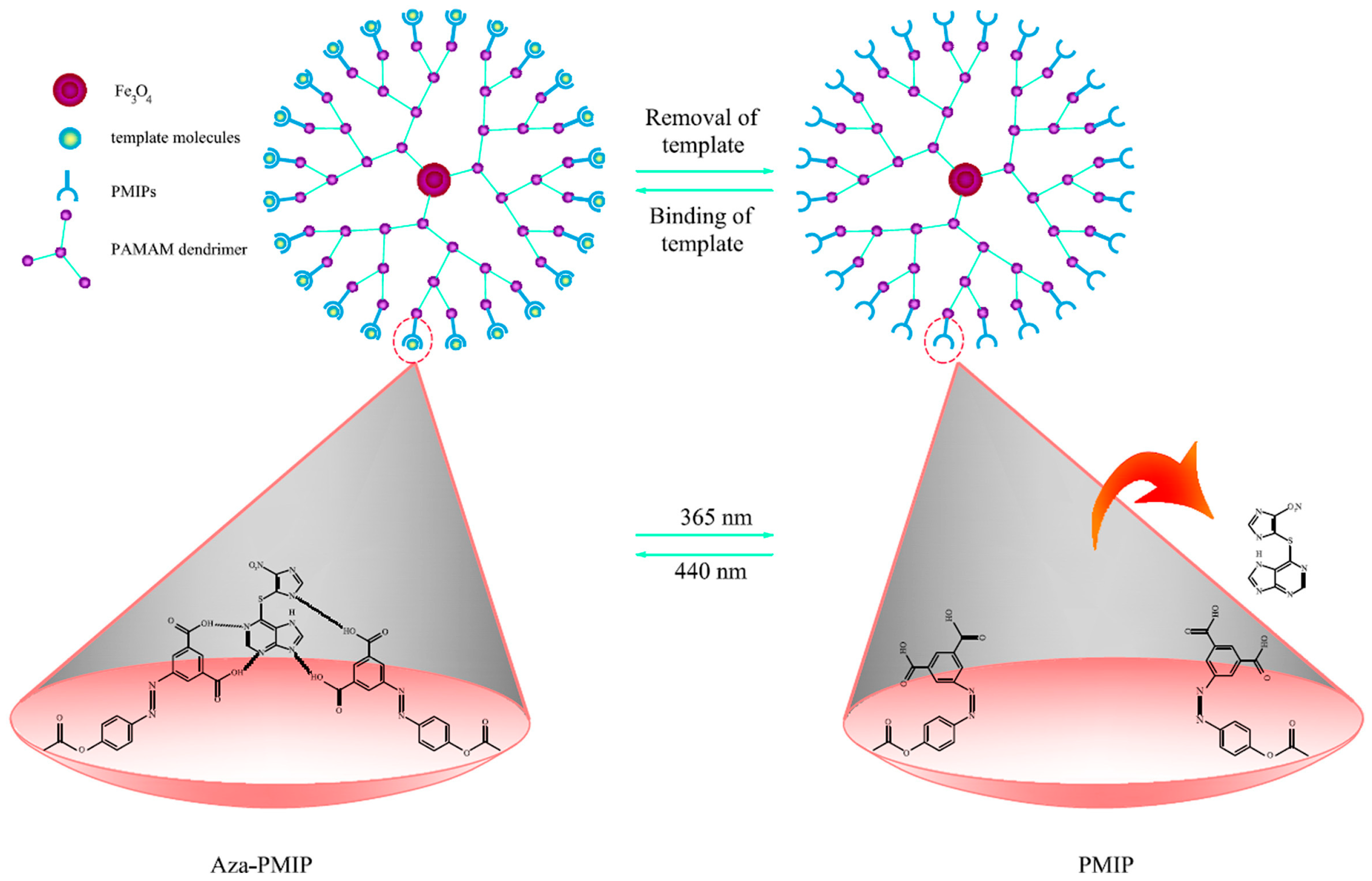
| Azathioprine | Plasma Urine | Sorbent: dendrimer-coated Fe3O4 NPs grafted to MIPs matrix, including 5-[(4-(methacryloyloxy)phenyl) diazenyl] isophthalic acid, and ethylene glycol dimethacrylate(dMNPs@PMIPs) | Magnetic field, Light | Column: C18 (30 × 3.9 mm, 10 μm); MP: Sodium 1-heptanesulfonate (1.6 g, pH 3.5)/MeOH (70:30, v/v); Flow Rate: 0.8 mL/min (40 °C); Injection volume: 20 μL; Detector: UV (at 286 nm) | MMIPs- d-SPE HPLC-UV | 95.85–102.71 | [89] |
| Methyl prednisolone acetate | Plasma Urine | Sorbent:polyester dendrimer-grafted, photo-responsive MIPs including 5-[(4, 3-(methacryloyloxy) phenyl) diazenyl] dihydroxy aniline and ethylene glycol dimethacrylate | Light | Column: Waters Corporation silica L3 (250 × 4.6 mm); MP: n-butyl chloride, Water-saturated n-butyl chloride, tetrahydrofuran, MeOH, and Glacial acetic acid (95:95:14:7:6); Flow Rate:1.0 mL/min; Injection volume: 10 μ; Detector: UV (254 nm) | MIPs- HPLC-UV | 96.8–104.2 | [91] |
| Imatinib mesylate | Plasma Urine | Sorbent: Synthesized Fe3O4 NPs, coated with PNVCL and grafted with chitosan, exhibit dual sensitivity to temperature and pH (Fe3O4@PNVCL-COOH) | Magnetic field, Temperature, pH | Column: BEH C18 (100 × 2.1 mm, 1.7 μm), BEH C18 VanGuard pre-column (5 × 2.1 mm, 1.7 μm), MP: FA(0.1%) in Water/MeOH, injection volume: 10 μL(4 °C), Detector: MS/MS with ESI-POS, Operating mode: MRM | MSPE-UPLC-MS/MS | 80–91 | [94] |
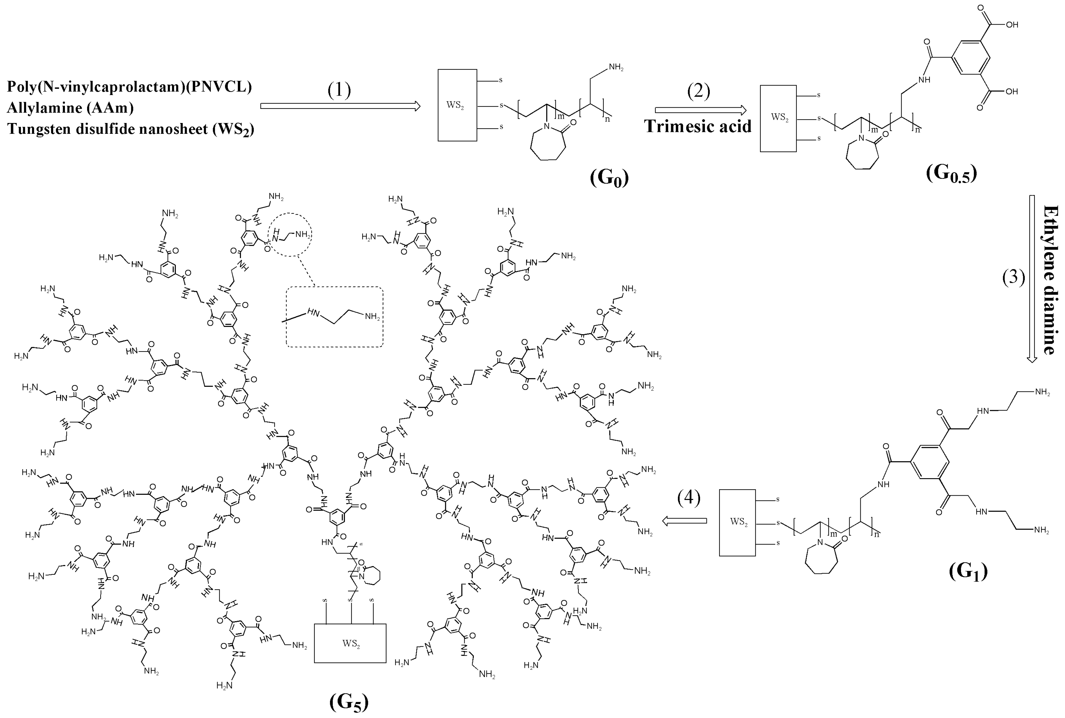
| Bicaltumide | Plasma Urine | Sorbent: NIR- and Thermo-sensitive NPs composed of WS2 nano-sheets and five generation of polymeric dendrimers | Temperature, NIR laser | Column: L1 (10 × 4 mm, 3 μm); MP: TFA (0.01%) in water/TFA (0.01%) in ACN (52:48); Flow Rate: 1.0 mL/min (25 °C); Injection volume: 10 μL; Detector: UV (at 270 nm) | SPE-HPLC-UV | 92.12–94.54 | [90] |
| Bicaltumide | Plasma Urine | Sorbent: Grafting of polymer chains including PNVCL, Allylamine, Allyl acetoacetate onto WS2 NPs (SMNA) | Temperature, NIR laser | Column: L1 (10 × 4 mm, 3 μm); MP:TFA (0.01%) in water/TFA (0.01%) in ACN (52:48); Flow Rate: 1.0 mL/min (25 °C); Injection volume:10 μ; Detector: UV (at 270 nm) | SPE-HPLC-UV | 92.08–94.17 | [95] |
4. Conclusions
Author Contributions
Funding
Acknowledgments
Conflicts of Interest
References
- Eliasson, E.; Lindh, J.D.; Malmström, R.E.; Beck, O.; Dahl, M.-L. Therapeutic drug monitoring for tomorrow. Eur. J. Clin. Pharmacol. 2013, 69, 25–32. [Google Scholar] [CrossRef] [PubMed]
- Niu, Z.; Zhang, W.; Yu, C.; Zhang, J.; Wen, Y. Recent advances in biological sample preparation methods coupled with chromatography, spectrometry and electrochemistry analysis techniques. TrAC-Trends Anal. Chem. 2018, 102, 123–146. [Google Scholar] [CrossRef]
- Wen, Y.; Chen, L.; Li, J.; Liu, D.; Chen, L. Recent advances in solid-phase sorbents for sample preparation prior to chromatographic analysis. TrAC-Trends Anal. Chem. 2014, 59, 26–41. [Google Scholar] [CrossRef]
- Maranata, G.J.; Surya, N.O.; Hasanah, A.N. Optimising factors affecting solid phase extraction performances of molecular imprinted polymer as recent sample preparation technique. Heliyon 2021, 7, e05934. [Google Scholar] [CrossRef] [PubMed]
- Zhang, Y.; Zhao, G.; Han, K.; Sun, D.; Zhou, N.; Song, Z.; Liu, H.; Li, J.; Li, G. Applications of molecular imprinting technology in the study of traditional Chinese medicine. Molecules 2022, 28, 301. [Google Scholar] [CrossRef] [PubMed]
- Drabińska, N.; Marcinkowska, M.A.; Wieczorek, M.N.; Jeleń, H.H. Application of Sorbent-Based Extraction Techniques in Food Analysis. Molecules 2023, 28, 7985. [Google Scholar] [CrossRef] [PubMed]
- Gałuszka, A.; Migaszewski, Z.; Namieśnik, J. The 12 principles of green analytical chemistry and the SIGNIFICANCE mnemonic of green analytical practices. TrAC-Trends Anal. Chem. 2013, 50, 78–84. [Google Scholar] [CrossRef]
- López-Lorente, Á.I.; Pena-Pereira, F.; Pedersen-Bjergaard, S.; Zuin, V.G.; Ozkan, S.A.; Psillakis, E. The ten principles of green sample preparation. TrAC-Trends Anal. Chem. 2022, 148, 116530. [Google Scholar] [CrossRef]
- Mandrioli, R.; Cirrincione, M.; Mladěnka, P.; Protti, M.; Mercolini, L. Green analytical chemistry (GAC) applications in sample preparation for the analysis of anthocyanins in products and by-products from plant sources. Adv. Sample Prep. 2022, 3, 100037. [Google Scholar] [CrossRef]
- Spietelun, A.; Marcinkowski, Ł.; de la Guardia, M.; Namieśnik, J. Recent developments and future trends in solid phase microextraction techniques towards green analytical chemistry. J. Chromatogr. A 2013, 1321, 1–13. [Google Scholar] [CrossRef]
- Das, S.S.; Bharadwaj, P.; Bilal, M.; Barani, M.; Rahdar, A.; Taboada, P.; Bungau, S.; Kyzas, G.Z. Stimuli-responsive polymeric nanocarriers for drug delivery, imaging, and theragnosis. Polymers 2020, 12, 1397. [Google Scholar] [CrossRef] [PubMed]
- Wojnicz, A.; Belmonte, C.; Koller, D.; Ruiz-Nuño, A.; Román, M.; Ochoa, D.; Abad-Santos, F. Effective phospholipids removing microelution-solid phase extraction LC-MS/MS method for simultaneous plasma quantification of aripiprazole and dehydro-aripiprazole: Application to human pharmacokinetic studies. J. Pharm. Biomed. Anal. 2018, 151, 116–125. [Google Scholar] [CrossRef]
- Tang, F.; Bada, H.; Ng, C.M.; Leggas, M. Validation of a HPLC/MS method for simultaneous quantification of clonidine, morphine and its metabolites in human plasma. Biomed. Chromatogr. 2019, 33, e4527. [Google Scholar] [CrossRef] [PubMed]
- Anilanmert, B.; Çavuş, F.; Narin, I.; Cengiz, S.; Sertler, Ş.; Özdemir, A.A.; Açikkol, M. Simultaneous analysis method for GHB, ketamine, norketamine, phenobarbital, thiopental, zolpidem, zopiclone and phenytoin in urine, using C18 poroshell column. J. Chromatogr. B Biomed. Appl. 2016, 1022, 230–241. [Google Scholar] [CrossRef]
- Woo, H.I.; Yang, J.S.; Oh, H.J.; Cho, Y.Y.; Kim, J.H.; Park, H.-D.; Lee, S.-Y. A simple and rapid analytical method based on solid-phase extraction and liquid chromatography–tandem mass spectrometry for the simultaneous determination of free catecholamines and metanephrines in urine and its application to routine clinical analysis. Clin. Biochem. 2016, 49, 573–579. [Google Scholar] [CrossRef]
- Zhao, M.; Wu, X.-J.; Fan, Y.-X.; Guo, B.-N.; Zhang, J. Development and validation of a UHPLC–MS/MS assay for colistin methanesulphonate (CMS) and colistin in human plasma and urine using weak-cation exchange solid-phase extraction. J. Pharm. Biomed. Anal. 2016, 124, 303–308. [Google Scholar] [CrossRef]
- Ahmed Baig, M.L.; Ali, S.A. LC-MS/MS method for the estimation of paliperidone in human plasma. Anal. Chem. Lett. 2017, 7, 228–240. [Google Scholar] [CrossRef]
- Koller, D.; Zubiaur, P.; Saiz-Rodríguez, M.; Abad-Santos, F.; Wojnicz, A. Simultaneous determination of six antipsychotics, two of their metabolites and caffeine in human plasma by LC-MS/MS using a phospholipid-removal microelution-solid phase extraction method for sample preparation. Talanta 2019, 198, 159–168. [Google Scholar] [CrossRef] [PubMed]
- Koller, D.; Vaitsekhovich, V.; Mba, C.; Steegmann, J.L.; Zubiaur, P.; Abad-Santos, F.; Wojnicz, A. Effective quantification of 11 tyrosine kinase inhibitors and caffeine in human plasma by validated LC-MS/MS method with potent phospholipids clean-up procedure. Application to therapeutic drug monitoring. Talanta 2020, 208, 120450. [Google Scholar] [CrossRef]
- Calucică, D.M.; Manda, C.-V.; Găman, A.M.; Răileanu, Ș.; Stanca, L.; Popescu, M.D.E.; Mateescu, O.G.; Biță, A.; Croitoru, O.; Neamțu, S.-D. Development of a SPE-LC-MS Method for the Quantitation of Palbociclib and Abemaciclib in Human Plasma. Molecules 2022, 27, 8604. [Google Scholar] [CrossRef]
- Alam, M.T.; Mujtaba, M.A.; Hussain, A.; Ali, A.; Imran, M.; Barkat, M.A.; Abdel-Gawad, S.A. Solid-Phase Extraction (SPE) Technique to Quantify Cefdinir in Human Plasma Using Liquid Chromatography–Tandem Mass Spectrometry (LC–MS/MS). J. Chromatogr. Sci. 2023, 61, 366–374. [Google Scholar] [CrossRef] [PubMed]
- Anastassiades, M.; Lehotay, S.J.; Štajnbaher, D.; Schenck, F.J. Fast and easy multiresidue method employing acetonitrile extraction/partitioning and “dispersive solid-phase extraction” for the determination of pesticide residues in produce. J. AOAC Int. 2003, 86, 412–431. [Google Scholar] [CrossRef] [PubMed]
- Ścigalski, P.; Kosobucki, P. Recent materials developed for dispersive solid phase extraction. Molecules 2020, 25, 4869. [Google Scholar] [CrossRef] [PubMed]
- Qian, M.-r.; Chen, Z.-m.; Tao, X.-x.; Yao, F.; Xu, X.-m. In-syringe dispersive solid phase filter extraction cleanup followed by liquid chromatography-triple quadrupole mass spectrometry for fast determination of colchicine in plasma/urine. J. Pharm. Biomed. Anal. 2023, 228, 115317. [Google Scholar] [CrossRef] [PubMed]
- Marzi Khosrowshahi, E.; Limuie Khosrowshahi, B.; Farajzadeh, M.A.; Jouyban, A.; Tuzen, M.; Afshar Mogaddam, M.R.; Nemati, M. Application of microcrystalline cellulose as an efficient and cheap sorbent for the extraction of metoprolol from plasma and wastewater before HPLC–MS/MS determination. Biomed. Chromatogr. 2022, 36, e5371. [Google Scholar] [CrossRef] [PubMed]
- Saito, K.; Kikuchi, Y.; Saito, R. Solid-phase dispersive extraction method for analysis of benzodiazepine drugs in serum and urine samples. J. Pharm. Biomed. Anal. 2014, 100, 28–32. [Google Scholar] [CrossRef] [PubMed]
- Fabrizi, G.; Fioretti, M.; Mainero Rocca, L. Dispersive solid-phase extraction procedure coupled to UPLC-ESI-MS/MS analysis for the simultaneous determination of thirteen cytotoxic drugs in human urine. Biomed. Chromatogr. 2016, 30, 1297–1308. [Google Scholar] [CrossRef] [PubMed]
- Júnior, E.F.; Caldas, E.D. Simultaneous determination of drugs and pesticides in postmortem blood using dispersive solid-phase extraction and large volume injection-programmed temperature vaporization-gas chromatography–mass spectrometry. Forensic Sci. Int. 2018, 290, 318–326. [Google Scholar] [CrossRef] [PubMed]
- Huang, W.; Qiu, Q.; Chen, M.; Shi, J.; Huang, X.; Kong, Q.; Long, D.; Chen, Z.; Yan, S. Determination of 18 antibiotics in urine using LC-QqQ-MS/MS. J. Chromatogr. B Biomed. Appl. 2019, 1105, 176–183. [Google Scholar] [CrossRef]
- Arthur, C.L.; Pawliszyn, J. Solid phase microextraction with thermal desorption using fused silica optical fibers. Anal. Chem. 1990, 62, 2145–2148. [Google Scholar] [CrossRef]
- Looby, N.; Vasiljevic, T.; Reyes-Garcés, N.; Roszkowska, A.; Bojko, B.; Wąsowicz, M.; Jerath, A.; Pawliszyn, J. Therapeutic drug monitoring of tranexamic acid in plasma and urine of renally impaired patients using solid phase microextraction. Talanta 2021, 225, 121945. [Google Scholar] [CrossRef]
- Hasegawa, C.; Kumazawa, T.; Uchigasaki, S.; Lee, X.-P.; Sato, K.; Terada, M.; Kurosaki, K. Determination of dextromethorphan in human plasma using pipette tip solid-phase extraction and gas chromatography–mass spectrometry. Anal. Bioanal. Chem. 2011, 401, 2215–2223. [Google Scholar] [CrossRef] [PubMed]
- Wan, Q.; Liu, H.; Deng, Z.; Bu, J.; Li, T.; Yang, Y.; Zhong, S. A critical review of molecularly imprinted solid phase extraction technology. J. Polym. Res. 2021, 28, 401. [Google Scholar] [CrossRef]
- Małysiak, M.; Kiljanek, T. Method of Glyphosate, AMPA, and Glufosinate Ammonium Determination in Beebread by Liquid Chromatography—Tandem Mass Spectrometry after Molecularly Imprinted Solid-Phase Extraction. Molecules 2022, 27, 5741. [Google Scholar] [CrossRef]
- Sun, D.; Song, Z.; Zhang, Y.; Wang, Y.; Lv, M.; Liu, H.; Wang, L.; Lu, W.; Li, J.; Chen, L. Recent advances in molecular-imprinting-based solid-phase extraction of antibiotics residues coupled with chromatographic analysis. Front. Environ. Chem. 2021, 2, 703961. [Google Scholar] [CrossRef]
- Combes, A.; Kadhirvel, P.; Bordron, L.; Pichon, V. Synthesis and characterization of molecularly imprinted polymers for the selective extraction of carbamazepine and analogs from human urine samples. Chromatographia 2019, 82, 287–295. [Google Scholar] [CrossRef]
- Zhou, H.; Chen, Q.; Song, X.; He, L.; Liu, R. Surface molecularly imprinted solid-phase extraction for the determination of vancomycin in plasma samples using HPLC–MS/MS. J. Anal. Chem. 2022, 38, 1171–1179. [Google Scholar] [CrossRef]
- Santos, M.G.; Tavares, I.M.C.; Boralli, V.B.; Figueiredo, E.C. Direct doping analysis of beta-blocker drugs from urinary samples by on-line molecularly imprinted solid-phase extraction coupled to liquid chromatography/mass spectrometry. Analyst 2015, 140, 2696–2703. [Google Scholar] [CrossRef] [PubMed]
- Sánchez-González, J.; García-Carballal, S.; Cabarcos, P.; Tabernero, M.J.; Bermejo-Barrera, P.; Moreda-Piñeiro, A. Determination of cocaine and its metabolites in plasma by porous membrane-protected molecularly imprinted polymer micro-solid-phase extraction and liquid chromatography—Tandem mass spectrometry. J. Chromatogr. A 2016, 1451, 15–22. [Google Scholar] [CrossRef]
- Ding, J.; Zhang, F.; Zhang, X.; Wang, L.; Wang, C.; Zhao, Q.; Xu, Y.; Ding, L.; Ren, N. Determination of roxithromycin from human plasma samples based on magnetic surface molecularly imprinted polymers followed by liquid chromatography-tandem mass spectromer. J. Chromatogr. B Biomed. Appl. 2016, 1021, 221–228. [Google Scholar] [CrossRef]
- El-Beqqali, A.; Abdel-Rehim, M. Molecularly imprinted polymer-sol-gel tablet toward micro-solid phase extraction: I. Determination of methadone in human plasma utilizing liquid chromatography–tandem mass spectrometry. Anal. Chim. Acta 2016, 936, 116–122. [Google Scholar] [CrossRef]
- El-Beqqali, A.; Andersson, L.I.; Jeppsson, A.D.; Abdel-Rehim, M. Molecularly imprinted polymer-sol-gel tablet toward micro-solid phase extraction: II. Determination of amphetamine in human urine samples by liquid chromatography–tandem mass spectrometry. J. Chromatogr. B Biomed. Appl. 2017, 1063, 130–135. [Google Scholar] [CrossRef]
- Sánchez-González, J.; Odoardi, S.; Bermejo, A.M.; Bermejo-Barrera, P.; Romolo, F.S.; Moreda-Piñeiro, A.; Strano-Rossi, S. Development of a micro-solid-phase extraction molecularly imprinted polymer technique for synthetic cannabinoids assessment in urine followed by liquid chromatography–tandem mass spectrometry. J. Chromatogr. A 2018, 1550, 8–20. [Google Scholar] [CrossRef]
- Sartore, D.M.; Medina, D.A.V.; Costa, J.L.; Lanças, F.M.; Santos-Neto, Á.J. Automated microextraction by packed sorbent of cannabinoids from human urine using a lab-made device packed with molecularly imprinted polymer. Talanta 2020, 219, 121185. [Google Scholar] [CrossRef] [PubMed]
- Guthrie, R.; Susi, A. A simple phenylalanine method for detecting phenylketonuria in large populations of newborn infants. Pediatrics 1963, 32, 338–343. [Google Scholar] [CrossRef]
- Marasca, C.; Protti, M.; Mandrioli, R.; Atti, A.R.; Armirotti, A.; Cavalli, A.; De Ronchi, D.; Mercolini, L. Whole blood and oral fluid microsampling for the monitoring of patients under treatment with antidepressant drugs. J. Pharm. Biomed. Anal. 2020, 188, 113384. [Google Scholar] [CrossRef] [PubMed]
- Protti, M.; Sberna, P.M.; Sardella, R.; Vovk, T.; Mercolini, L.; Mandrioli, R. VAMS and StAGE as innovative tools for the enantioselective determination of clenbuterol in urine by LC-MS/MS. J. Pharm. Biomed. Anal. 2021, 195, 113873. [Google Scholar] [CrossRef]
- Zimmermann, S.; Aghai, F.; Schilling, B.; Kraus, S.; Grigoleit, G.U.; Kalogirou, C.; Goebeler, M.-E.; Jung, P.; Pelzer, T.; Klinker, H.; et al. Volumetric absorptive microsampling (VAMS) for the quantification of ten kinase inhibitors and determination of their in vitro VAMS-to-plasma ratio. J. Pharm. Biomed. Anal. 2022, 211, 114623. [Google Scholar] [CrossRef] [PubMed]
- Paniagua-González, L.; Díaz-Louzao, C.; Lendoiro, E.; Otero-Antón, E.; Cadarso-Suárez, C.; López-Rivadulla, M.; Cruz, A.; de-Castro-Ríos, A. Volumetric Absorptive Microsampling (VAMS) for assaying immunosuppressants from venous whole blood by LC–MS/MS using a novel atmospheric pressure ionization probe (UniSpray™). J. Pharm. Biomed. Anal. 2020, 189, 113422. [Google Scholar] [CrossRef]
- Barco, S.; Bandettini, R.; Maffia, A.; Tripodi, G.; Castagnola, E.; Cangemi, G. Quantification of piperacillin, tazobactam, meropenem, ceftazidime, and linezolid in human plasma by liquid chromatography/tandem mass spectrometry. J. Chemother. 2015, 27, 343–347. [Google Scholar] [CrossRef]
- Moorthy, G.S.; Vedar, C.; Zane, N.R.; Downes, K.J.; Prodell, J.L.; DiLiberto, M.A.; Zuppa, A.F. Development and validation of a volumetric absorptive microsampling-liquid chromatography mass spectrometry method for the analysis of cefepime in human whole blood: Application to pediatric pharmacokinetic study. J. Pharm. Biomed. Anal. 2020, 179, 113002. [Google Scholar] [CrossRef] [PubMed]
- Mandrioli, R.; Mercolini, L.; Protti, M. Blood and plasma volumetric absorptive microsampling (VAMS) coupled to LC-MS/MS for the forensic assessment of cocaine consumption. Molecules 2020, 25, 1046. [Google Scholar] [CrossRef] [PubMed]
- Capriotti, A.L.; Cavaliere, C.; La Barbera, G.; Montone, C.M.; Piovesana, S.; Laganà, A. Recent applications of magnetic solid-phase extraction for sample preparation. Chromatographia 2019, 82, 1251–1274. [Google Scholar] [CrossRef]
- Materón, E.M.; Miyazaki, C.M.; Carr, O.; Joshi, N.; Picciani, P.H.; Dalmaschio, C.J.; Davis, F.; Shimizu, F.M. Magnetic nanoparticles in biomedical applications: A review. Appl. Surf. Sci. Adv. 2021, 6, 100163. [Google Scholar] [CrossRef]
- Cai, P.; Xiong, X.; Li, D.; Zhou, Y.; Xiong, C. Magnetic solid-phase extraction coupled with UHPLC–MS/MS for four antidepressants and one metabolite in clinical plasma and urine samples. Bioanalysis 2020, 12, 35–52. [Google Scholar] [CrossRef] [PubMed]
- Heidari, H.; Limouei-Khosrowshahi, B. Magnetic solid phase extraction with carbon-coated Fe3O4 nanoparticles coupled to HPLC-UV for the simultaneous determination of losartan, carvedilol, and amlodipine besylate in plasma samples. J. Chromatogr. B Biomed. Appl. 2019, 1114, 24–30. [Google Scholar] [CrossRef] [PubMed]
- Taghvimi, A.; Hamishehkar, H. Carbon coated magnetic nanoparticles as a novel magnetic solid phase extraction adsorbent for simultaneous extraction of methamphetamine and ephedrine from urine samples. J. Chromatogr. B Biomed. Appl. 2017, 1041, 113–119. [Google Scholar] [CrossRef] [PubMed]
- Zhang, J.; Liu, D.; Meng, X.; Shi, Y.; Wang, R.; Xiao, D.; He, H. Solid phase extraction based on porous magnetic graphene oxide/β-cyclodextrine composite coupled with high performance liquid chromatography for determination of antiepileptic drugs in plasma samples. J. Chromatogr. A 2017, 1524, 49–56. [Google Scholar] [CrossRef] [PubMed]
- Yang, F.; Zou, Y.; Ni, C.; Wang, R.; Wu, M.; Liang, C.; Zhang, J.; Yuan, X.; Liu, W. Magnetic dispersive solid-phase extraction based on modified magnetic nanoparticles for the detection of cocaine and cocaine metabolites in human urine by high-performance liquid chromatography–mass spectrometry. J. Sep. Sci. 2017, 40, 4234–4245. [Google Scholar] [CrossRef]
- Kamaruzaman, S.; Sanagi, M.M.; Yahaya, N.; Wan Ibrahim, W.A.; Endud, S.; Wan Ibrahim, W.N. Magnetic micro-solid-phase extraction based on magnetite-MCM-41 with gas chromatography–mass spectrometry for the determination of antidepressant drugs in biological fluids. J. Sep. Sci. 2017, 40, 4222–4233. [Google Scholar] [CrossRef]
- Ebrahimi Rahmani, M.; Ansari, M.; Kazemipour, M.; Nateghi, M. Selective extraction of morphine from biological fluids by magnetic molecularly imprinted polymers and determination using UHPLC with diode array detection. J. Sep. Sci. 2018, 41, 958–965. [Google Scholar] [CrossRef] [PubMed]
- Attallah, O.A.; Al-Ghobashy, M.A.; Ayoub, A.T.; Nebsen, M. Magnetic molecularly imprinted polymer nanoparticles for simultaneous extraction and determination of 6-mercaptopurine and its active metabolite thioguanine in human plasma. J. Chromatogr. A 2018, 1561, 28–38. [Google Scholar] [CrossRef] [PubMed]
- Safari, M.; Shahlaei, M.; Yamini, Y.; Shakorian, M.; Arkan, E. Magnetic framework composite as sorbent for magnetic solid phase extraction coupled with high performance liquid chromatography for simultaneous extraction and determination of tricyclic antidepressants. Anal. Chim. Acta 2018, 1034, 204–213. [Google Scholar] [CrossRef] [PubMed]
- Wang, R.; Cui, Y.; Hu, F.; Liu, W.; Du, Q.; Zhang, Y.; Zha, J.; Huang, T.; Fizir, M.; He, H. Selective recognition and enrichment of carbamazepine in biological samples by magnetic imprinted polymer based on reversible addition-fragmentation chain transfer polymerization. J. Chromatogr. A 2019, 1591, 62–70. [Google Scholar] [CrossRef] [PubMed]
- Yilmaz, E.; Salem, S.; Sarp, G.; Aydin, S.; Sahin, K.; Korkmaz, I.; Yuvali, D. TiO2 nanoparticles and C-Nanofibers modified magnetic Fe3O4 nanospheres (TiO2@Fe3O4@C–NF): A multifunctional hybrid material for magnetic solid-phase extraction of ibuprofen and photocatalytic degradation of drug molecules and azo dye. Talanta 2020, 213, 120813. [Google Scholar] [CrossRef] [PubMed]
- Lu, Q.; Guo, H.; Zhang, Y.; Tang, X.; Lei, W.; Qi, R.; Chu, J.; Li, D.; Zhao, Q. Graphene oxide-Fe3O4 nanocomposite magnetic solid phase extraction followed by UHPLC-MS/MS for highly sensitive determination of eight psychoactive drugs in urine samples. Talanta 2020, 206, 120212. [Google Scholar] [CrossRef] [PubMed]
- Jia, M.; Zhu, Y.; Guo, D.; Bi, X.; Hou, X. Surface molecularly imprinted polymer based on core-shell Fe3O4@MIL-101(Cr) for selective extraction of phenytoin sodium in plasma. Anal. Chim. Acta 2020, 1128, 211–220. [Google Scholar] [CrossRef] [PubMed]
- Bashir, K.; Chen, G.; Han, J.; Shu, H.; Cui, X.; Wang, L.; Li, W.; Fu, Q. Preparation of magnetic metal organic framework and development of solid phase extraction method for simultaneous determination of fluconazole and voriconazole in rat plasma samples by HPLC. J. Chromatogr. B Biomed. Appl. 2020, 1152, 122201. [Google Scholar] [CrossRef] [PubMed]
- Shaban, M.; Ghaffary, S.; Hanaee, J.; Karbakhshzadeh, A.; Soltani, S. Synthesis and characterization of new surface modified magnetic nanoparticles and application for the extraction of letrozole from human plasma and analysis with HPLC-fluorescence. J. Pharm. Biomed. Anal. 2021, 193, 113659. [Google Scholar] [CrossRef]
- Banan, K.; Ghorbani-Bidkorbeh, F.; Afsharara, H.; Hatamabadi, D.; Landi, B.; Keçili, R.; Sellergren, B. Nano-sized magnetic core-shell and bulk molecularly imprinted polymers for selective extraction of amiodarone from human plasma. Anal. Chim. Acta 2022, 1198, 339548. [Google Scholar] [CrossRef]
- Akyol, E.; Ulusoy, H.İ.; Yilmaz, E.; Polat, Ü.; Soylak, M. Application of magnetic solid-phase extraction for sensitive determination of anticancer drugs in urine by means of diamino benzidine tetrachlorohydrate modified magnetic nanoparticles. Pharmacol. Rep. 2023, 75, 456–464. [Google Scholar] [CrossRef] [PubMed]
- Musarurwa, H.; Tavengwa, N.T. Thermo-responsive polymers and advances in their applications in separation science. Microchem. J. 2022, 179, 107554. [Google Scholar] [CrossRef]
- Lanzalaco, S.; Armelin, E. Poly(N-isopropylacrylamide) and copolymers: A review on recent progresses in biomedical applications. Gels 2017, 3, 36. [Google Scholar] [CrossRef] [PubMed]
- Hoffman, A.S. Stimuli-responsive polymers: Biomedical applications and challenges for clinical translation. Adv. Drug Deliv. Rev. 2013, 65, 10–16. [Google Scholar] [CrossRef] [PubMed]
- Kazemi, A.; Ahmad Panahi, H.; Safaeijavan, R. Thermosensitive molecularly imprinted poly(1-vinyl-2-pyrrolidone/methyl methacrylate/N-vinylcaprolactam) for selective extraction of imatinib mesylate in human biological fluid. J. Sep. Sci. 2020, 43, 614–621. [Google Scholar] [CrossRef] [PubMed]
- Gong, C.-B.; Wei, Y.-B.; Liu, L.-T.; Zheng, A.-X.; Yang, Y.-H.; Chow, C.-f.; Tang, Q. Photoresponsive hollow molecularly imprinted polymer for trace triamterene in biological samples. Mater. Sci. Eng. C 2017, 76, 568–578. [Google Scholar] [CrossRef]
- Gomar, M.; Panahi, H.A.; Pournamdari, E. Synthesis and Characterization of Thermosensitive Molecularly Imprinted Poly[allylacetoacetate/N-vinyl caprolactam] for Selective Extraction of Gemcitabine in Biological Samples. ChemistrySelect 2018, 3, 2571–2577. [Google Scholar] [CrossRef]
- Yazdanian, N.; Akbari-Adergani, B.; Kazemipour, M.; Panahi, H.A.; Javanbakht, M. Improving the determination of celecoxib in body fluids and pharmaceuticals using a new selective and thermosensitive molecularly imprinted poly(vinylidene fluoride) membrane. Anal. Methods 2020, 12, 2185–2195. [Google Scholar] [CrossRef]
- Zeng, H.; Yu, X.; Wan, J.; Cao, X. Synthesis of molecularly imprinted polymers based on boronate affinity for diol-containing macrolide antibiotics with hydrophobicity-balanced and pH-responsive cavities. J. Chromatogr. A 2021, 1642, 461969. [Google Scholar] [CrossRef]
- Zafarghandi, S.S.; Panahi, H.A.; Nezhati, M.N. Preparation of pH-Sensitive Molecularly Imprinted Polymer via Dual-Monomer for Selective Solid-Phase Extraction of Ribavirin from Human Urine and Pharmaceutical Samples. ChemistrySelect 2022, 7, e202104038. [Google Scholar] [CrossRef]
- Feng, L.; Wang, Y.; Luo, Z.; Huang, Z.; Zhang, Y.; Guo, K.; Ye, D. Dual stimuli-responsive nanoparticles for controlled release of anticancer and anti-inflammatory drugs combination. Chem. Eur. J. 2017, 23, 9397–9406. [Google Scholar] [CrossRef] [PubMed]
- Naghibi, S.; Sahebi, H. Employment of modified Fe3O4 nanoparticles using thermo-sensitive polymer for extraction and pre-concentration of cefexime in biological samples. Biomed. Chromatogr. 2018, 32, e4082. [Google Scholar] [CrossRef] [PubMed]
- Taghvimi, A.; Ghorbani, M.; Hamishehkar, H. Synthesis of a novel polymeric magnetic solid phase extraction adsorbent for selective extraction of amphetamine from urine samples coupled with high performance liquid chromatography. Drug Test. Anal. 2018, 10, 832–838. [Google Scholar] [CrossRef] [PubMed]
- Hao, Y.; Meng, J.; Wang, S. Photo-responsive polymer materials for biological applications. Chin. Chem. Lett. 2017, 28, 2085–2091. [Google Scholar] [CrossRef]
- Mu, C.; Xiang, J.; Liu, Z. Photodetectors based on sensitized two-dimensional transition metal dichalcogenides—A review. J. Mater. Sci. 2017, 32, 4115–4131. [Google Scholar] [CrossRef]
- Bandara, H.D.; Burdette, S.C. Photoisomerization in different classes of azobenzene. Chem. Soc. Rev. 2012, 41, 1809–1825. [Google Scholar] [CrossRef]
- Di Martino, M.; Sessa, L.; Diana, R.; Piotto, S.; Concilio, S. Recent Progress in Photoresponsive Biomaterials. Molecules 2023, 28, 3712. [Google Scholar] [CrossRef] [PubMed]
- Yang, P.C.; Wang, Y.H.; Wu, H. Synthesis, characterization, and photochemical properties of chromophores containing photoresponsive azobenzene and stilbene. J. Appl. Polym. Sci. 2012, 124, 4193–4205. [Google Scholar] [CrossRef]
- Alaei, H.S.; Tehrani, M.S.; Husain, S.W.; Panahi, H.A.; Mehramizi, A. Photoresponsive molecularly imprinted dendrimer-based magnetic nanoparticles for photo-regulated selective separation of azathioprine. React. Funct. Polym. 2019, 136, 58–65. [Google Scholar] [CrossRef]
- Mahdavijalal, M.; Ahmad Panahi, H.; Niazi, A.; Tamaddon, A. Near-infrared light responsive dendrimers facilitate the extraction of bicalutamide from human plasma and urine. Biotechnol. J. 2021, 16, 2100299. [Google Scholar] [CrossRef]
- Baimani, N.; Aberoomand Azar, P.; Waqif Husain, S.; Ahmad Panahi, H.; Mehramizi, A. Ultrasensitive separation of methylprednisolone acetate using a photoresponsive molecularly imprinted polymer incorporated polyester dendrimer based on magnetic nanoparticles. J. Sep. Sci. 2019, 42, 1468–1476. [Google Scholar] [CrossRef] [PubMed]
- Parham, N.; Panahi, H.A.; Feizbakhsh, A.; Moniri, E. Synthesis of high generation thermo-sensitive dendrimers for extraction of rivaroxaban from human fluid and pharmaceutic samples. J. Chromatogr. A 2018, 1545, 12–21. [Google Scholar] [CrossRef] [PubMed]
- Morovati, A.; Panahi, H.A.; Yazdani, F. Grafting of allylimidazole and n-vinylcaprolactam as a thermosensitive polymer onto magnetic nano-particles for the extraction and determination of celecoxib in biological samples. Int. J. Pharm. 2016, 513, 62–67. [Google Scholar] [CrossRef] [PubMed]
- Sahebi, H.; Pourmortazavi, S.M.; Zandavar, H.; Mirsadeghi, S. Chitosan grafted onto Fe3O4@poly(N-vinylcaprolactam) as a new sorbent for detecting Imatinib mesylate in biosamples using UPLC-MS/MS. Analyst 2019, 144, 7336–7350. [Google Scholar] [CrossRef] [PubMed]
- Mahdavijalal, M.; Panahi, H.A.; Niazi, A.; Tamaddon, A. Preparation of responsive nano-adsorbent to near-infrared laser based on tungsten disulfide for bicalutamide extraction in human biological fluids. J. Pharm. Biomed. Anal. 2022, 215, 114759. [Google Scholar] [CrossRef]

| Analyte(s) | Matrices | Extraction Conditions | Analysis Conditions | Analysis Method | LOD (ng/mL) | Ref. |
|---|---|---|---|---|---|---|
| GHB, Ketamine, Norketamine, Phenobarbital, Thiopental, Zolpidem, Zopiclone, Phenytoin | Urine | Sorbent: SPE Strata Screen C18 Cartridge/LLE; Sample Volume: 400 μL; Extraction solvent: EA, DE, H (1:1:1, v/v/v); Sorbent: SPE Strata Screen C18 Cartridge/LLE; Sample Volume: 400 μL; Extraction solvent: EA, DE, H (1:1:1, v/v/v) | Column: Poroshell C18, 100 × 3.0 mm, 2.7 µm; MP: FA, ACN(50:50 v/v); Flow rate: 0.3 mL/min; Injection volume: 10.0 μL (60 °C); Detector: MS/MS with ESI; operating mode: MRM | SPE-HPLC-MS/MS | 0.59–49.5 | [14] |
| Catecholamines, Metanephrines | Urine | Sorbent: SPE Strata-X-CW; Extraction solvent: FA, MeOH (2:98 v/v); Sample Volume: ≥1000 μL | Column: Unison C18 (100 × 2.0 mm, 3 μm); MP: water/FA (99.9:0.1, v/v) and ACN-FA (99.9:0.1, v/v); Flow rate: 0.3 mL/min; Injection volume: 1.0 μL (40 °C); Detector: MS/MS with iFunnel Technology source and ESI-POS; Operating mode: MRM | SPE-HPLC-MS/MS | 3.5–7.4 | [15] |
| Colistin, Methanesulphonate Colistin | Plasma Urine | Sorbent: SPE Oasis WCX 96-well plate; Sample Volume: 420 μL; Extraction solvent: ACN/water (30/70 v/v), and (6%) FA | Column: Phonomenex Kinetex XB-C18 (100 × 2.1 mm, 2.6 µm); MP: ACN/MeOH (1:1, v/v) and 0.1% FA in Water; Flow rate: 0.4 mL/min; Injection volume: 5.0 μL (30 °C); Detector: MS/MS with ESI-POS; Operating mode: SRM | SPE- UHPLC- MS/MS | 13–25.1 | [16] |
| Paliperidone | Plasma | Sorbent: SPE Oasis PRiME HLB, SPE cartridge (30 mg); Sample Volume: 250 μL | Column: Thermo Betabasic (100 × 4.6 mm, 8.5 μm), MP: MeOH/NH4OAc (70:30 v/v); Flow rate:1.0 mL/min; Detector: MS/MS with ESI-POS; Operating mode: SRM | SPE-HPLC-MS/MS | - | [17] |
| Aripiprazole, Dehydro-aripiprazole | Plasma | Sorbent: SPE Oasis PRiME HLB 96-well elution plate (3 mg); Sample volume: 200 μL; Extraction solvent: ACN/MeOH/NH4FA (5 mM, pH 4.0) (8:1:1, v/v/v) | Column: ACE C18-PFP (4.6 × 100 mm, 3 μm); MP: NH4FA(5 mM,pH 4.0)/ACN (65:35, v/v); Flow rate: 0.6 mL/min; Injection Volume:5.0 μL (25 °C); Detector: MS/MS with ESI-POS; Operating mode: MRM | SPE-HPLC- MS/MS | - | [12] |
| Clonidine, Morphine | Plasma | Sorbent: SPE Strata-X polymeric (30 mg/mL); Extraction solvent: ACN/MeOH (1:1 v/v); Sample Volume: 150 μL (plasma), 10 μL (urine) | Column: Inertsil ODS3 C18, (100 × 3 mm, 4 μm); MP: FA (0.1%) in Water + FA (0.1%) in MeOH; Flow rate: 0.2 mL/min; Injection volume:10.0 μL (25 °C); Detector: MS/MS with Turbo Ion Spray; Operating mode: MRM | SPE-HPLC- MS/MS | 0.2–0.15 | [13] |
| Aripiprazole, Dehydro-aripiprazole, Olanzapine, Risperidone, Paliperidone, Quetiapine, Clozapine, Caffeine | Plasma | Sorbent: HLB-Oasis PRiME HLB 96-well μElution Plate; Extraction Solvent: ACN with 0.1% FA; Sample Volume: 200 µL | Column: ACE C18-PFP (pentafluorophenyl) (100 × 4.6 mm, 3 μm); MP: FA (0.2%)-ACN (pH 3.0) (65:35, v/v); Flow rate: 0.6 mL/min (25 °C); Injection Volume: 5 μL; Detector: MS/MS with ESI-POS; Operating mode: MRM | µ-SPE- HPLC- MS/MS | - | [18] |
| Imatinib,Dasatinib, Nilotinib,Bosutinib, Ponatinib,Ruxolitinib, Ibrutinib,Filgotinib, Tofacitinib,Baricitinib, Peficitinib,Caffeine | Plasma | Sorbent: PRiME μ-SPE MCX (mixed-mode cation exchange); Extractin Solvent: 5% NH4OH in MeOH solution (1:1, v/v) and water | Column: Poroshell EC-C18 (75 × 2.1 mm, 2.7 μm); MP: Water/ACN with 0.1% FA (82:12, v/v); Flow rate: 0.5 mL/min (60 °C); Injection volume: 5 μL; Detector: MS/MS with ESI-POS; Operating mode: dMRM | µ-SPE- HPLC- MS/MS | - | [19] |
| Palbociclib, Abemaciclib | Plasma | Sorbent: SPE Oasis PRiME HLB; Extraction Solvent: MeOH; Sample Volume: ≥200 µL | Column: Waters CORTECS C18 (50 × 4.6 mm, 2.7 μm); MP: NH4OAc/Acetic acid (10 mM) and ACN; Flow rate: 0.8 mL/min; Injection volume: 40.0 μL (40 °C); Detector: MS/MS with ESI-POS; Operating mode: SRM | SPE-HPLC-MS/MS | - | [20] |
| Cefdinir | plasma | Sorbent: Bond Elut Plexa, PCX 30 mg/cc; Extraction Solvent:ACN/MeOH(50:50) | Column: Sigma Aldrich, Kromasil C18 with security guard C18 (4 × 3 mm); Flow rate: 1.0 mL/min; Injection volume: 15.0 μL (25 °C); MP: ACN/MeOH/Water (0.01% TFA) (45:45:10 v/v/v); Detector: MS/MS with ESI-POS; operating mode: MRM | SPE-HPLC-MS/MS | - | [21] |
| Analyte(s) | Matrices | Extraction Conditions | Analysis Conditions | Analysis Method | LOD (ng/mL) | Ref. |
|---|---|---|---|---|---|---|
| Bromazepam, Nitrazepam, Lorazepam, Alprazolam, Triazolam, Flunitrazepam, Nimetazepam, Etizolam, Diazepam | Serum Urine | Sorbent: Oasis HLB gel (50 μL) into a micro test tube; Extraction solvent: MeOH | Column: Poroshell 120 EC-C18 column (100 × 4.6 mm, 2.7 μm); MP: NH4FA (pH 3.0, 10 mM)/ACN (70:30, v/v); Flow rate: 0.5 mL/min (40 °C); Detector: TOF MS with ESI-POS | d-SPE HPLC–TOF | 1–10 | [26] |
| Cyclophosphamide, Cytarabine, Dacarbazine, Doxorubicin, Epirubicin, Etoposide, 5-Fluorouracil, Gemcitabine, Ifosfamide, Methotrexate, Paclitaxel, Vinblastine, Vincristine | Urine | Sorbent: mixture of Oasis HLB® and C18; Extraction solvent: H2O–MeOH (50:50, v/v) | Column: Kinetex PFP (100 × 2.10 mm; 2.6 μm); MP: ACN/water and (0.1%) FA; Flow rate: 0.5 mL/min (20 °C); Detector: MS/MS with Turbo ESI source; Operating mode: SRM | d-SPE HPLC–MS/MS | 0.01–33.3 | [27] |
| Cocaine, 3,4-Methylenedioxy Methamphetamin | Post mortem blood | Sorbent: Primary Secondary Amine; Extraction Solvent: ACN, EA | Capillary Column: DB-1 ms (30 × 0.25 mm × 0.25 mm); Carrier Gas: Helium, Run Time: 20.5 min; Temperature Ramp: 20 °C/min to 200 °C, 10 °C/min to 300 °C; Run Time: 20.5 min | d-SPE- GC-MS | 0.02–0.03 | [28] |
| Amoxicillin,Penicillin, Tylosin artrate, Roxithromycin, Clarithromycin, Azithromycin, Erythromycin,Tetracycline, Chlortetracycline, Ofloxacin, Enrofloxacin,Ciprofloxacin, Terramycin,Norfloxacin, Sulfadiazine, Sulfamethazine, Trimethoprim, Olaquindox, | Urine | Sorbent: roQ™ QuEChERS Dispersed SPE kit; Extraction solvent: MeOH/FA; Sample Volume: 200 μL | Column: RP Gemini NX-C18 (50 × 2 mm, 3 μm); MP: Water with (0.1%) FA/MeOH with (0.1%) FA; Flow rate: 0.5 mL/min (31 °C); Injection Volume: 5 μL; Detector: MS/MS with ESI-POS; Operating mode: MRM | d-SPE HPLC–MS/MS | 0.11–14.29 | [29] |
| Metoprolol | Plasma | Sorbent: Microcrystalline cellulose (MCC); Extraction Solvent: MeOH | Column: C18 (Agilent 15 × 4.6 mm, 5 μm); MP: MeOH/water (25:75, v/v) and (0.1%) FA; Flow rate: 0.3 mL/min (40 °C); Detector: MS/MS with ESI-POS; Operating mode: MRM | d-SPE HPLC–MS/MS | 0.3 | [25] |
| Colchicine | Plasma Urine | Sorbent: Magnesium Sulfate (MgSO4) and primary secondary amine (PSA); Extraction Solvent: Water/FA in Water/MeOH/ACN | Column: Waters XBridge™ BEH C18 (100 × 2.1 mm, 2.5 μm); MP: Ammonia/MeOH; Flow rate: 0.35 mL/min (30 °C); Detector: MS/MS with ESI-POS; Operating mode: MRM | d-SPE HPLC–MS/MS | 0.6 | [24] |
| Analyte(s) | Matrices | Extraction Conditions | Stimulus | Analysis Conditions | Analysis Method | Recovery (%) | Ref. |
|---|---|---|---|---|---|---|---|
| Ephedrine Methamphetamine | Urine | Sorbent: Carbon-coated Fe3O4 NPs (C/MNPs) | Magnetic field | Column: Analytical C18 column (25 × 4.6 mm, 10 μm);MP: ACN/phosphate buffer (10 mM, pH = 3.5); Flow Rate: 1.5 mL/min; Injection Volume: 20 μL | MSPE- HPLC- UV | 98.71–97.87 | [57] |
| Phenytoin, Carbamazepine, Diazepam | Plasma Urine | Sorbent: porous magnetic graphene oxide-cyclodextrin polymers (MGO-CDP) | Magnetic field | Column: ODS2 C18 (250 × 4.6 mm, 5 μm); Flow Rate: 1.0 mL/min (35 °C); Injection volume: 20 μL; Detector: DAD (230 nm) | MSPE - HPLC- DAD | 77.4–87.5 92.4–97.0 6.9–100.9 | [58] |
| Cocaine and its metabolites | Urine | Sorbent: PLS@SMPS@Fe3O4; Extraction Solvent: MeOH, ACN(4:1, v/v); Sample Volume: 1000 μL | Magnetic field | Column: XDB C18 (4.5 × 150 mm, 5 μm); Guard Column: XDB C18, (4.5 × 12.5 mm, 5 μm); MP: (2 mM) NH4FA and (0.05%) FA in Water)/(2 mM) NH4FA and (0.05%) FA in ACN; Injection Volume: 5 μL, Detector; MS/MS with ESI-POS, Operating mode: MRM | M-d-SPE HPLC–MS | 75.1–96.3 | [59] |
| Amitriptyline, Chlorpromazine | Plasma Urine | Sorbent: Magnetite-MCM-41 (Fe3O4) (15 mg); Extraction Solvent: ACN; Sample Volume:15 mL | Magnetic field | Column: (25 × 0.2 mm, 0.33 μm); Temperature: 220 °C for 3 min, raised to 270 °C at 10 °C/min, then 270 °C for 3 min; Detector: Agilent Technology 5973 inert mass; Carrier Gas: Helium; Flow rate: 1 mL/min | MSPME-GC/MS | 0.008–0.01 | [60] |
| Morphine | Plasma Urine | Sorbent: MMIPs composed of Fe3O4 NPs coated with SiO2-NH2 (Fe3O4/SiO2-NH2) | Magnetic field | Column: XDB-C18 column (50 × 4.6 mm,1.8 μm); MP: Acetate buffer (10 mM, pH 4.0, and (0.1% w/v) 1-octanesulfonic acid sodium) mixed with ACN (60:40, v/v); Flow Rate: 1.0 mL/min (25 °C); Injection Volume: 20 μL; Detector: Diode array (280 nm) | MMIPs- UHPC- DAD | 84.8–105.5 94.9–102.8 | [61] |
| 6-mercaptopurine | Plasma | Sorbent: vinyl-modified Fe3O4 NPs; modified by a silica layer, and functionalized by methacryloxypropyl trimethoxysilane (Fe3O4@MPS) | Magnetic field | Column: Acquity UPLC BEH shield RP (150 × 2.1mm, 1.7 μm); MP: ACN/FA (0.1%) (85:15 v/v); Flow Rate: 0.75 mL/min (40 °C); Detector: MS/MS with ESI-POS; Operating mode: MRM | MMIPs- HPLC–MS/MS | 85.94–97.62 | [62] |
| Tricyclic, Amitriptyline, Imipramine | Plasma Urine | Sorbent: Combination of Fe3O4 and TMU-10 (porous shell) for MSPE process (Fe3O4@TMU-10) | Magnetic field | Column: ODS (250 × 4.6 mm, 5 μm); MP: phosphate buffer (10 mM, pH 4.0) and KClO4(25 mM)/ACN(65:35 v/v); Flow Rate: 1.0 mL/min; Detector: UV (220 nm) | MSPE- HPLC–UV | 90.5–99 | [63] |
| Losartan Carvedilol Amlodipine | Plasma | Sorbent: carbon-coated Fe3O4 magnetic NPs (C/Fe3O4) | Magnetic field | Column: Eurospher 100–5 C18 with precolumn Vertex Plus Column (250 × 4.6mm, 5 μm); MP: MeCN/MeOH/phosphate buffer (25 mM) (36.5:20:43.5); Flow Rate: 0.1 mL/min; Injection Volume: 20 μL, Detector: UV(240nm) | MSPE- HPLC- UV | 62.11–96.81 | [56] |
| Carbamazepine | Plasma Urine | Sorbent: Synthesized MMIPs using 4-vinyl pyridine, divinylbenzene, dimethylf ormamide, and coated on magnetic chitosan NPs (Fe3O4@CS@MIP) | Magnetic field | Column: Hedera ODS-2 (250 × 4.6mm, 5 µm); MP: FA (0.2%)/TEA (0.5%) and organic phase (40%)(MeOH); Flow Rate: 1.0 mL/min (35 °C); Detector: DAD (285 nm) | MMIPs- HPLC–DAD | 88.22–101.18 | [64] |
| Venlafaxine Paroxetine Fluoxetine Norfluoxetine Sertraline | Plasma Urine | Sorbent: C18-functionalized magnetic silica NPs (C18-Fe3O4@SiO2 NPs) | Magnetic field | Column: ZORBAX Eclipse Plus C18 (50 × 2.1 mm, 1.8 μm); MP: ACN and (0.1%) FA; Flow Rate: 0.40 mL/min (25 °C); Injection Volume: 2 μL; Detector: MS/MS with ESI-POS; Operating mode: MRM | MSPE- UHPLC–MS/MS | 89.1–110.9 | [55] |
| Ibuprofen | Urine | Sorbent: TiO2 NPs and C-anofibers modified magnetic Fe3O4 nanospheres (TiO2@Fe3O4@C-NFs) | Magnetic field | Column: C18 (150 × 4.6 mm, 5 μm); MP: ACN/MeOH (50:50 v/v); Flow Rate: 1.2 mL/min (25 °C); Injection Volume: 20 μL | MSPE HPLC- DAD | 97–100 | [65] |
| Morphine,6-onoacetylmorphine,Amphetamine, Methamphetamine, Codeine, Cocaine, Dolantin, Benzoylecgonine | Urine | Sorbent: Graphene oxide–Fe3O4 nanocomposite (GO–Fe3O4) | Magnetic field | Column: ACQUITY UPLC BEH C18 (100 × 2.1 mm, 1.7 μm); MP: NH4FA (10mM) and FA (0.1%) in water/MeOH; Detector: MS/MS with ESI-POS | MSPE- UHPLC-MS/MS | 80.4–105.5 | [66] |
| Phenytoin Sodium | Plasma | Sorbent: Synthesized Fe3O4 NPs with porous structure MIL-101(Cr) shell, and Phe-imprinted polymer (Fe3O4@MIL-101(Cr)@MIP) | Magnetic field | Column: Kromasil C18 (150 × 4.6 mm, 5 μm); MP: Water/MeOH (55:45 (v/v)); Flow Rate: 1.0 mL/min (30 °C); Injection volume: 20 μL; Detector: UV (220 nm) | MMIPs HPLC–UV | 89.1–101 | [67] |
| Fluconazole Voriconazole | Rat Plasma | Sorbent: Modified Fe3O4NPs with TEOS and APTES and being combined with MOF-5 (Fe3O4@NH2) | Magnetic field | MP: MeOH/Water (60:40, v/v); Flow Rate: 1.0 mL/min (30 °C); Injection volume: 10 μL; Detector: UV (210 nm) | MSPE- HPLC–UV | 86.8–78.6 | [68] |
| Letrozole | Plasma | Sorbent: Acetic Acid-Functionalized Magnetic NPs (AA-FSLN-MNPs) | Magnetic field | Column: RP Intersil column (ODS, octadecylsilane) C18 (250 × 4.6 mm, 5 μm); MP: ACN/phosphate buffer (10 mM, pH = 5.5) (50/50 (v/v)); Flow Rate: 1.5 mL/min (25 °C); Injection Volume: 20 μL; Detector: FL (λex = 240 nm, λem = 296 nm) | MSPE- HPLC-FL | 93.5–104 | [69] |
| Amiodarone Lidocaine | Plasma | Sorbent: synthesized Fe3O4 NPs coated with silica and tetraethyl orthosilicate was used for MIPs (MNP-SMIPs) | Magnetic field | Amiodarone: Column: Perfectsil C4 (150 × 3.9 mm, 5 mm); MP: ACN/phosphate (0.05 M, pH 6); Flow Rate: 1.5 mL/min (25 °C), Detector: UV (240 nm). Lidocaine: Column: Perfectsil C18(300 × 3.9mm, 5 mm); MP: ACN/glacial acetic acid (0.9 M, pH 3.4); Flow Rate: 1.5 mL/min (25 °C); Detector: UV (254 nm) | MMIPs HPLC–UV | 91.38–97.33 | [70] |
| Paclitaxel | Urine | Sorbent: Synthesized by modifying Fe3O4 NPs with diamino benzidine tetra chloro hydrate (DABTC-Fe3O4 NPs) | Magnetic field | Column: RP Inertsil ODS-3 C18 (250 × 4.6 mm, 5 μm), MP: ACN/Methyl alcohol (1:1 (v/v)) | MSPE- HPLC–DAD | 99–105 | [71] |
| Analyte(s) | Matrices | Extraction Conditions | Stimulus | Analysis Conditions | Analysis Method | Recovery (%) | Ref. |
|---|---|---|---|---|---|---|---|
| Triamterene | Plasma Urine | Sorbent: photo-sensitive hollow MIPs, composed of silica micro spheres, a water-soluble azobenzene derivative, ethylene glycol dimethacrylate, and triamterene (PHMIP) | Light | Column: Phenomenex Luna 5u C18 (250 × 4.6mm, 5 μm); MP: MeOH/Water (65:35, v:v); Flow Rate:0.4 mL/min; Detector: UV (at 273, and 360 nm) | MIPs- HPLC-UV | 93.4–98.5 | [76] |
| Gemcitabine | Serum Urine | Sorbent: Thermo-responsive MIPs made of PNVCL, allylaceto acetate, N,N′-methyl enebisacryl amide, and azobis isobutyronitrile as an initiator (MIP-AA/VC) | Temperature | - | MIPs - HPLC-UV | 75–95 | [77] |
| Imatinib mesylate | Plasma Urine | Sorbent: Thermo-sensitive MIPs composed of PNVCL, 1-vinyl-2-pyrrolidone, methyl methacrylate, and N,N′-ethylenebisacrylamide | Temperature | Column: Zorbax Extend C18 (15 × 4.6 mm, 3 μm); MP:(A)Sodium 1-octanesulfonate monohydrate in water/ACN and sulfuric acid(7:3 v/v); (B) Sodium 1-octanesulfonate monohydrate in water/ACN and sulfuric acid(1:9 v/v); Flow Rate: 2.0 mL/min (25 °C); Injection volume: 50 μL; Detector: UV (260nm) | MIPs - HPLC-UV | 90 | [75] |
| Celecoxib | Plasma Urine | Sorbent: Thermo-sensitive MIPs composed of PNVCL, 2-hydroxy ethyl methacrylate, Ethylene glycol dimethacrylate (MIMs) | Temperature | Column: L1 (25 × 4.6 mm, 5 μm); MP: MeOH, ACN, and phosphate buffer (pH = 3) (3:1:6 (v/v)); Flow Rate: 1.5 mL/min (25 °C); Injection volume: 25 μL; Detector: UV (215 nm) | MIPs - HPLC- UV | 93–91 | [78] |
| Antibiotics | Biological Samples | Sorbent: pH-sensitive MIPs composed of 4-vinyl phenyl boronic acid, dimethyl amino ethyl methacrylate, N,N′-methylene bisacrylamide, and ethylene glycol dimethacrylate (tylosin-MIP (D13)) | pH | For Tylosin: Column: C18 (250 × 4.6mm,5 μm); MP: mono potassium phosphate solution (0.025 mol/L, pH 2.5)/ACN (7:3, v/v); Flow Rate: 1.0 mL/min (25 °C); Detector: UV (290 nm) For Spiramysin: Column: C18 (250 × 4.6 mm, 5 μm); MP: KH2PO4-K2HPO4 buffer (0.0167 mol/L, pH 6.5)/ACN (6:4, V/V); Flow Rate: 1.0 mL/min (40 °C); Detector: UV (232nm) | MIPs - HPLC-UV | 75.6 | [79] |
| Ribavirin | Urine | Sorbent: pH-sensitive MIPs composed of 3-allyloxy-1, 2-propanediol, acrylic acid, Divinylbenzene, N,N′-methylene bisacrylamide (MIP Adsorbent) | pH | - | MIPs - HPLC-UV | 102 | [80] |
Disclaimer/Publisher’s Note: The statements, opinions and data contained in all publications are solely those of the individual author(s) and contributor(s) and not of MDPI and/or the editor(s). MDPI and/or the editor(s) disclaim responsibility for any injury to people or property resulting from any ideas, methods, instructions or products referred to in the content. |
© 2024 by the authors. Licensee MDPI, Basel, Switzerland. This article is an open access article distributed under the terms and conditions of the Creative Commons Attribution (CC BY) license (https://creativecommons.org/licenses/by/4.0/).
Share and Cite
Mahdavijalal, M.; Petio, C.; Staffilano, G.; Mandrioli, R.; Protti, M. Innovative Solid-Phase Extraction Strategies for Improving the Advanced Chromatographic Determination of Drugs in Challenging Biological Samples. Molecules 2024, 29, 2278. https://doi.org/10.3390/molecules29102278
Mahdavijalal M, Petio C, Staffilano G, Mandrioli R, Protti M. Innovative Solid-Phase Extraction Strategies for Improving the Advanced Chromatographic Determination of Drugs in Challenging Biological Samples. Molecules. 2024; 29(10):2278. https://doi.org/10.3390/molecules29102278
Chicago/Turabian StyleMahdavijalal, Mohammadreza, Carmine Petio, Giovanni Staffilano, Roberto Mandrioli, and Michele Protti. 2024. "Innovative Solid-Phase Extraction Strategies for Improving the Advanced Chromatographic Determination of Drugs in Challenging Biological Samples" Molecules 29, no. 10: 2278. https://doi.org/10.3390/molecules29102278