


Antibodies 2026, 15(3), 42; https://doi.org/10.3390/antib15030042 - 18 May 2026
Abstract
Background: Immunotherapy has become an integral part of systemic treatment for cervical cancer (CC). This study assessed the safety profile of cemiplimab and the association between immune-related adverse events (irAEs) and treatment outcomes in patients with persistent, recurrent or metastatic CC. Methods: This
[...] Read more.
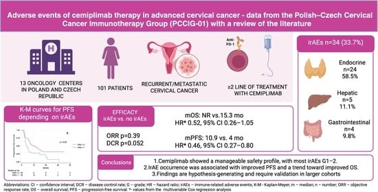
Background: Immunotherapy has become an integral part of systemic treatment for cervical cancer (CC). This study assessed the safety profile of cemiplimab and the association between immune-related adverse events (irAEs) and treatment outcomes in patients with persistent, recurrent or metastatic CC. Methods: This ambispective, multicenter, real-world cohort study included 101 patients treated in 13 reference oncology centers as part of the PCCIG-01 study. We evaluated the frequency and severity of irAEs and their association with progression-free survival (PFS) and overall survival (OS). Survival outcomes were analyzed using the Kaplan–Meier method and Cox proportional hazards models, with p < 0.05 considered statistically significant. Results: After a median follow-up of 7.5 months, adverse events occurred in 45 patients (44.6%) and were mostly grade (G) 1–2. IrAEs were observed in 34 patients (33.7%). Endocrine toxicities predominated (n = 24, 58.5% of irAEs), followed by hepatic (n = 5, 12.2%) and gastrointestinal events (n = 4, 9.8%). G3 irAEs occurred in 8 patients (7.9%). Median PFS was 3.9 months (95% CI 2.9–5.6) in patients without irAEs and 10.9 months (95% CI 5.7–16.3) in those with irAEs (p = 0.03). Median OS was 15.3 months (95% CI 8.6–25.9) in patients without irAEs and was not reached in those with irAEs (95% CI 11.6-NR; p = 0.11). The development of irAEs was associated with a 54% reduction in the risk of progression (HR 0.46, 95% CI 0.27–0.80), with no statistically significant impact on OS. Conclusions: In exploratory analyses, the occurrence of irAEs was associated with improved PFS in cemiplimab-treated patients with persistent, recurrent or metastatic CC. Cemiplimab showed a manageable safety profile, with most toxicities being G1–G2.
Full article
(This article belongs to the Section Antibody-Based Therapeutics)
►
Show Figures
Graphical abstract


