Review on Silver Nanoparticles as a Novel Class of Antibacterial Solutions
by
Corina Michaela Crisan
1, 
Teodora Mocan
2,3,*,
Meda Manolea
1,3,
Lavinia Iulia Lasca
4,
Flaviu-Alexandru Tăbăran
3,4,5 and
Lucian Mocan
1,3 1
3-rd Surgery Clinic, University of Medicine and Pharmacy, 400162 Cluj-Napoca, Romania
2
Physiology Discipline, University of Medicine and Pharmacy, 400006 Cluj-Napoca, Romania
3
Nanomedicine Department, Regional Institute of Gastroenterology and Hepatology, 400162 Cluj-Napoca, Romania
4
Emergency Hospital, 410169 Oradea, Romania
5
Pathology Department, University of Agriculture Sciences and Veterinary Medicine, 400372 Cluj-Napoca, Romania
*
Author to whom correspondence should be addressed.
Appl. Sci. 2021, 11(3), 1120; https://doi.org/10.3390/app11031120
Submission received: 25 November 2020
/
Revised: 15 January 2021
/
Accepted: 20 January 2021
/
Published: 26 January 2021
(This article belongs to the Special Issue Synthesis and Biomedical Application of Nanoparticles)
Abstract
:Nanomaterials represent a promising novel class of materials to be used as antibacterial solutions. Inhomogeneity of synthesis and characterization methods, as well as resulting variate physical and chemical properties make selection of proper nanostructure difficult when designing antimicrobial experiments. Present study focuses on the already existing evidence regarding silver nanoparticles and their antibacterial applications, with focus on various modulatory factors of reported antimicrobial efficiency. Present paper focuses on synthesis and characterization methods, factors modulating antibacterial efficiency, laboratory quantification procedures, as well as up–to-date knowledge on mechanisms of antibacterial action for silver nanoparticles. Moreover, challenges and future prospects for antimicrobial applications of silver nanoparticles are reviewed and discussed.
1. Introduction
Nanotechnology is a field of technology that works with materials presenting at least one dimension ranging between 1 nm and 100 nm for the purpose of obtaining new materials and objects. Nano materials are very different when compared with macroscopic materials, because of their unique, superior and indispensable properties, and they have recently became a research focus due to their distinct characteristics. One of the key differences is their high surface-to-volume ratio as well as an increased number of atoms present in grain boundaries. Due to their unique properties and qualities, nanomaterials are an important part of developing new devices which can be used in many fields such as: medicine, physics, biology, biomedicine, pharmacy, cosmetics and various product industries [1]. Recently, nanotechnology has proven great potential and efficient results in treating bacterial infections. In 1870, English doctor John Scott Burdon Sanderson discovered a connection between mold and bacteria cultivation. Soon after, in 1928, the accidental discovery of penicillin is attributed to Alexander Fleming. Penicillin was first used in 1942, having a huge impact on antibacterial treatment because up to that moment, most infectious diseases were deadly. When penicillin started to be used, it had an effect against Gram positive bacteria. Nowadays, a lot of bacteria strains have become immune due to the irrational use of antibiotics.
Following the first reports, various drugs have been developed, exerting antibacterial, antifungal and antiviral properties with great life-saving impact. Excessive use of antibiotics had two major effects: first, it led to a higher microbial resistance and second, to the appearance of multiresistant bacteria, endangering the lives of many patients. An immediate consequence was a need to develop further technologies to allow the generation of new substances with antibacterial properties while maintaining low toxicity levels suitable for applications in medicine. The latest antibacterial drugs have been developed around 1980s. Since then, no significant discovery was reported. Vancomycin is one of the first antibiotics that have been used in infections treatment against methicillin resistant Staphylococcus aureus for more than 50 years. The evolution of bacteria in time leads to more Vancomycin-resistant strains appearance. As a consequence, there is a constant need for the development of new drugs. One possible concept of drug improvement is effect enhancement by means of nanoparticles, thus allowing the binding of metals, proteins, phospholipids and antibodies [2].
Materials such as metal nanoparticles [3], metal oxide nanoparticles [4], carbon nanomaterials [5] and their composites have been intensively used as new antibacterial agents as a result of their small sizes, unique chemical and physical properties, and high specific surface. Silver nanoparticles proved to have unique antibacterial properties which resulted in their extensive use in fields such as: biomedicine, pharmacy or cosmetic industry [6]. Excellent antimicrobial properties, lowered toxicity and better biocompatibility have been reported to be exerted by silver nanoparticles, as compared to other metallic particles. The observations and data on the broad-spectrum antibacterial nature of AgNPs have been providing a fundament for more antibacterial-oriented applications. It has even been recently reported that Ag nanoparticles have an increased ability to inhibit bacterial growth as compared to antibiotics and that no or limited side effects are induced by AgNPs exposure [1,6].
2. Metal Nanoparticle Synthesis
Metal nanoparticles can be produced using: green method-using plants, leaf, flowers, extract of plants, prokaryotic bacterial cells and eukaryotic fungi or using various chemicals and reagents. Also, physical methods have been developed, and will be included in our review.
2.1. Green Methods
Synthesis can be performed in one step by using biological organisms such as bacteria, molds, algae and plants. The nanoparticle synthesis is performed by reduction using molecules in plants and microorganisms such as proteins and enzymes. The green synthesis of metal nanoparticles has the advantage of using more eco-friendly materials, and tends to be cheaper than chemical methods. The above mentioned plants are used as a reducing agent to transform the metallic silver precursor into metallic nanoparticles. They also represent a renewable source of bioactive molecules in obtaining metallic nanoparticles. The method has the ability to minimize the resulting toxicity and it has a small impact on the environment [7].
A recent study [8] used rind extract of Garcinia mangostana was used to obtain silver nano particles. The authors separated the rind, washed to remove impurities and then dried for 10 days at room temperature. The dried rind was then blended and sieved to obtain particles of uniform size. 1.5 g of the obtained particles were suspended in 50 mL of distilled water at 50–70 °C for 15 min.
One of the challenges that arise with using the green method is to control the nanoparticles’ shape and size. Temperature is thought of having the biggest impact on the material synthesis [8]. The authors discovered that at lower temperatures, nanoparticles were not produced. However, by increasing the temperature up to 80 °C, the authors observed the formation of metallic nanoparticles. Moreover, when the temperature is around 100 °C, the reaction rate is very high and prevents the particles from growing. The shape of nanoparticles is influenced by temperature, starting with small spherical particles at lower temperature and nanorods and platelet nanoparticles at higher temperatures. Reaction time is another variable that should be considered when performing the synthesis. Nishanthi et al [8] noted that silver and platinum nanoparticles have formed within 10 min while gold nanoparticles appeared seconds after adding the rind extract. Another study investigated the effect of pH on nanoparticles formation. The authors discovered that at a pH of 5, the synthesis of silver nanoparticles is low-rate process, while at a pH of 9, the production is intense [9].
2.2. Chemical Methods
Chemical methods used for the synthesis of nanoparticles include: chemical reduction, photo induced reduction, micro emulsion (see Figure 1), microwave assisted synthesis, UV initiated photo reduction, electrochemical synthetic approach, irradiation methods [10].
The authors propose a multi-step synthesis method, including (1) dispersion of water droplets in a continuous oil phase, (2) stabilization by means of surfactant molecules at the water/oil interface, (3) mixing the two microemulsions, (4) collision of water droplets with each other and exchange of reactants, (5) nucleation reaction, (6) growth of silver nanocrystals, (7) adsorption of surfactant molecules onto the surface of nanocrystals when the size of newly prepared nanocrystals reached comparable size with that of liquid droplets [11].
Out of the chemical methods, the wet chemical reduction stands due to its cost-effectiveness, low impurity factors, thermal stability and ability to use a multitude of stabilizers to enhance the nanoparticle’s stability. However, the chemical methods are in general toxic, need more energy for the producing process, more chemical substances and it is more expensive than green methods [8].
One of the most used protocols are the ones reported by Turkevich and Khan. The Turkevich methods comprises the following steps: 9 mg of AgNO3 is dissolved in 49 mL of ultra-pure water and the resulting solution is heated to 100 °C while stirring. Next, 1 mL of 38.8 mM sodium citrate solution is added, and the mixture is brought to room temperature after 45 min, with removal of large particles by using a centrifuge for 1 h at 500 rpm. Storage of the obtained pure and stable Ag nanoparticles at 4 °C. Hu Tian et al [12] used Turkevich’s method to test the antibacterial activity of the resulted nanoparticles against S. aureus. They have discovered that more than 1 mg/L of AgNP could stop bacterial growth.
A group of authors studied the reduction of Ag+ ions by cysteine in presence of poly vinyl alcohol (PVA). In order to obtain Ag nanoparticles, Khan et al used an aqueous solution consisting of AgNO3 and cysteine. The authors have discovered the effect of PVA on particle size, 30 nm in absence of PVA and 7 nm in the presence of PVA. The obtained nanoparticles were spherical, cross-linking and aggregated. However, a few disadvantages of the above methods are: high number of chemicals involved, resulting morphology, resulting different sizes and shapes of particles [13].
2.3. Physical Methods
The most important physical synthesis methods are: evaporation-condensation and laser ablation.
The advantages of using a physical method compared to a chemical one are the absence of solvent contamination and the uniformity of nanoparticles’ distribution. Using the evaporation-condensation method, very small nanoparticles (6.2–21.5 nm and 1.23–1.88 nm) can be obtained. However, the process is energy-consuming (energy is needed to raise the operating temperature) and time-consuming [14].
The laser ablation method is dependent on the wavelength of the laser, the duration of the laser pulses, the laser fluence, the ablation time and the liquid medium. The authors obtained nanospheres (20–50 nm) in water with femtosecond laser pulses at 800 nm [14].
2.4. Synthesis Methods for Silver Nanoparticles for Antibacterial Applications
Several methods of obtaining metallic nanoparticles have described, such as chemical, physical and green methods, each yielding different chemical and physical properties. By using these methods, nanoparticles have been obtained with sizes ranging from 3 to 100 nm, with different shapes: spherical, cubical, triangular, pentagonal, hexagonal and nano-wire. All referenced studies show that the size of nanoparticles directly influences the antibacterial activity (Table 1).
3. Characterization Methods for Metal Nanoparticles
In Table 2 several nanoparticles characteristics are presented along with the techniques that can be used to observe the respective characteristics (Table 2). Physical and chemical properties of nanoparticles need to be observed when trying to characterize nanoparticles. The chemical composition and concentration are not enough to distinguish nanoparticles. Their size, shape and surface properties also need to be depicted for adequate experimental reproducibility.
Several methods of characterization are presented by Mourdikoudis et al [33]. X-ray diffraction (XRD) is one of the most used techniques to provide information about crystalline structure, nature of the phase, lattice parameters and crystalline grain size. X-ray Diffraction has been proposed only during late years for usage in nanoparticle characterisation [34]. Although it has prove it utility, some limitations have been reported, such as cystal growing difficulty, increased effort to obtain data on single conformation as well as it low intensity [35].
Moreover, similar X-ray–based methods are presented by the authors such as: X-ray absorption spectroscopy suitable for determination of the X-ray absorption coefficient, or X-ray photoelectron spectroscopy for surface chemical analysis.
Transmission Electron Microscopy (TEM) is a microscopy technique that uses uniform current electron beams which either transmit or scatter the electrons from a thin sample. The interaction is dependent on the nanoparticles’ size, sample density and elemental composition. This is the most used technique to analyze size and shape, because of its ability to provide an accurate estimation of nanoparticle homogeneity. It does, however, have some limitations when quantifying a large number of particles and it can produce unreliable images due to orientation effects [33].
High Resolution Transmission Electron Microscopy (HRTEM) uses both transmitted and scattered electrons to produce the images. Thanks to its high resolution, HRTEM became the most used technique used to characterize the internal structure of nanoparticles [33].
Atomic Force Microscopy (AFM) is used to create three dimensional images of surfaces. It has the advantage of being cost effective and space saving and it is mainly used to detect nanoparticles’ size [33].
Both electron microscopy techniques (e.g., TEM) and AFM have demonstrated to be very useful in AgNPs characterization. While quantitative assessment of size and shape could be obtained in any of the two techniques, AFM can have the advantage of real-time assessment of interaction of silver nanoparticles with the lipid bilayer of bacterial membrane [36].
UV-VIS spectroscopy is a method that uses the intensity of reflected light from a reference material and compares it to the intensity reflected from the sample. The size, shape, concentration, agglomeration state and refractive index are optical properties that can be observed using UV-VIS [33]. UV-VIS represents an extensively used tool for silver nanoparticle characterization. Particularity of AgNPs is the close proximity of valence and conduction band, thus allowing the free movement of electrons. The absorbance surface plasmon resonance band, coming from their collective oscillation was widely observed and reported for diameters below 100 nm. Several factors may affect absorbance of AgNPs, such as chemical environment, diameter of particle, dimension [37,38].
FTIR spectroscopy is also widely used in silver nanoparticle characterisation having the advantage of increased signal-to noise ratio as well as reduced thermal deterioration of the sample [39]. Limitations exist, however, the recent update in the technique, attenuated total reflection (ATR)-FTIR spectroscopy, provides new features for proper evaluation of chemical features at the surface of nanoparticles [40].
Nuclear Magnetic Resonance (NMR) is a technique that uses a strong magnetic field to measure the energy difference between spin-ups and downs of nuclei. This transition can be measured using electromagnetic radiation in the radio wave range. This technique has limitations when trying to characterize ferri or ferro-magnetic materials [33].
Zeta potential is used to analyze the stability of colloidal dispersions. Particles with high positive or negative charge, tend to repel each other and form stable colloidal solutions [33].
Electrophoretic mobility (EPM) is used for the surface charge of nanoparticles. High EPM values were correlated with stable nanoparticles for long periods of time while low EPM values are associated with large aggregations of iron oxide nanoparticles in relation to nanoparticle concentration [33].
Dynamic light scattering (DLS) is a method used to measure nano and sub micrometer sizes of nanoparticles in colloidal suspensions. In order to avoid multiple scattering, a low concentration of nanoparticles is used in DLS. DLS has proven very useful for real time observation of the aggregation process due to its ability to measure the previously formed cluster sizes [8]. Combining the DLS technique with differential centrifugal sedimentation (DCS) can provide confirmation of the absence of that samples are not aggregated, the measured values are accurate, even though it lacks the necessary resolution when working with small aggregates [33]. Assessement of particle size using DLS results in slightly larger dimensions as compared to TEM. Although advantages make it a widely used technique due to ability to asess various dimensions of nanoparticles in the same time, DLS has also been reported to present sample specific drawbacks, some of them could also be applicable to silver nanoparticles [41].
The light emitted by molecules that absorb photons is measured using photoluminescence (PL). PL is a good technique for studying quantum dots and metal nanoclusters and can be successfully used in optical labeling applications due to its lack of photo bleaching and photo blinking [33].
Superconducting quantum interference device magnetometry (SQUID) is using to measure magnetic properties of nanoparticles. SQUID can measure magnetization saturation, magnetization remanence and blocking temperature. Moreover, vibrating sample magnetometry (VSM) is also used to measure magnetic properties as a function of magnetic field, temperature and time [33].
4. Silver Nanoparticles and Their Antibacterial Role
For the last 30 years, the pharmaceutical industries have focused on developing new antibiotics that have the ability to better fight against DNA replication, protein synthesis and bacterial cell wall synthesis. Despite this progress, a high fatality rate is still present due to the rise of antimicrobial resistance in bacterial infections. Bacterial resistance to the traditional antibiotics is a great healthcare issue with huge impact worldwide [42].
There is an increasing growth on using nano materials and they are becoming an important part of our lives. Efforts are made towards discovering non-toxic and cost-effective materials that can be used for various applications in industry, medical, pharmaceutical and cosmetic domains. An important application of such materials is drug-resistant bacteria and diseases control [43]. Nanoparticles obtained from silver, gold, platinum and semiconductors can be used with success as delivery agents for carrying small molecules such as drugs. Nanoparticles became highly effective against bacteria due to their antimicrobial activity because of their large surface area allowing high synergy arising from multivalent interactions [44].
Among the variety of engineered nanoparticles that have been used in antibacterial treatments, silver (Ag) nanoparticles is the most widely used antibacterial nano agent because of its broad-spectrum antimicrobial properties and strong antimicrobial effectiveness against multiple bacteria, viruses, and fungi [45]. The first evidence of using silver in medicine dates back to 1881, when it was used to prevent eye infections in neonates, and later on, in 1901 as internal antisepsis. Nowadays, drugs containing silver such as silver nitrate and silver sulfadiazine are commonly used to treat dermal burns, wounds and to remove warts [1].
Scientists agree that Ag nanoparticles interact with the bacterial cell envelope, however it is still unknown what the primary cellular target is [46]. Silver nanoparticles are antibacterial agents, capable of fighting against 650 types of diseases. Recently, many publications discovered proof for the antibacterial activity of Ag nanoparticles combined with common antibiotics, especially against multidrug resistant bacteria including Staphylococcus aureus and Escherichia coli [6,42,47,48].
An increased antibacterial activity can be observed when combining Ag nanoparticles and antibiotics, especially against drug-resistant bacteria [6]. In recent times, this combination has been considered a potential method to overcome bacterial drug resistance. Binding to various antibacterial agents for obtaining a higher antimicrobial activity was proposed. Several studies have been studying Vancomicin capped with Ag nanoparticles. While some autors reveal improved effects against both Gram -positive (S. aureus) and Gram- negative (E. coli) [44]. Recent studies show that the nanomaterial is capable of developing a better antibacterial activity against Gram positive bacteria but not against Gram negative bacteria. During the First World War, silver was the most used substance in fighting and treating soldiers’ infections [2].
Silver nanoparticles have proven a high antimicrobial activity and a low cytotoxicity levels when compared to particles obtained from other heavy metals like gold, platinum and zinc. They can bind to cells and restrict enzyme activity, destabilize the cell membrane and eventually lead to cell death [45].
Studies have demonstrated that AgNP could induce: cyto-toxicity, geno-toxicity, inflammatory response, DNA damage, ultimately cell death. Cell’s membrane and neuronal structures prolonged contact may lead to: skin diseases, argyria disease (blue skin) [45].
4.1. Factors Impacting the Antibacterial Activity of Silver Nanoparticles
Antibacterial activity can be influenced by: size, shape, surface chemistry [45].
4.1.1. Nanoparticle Size
Numerous studies show that there is an inverse correlation between dimensions of nano silver particles (5–9 nm) and their antibacterial activity. Zheng et al found that there is significant increase in antibacterial activity especially below 10 nm [45]. The smaller dimensions of nano particles are associated with easier bacterial cell’s wall penetration, and more intense destruction by reactive oxygen species (ROS) accumulation. Also, recent studies suggest the small and medium-size functionalized silver nanoparticles severely impact mitochondrial electron transport, phagocytosis, autophagy, organelle integrity and organization (e.g., microtubules). Pronounced transcriptional responses were also observed for medium-size silver nanoparticles [49].
Due to their small hydrodynamic diameter (less than 6 nm), the silver nanoparticles can easily pass through the kidneys, being eliminated from the body through the urinary system, which greatly reduced the risk of long-term toxicity [45].
4.1.2. Nanoparticle Shape
As presented by Tang et al. [45] the shape also plays an important part in the antibacterial activity. They have analyzed three shapes: spherical, rod-shaped and truncated triangular and their effectiveness against E. coli in solution and agar plates. The study concluded that in terms of biocidal activity truncated triangular nano silver particles ranked the highest followed by spheres and finally rods.
Observation of the membrane integrity lead to the conclusion that all shape types were able to bond and eventually damage the cell’s membrane. However, the truncated triangular shape has the highest number of facets which help with bacterial interaction and increased surface binding, cell uptake and bacteria death [45].
Other authors have investigated the effect of nano particles’ shape against S. aureus. They have concluded that nano platelets have the highest toxicity, followed by nano spheres, nano rods and nano cubes [50].
The study of antibacterial activity is usually conducted in liquid environments in either in vitro or in vivo conditions, therefore the stability (dispersion and chemical – release of ions) of silver nano particles is an issue because it affects its properties and its antibacterial activity as well. When observing the dispersion stability different solutions were used and the aggregation process was monitored using DLS (dynamic light scattering) and UV-vis spectra and TEM (transmission electron microscopy). The authors concluded that SDS (sodium dodecil sulfate) silver nano particles had the highest antibacterial activity due to their dispensability and cell membrane interaction [45].
4.1.3. Nanoparticle Surface Chemistry
Surface charge also plays an important role into the antibacterial activity. Different coated silver nano particles were used, with values ranging from −38 mV to +40 mV, to test the antibacterial activity against Bacillus species. An important step is purification to accurately measure toxicity levels, which decreased the antibacterial activity [45].
Silver ion release affects antibacterial activity of silver nano particles. This release can be triggered by oxidative dissolutions combining silver nano particles with oxygen. Silver ions also have high affinity to electron donating groups which can be easily found in membranes and proteins. Silver ions are capable of combining with DNA, RNA and peptides to form a barrier that stops cell division and reproduction [45]. Attachment of functional groups has been recently reported to differently alter the transcriptome. While negative bovine serum albumin-functionalized nanoparticles and neutral polyvinylpyrrolidone- binded silver nanoparticles significantly impair and down-regulate ribosomal translation and protein synthesis, while the positive, chitosan-based nanoparticles induce limited effects over the respective mechanisms [49].
4.2. Antibacterial Mechanisms Activated by Silver Nanoparticles
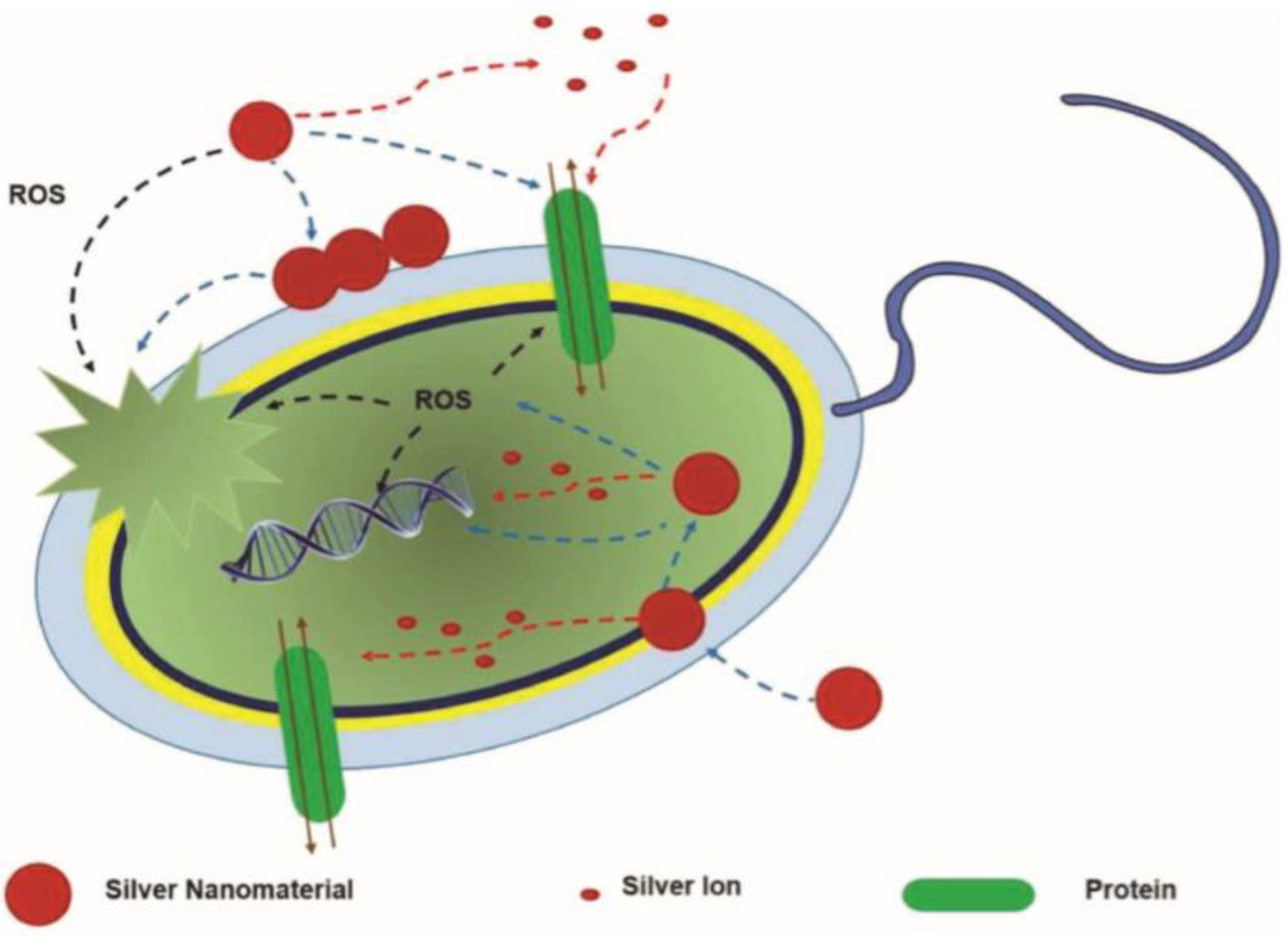
Reactive oxygen species (ROS) is created as a result of oxidative stress, a toxic effect of silver nano particles and ions. Such ROS are: superoxide anions, hydrogen peroxide, hydroxyl radical, singlet oxygen, hypochlorous acid [45]. Authors [51] studied the effect of Ag nanoparticles on intracellular ROS. The authors have discovered that the production of ROS is dose and surface- dependent. The action of Ag nanoparticles on bacterial cells begins with the appearance of oxidative stress, then membrane damage and ending with cell death. Cell death occurs due to the interaction between the active oxygen species and DNA, respiratory enzymes and cell membrane (Figure 2).
Potential mechanisms in nanosilver activity are (1) attachment to cell membrane, to protein component of the membrane and DNA structure, resulting in function alteration (blue interrupted line marks the mechanism); (2) silver ions release, impacting the membrane, nucleic acids( DNA) and protein content of the cell (red interrupted line marks the described mechanism); (3) oxidative stress mounting as a result of reactive oxygen species release with effects on cell membrane, nuclei acids (DNA) and protein content of the membrane (the black interrupted line marks the mechanism) [45].
Silver ions present in the medium can interact with the negatively charged bacterial cell and may promote the appearance of pores in the cell membrane. These pores result in a loss of proteins and nucleic acids, affect the basic metabolic functions and thus lead to cell death. The level of cell membrane disruption is correlated with the dosage of Ag nanoparticles [51]. A different cause of cell death is the reaction between colloidal silver and sulfhydryl groups present on the cell wall generating disulfide bridges which blocks cell respiration. Silver ions and nanoparticles can generate too many free radicals which result in hyper oxidation of: proteins, lipids, DNA, cell membrane [45].
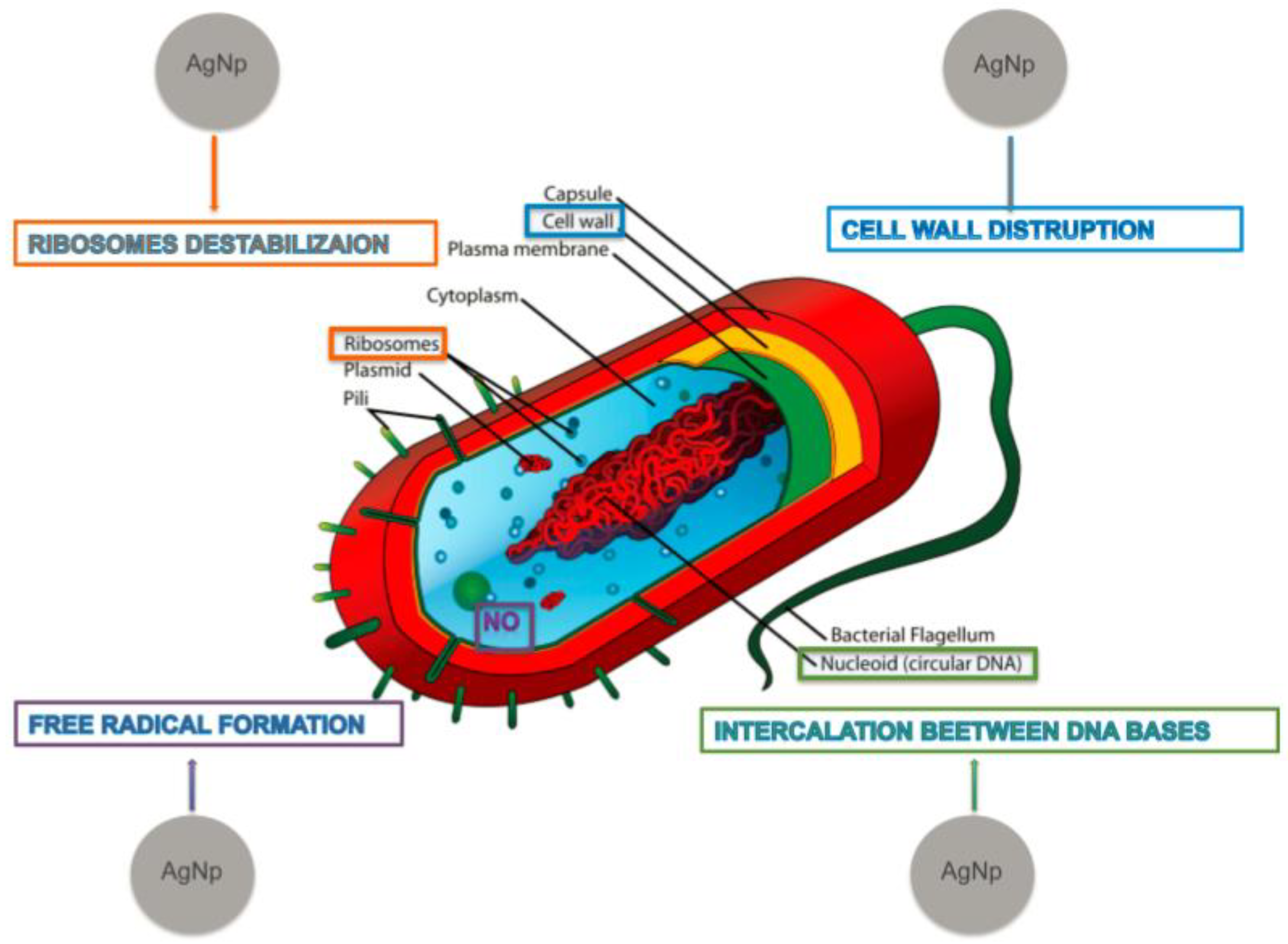
Silver nanoparticles antimicrobial action is exerted at different cell compartments and follows multiple mechanisms. (1) Interaction with the cell membrane, using physical interaction, lipid bilayer disturbance and/or cationic ion release. (2) Contact and reaction of cationic silver with bacterial DNA, resulting in structural changes. (3) Interaction with cytoplasmic structures, including protein synthesis ribosomal machinery, causing block of translation and transcription with reduced protein synthesis, (4) ROS formation, resulting in oxidative stress-based bactericidal action (see Figure 3) [52].
Gram positive and Gram-negative bacteria react differently to Ag nanoparticles. Gram positive bacteria (Staphylococcus aureus, Lysteria monocytogenes, Bacillus cereus) have a greater resistance to Ag nanoparticles than Gram negative bacteria (E. coli, Salmonella Typhimurium, Pseudomonas aeruginosa) due to their ticker cell walls [30,52].
The thicker cell envelope in the Gram-positive bacteria is due to a thick layer of peptidoglycan (see Figure 3). Peptidoglycan is a polymer consisting of sugars and amino acids which is present around the plasma membrane acting as a cell wall. This polymer might prevent the silver ions from entering the vital cellular structure and cytoplasm. Its negative charge may also be responsible for trapping positively charged silver ions [43].
Franci et al. studied the potential of silver nanoparticles as antibacterial agents upon several bacteria. According to the collected data by authors, some of the most important mechanisms underlying the antibacterial effect of nanoparticles are alteration of cell and cytoplasm, alteration of membrane permeability and respiration, inhibition of bacterial DNA replication, bacterial cytoplasm membranes damage, and irreversible damage on bacterial cells. Electron transport interference, with alteration of intracellular ATP levels has also been cited as being a significant mechanism of action [52].
4.3. Laboratory Methods for Quantification of Antibacterial Activity
Antibacterial activity is observed using: zone of inhibition plates, minimal inhibitory concentration test, minimal bactericidal concentration test.
Zone of inhibition (ZOI) plates is a test used for assessing antibiotic sensitivity of bacteria. Antibiotics are placed in agar plates and then left to incubate. After the incubation period, the growth inhibition or bacterial destruction is measured by examination of the empty zone surrounding the antibiotic (inhibition zone) [9].
Several advantages make the zone inhibition test one of the most used laboratory methods, such as being a low-price and fast method, suitable for water soluble antimicrobial efficiency, usable for testing liquids, coated surfaces as well as antimicrobial impregnated solids. However, a number of disadvantages of zone of inhibition plates test have also been identified. The method does not directly assess bacterial death, but only indirectly by measuring the growth inhibition zone. Moreover, it cannot be used for viruses, it is not a real quantitative method although it does allow measurement of the diameter of the zone.
Minimal inhibitory concentration (MIC) is the lowest concentration of a chemical which prevents the growth of bacteria. In order to determine MIC, solutions with different concentrations are incubated with bacteria and the results measured using agar dilution or broth micro dilution [53]. Relatively easy preparation and execution, small scale experiment adequacy, requirement of little preparation, as well as possible low test turnaround times are some of the advantages. Minor variations (incubation period, concentration) lead to large variation of MIC, bacteria were prevented from growing (but not killed), meaning the initial quantity of bacteria can still be present—are just a few disadvantages of MIC.
Minimal bactericidal concentration (MBC) is the lowest concentration of a chemical that results in bacteria death. A similar approach to MIC is used to determine MBC, resulting in a protocol that presents several advantages. It allows the determination of minimum concentration of agent that achieves bacteria death, can be used to rank antimicrobial agents by potency. The test parameters are easy to control and the assay can test multiple antimicrobial agents under the same conditions and observe their effect.
However, some disadvantages of MBC include the impossibility of individual bacteria assay. Also, tests will not determine the needed concentration to counterattack bacteria in short time and it has been demonstrated that different growth media will impact MBC values.
Due to a more facile protocol, MIC is easier to be determined as compared to MBC, and it is therefore used clinically. In normal conditions, the antimicrobial effect of drugs is similar for comparable concentration thanks to the host immune system fighting against bacteria (in non-multiplication conditions). When MBC is much higher than MIC, the toxicity increases and may be harmful for the human cell [54].
4.4. Results of Studies
The most studied bacteria is represented by Staphylococcus aureus (Gram positive) and Escherichia coli (Gram negative). Other strains include Multiresistant Staphylococcus aureus (MRSA), Pseudomonas aeruginosa, Klebsiella pneumoniae, Staphylococcus epidermidis, Listeria monocytogenes, Vibrio cholerae, Proteus mirabilis, Enterobacter and Enterococcus faecalis. Due to the irrational use of antibiotics, bacteria have developed a resistance to classical antibiotics over time, thus leading to the appearance of a new generation of antibiotics.
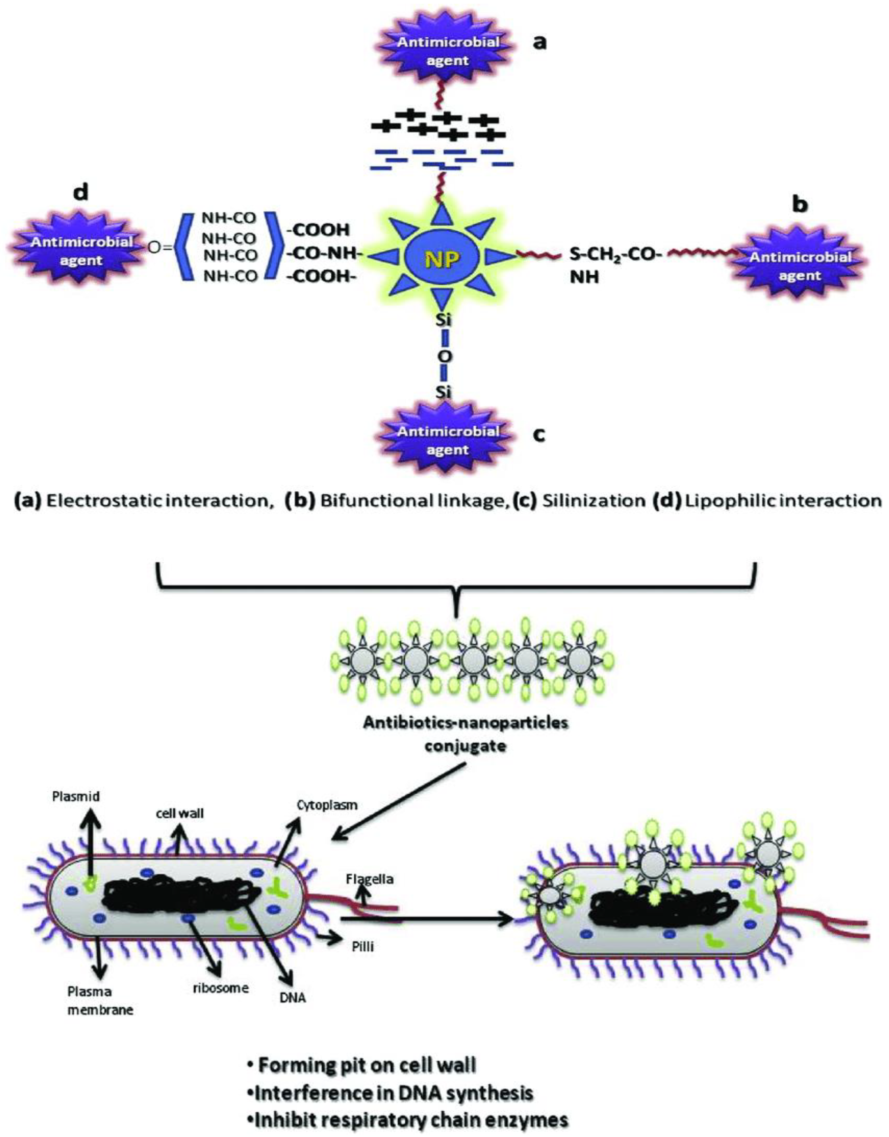
By combining a small quantity of antibiotics with metallic nanoparticles (Ag, Au, Pt), the antibacterial activity is greatly enhanced, especially when fighting multi strain resistant bacteria (see Figure 4).
The authors describe (1) The biogenic synthesis of bimetallic silver- gold nanoparticles and their functionalization with standard antibiotics (viz., bacitracin, chloramphenicol, erythromycin, gentamicin, kanamycin, streptomycin) and (2) The antibacterial effect of the resulting compound. Synergistic action of (a) biogenic metabolites formed along the synthesis process,(b) physical interaction of silver nanoparticles with cell structures, (c) cationic silver release, and (d) synthetic antibiotic specific mechanism result in enhanced antibacterial effect by attacking different structural and functional regions of the cell (flagelia, pili, membrane, cell wall, cytoplasm, DNA, plasmid, ribosomes) in various bacterial strains (E.coli, B subtilis, K. pneumoniae) [55].
Silver nanoparticles have attracted a lot of attention due to their important antibacterial activity against several bacteria types. They do not possess a toxic side effect for humans for doses up to 350 µg/day [44].
Antibacterial Activity of Silver Nanoparticles
Different results were reported by various studies concerning the Staphylococcus aureus and Escherichia coli antimicrobial effects. In the following table, the correlation between nanoparticle size and the obtained MIC values is presented (Table 3) [56].
Zone of inhibition differed significantly across the various synthesis methods, dimensions and shapes. The tables below summarize the zone of inhibition values obtained when testing the antibacterial activity of silver nanoparticles against S. aureus and E. coli. Both green and chemical methods have been reported, with different results (Table 4).
Similarly, the table below reveals the MIC values obtained for various synthesis methods, shape and dimensions of nanoparticles regarding their effect against S. aureus and E. coli (see Table 5).
The previous two tables show the antibacterial activity of silver nanoparticles using the MIC test. Regarding the ZOI test, we can observe a connection between their size and shape and the obtained MIC values.
One of the most difficult challenges for researchers is the increasing resistance to antibiotics of bacteria such as Staphylococcus aureus, Enterococcus faecium, Escherichia coli, Pseudomonas aeruginosa, Klebsiella pneumoniae, and Enterobacter species. Due to this increasing resistance to drugs, researchers had to look for alternatives that would allow them to surpass this resistance without side effects. Bacteria have developed various mechanisms to fight against antibiotics such as: cell through efflux pumps, inactivation of antibiotics, and alteration to metabolic pathway [65].
We reviewed over 60 articles that studied silver nanoparticles. Along the study, we have depicted the multiple methods used for silver nanoparticles synthesis and the substances used in this process. Depending on the method and substances, the resulting nanoparticles have different characteristics which have a different antibacterial activity. We focused on searching relevant data on the antibacterial activity of silver nanoparticles on Staphylococcus aureus and Escherichia coli. We have identified multiple articles that used the inhibition zones and minimal inhibitory concentration to determine the success of using silver nanoparticles against these bacteria. In our analysis, we divided the silver nanoparticles based on their obtaining method and then assessed the ZOI and MIC against S. aureus and E. coli. Our study may provide support for selection of design, synthesis methods and antibacterial analytical methods in accordance to intended antibacterial application.
5. Challenges and Future Prospects
The main challenge in predicting the antibacterial efficiency of a newly synthesized silver nanoparticle resides in the inhomogeneity of data in up-to-date literature. The large variety of sizes, shapes, synthesis methods, surface chemistry, the wide spectrum for choices of characterization techniques make data difficult to integrate and analyze. Moreover, the variety of bacterial strains used for testing result in a wide spectrum of identified bacterial cell targets and pathogenetic bacterial mechanisms. However, consensus exist on the benefit of usage of silver nanoparticles as antibacterial agent, and their potential to represent a promising tool against resistance [66]. The physical interaction with bacterial cell membrane, coupled with ionic silver elution, may serve as supplementary mechanisms in antibiotic therapy, resulting in enhancing standard antibiotic treatment effects [67]. Also, future directions of use may benefit from the already validated data on antimicrobial effects of silver nanoparticles. Medical devices with AgNPs films would represent a new step in using their antibacterial protection [68,69]. Usage in dental amaglams and various dental treatment and hygiene products could also represent a future direction [70]. Use of silver nanoparticles in textiles and regenerative–intend films (wound healing, burn treatment) may open new doors for the development of the field [71,72]. Osteo-synthesis products are also to be benefit from the antimicrobial effect of silver nanoparticles [73,74]. Also, non-medical, environmental applications are increasingly appealing (water, soil treatment) [75].
6. Conclusions
Nano silver particles have proven very useful in different domains and have already started to be used in medicine. They can be obtained using chemical, physical and green methods. The resulting nanoparticles have different characteristics (size, shape, surface chemistry) which influence their ability to fight bacteria. They constitute a new research direction that concerns a new class of materials with promising applications in biological, biomedical and pharmaceutical domains, and it has proved great potential and implications in treatment of bacterial infections. Combining silver nanoparticles with small quantities of antibiotics increases their antimicrobial efficiency yielding excellent results in vitro, thus holding promises for efficient in vivo bacterial eradication.
Author Contributions
Conceptualization, C.M.C. and L.M. and F.-A.T. Methodology, T.M. Data collection M.M. and L.I.L. Writing—C.M.C., L.I.L.—Original draft preparations, T.M., F.-A.T.—Review and editing. Supervision, L.M. All authors have read and agreed to the published version of the manuscript.
Funding
The authors wish to acknowledge financial support from the “Iuliu Hatieganu” University of Medicine and Pharmacy, Cluj-Napoca, Romania, grant contract no. 1529/16/18.01.2019. This work was also supported by the Romanian National Authority for Scientific Research and Innovation, CNCS-UEFISCDI, project numbers PN-III-P1-1.1-TE-2016-2161, PN-III-P2-2.1-PED-2019-0844, PN-III-P2-2.1-PED-2019-0997, PN-III-P2-2.1-PED-2019-3373.
Institutional Review Board Statement
Not applicable.
Informed Consent Statement
Not applicable.
Conflicts of Interest
The authors declare no conflict of interest.
References
- Annamalai, J.; Nallamuthu, T. Green synthesis of silver nanoparticles: Characterization and determination of antibacterial potency. Appl. Nanosci. 2016, 6, 259–265. [Google Scholar] [CrossRef] [Green Version]
- Esmaeillou, M.; Zarrini, G.; Rezaee, M.A. Vancomycin Capped with Silver Nanoparticles as an Antibacterial Agent against Multi-Drug Resistance Bacteria. Adv. Pharm. Bull. 2017, 7, 479. [Google Scholar] [CrossRef] [Green Version]
- da Silva, G.S.E.; Cabral, R.L.B.; de Sena Pereira, N.; do Nascimento, J.H.O. Antibacterial Effect of Silver Nanoparticles on Klebsiella spp. Healthc. Rev. 2020, 1, 8–15. [Google Scholar] [CrossRef]
- Keshavarz, M.; Chowdhury, A.R.H.; Kassanos, P.; Tan, B.; Venkatakrishnan, K. Self-assembled N-doped Q-dot carbon nanostructures as a SERS-active biosensor with selective therapeutic functionality. Sens. Actuators B Chem. 2020, 323, 128703. [Google Scholar] [CrossRef]
- Alavi, M.; Jabari, E.; Jabbari, E. Functionalized carbon-based nanomaterials and quantum dots with antibacterial activity: A review. Expert Rev. Anti Infect. Ther. 2020, 1, 35–44. [Google Scholar] [CrossRef]
- Yuwen, L.; Sun, Y.; Tan, G.; Xiu, W.; Zhang, Y.; Weng, L.; Teng, Z.; Wang, L. MoS 2@ polydopamine-Ag nanosheets with enhanced antibacterial activity for effective treatment of Staphylococcus aureus biofilms and wound infection. Nanoscale 2018, 10, 16711–16720. [Google Scholar] [CrossRef]
- Rauwel, P.; Küünal, S.; Ferdov, S.; Rauwel, E. A review on the green synthesis of silver nanoparticles and their morphologies studied via TEM. Adv. Mater. Sci. Eng. 2015, 1–9. [Google Scholar] [CrossRef] [Green Version]
- Nishanthi, R.; Malathi, S.; Palani, P. Green synthesis and characterization of bioinspired silver, gold and platinum nanoparticles and evaluation of their synergistic antibacterial activity after combining with different classes of antibiotics. Mater. Sci. Eng. C 2019, 96, 693–707. [Google Scholar]
- Chitra, K.; Annadurai, G. Antibacterial activity of pH-dependent biosynthesized silver nanoparticles against clinical pathogen. Biomed. Res. Int. 2014, 2014, 725165. [Google Scholar] [CrossRef] [Green Version]
- Raza, M.A.; Kanwal, Z.; Rauf, A.; Sabri, A.N.; Riaz, S.; Naseem, S. Size-and shape-dependent antibacterial studies of silver nanoparticles synthesized by wet chemical routes. Nanomaterials 2016, 6, 74. [Google Scholar] [CrossRef] [Green Version]
- Feng, A.; Cao, J.; Wei, J.; Chang, F.; Yang, Y.; Xiao, Z. Facile Synthesis of Silver Nanoparticles with High Antibacterial Activity. Materials 2018, 11, 2498. [Google Scholar] [CrossRef] [PubMed] [Green Version]
- Tian, H.; Liao, Q.; Liu, M.; Hou, J.; Zhang, Y.; Liu, J. Antibacterial activity of silver nanoparticles target sara through srna-teg49, a key mediator of hfq, in staphylococcus aureus. Int. J. Clin. Exp. Med. 2015, 8, 5794–5799. [Google Scholar] [PubMed]
- Khan, S.; Ahmad, K.; Ahmad, A.; Raish, M.; Jan, B.L.; Khan, A.; Khan, M.S. Biogenic pentagonal silver nanoparticles for safer and more effective antibacterial therapeutics. Int. J. Nanomed. 2018, 13, 7789. [Google Scholar] [CrossRef] [PubMed] [Green Version]
- Iravani, S.; Korbekandi, H.; Mirmohammadi, S.V.; Zolfaghari, B. Synthesis of silver nanoparticles: Chemical, physical and biological methods. Res. Pharm. Sci. 2014, 9, 385–406. [Google Scholar]
- Liu, M.; Zhang, H.; Song, X.; Wei, C.; Xiong, Z.; Yu, F.; Li, C.; Ai, F.; Guo, G.; Wang, X. Nacl: For the safer in vivo use of antibacterial silver based nanoparticles. Int. J. Nanomed. 2018, 13, 1737. [Google Scholar] [CrossRef] [Green Version]
- Teodoro, K.B.; Sanfelice, R.C.; Mattoso, L.; Correa, D.S. Cellulose Whiskers Influence the Morphology and Antibacterial Properties of Silver Nanoparticles Composites. J. Nanosci. Nanotechnol. 2018, 18, 4876–4883. [Google Scholar] [CrossRef]
- Senthil, B.; Devasena, T.; Prakash, B.; Rajasekar, A. Non-cytotoxic effect of green synthesized silver nanoparticles and its antibacterial activity. J. Photochem. Photobiol. B Biol. 2017, 177, 1–7. [Google Scholar] [CrossRef]
- Shankar, S.; Rhim, J.; Won, K. Preparation of poly (lactide)/lignin/silver nanoparticles composite films with UV light barrier and antibacterial properties. Int. J. Biol. Macromol. 2018, 107, 1724–1731. [Google Scholar] [CrossRef]
- Long, Y.M.; Hu, L.G.; Yan, X.T.; Zhao, X.C.; Zhou, Q.F.; Cai, Y.; Jiang, G.B. Surface ligand controls silver ion release of nanosilver and its antibacterial activity against Escherichia coli. Int. J. Nanomed. 2017, 12, 3193–3206. [Google Scholar] [CrossRef] [Green Version]
- Kasithevar, M.; Periakaruppan, P.; Muthupandian, S.; Mohan, M. Antibacterial efficacy of silver nanoparticles against multi-drug resistant clinical isolates from post-surgical wound infections. Microb. Pathog. 2017, 107, 327–334. [Google Scholar] [CrossRef]
- Składanowski, M.; Golinska, P.; Rudnicka, K.; Dahm, H.; Rai, M. Evaluation of cytotoxicity, immune compatibility and antibacterial activity of biogenic silver nanoparticles. Med. Microbiol. Immunol. 2016, 205, 603–613. [Google Scholar] [CrossRef] [PubMed] [Green Version]
- Deng, H.; McShan, D.; Zhang, Y.; Sinha, S.S.; Arslan, Z.; Ray, P.C.; Yu, H. Mechanistic study of the synergistic antibacterial activity of combined silver nanoparticles and common antibiotics. Environ. Sci. Technol. 2016, 50, 8840–8848. [Google Scholar] [CrossRef] [PubMed] [Green Version]
- Panáček, A.; Smékalová, M.; Kilianová, M.; Prucek, R.; Bogdanová, K.; Večeřová, R.; Kolář, M.; Havrdová, M.; Płaza, G.; Chojniak, J. Strong and nonspecific synergistic antibacterial efficiency of antibiotics combined with silver nanoparticles at very low concentrations showing no cytotoxic effect. Molecules 2016, 21, 26. [Google Scholar] [CrossRef] [PubMed] [Green Version]
- Hortigüela, M.J.; Yuste, L.; Rojo, F.; Aranaz, I. Green synthesis of hierarchically structured silver-polymer nanocomposites with antibacterial activity. Nanomaterials 2016, 6, 137. [Google Scholar] [CrossRef] [PubMed] [Green Version]
- Kujda, M.; Oćwieja, M.; Adamczyk, Z.; Bocheńska, O.; Braś, G.; Kozik, A.; Bielańska, E.; Barbasz, J. Charge stabilized silver nanoparticles applied as antibacterial agents. J. Nanosci. Nanotechnol. 2015, 15, 3574–3583. [Google Scholar] [CrossRef]
- Ghorbani, P.; Soltani, M.; Homayouni-Tabrizi, M.; Namvar, F.; Azizi, S.; Mohammad, R.; Moghaddam, A.B. Sumac silver novel biodegradable nano composite for bio-medical application: Antibacterial activity. Molecules 2015, 20, 12946–12958. [Google Scholar] [CrossRef] [Green Version]
- Padmos, J.D.; Boudreau, R.T.; Weaver, D.F.; Zhang, P. Impact of protecting ligands on surface structure and antibacterial activity of silver nanoparticles. Langmuir 2015, 31, 3745–3752. [Google Scholar] [CrossRef]
- Sun, W.; Qu, D.; Ma, Y.; Chen, Y.; Liu, C.; Zhou, J. Enhanced stability and antibacterial efficacy of a traditional Chinese medicine-mediated silver nanoparticle delivery system. Int. J. Nanomed. 2014, 9, 5491–5502. [Google Scholar] [CrossRef] [Green Version]
- Zhang, G.; Liu, Y.; Gao, X.; Chen, Y. Synthesis of silver nanoparticles and antibacterial property of silk fabrics treated by silver nanoparticles. Nanoscale Res. Lett. 2014, 9, 216. [Google Scholar] [CrossRef] [Green Version]
- Paredes, D.; Ortiz, C.; Torres, R. Synthesis, characterization, and evaluation of antibacterial effect of Ag nanoparticles against Escherichia coli O157:H7 and methicillin-resistant Staphylococcus aureus (MRSA). Int. J. Nanomed. 2014, 9, 1717–1729. [Google Scholar] [CrossRef] [Green Version]
- Thomas, R.; Nair, A.P.; Soumya, K.; Mathew, J.; Radhakrishnan, E. Antibacterial activity and synergistic effect of biosynthesized AgNPs with antibiotics against multidrug-resistant biofilm-forming coagulase-negative staphylococci isolated from clinical samples. Appl. Biochem. Biotechnol. 2014, 173, 449–460. [Google Scholar] [CrossRef] [PubMed]
- Matai, I.; Sachdev, A.; Dubey, P.; Kumar, S.U.; Bhushan, B.; Gopinath, P. Antibacterial activity and mechanism of Ag–ZnO nanocomposite on S. aureus and GFP-expressing antibiotic resistant E. coli. Colloids Surf. B Biointerfaces 2014, 115, 359–367. [Google Scholar] [CrossRef] [PubMed]
- Mourdikoudis, S.; Pallares, R.M.; Thanh, N.T. Characterization techniques for nanoparticles: Comparison and complementarity upon studying nanoparticle properties. Nanoscale 2018, 10, 12871–12934. [Google Scholar] [CrossRef] [PubMed] [Green Version]
- Das, R.; Ali, E.; Abd Hamid, S.B. Current applications of X-ray powder diffraction—A review. Rev. Adv. Mater. Sci. 2014, 38, 95–109. [Google Scholar]
- Chapman, H.N.; Fromme, P.; Barty, A.; White, T.A.; Kirian, R.A.; Aquila, A.; Hunter, M.S.; Schulz, J.; DePonte, D.P.; Weierstall, U. Femtosecond X-ray protein nanocrystallography. Nature 2011, 470, 73–77. [Google Scholar] [CrossRef]
- Dobrovolskaia, M.A.; Aggarwal, P.; Hall, J.B.; McNeil, S.E. Preclinical studies to understand nanoparticle interaction with the immune system and its potential effects on nanoparticle biodistribution. Mol. Pharm. 2008, 5, 487–495. [Google Scholar] [CrossRef] [Green Version]
- Link, S.; El-Sayed, M.A. Optical properties and ultrafast dynamics of metallic nanocrystals. Annu. Rev. Phys. Chem. 2003, 54, 331–366. [Google Scholar] [CrossRef] [Green Version]
- He, R.; Qian, X.; Yin, J.; Zhu, Z. Preparation of polychrome silver nanoparticles in different solvents. J. Mater. Chem. 2002, 12, 3783–3786. [Google Scholar] [CrossRef]
- Kumar, S.; Barth, A. Following enzyme activity with infrared spectroscopy. Sensors 2010, 10, 2626–2637. [Google Scholar] [CrossRef] [Green Version]
- Demathieu, C.; Chehimi, M.M.; Lipskier, J.; Caminade, A.; Majoral, J. Characterization of dendrimers by X-ray photoelectron spectroscopy. Appl. Spectrosc. 1999, 53, 1277–1281. [Google Scholar] [CrossRef]
- Dieckmann, Y.; Cölfen, H.; Hofmann, H.; Petri-Fink, A. Particle size distribution measurements of manganese-doped ZnS nanoparticles. Anal. Chem. 2009, 81, 3889–3895. [Google Scholar] [CrossRef] [PubMed] [Green Version]
- Surwade, P.; Ghildyal, C.; Weikel, C.; Luxton, T.; Peloquin, D.; Fan, X.; Shah, V. Augmented antibacterial activity of ampicillin with silver nanoparticles against methicillin-resistant Staphylococcus aureus (MRSA). J. Antibiot. 2019, 72, 50. [Google Scholar] [CrossRef] [PubMed]
- Shim, J.; Mazumder, P.; Kumar, M. Corn cob silica as an antibacterial support for silver nanoparticles: Efficacy on Escherichia coli and Listeria monocytogenes. Environ. Monit. Assess 2018, 190, 583. [Google Scholar] [CrossRef] [PubMed]
- Kaur, A.; Preet, S.; Kumar, V.; Kumar, R.; Kumar, R. Synergetic effect of vancomycin loaded silver nanoparticles for enhanced antibacterial activity. Colloids Surf. B Biointerfaces 2019, 176, 62–69. [Google Scholar] [CrossRef]
- Tang, S.; Zheng, J. Antibacterial Activity of Silver Nanoparticles: Structural Effects. Adv. Healthc. Mater. 2018, 7, 1701503. [Google Scholar] [CrossRef] [PubMed]
- Bondarenko, O.M.; Sihtmäe, M.; Kuzmičiova, J.; Ragelienė, L.; Kahru, A.; Daugelavičius, R. Plasma membrane is the target of rapid antibacterial action of silver nanoparticles in Escherichia coli and Pseudomonas aeruginosa. Int. J. Nanomed. 2018, 13, 6779. [Google Scholar] [CrossRef] [Green Version]
- Vasil’kov, A.; Dovnar, R.; Smotryn, S.; Iaskevich, N.; Naumkin, A. Plasmon Resonance of Silver Nanoparticles as a Method of Increasing Their Antibacterial Action. Antibiotics 2018, 7, 80. [Google Scholar] [CrossRef] [Green Version]
- Elbehiry, A.; Al-Dubaib, M.; Marzouk, E.; Moussa, I. Antibacterial effects and resistance induction of silver and gold nanoparticles against Staphylococcus aureus-induced mastitis and the potential toxicity in rats. MicrobiologyOpen 2018, 8, e00698. [Google Scholar] [CrossRef] [Green Version]
- Roelofs, D.; Makama, S.; De Boer, T.E.; Vooijs, R.; Van Gestel, C.A.; Van Den Brink, N.W. Surface coating and particle size are main factors explaining the transcriptome-wide responses of the earthworm Lumbricus rubellus to silver nanoparticles. Environ. Sci. Nano 2020, 7, 1179–1193. [Google Scholar] [CrossRef]
- Helmlinger, J.; Sengstock, C.; Groß-Heitfeld, C.; Mayer, C.; Schildhauer, T.; Köller, M.; Epple, M. Silver nanoparticles with different size and shape: Equal cytotoxicity, but different antibacterial effects. RSC Adv. 2016, 6, 18490–18501. [Google Scholar] [CrossRef] [Green Version]
- Ahmad, A.; Wei, Y.; Syed, F.; Tahir, K.; Rehman, A.U.; Khan, A.; Ullah, S.; Yuan, Q. The effects of bacteria-nanoparticles interface on the antibacterial activity of green synthesized silver nanoparticles. Microb. Pathog. 2017, 102, 133–142. [Google Scholar] [CrossRef] [PubMed]
- Franci, G.; Falanga, A.; Galdiero, S.; Palomba, L.; Rai, M.; Morelli, G.; Galdiero, M. Silver nanoparticles as potential antibacterial agents. Molecules 2015, 20, 8856–8874. [Google Scholar] [CrossRef] [PubMed] [Green Version]
- Wiegand, I.; Hilpert, K.; Hancock, R.E. Agar and broth dilution methods to determine the minimal inhibitory concentration (MIC) of antimicrobial substances. Nat. Protoc. 2008, 3, 163. [Google Scholar] [CrossRef] [PubMed]
- Emami-Karvani, Z.; Chehrazi, P. Antibacterial activity of ZnO nanoparticle on gram-positive and gram-negative bacteria. Afr. J. Microbiol. Res. 2011, 5, 1368–1373. [Google Scholar]
- Baker, S.; Pasha, A.; Satish, S. Biogenic nanoparticles bearing antibacterial activity and their synergistic effect with broad spectrum antibiotics: Emerging strategy to combat drug resistant pathogens. Saudi Pharm. J. 2017, 25, 44–51. [Google Scholar] [CrossRef] [Green Version]
- Wolny-Koładka, K.A.; Malina, D.K. Eco-friendly approach to the synthesis of silver nanoparticles and their antibacterial activity against Staphylococcus spp. and Escherichia coli. J. Environ. Sci. Health Part A 2018, 53, 1041–1047. [Google Scholar] [CrossRef]
- Jameel, M.S.; Aziz, A.A.; Dheyab, M.A. Green synthesis: Proposed mechanism and factors influencing the synthesis of platinum nanoparticles. Green Process. Synth. 2020, 9, 386–398. [Google Scholar] [CrossRef]
- Chumpol, J.; Siri, S. Simple green production of silver nanoparticles facilitated by bacterial genomic DNA and their antibacterial activity. Artif. Cells Nanomed. Biotechnol. 2018, 46, 619–625. [Google Scholar] [CrossRef]
- Lin, Y.; Xiong, K.; Lu, Z.; Liu, S.; Zhang, Z.; Lu, Y.; Fu, R.; Wu, D. Functional nanonetwork-structured polymers and carbons with silver nanoparticle yolks for antibacterial application. Chem. Commun. 2017, 53, 9777–9780. [Google Scholar] [CrossRef]
- Wu, J.; Zheng, Y.; Wen, X.; Lin, Q.; Chen, X.; Wu, Z. Silver nanoparticle/bacterial cellulose gel membranes for antibacterial wound dressing: Investigation in vitro and in vivo. Biomed. Mater. 2014, 9, 035005. [Google Scholar] [CrossRef]
- Van Viet, P.; Sang, T.T.; Bich, N.H.N.; Thi, C.M. An improved green synthesis method and Escherichia coli antibacterial activity of silver nanoparticles. J. Photochem. Photobiol. B Biol. 2018, 182, 108–114. [Google Scholar] [CrossRef] [PubMed]
- Soleimani, M.; Habibi-Pirkoohi, M. Biosynthesis of silver nanoparticles using chlorella vulgaris and evaluation of the antibacterial efficacy against staphylococcus aureus. Avicenna J. Med. Biotechnol. 2017, 9, 120. [Google Scholar] [PubMed]
- Muniyan, A.; Ravi, K.; Mohan, U.; Panchamoorthy, R. Characterization and in vitro antibacterial activity of saponin-conjugated silver nanoparticles against bacteria that cause burn wound infection. World J. Microbiol. Biotechnol. 2017, 33, 147. [Google Scholar] [CrossRef] [PubMed]
- Lee, W.; Kim, K.; Lee, D.G. A novel mechanism for the antibacterial effect of silver nanoparticles on Escherichia coli. Biometals 2014, 27, 1191–1201. [Google Scholar] [CrossRef] [PubMed]
- Soto, S.M. Role of efflux pumps in the antibiotic resistance of bacteria embedded in a biofilm. Virulence 2013, 4, 223–229. [Google Scholar] [CrossRef] [Green Version]
- Lu, J.; Wang, Y.; Jin, M.; Yuan, Z.; Bond, P.; Guo, J. Both silver ions and silver nanoparticles facilitate the horizontal transfer of plasmid-mediated antibiotic resistance genes. Water Res. 2020, 169, 115229. [Google Scholar] [CrossRef]
- Salleh, A.; Naomi, R.; Utami, N.D.; Mohammad, A.W.; Mahmoudi, E.; Mustafa, N.; Fauzi, M.B. The potential of silver nanoparticles for antiviral and antibacterial applications: A mechanism of action. Nanomaterials 2020, 10, 1566. [Google Scholar] [CrossRef]
- Mohamed, N.; Madian, N.G. Evaluation of the mechanical, physical and antimicrobial properties of chitosan thin films doped with greenly synthesized silver nanoparticles. Mater. Today Commun. 2020, 25, 101372. [Google Scholar] [CrossRef]
- Keshavarz, M.; Tan, B.; Venkatakrishnan, K. Label-free SERS quantum semiconductor probe for molecular-level and in vitro cellular detection: A Noble-metal-free methodology. ACS Appl. Mater. Interfaces 2018, 10, 34886–34904. [Google Scholar] [CrossRef]
- Patel, E.; Pradeep, P.; Kumar, P.; Choonara, Y.E.; Pillay, V. Oroactive dental biomaterials and their use in endodontic therapy. J. Biomed. Mater. Res. Part B Appl. Biomater. 2020, 108, 201–212. [Google Scholar] [CrossRef]
- Hoang, M.H.; Thanh, H.T.T.; Nguyen, D.N.; Van Nguyen, T.; Chyshankou, I.; Liubimau, A.; Dobysh, U.; Kulikouskaya, V. Antimicrobial ultrathin film based on well-defined silver nanoparticles and polylactide. Adv. Nat. Sci. Nanosci. Nanotechnol. 2020, 11, 025014. [Google Scholar] [CrossRef]
- Balakrishnan, S.B.; Thambusamy, S. Preparation of silver nanoparticles and riboflavin embedded electrospun polymer nanofibrous scaffolds for in vivo wound dressing application. Process Biochem. 2020, 88, 148–158. [Google Scholar] [CrossRef]
- Wenhao, Z.; Zhang, T.; Yan, J.; Li, Q.; Xiong, P.; Li, Y.; Cheng, Y.; Zheng, Y. In vitro and in vivo evaluation of structurally-controlled silk fibroin coatings for orthopedic infection and in-situ osteogenesis. Acta Biomater. 2020, 116, 223–245. [Google Scholar] [CrossRef] [PubMed]
- Abdelaziz, D.; Hefnawy, A.; Al-Wakeel, E.; El-Fallal, A.; El-Sherbiny, I.M. New biodegradable nanoparticles-in-nanofibers based membranes for guided periodontal tissue and bone regeneration with enhanced antibacterial activity. J. Adv. Res. 2020, 28, 51–62. [Google Scholar] [CrossRef]
- Fiorati, A.; Bellingeri, A.; Punta, C.; Corsi, I.; Venditti, I. Silver Nanoparticles for Water Pollution Monitoring and Treatments: Ecosafety Challenge and Cellulose-Based Hybrids Solution. Polymers 2020, 12, 1635. [Google Scholar] [CrossRef]
Figure 1.
Schematic of the reaction process with a reverse micro emulsion method.
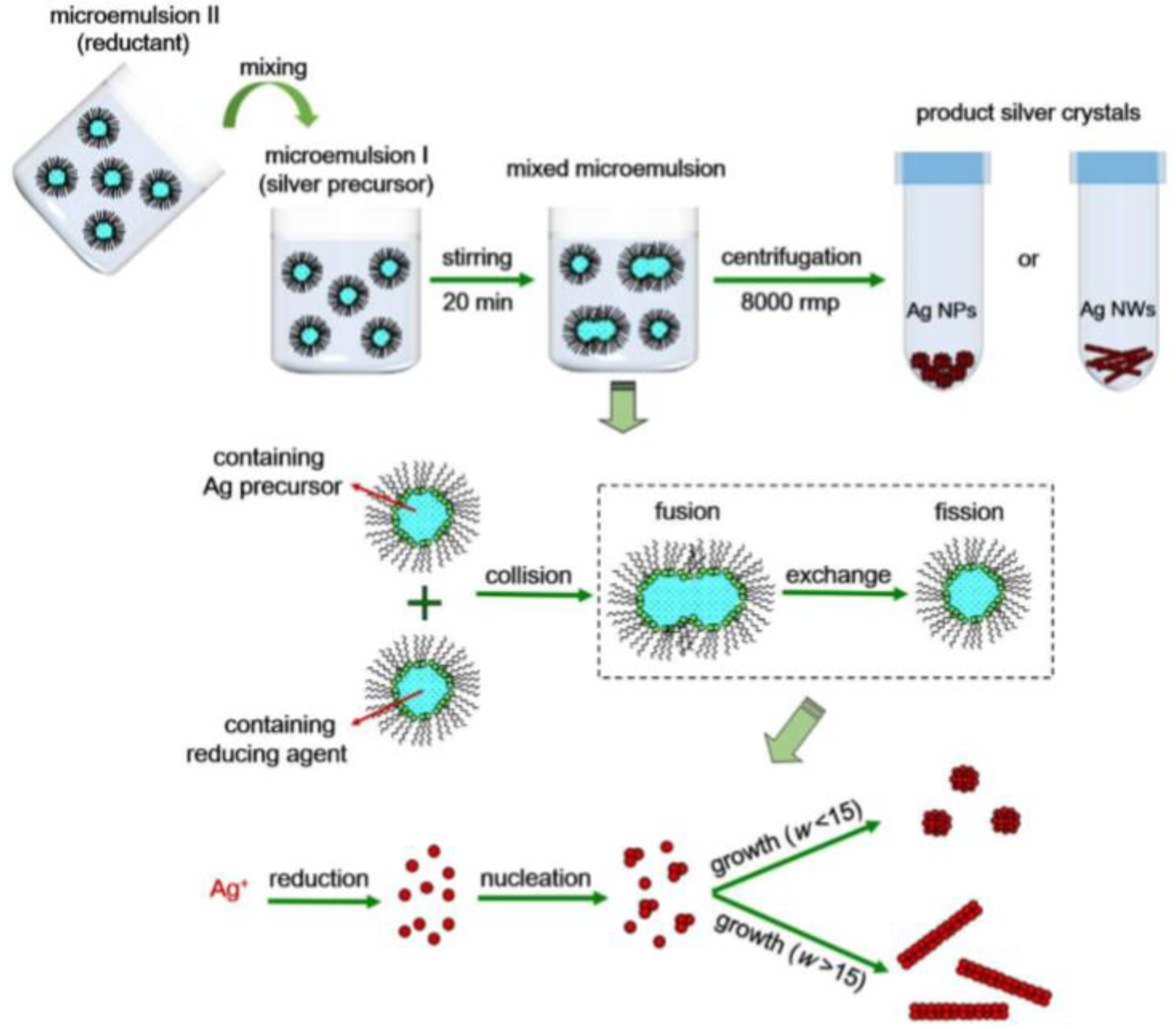
Figure 2.
Interaction between silver nanoparticles and cells.

Figure 3.
Silver nanoparticles’ toxic action.

Figure 4.
Antibiotic conjugation with nanoparticles.


Table 1.
Silver nanoparticles characterized using UV-VIS obtained with various methods.
| Synthesis Method | Chemicals | Size (nm) | Shape | UV-VIS | Ref. |
|---|---|---|---|---|---|
| Chemical | Silver nitrate, potassium sodium tartrate tetrahydrate, ammonia | 12.16 ± 4.13 | Spherical | 422 | [15] |
| Chemical | Silver nitrate, sodium citrate, sodium borohydride, cotton cellulose whiskers | 15 ± 5 | Spherical | 425 | [16] |
| Chemical | Silver nitrate, N-Hydroxy succinimide, Thioglycolic acid, 1-3 diethylamino propyl-e-ethyl carbodiimide hydrochloride, 3 N-Morpholinopropanesulfonic acid, sodium borohydride | 16–25 | Spherical | 398 | [2] |
| Green | Silver nitrate, ethanol, fenugreek leaf extract | 20–30 | Spherical | 410 | [17] |
| Chemical | Silver nitrate, organosolv lignin, chloroform, ethanol, polylactide PLA | <100 | Spherical | 450 | [18] |
| Chemical | Silver nitrate, sodium citrate, tannic acid | 10.2 ± 2.3 | Spherical | 392 | [19] |
| Green | Silver nitrate, aqueous leaf extract of Corchorus capsularis, | 20.52 | Spherical | 435 | [20] |
| Green | Silver nitrate, streptomycessp NH28 strain | 50 ± 25 | Spherical | 421 | [21] |
| Green | Silver nitrate, sodium borohydride, sodium citrate, sodium hydroxide | 29.8 ± 6.4 | Spherical | 392 | [22] |
| Chemical | Silver nitrate, ammonia, sodium hydroxide, D-maltose monohydrate, gelatin | 26 | Spherical | 410 | [23] |
| Green | Silver nitrate, chondroitin sulfate scaffolds | 20–40 | Spherical | 445 | [24] |
| Chemical | Silver nitrate, trisodium citrate | 30–80 | Spherical | 426 | [10] |
| Chemical | Silver nitrate, polyvinyl pyrrolidone, trisodium citrate, hydrogen peroxide, sodium borohydride | 150 | Triangular | 392 | [10] |
| Chemical | Silver nitrate, sodium borohydride | 25–70 | Spherical | 403 | [10] |
| Chemical | Silver nitrate, trisodium citrate, sodium borohydride | 15–50 | Spherical | 397 | [10] |
| Chemical | Silver nitrate, polyvinyl pyrrolidone, trisodium citrate, hydrogen peroxide, sodium borohydride, performed in a dark environment | 30–200 | Spherical | 504–735 | [10] |
| Chemical | Silver nitrate, sodium borohydride, trisodium citrate, gallic acid, tannin, hydroxylamine hydrochloride, sodium hypophosphite, sodium hexametaphosphate, sodium tripolyphosphate | 10–20 | Spherical | 390 | [25] |
| Green | Silver nitrate, extract of sumac fruits, | 15–30 | Spherical | 438 | [26] |
| Chemical | Silver nitrate, polyvinyl pyrrolidone, sodium borohydride | 7.7 ± 1.5 | Spherical | 403 | [27] |
| Chemical | Silver nitrate, sodium borohydride, L-cysteine | 7.6 ± 1.6 | Spherical | 396 | [27] |
| Green | Silver nitrate, Polygonum cuspidatum, Fagopyrum dibotrys, sanguisorbaofficinolis, agrimoniapilosa, Hedyotisdiffusa, Rheum palmatum, Geroniumwilfordii, phospforic acid, trisodium citrate | 36–55 | Spherical | 425 | [28] |
| Green | Silver nitrate, sodium chloride, hydrochloric acid, sodium hydroxide, pH 5 | 60–110 | Hexagonal | 480 | [9] |
| Green | Silver nitrate, sodium chloride, hydrochloric acid, sodium hydroxide, pH 9 | 10–40 | Spherical | 420 | [9] |
| Chemical | Silver nitrate, multi amino compound | 26 | Spherical | 400–420 | [29] |
| Chemical | Silver nitrate, L-cysteine, cetyltrimethylammonium bromide, polyvinyl alcohol | 55 | Spherical | 420 | [30] |
| Green | Silver nitrate, peptone, beef and yeast extract, sodium chloride | 14–42 | Spherical | 440 | [31] |
| Chemical | Silver nitrate, citric acid, zinc nitrate | 53.07 | Spherical | 410 | [32] |
Table 2.
Nanoparticle characterization techniques.
| Parameter | Suitable Technique |
|---|---|
| Size | Transmission electron microscopy (TEM), X-Ray Diffraction (XRD), Dynamic light scattering (DLS), UV-Vis Spectroscopy (UV-Vis), Nuclear Magnetic Resonance (NMR) |
| Shape | Transmission electron microscopy TEM, High-Resolution Transmission Electron Microscopy (HRTEM), Atomic force microscopy (AFM) |
| Crystal structure | X-Ray Diffraction (XRD) |
| Surface charge | Zeta potential, Electrophoretic mobility (EPM) |
| Size distribution | Dynamic light scattering (DLS), Differential centrifugal sedimentation (DCS) Atomic forme microscopy (AFM) |
| Optical properties | UV-Vis Spectroscopy (UV-Vis), Photoluminescence (PL) |
| Magnetic properties | Superconducting quantum interference device magnetometry (SQUID), Vibrating sample magnetometry (VSM) |
Table 3.
Correlation between nanoparticles’ size and MIC in S. aureus and E. coli cultures.
| Nanoparticle Size (Nanometers) | MIC (µg/mL) on Staphylococcus aureus | MIC (µg/mL) on E. coli |
|---|---|---|
| 4 | 13.5 | 6.75 |
| 10 | 50 | - |
| 5–20 | 53 | 27 |
| 30 | 20–80 | 80–320 |
Table 4.
ZOI results on S. aureus and E. coli of silver nanoparticles.
| Size (nm) | Synthesis Method | Substances Used in Synthesis | Shape | ZOI S. aureus (mm) | ZOI E. coli (mm) | Ref. |
|---|---|---|---|---|---|---|
| 23 | Green | Silver nitrate, chloroauric acid, Hex chloroplatinic acid, rind extract of garcinia mangostana | Spherical | 12 ± 3 | [8,57] | |
| 20–30 | Green | Silver nitrate, ethanol, fenugreek leaf extract | Spherical | 12 | 16 | [17] |
| 15 ± 7.6 | Green | Silver nitrate, glucose, genomic DNA | Spherical | 7.03 ± 0.5 | [58] | |
| 15–20 | Green | Silver nitrate, aqueous rhizome extract of coptischinesis | Cubical | 12 ± 1.2 | [51] | |
| 18.94 | Green | Silver nitrate, aqueous extract of chlorella vulgaris | Spherical | 15 | 20 | [1] |
| 15–30 | Green | Silver nitrate, extract of sumac fruits | Spherical | 14.3 ± 0.32 | [26] | |
| 60–110 | Green | Silver nitrate, sodium chloride, hydrochloric acid, sodium hydroxide, pH 5 | Hexagonal | 13 | [9] | |
| 10–40 | Green | Silver nitrate, sodium chloride, hydrochloric acid, sodium hydroxide, pH 9 | Spherical | 13 | [9] | |
| 20 | Chemical | Silver nitrate, silica, polystyrene | Spherical | 12.1 | 13.5 | [59] |
| 30–80 | Chemical | Silver nitrate, trisodium citrate | Spherical | 1 | [10] | |
| 150 | Chemical | Silver nitrate, polyvinyl pyrrolidone, trisodium citrate, hydrogen peroxide, sodium borohydride | Triangular | 3 | [10] | |
| 25–70 | Chemical | Silver nitrate, sodium borohydride | Spherical | 1.6 | [10] | |
| 15–50 | Chemical | Silver nitrate, trisodium citrate, sodium borohydride | Spherical | 8 | [10] | |
| 30–200 | Chemical | Silver nitrate, polyvinyl pyrrolidone, trisodium citrate, hydrogen peroxide, sodium borohydride, performed in a dark environment | Spherical | 0.8 | [10] | |
| 10–100 | Chemical | Silver nitrate, sodium hydroxide, diamine tetra acetic acid, ethylene tetrazolium bromide, 3-4-5-dymethyl-2-thiazolyl-2-5-diphenyl 2h trypsin | Cubical | 3.46 | [60] | |
| 55 | Chemical | Silver nitrate, L-cysteine, cetyltrimethylammonium bromide, polyvinyl alcohol | Spherical | 9 | 7 | [30] |
| 14–42 | Chemical | Silver nitrate, peptone, beef and yeast extract, sodium chloride | Spherical | 18 | [31] |
Table 5.
MIC results on S. aureus and E. coli of silver nanoparticles obtained with chemical methods.
Table 5.
MIC results on S. aureus and E. coli of silver nanoparticles obtained with chemical methods.
| Size (nm) | Used Substances | Shape | MIC S. aureus (µg/mL) | MIC E. coli (µg/mL) | Ref |
|---|---|---|---|---|---|
| 23 ± 2 | Silver nitrate, air borne fungus | Pentagonal | 9.23 | 5.063 | [13] |
| 70.4 ± 0.4 | Silver nitrate, gum arabic, ascorbic acid, sodium hydroxide | Spherical | 430 | 400 | [56] |
| 16 ± 2 | Silver nitrate, ethanol, sodium hydroxide, polyvinyl pyrrolidone | Spherical | 35 ppm | [61] | |
| 20–30 | Silver nitrate, ethanol, fenugreek leaf extract | Spherical | 12.5 | 6.25 | [17] |
| 9–11 | Silver nitrate, Chlorella vulgaris | Spherical | 50 | [62] | |
| 4–15 | Silver nitrate, fenugreek extract | Spherical | 10 | 25 | [63] |
| 15 ± 7.6 | Silver nitrate, glucose, genomic DNA | Spherical | 250 | 250 | [58] |
| 15–20 | Silver nitrate, aqueous rhizome extract of coptischinesis | Cubical | 25 | [51] | |
| 20–70 | Silver nitrate, sodium bis(2-ethylhexyl) sulfosuccinate (AOT), ascorbic acid (molar ratio of water to AOT of 3–15) | Spherical | 256 | 512 | [11] |
| 125 | Silver nitrate, AOT, ascorbic acid (molar ratio of water to AOT of 25) | Nanowires | 32 | 32 | [11] |
| 10–20 | Silver nitrate, sodium borohydride, polyvinylpyrrolidone | Spherical | 14.7 ± 1.19 | [48] | |
| 15 ± 5 | Silver nitrate, sodium citrate, sodium borohydride, cotton cellulose whiskers | Spherical | 14.2 | 7.1 | [16] |
| 16–25 | Silver nitrate, N-Hydroxy succinimide, Thioglycolic acid, 1-3 diethylamino propyl-e-ethyl carbodiimide hydrochloride, 3 N-Morpholinopropanesulfonic acid, sodium borohydride | Spherical | 0.8 | 1.6 | [2] |
| 20 | Silver nitrate, silica, polystyrene | Spherical | 75 | 50 | [59] |
| 20–40 | Silver nitrate, chondroitin sulfate scaffolds | Spherical | 6.2 | [24] | |
| 3 | Solid silver, nitric acid, sodium chloride | Spherical | 2 | [64] | |
| 55 | Silver nitrate, L-cysteine, cetyltrimethylammonium bromide, polyvinyl alcohol | Spherical | 0.25 | 0.25 | [30] |
| 53 | Silver nitrate, citric acid, zinc nitrate | Spherical | 60 | 550 | [32] |
Publisher’s Note: MDPI stays neutral with regard to jurisdictional claims in published maps and institutional affiliations. |
© 2021 by the authors. Licensee MDPI, Basel, Switzerland. This article is an open access article distributed under the terms and conditions of the Creative Commons Attribution (CC BY) license (http://creativecommons.org/licenses/by/4.0/).
Share and Cite
MDPI and ACS Style
Crisan, C.M.; Mocan, T.; Manolea, M.; Lasca, L.I.; Tăbăran, F.-A.; Mocan, L. Review on Silver Nanoparticles as a Novel Class of Antibacterial Solutions. Appl. Sci. 2021, 11, 1120. https://doi.org/10.3390/app11031120
AMA Style
Crisan CM, Mocan T, Manolea M, Lasca LI, Tăbăran F-A, Mocan L. Review on Silver Nanoparticles as a Novel Class of Antibacterial Solutions. Applied Sciences. 2021; 11(3):1120. https://doi.org/10.3390/app11031120
Chicago/Turabian StyleCrisan, Corina Michaela, Teodora Mocan, Meda Manolea, Lavinia Iulia Lasca, Flaviu-Alexandru Tăbăran, and Lucian Mocan. 2021. "Review on Silver Nanoparticles as a Novel Class of Antibacterial Solutions" Applied Sciences 11, no. 3: 1120. https://doi.org/10.3390/app11031120
Note that from the first issue of 2016, this journal uses article numbers instead of page numbers. See further details here.