Article

Concentration-Dependent Reinforcement and Structural Modulation of Silk Fibroin Films Induced by Mulberry Leaf Extract for Sustainable Bio-Based Materials

by
Fatma Tuba Kirac Demirel
1,2,*,
Adnan Fatih Dagdelen
1 and
Yasemin Sahan
2
1
Department of Food Engineering, Bursa Technical University, Bursa 16310, Türkiye
2
Department of Food Engineering, Bursa Uludag University, Bursa 16059, Türkiye
*
Author to whom correspondence should be addressed.
Macromol 2026, 6(2), 27; https://doi.org/10.3390/macromol6020027
Submission received: 25 March 2026 / Revised: 10 April 2026 / Accepted: 20 April 2026 / Published: 24 April 2026

Abstract

Fibroin-based films represent a promising platform for sustainable and bio-derived materials. Existing literature has mainly focused on isolated molecules, plasticizers, or chemical cross-linkers, and the function of complex, multi-component natural extracts as structure-modulating agents in fibroin films remains poorly understood. In this study, edible films containing mulberry leaf extract (MLE; 2–8 wt%) and fibroin (8 wt%) were prepared by solution casting, and their structures were investigated using spectroscopic, morphological, thermal, mechanical, and barrier property analyses. The results reveal that MLE induces concentration-dependent changes in film performance through multicomponent, non-covalent interactions with the fibroin. An approximately 187% increase in tensile strength was achieved at high MLE concentration, confirming effective physical reinforcement. The water vapor transmission rate decreased markedly from 0.888 to 0.170 g·h−1·m−2, indicating an enhanced moisture barrier, whereas oxygen permeability increased at higher extract loadings, suggesting localized chain rearrangements. High optical transparency in the visible region was maintained (79.95–83.77%), while UV response was selectively altered with extract concentration. Overall, the 8MLE formulation exhibited the most balanced performance. This study demonstrates that plant-derived extracts can serve as effective natural modifiers for tailoring fibroin film properties without inducing crystallization, offering a sustainable strategy for designing bio-based and edible protein film systems.

Graphical Abstract

1. Introduction

Silk fibroin is a fibrous structural protein from silkworm cocoons, accounting for 60–80% of their protein content. Its unique molecular structure, rich in glycine and alanine residues, allows it to form polymer networks that are stabilized predominantly by hydrogen bonding and chain entanglement, resulting in mechanical robustness and thermal stability [1,2]. These properties, combined with its biodegradability, non-toxicity, and cost-effectiveness—especially when sourced from industrial silk waste—position silk fibroin as a sustainable and bio-based polymer for various applications, including film-forming systems and composite materials [3,4].
Silk fibroin films exhibit physical properties governed by intermolecular interactions, especially hydrogen bonding between peptide chains. Modulating these interactions can tailor mechanical performance, thermal response, and structural organization, often without the need for irreversible covalent crosslinking [5].
Recent studies emphasize that while crystallinity significantly influences material properties, substantial reinforcement can also be achieved by enhancing interactions within non-crystalline regions. This facilitates superior stress-transfer mechanisms relevant to sustainable film and coating technologies [6,7].
Naturally derived plant extracts represent an attractive class of multifunctional modifiers for protein-based polymer systems, particularly for the development of bio-based and potentially edible material platforms. Unlike isolated polyphenols, crude plant extracts comprise a diverse range of secondary metabolites, including polyphenols, flavonoids, alkaloids, and carbohydrates, each capable of interacting with protein chains through multiple physicochemical pathways. These interactions collectively promote physical crosslinking, restrict segmental motion, and alter stress dissipation mechanisms within polymer networks. However, the contribution of such multicomponent systems to reinforcement behavior is less understood than that of single-molecule additives [8,9].
Mulberry (Morus alba) leaf extract (MLE) is particularly noteworthy due to its complex chemical composition, which includes not only polyphenolic compounds but also nitrogen-containing secondary metabolites such as 1-deoxynojirimycin (DNJ). DNJ possesses multiple hydroxyl groups and a heterocyclic structure capable of interacting with peptide backbones and side chains through a combination of polar interactions and steric effects. Despite extensive investigation in nutritional contexts, the role of DNJ-containing extracts in modifying protein-based polymer networks has received minimal attention [10,11]. The functional novelty of MLE stems from its unique dual-component signature, comprising the iminosugar DNJ alongside a diverse polyphenolic matrix [12]. Unlike other plant-derived modifiers that rely solely on polyphenols, DNJ acts as a specialized structural anchor; its polyhydroxylated heterocyclic ring offers multivalent hydrogen-bonding opportunities with the silk fibroin peptide backbone [13]. This synergy between DNJ and flavonoids enables a complex interaction landscape that promotes network densification within the amorphous regions of the fibroin matrix [14]. Exploring this untapped synergy offers a novel lever for the rational design of functional protein films. Recently, Ottaviano et al. [15] reported silk fibroin scaffolds enriched with mulberry fruit extract for regenerative medicine. However, structural modulation of silk fibroin films using leaf-derived extracts and the subsequent enhancement of their mechanical and barrier properties for sustainable packaging remain largely unexplored. Unlike 3D scaffolds produced via freeze-drying, our study focuses on functional bio-based films developed through solvent-casting, highlighting the reinforcement mechanism induced specifically by mulberry leaf extract. This distinction is significant, as the phytochemical profile of mulberry leaves, particularly regarding specific alkaloids like DNJ and distinct polyphenolic compositions, differs substantially from that of the fruit, offering unique intermolecular interaction pathways with the silk fibroin matrix. Existing studies involving MLE have largely focused on application-driven outcomes, often within polysaccharide- or synthetic-polymer-based composites, where the intrinsic contribution of extract–protein interactions is masked by plasticizers, surfactants, or chemically dissimilar matrices [16].
Similarly, silk fibroin-based film studies have predominantly emphasized end-use functionality, frequently correlating improved performance with crystallinity enhancement or additive-induced phase transitions. While valuable, such approaches tend to overlook reinforcement mechanisms originating from multivalent, non-covalent interactions within largely amorphous polymer regions [17,18]. Consequently, a systematic understanding of how complex metabolite systems influence silk fibroin organization and reinforcement remains lacking.
In the present study, MLE was incorporated into silk fibroin films to investigate concentration-dependent reinforcement mechanisms. Contrary to crystallinity-based models, this work aims to clarify structure-property correlations based on network densification and intermolecular interaction modulation [11,19]. A comprehensive characterization strategy was employed to correlate molecular-level interactions with macroscopic behavior. The results offer new perspectives on using natural extracts as reinforcing additives and provide a mechanistic platform for the design of sustainable, bio-based protein films.

2. Materials and Methods

2.1. Materials

Young mulberry leaves (Morus alba) were collected between May–June from a private orchard in Bursa, Türkiye (40°13′41.6″ N, 29°07′28.0″ E). The species was identified, and a voucher specimen was deposited at the Bursa Technical University, Faculty of Forestry Herbarium (BUTOF), under the accession number BUTOF224. These leaves were selected for their high levels of 1-deoxynojirimycin (DNJ) and phenolic content [20]. Leaves of uniform size and color, free from visible damage, were washed with distilled water and air-dried at ambient temperature for 48 h in the dark. The dried leaves were ground, sieved through a 420 µm mesh, and stored at 4 °C. Raw silkworm cocoons (Bombyx mori) were supplied by Kozabirlik (Bursa, Türkiye). Prior to processing, all raw materials were stored in light-impermeable packaging at −18 °C.

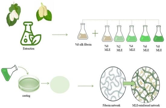
2.2. Preparation of Mulberry Leaf Extract

Dried leaf powder (100 g) was extracted with 1000 mL of a solvent mixture (90% ethanol, 0.5% phosphoric acid, and 9.5% deionized water, w/w) at 40 °C for 60 min at 170 rpm. After coarse filtration, the solvent was removed under reduced pressure using a rotary evaporator (model R-3, BUCHI, Flawil, Switzerland). The resulting concentrated extract was freeze-dried and stored at 4 °C.
The bioactive profile of mulberry leaf extract was characterized using established protocols. The DNJ content was determined via HPLC (Agilent Infinity 1260, Santa Clara, CA, USA)) following the method of Zhang et al. [21]. Total phenolic content (TPC) was measured as gallic acid equivalents (GAE) at 750 nm using a UV-VIS spectrometer (model 3660UV, Rigol, Suzhou, China) according to Lamuela-Raventós [22]. The antioxidant capacity was evaluated through CUPRAC (Cupric ion reducing antioxidant capacity) and DPPH (2,2-diphenyl-1-picrylhydrazyl free radical scavenging) assays, following the methodologies of Apak et al. [23] and Dawidowicz et al. [24], with absorbance values recorded at 450 nm and 517 nm, respectively.

2.3. Preparation of Silk Fibroin Solution

Silk fibroin was extracted by modifying reported protocols [25,26]. Raw cocoons were degummed in an aqueous sodium carbonate solution (0.02 M) for 30 min to remove sericin. The fibers were rinsed with deionized water and impurities and air-dried overnight.
Purified fibroin was dissolved in Ajisawa solution (CaCl2/ethanol/water) at 70 °C for 5 h. To remove the chaotropic salt, the solution was transferred into dialysis tubing (MWCO 3.5 kDa) and dialyzed against deionized water for 48 h. The resulting suspension was purified by centrifugation (9000 rpm, 20 min, 4 °C) and freeze-dried.

2.4. Film Fabrication

Edible films were prepared by a solution-casting method. Silk fibroin solution (8%, w/v) was mixed with MLE at concentrations of 0, 2, 4, 6, and 8% (w/w) and homogenized. After degassing, 4 mL aliquots were cast into 100 mm Petri dishes and dried overnight at ambient temperature. Films were conditioned at 23 °C and 50% relative humidity for 48 h in a controlled climate chamber (model MIT-120, Mikrotest, Ankara, Türkiye) prior to testing [26].

Structural Characterization

Fourier transform infrared spectroscopy (FTIR, model Alpha II, Bruker Optics, Ettlingen, Germany) was used. Spectra were recorded over the range of 4000–400 cm−1 at a resolution of 4 cm−1, with 16 scans. Spectral variations associated with interactions between silk fibroin and MLE components were examined [27]. Structural transitions were quantified via peak deconvolution of the 1016–1633 cm−1 region using OPUS software (version 8.5, Bruker Optics, Ettlingen, Germany). Following the protocols and area-ratio equations described by Ottaviano et al. [15], the relative contents of crystalline (β-sheet) and amorphous fractions, as well as other functional groups, were determined through the Levenberg–Marquardt algorithm.

2.5. Optical and Color Characterization

The light transmittance of the films in the UV–visible region was measured using a UV–Vis spectrophotometer (model 3660UV, Rigol, Suzhou, China). Film specimens were cut into rectangular strips (10 × 40 mm) and placed in quartz cuvettes. Transmittance spectra were recorded over the wavelength range of 200–800 nm with a wavelength interval of 20 nm [28].
Film opacity was determined based on the absorbance measured at 600 nm and the corresponding film thickness. Opacity values were calculated according to the following equation:
O p a c i t y ( a b s · m m 1 ) = A 600 d
where A600 represents the absorbance at 600 nm, and d is the film thickness (mm) [28].
Color parameters of the films were determined using a colorimeter (UltraScan VIS, HunterLab, Reston, VA, USA). Prior to measurements, the instrument was calibrated according to the manufacturer’s instructions. The CIELAB color coordinates (L*, a*, and b*) were measured at fifteen different positions on each film sample, and mean values were reported. The total color difference (ΔE) was calculated relative to a standard white reference (L* = 100.03, a* = −0.01, b* = 0.00) [29].

2.6. Film Thickness Measurement

Film thickness was measured using an external micrometer (Mitutoyo, model 293-IP-54, Mitutoyo, Kawasaki, Japan) and reported in millimeters. Measurements were taken at seven different positions on each film sample, and the average value was used for further analysis.

2.7. Morphological Characterization

Surface and cross-sectional morphologies were examined by scanning electron microscopy (SEM, model Gemini 300, Zeiss, Oberkochen, Germany) at 5.0 kV. Surface images were acquired at 5000× magnification (WD ≈ 13.5 mm), while cross-sectional images were obtained at 1000× magnification (WD ≈ 9.6 mm).

2.8. Thermal Characterization

The thermal stability of the films was evaluated by differential scanning calorimetry (DSC, model DSC250, TA Instruments, New Castle, DE, USA) and thermogravimetric analysis (TGA, model SDT 650, TA Instruments, New Castle, DE, USA). DSC measurements were conducted under a nitrogen atmosphere at a heating rate of 10 °C/min over a temperature range of −80 to 200 °C. TGA measurements were performed at a heating rate of 10 °C/min from 25 to 600 °C under an air flow of 50 mL/min, following previously reported procedures [30].

2.9. Crystalline Structure Characterization

The crystalline structure of the films was analyzed using X-ray diffraction (XRD; model D8 Discovery, Bruker AXS, Karlsruhe, Germany) with monochromatized Cu Kα radiation. Diffractograms were recorded over a 2θ range of 5–30°.

2.10. Measurement of Water Vapor Transmission Rate and Permeability

Water vapor transmission rate (WVTR), water vapor permeability (WVP) and permeability coefficient (P) of the films were determined using a gravimetric method [31]. Film samples were cut into circular specimens (60 mm diameter) and sealed onto the mouths of test cups containing dried silica gel, ensuring no direct contact between the film and desiccant. The film edges were sealed with water-resistant wax to prevent vapor leakage, and the cups were tightly closed without damaging the film surface. The assembled cups were weighed and placed in a controlled climate chamber (model MIT-120, Mikrotest, Ankara, Türkiye) maintained at 23 °C and 50% relative humidity. Weight changes were recorded at 2 h intervals over a 24 h period, with at least eight measurements collected for each sample.
WVTR and WVP values were calculated according to the following equations:
W V T R   g m 2 . h = w A . t
W V P   g . m m 2 . h . P a = w . d A . t . P 0 . R H
P   g . m m m 2 . h . k P a = W V P . d
where w is the mass change (g), A is the exposed film area (m2), t is time (h), d is film thickness (m), P0 is the saturated water vapor pressure at the test temperature (Pa), and ΔRH is the relative humidity difference across the film.

2.11. Mechanical Testing

Mechanical properties of the films were evaluated using a texture analyzer (TA-HD Plus, Stable Micro Systems, Godalming, UK). Tensile strength (TS) and elongation at break (EAB) were determined according to ASTM standards [32]. Film strips (10 × 80 mm) were conditioned at 23 °C and 50% relative humidity for 48 h prior to testing. Tensile measurements were performed at a crosshead speed of 2 mm/s. Tensile strength was calculated by dividing the maximum force at break by the cross-sectional area of the film, while elongation at break was expressed as the percentage change in length relative to the initial length. Puncture force (PF), puncture distance (PD) and Young’s modulus (E) were measured using circular film specimens (50 mm diameter) conditioned under the same temperature and humidity conditions. The samples were mounted on a circular sample holder and punctured using a spherical probe at a speed of 0.2 mm/s. The maximum force and deformation at puncture were recorded [33].

2.12. Measurement of Oxygen Permeability

Oxygen permeability of the films was measured using an oxygen transmission rate analyzer (VAC-V2, Labthink Instruments, Jinan, China) in accordance with ISO standards [34]. Film specimens were cut into circular samples with a diameter of 12 cm, and film thickness was measured at ten different locations prior to testing. All measurements were conducted at 23 °C. The oxygen gas transmission rate (O2GTR) was recorded for each sample. Oxygen permeability (PO2) was calculated by normalizing the O2GTR values to the oxygen partial pressure difference across the film, according to the following equation:
P O 2 = O 2   G T R ( c m 3 ) / ( m 2 · d ) p   ( P a )
where p represents the oxygen partial pressure difference (Pa). To enable comparison under different experimental conditions, thickness-dependent oxygen permeability coefficients (Dk, cm3·cm/cm2·s·Pa) were calculated by multiplying the O2GTR values by the corresponding film thickness.

2.13. Statistical Analysis

All experiments were conducted in triplicate (n = 3). Before analysis, the assumptions of normality and homogeneity of variance were verified using the Shapiro–Wilk and Levene tests, respectively. Data were then subjected to one-way analysis of variance (ANOVA), and significant differences between means were determined using Duncan’s multiple range test at a 95% confidence interval (p < 0.05). Statistical analysis was performed using the SPSS software package (SPSS for Windows, version 26.0, SPSS Inc., Chicago, IL, USA).

3. Results and Discussion

3.1. Bioactive Profile of Mulberry Leaf Extract

The functional performance and structural impact of plant extracts on biopolymer matrices are intrinsically linked to their specific chemical constituents. To establish a mechanistic understanding of the interactions between MLE and silk fibroin, the bioactive profile of the extract was first quantitatively characterized (Table 1). The analysis reveals a significant concentration of DNJ (5.81 mg/g) and a robust total phenolic content (181.40 mg GAE/g). These components are pivotal for the reinforcement of the films; specifically, the iminosugar structure of DNJ and the abundant hydroxyl groups of the phenolics function as multivalent cross-linking agents. These constituents facilitate extensive hydrogen bonding with the fibroin peptide backbone, effectively driving the transition toward a more ordered crystalline β-sheet structure, as further evidenced by our FTIR deconvolution results. Moreover, the antioxidant activity of MLE demonstrated competitive free-radical scavenging and reducing power. This potent bioactivity, integrated with the high DNJ content, confirms that MLE serves as a functional reinforcement agent that actively stabilizes the silk fibroin’s secondary structure, rather than acting as a passive physical filler.
The rich bioactive profile of MLE provides a versatile platform for modulating the silk fibroin network. These specific functional groups are expected to interfere with the fibroin’s native assembly, potentially inducing significant molecular rearrangements. To determine if these phytochemical-driven interactions lead to physical changes, such as network densification, the structural, morphological, and mechanical properties of the films are investigated in the following sections.

3.2. Structural Characterization

The FTIR spectra of silk fibroin films with varying MLE concentrations exhibited concentration-dependent shifts in band intensity, width, and position (Figure 1). These variations reflect modifications in the intermolecular interactions within the fibroin network. The absence of new absorption bands suggests that MLE incorporation modulates existing interactions rather than inducing new chemical bonds. The broad band at 3277–3282 cm−1, assigned to overlapping N–H and O–H stretching vibrations, is characteristic of hydrogen-bonded systems and reflects peptide N–H stretching associated with β-sheet stabilization. With increasing MLE content, progressive band broadening and intensity enhancement were observed, attributable to hydroxyl-rich extract constituents such as polyphenols and flavonoids. These components contribute additional hydrogen-bond donors, promoting interactions with fibroin amino groups and increasing the fraction of bound water within the matrix [35,36].
In the amide I region (1617–1633 cm−1), which is dominated by C=O stretching vibrations of the peptide backbone and is commonly associated with β-sheet-rich crystalline domains, subtle changes in band shape and intensity were detected. Contributions from extract-derived aromatic structures and carbonyl-containing phenolic compounds may overlap within this region, particularly in the 1610–1620 cm−1 range, where flavonoids and phenolic acids are known to exhibit vibrational modes. Additionally, hydrated components of the plant extract, including carbohydrate derivatives, may further influence this region [37,38].
The amide II band (1534–1544 cm−1), arising primarily from N–H bending and C–N stretching, exhibited a non-monotonic response to MLE concentration. While an increase in band intensity was observed for films containing 2% and 4% MLE, a relative decrease occurred at higher extract loadings (6% and 8% MLE). This behavior suggests that moderate extract concentrations optimize extract–fibroin interactions within amorphous regions. Conversely, excessive loading may reduce interaction efficiency due to steric hindrance, aggregation, or partial disruption of the fibroin quaternary organization [39]. In addition to protein-related vibrations, this region may receive contributions from extract constituents, including flavonoids with aromatic structures, nitrogen-containing secondary metabolites such as alkaloids, and low-molecular-weight protein fragments, all of which can participate in N–H- or C–N-related vibrational modes [40,41].
With increasing MLE concentration, systematic intensity shifts across several FTIR regions reflect the integration of extract-derived functional groups into the fibroin matrix. The 1430–1450 cm−1 region, associated with C–H bending and carboxylate (COO) vibrations, showed enhanced intensity due to aliphatic and carboxyl-containing compounds in MLE, such as organic acids and phenolic derivatives [42]. Concurrently, modulation of the amide III band (1220–1235 cm−1), which is sensitive to fibroin secondary structure, suggests overlapping contributions from flavonoid aromatic vibrations and phosphorus-containing organic constituents. A pronounced, concentration-dependent increase was also detected in the 1016–1033 cm−1 region, attributed to C–O and C–C stretching vibrations of carbohydrate- and phenolic-rich components of MLE, which are only weakly expressed in native fibroin [43]. Overall, the FTIR results indicate that MLE modifies the silk fibroin network through multicomponent physical interactions involving polyphenols, flavonoids, carbohydrates, and nitrogen-containing secondary metabolites, altering the organization of ordered and less ordered domains without inducing new crystalline phases, in agreement with characteristic fibroin absorption features reported in the literature [44]. To quantitatively validate these structural transitions, a deconvolution analysis of the amide regions was performed (Table S1). The results demonstrate that MLE acts as a potent structural modulator; specifically, the crystalline β-sheet content increased from 35.20% in pure fibroin to 41.60% at 4% MLE loading. This numerical increase provides direct evidence that the bioactive components of MLE—particularly DNJ and phenols—act as molecular anchors that stabilize the crystalline nanodomains through multivalent hydrogen bonding. The slight plateauing of this effect at 6% and 8% MLE (38.10% and 37.70%, respectively) further supports the interpretation of a non-monotonic interaction efficiency due to steric hindrance or localized aggregation at higher loadings. These molecular-level modifications are expected to influence the light-matter interactions and the resulting optical performance of the composite films.

3.3. Optical and Color Properties

Light transmittance in the visible region (400–800 nm) is a key indicator of film transparency and optical performance. All silk fibroin–based films exhibited high visible light transmittance values ranging from 79.95 to 83.77%, indicating overall good transparency (Figure 2).
The control fibroin film (F) showed the highest transmittance (83.77%), confirming the intrinsically transparent nature of silk fibroin films. Films with 2% and 4% MLE maintained comparable transparency (82.18% and 80.79%, respectively), while further increases in extract content led to a slight decrease in transmittance for 6% and 8% MLE-containing films. Despite this reduction, visible light transmittance remained relatively high across all formulations, indicating that MLE incorporation only marginally affected film clarity. These results align with the visual appearance of the films (Figure 3), which remained optically clear across the entire concentration range. The high visible-light transmittance observed for all films is consistent with their predominantly amorphous structure, as confirmed by XRD analysis, since the absence of crystalline domains minimizes light scattering.
In the UV region (200–315 nm), distinct differences in optical behavior were observed. Neat fibroin films exhibited low UV transmittance (13.03%), demonstrating effective UV-blocking capability. Incorporating low levels of MLE (2% and 4%) moderately increased UV transmittance to 17.72% and 20.80%, respectively. However, 6% and 8% MLE loadings resulted in substantially higher values (66.58% and 67.59%). These results indicate that increasing MLE concentration reduces UV-shielding efficiency, likely due to structural and compositional changes within the film matrix. Consequently, neat and low-MLE films are better suited for packaging light-sensitive products.
The observed optical behavior aligns well with previous reports on silk fibroin–based films, which are known for high transparency in the visible range. A study reported visible light transmittance values exceeding 93% for regenerated silk fibroin films [45], while composite fibroin films typically exhibit transmittance values between 60% and 80% depending on formulation [46]. Similarly, silk fibroin films developed for biomedical applications have been shown to maintain visible light transmittance above 90% [47]. The transparency levels obtained in the present study therefore fall within the expected range for fibroin-based systems. Changes in transparency and UV response can also be associated with the presence of bioactive compounds in MLE. Plant-derived antioxidants can absorb light in the UV and near-visible regions, leading to reduced transparency at shorter wavelengths [48]. In line with this, the gradual reduction in transparency at wavelengths below approximately 450 nm with increasing MLE concentration is consistent with the enhanced antioxidant capacity of the films.
Opacity and color measurements further supported these observations (Table 2). The control fibroin film (F) exhibited the lowest opacity value (0.93 abs·mm−1), corresponding to the highest transparency. A slight increase in opacity was observed with increasing MLE content, indicating partial light scattering by extract-derived components within the film matrix. Similar trends have been reported for fibroin-based composite films containing natural extracts or plasticizers [48,49]. Color analysis revealed that increasing MLE concentration significantly affected film appearance. The lightness parameter (L*) decreased progressively with extract addition, indicating darker films, while a* values shifted toward more negative values, reflecting a slight increase in greenish tones. In parallel, b* values increased markedly with MLE concentration, confirming the development of yellowish coloration associated with natural pigments present in mulberry leaf extract. Consequently, chroma (C*) and total color difference (ΔE) values increased with extract content, demonstrating enhanced color intensity. These changes are attributed to the inherent pigments and secondary metabolites of MLE, which effectively modulate the optical and color characteristics of silk fibroin films.
Beyond these optical shifts, the distribution of MLE within the fibroin matrix and its effect on the cross-sectional morphology were further examined to understand the internal arrangement of the films.

3.4. Film Thickness

The neat silk fibroin film (F) exhibited the lowest thickness (0.034 mm), confirming the formation of thin, compact films in the absence of additives. Incorporation of MLE led to a concentration-dependent increase in thickness, reaching 0.062 mm for the 8MLE film, a 126% increase relative to the control (Table 2). This trend indicates physical integration of extract components into the fibroin matrix, resulting in volumetric expansion. These values align with reported fibroin films prepared by solvent casting [50] and with mulberry-extract-containing polysaccharide films, where extract addition increased thickness through solid incorporation [51].

3.5. Morphological Analysis

SEM images showed concentration-dependent morphological changes (Figure 4). The neat fibroin film (F) exhibited a uniform and compact structure with distinguishable fibril-like features and aligned surface patterns, indicative of an ordered molecular regenerated silk fibroin [52]. The cross-section of the control film displayed a dense and homogeneous internal structure, suggesting strong inter-fibrillar cohesion.
Progressive morphological modifications occurred with MLE addition. At low extract concentrations (2MLE), minor irregularities appeared in the cross-section, although the overall matrix integrity was largely preserved. Increasing the extract concentration to 4MLE resulted in more pronounced heterogeneity and partial layering within the cross-section, indicating that extract components interfere with the native packing of fibroin chains.
At higher MLE contents (6MLE and 8MLE), the cross-sectional morphology became increasingly heterogeneous, with visible densification and layered regions. This behavior suggests extract-induced rearrangement of polymer chains and possible local phase separation within the matrix at elevated concentrations. In contrast, surface images at higher magnifications revealed that all films maintained a generally homogeneous surface with comparable nano-scale porosity. Notably, increasing MLE concentration led to progressive surface smoothing and a reduction in fibril-like features, consistent with effective interaction between extract components and fibroin chains, which alters surface energy and suppresses fibrillar aggregation.
Overall, MLE incorporation modulated the micro- and nano-scale organization of silk fibroin films by smoothing surface morphology while inducing internal structural heterogeneity at higher concentrations. Similar surface homogenization has been reported for silk fibroin films containing plant extracts, where favorable compatibility between extract components and the protein matrix resulted in uniform surfaces [48]. The observed morphological trends support the conclusion that MLE interacts effectively with fibroin chains, influencing film structure in a concentration-dependent manner. This concentration-dependent internal arrangement is expected to play a critical role in the thermal stability and degradation behavior of the composite films.

3.6. Thermal Properties

The thermal behavior of films was evaluated by DSC and TGA to assess the influence of MLE on thermal stability. DSC thermograms (Figure 5) revealed a broad endothermic peak between 90 and 100 °C for all films, attributed to the evaporation of free and bound water within the fibroin matrix. The neat fibroin film (F) exhibited the highest endothermic peak temperature (Te 97.2 °C) with a sharper profile, indicating a more stable hydrated structure. Upon MLE incorporation, Te shifted toward lower temperatures (95.8 °C for 2MLE to 90.5 °C for 8MLE), and the peaks broadened. This suggests that extract addition modifies fibroin–water and fibroin–polymer interactions, reducing the thermal energy required for moisture loss, which is consistent with the FTIR findings. Similar low-temperature endothermic behavior is reported for regenerated fibroin systems and is primarily associated with moisture loss rather than melting transitions [53].
Structural degradation was observed above 150 °C, with no distinct melting peak (Tm) appearing in the DSC thermograms. This indicates the absence of a well-defined crystalline melting transition within the investigated temperature range, which is typical of regenerated silk fibroin systems, where thermal decomposition precedes melting. Furthermore, no clear crystallization exotherm was detected. Although weak thermal events around 170–180 °C have previously been attributed to moisture-plasticized glass transition (Tg) behavior in fibroin films, no distinct Tg could be resolved under the present experimental conditions [54,55].
TGA thermograms (Figure 6A) further supported these observations. All films exhibited an initial mass loss of ~7% below 150 °C, corresponding to moisture evaporation. With increasing MLE content, the residual mass in this region increased, indicating enhanced water retention. This is attributed to the interactions between hydroxyl-rich extract components and fibroin chains, which stabilize the water molecules within the matrix. The DTG curves (Figure 6B), derived from the TGA profiles, allowed for the identification of primary thermal transition regions. The main thermal degradation occurred above 300 °C, corresponding to peptide bond cleavage and backbone decomposition. The onset of major degradation ranged from 308 °C to 319 °C, with MLE-containing films showing slightly higher residual mass compared to the neat fibroin film, suggesting that extract incorporation preserves or slightly improves high-temperature thermal stability.
To further correlate these thermal transitions with the underlying molecular arrangement, the crystalline structure of the films was examined using XRD analysis.

3.7. Crystalline Structure

XRD patterns of all silk fibroin films (Figure 7), including those containing different concentrations of MLE, were characterized by broad and diffuse diffraction features without sharp crystalline peaks, indicating a predominantly amorphous structure. The absence of intense and narrow reflections typically associated with crystalline domains confirms the lack of long-range molecular order in the films. Accordingly, crystallinity indices could not be reliably calculated, as such analyses depend on the presence of well-defined crystalline peaks.
A broad halo centered at ~18.9° (2θ), attributed to the amorphous phase of regenerated fibroin, increased in intensity with increasing MLE content. This trend suggests progressive integration of extract biomolecules into the fibroin matrix, leading to enhanced molecular disorder. Minor diffraction features observed around 12.0°, 24.4°, and 30.2° exhibited slight shifts with increasing MLE concentration, which reflect local strain effects or weak molecular interactions induced by extract incorporation rather than the formation of new crystalline phases. The absence of sharp diffraction peaks, despite FTIR-detected changes in amide bands, indicates that MLE induces local rearrangements and short-range ordering rather than long-range crystalline organization.
These results are consistent with previous reports on solution-cast silk fibroin films, where dissolution in CaCl2-based systems disrupts intermolecular hydrogen bonding within β-sheet domains and suppresses recrystallization during film formation. Zhang et al. [56] demonstrated that while raw and degummed silk exhibit characteristic β-sheet reflections at ~16.7° and ~20.5°, cast fibroin films display a broad amorphous halo spanning approximately 16–30°, similar to the patterns observed in the present study. The use of solvent casting in combination with extract incorporation therefore favors amorphous network formation over ordered crystalline assembly.
Silk fibroin typically consists of coexisting crystalline (β-sheet) and amorphous (random coil) domains [57]. The predominance of broad, diffuse diffraction features in the present samples indicates a disruption of ordered crystalline regions and the dominance of an amorphous structure. This behavior suggests that the native crystalline organization of fibroin was altered during film preparation, likely as a consequence of solution processing steps such as dissolution, casting, drying, and extract incorporation. MLE, rich in phenolic compounds, hinders the reorganization of fibroin chains into ordered domains. Interactions between extract components and fibroin interfere with interchain packing, suppressing β-sheet formation [58]. In addition, the solvent-casting process and drying conditions employed during film fabrication are known to favor kinetically trapped, amorphous structures by limiting chain mobility during solidification. Collectively, these factors contribute to the amorphous nature of the fibroin–extract films observed in the XRD patterns [59].
The predominance of an amorphous structure has significant implications for film performance. Amorphous fibroin networks generally exhibit improved flexibility, homogeneity, and optical clarity [56,58], which is consistent with the smooth surface morphology observed in SEM images and the high visible-light transmittance of the films. Moreover, when evaluated alongside water vapor permeability results, the amorphous structure appears compatible with reduced permeability at higher MLE contents, suggesting that molecular disorder combined with extract–polymer interactions contributes to denser diffusion pathways. From an application perspective, the amorphous nature of the films supports their suitability for flexible, transparent, and biodegradable packaging systems. While amorphous structures may exhibit lower thermal resistance and mechanical strength compared to highly crystalline materials, the present results indicate that controlled extract incorporation enables functional performance without reliance on crystallinity-driven reinforcement mechanisms [60,61].

3.8. Water Vapor Transmission Rate and Water Vapor Permeability

Water vapor transport behavior provides indirect insight into the free volume, chain packing, and intermolecular interactions within polymeric film networks [62]. As shown in Table 3, neat silk fibroin films exhibited the highest water vapor transmission and permeability values, indicating relatively open diffusion pathways within the protein matrix. MLE incorporation led to a pronounced and concentration-dependent reduction in water vapor transmission rate (WVTR), water vapor permeability (WVP), and permeability coefficient (P). Even at low extract loading (2MLE; 0.35 g·m−2·h−1), WVTR decreased substantially, suggesting that extract incorporation effectively restricts water vapor diffusion through the fibroin network. With increasing MLE concentration, all transport parameters decreased progressively, reaching minimum values for the 8MLE-containing film (0.17 g·m−2·h−1).
The observed reduction in water vapor transport can be attributed to structural densification of the silk fibroin matrix induced by extract incorporation. The presence of polyphenols and other secondary metabolites in MLE is expected to enhance intermolecular interactions with fibroin chains, thereby reducing free volume and limiting diffusion pathways for water molecules. This effect becomes more pronounced at higher extract concentrations, consistent with the monotonic decrease observed in WVTR, WVP, and p values [63,64]. Similarly to the effects observed with grape seed extract in zein-based films, the integration of polyphenol-rich extracts like MLE promotes the formation of a multifunctional network that enhances antioxidant capacity while modulating moisture migration through the protein matrix [65]. The reduction in water vapor transport despite increased structural heterogeneity can be attributed to the polar nature of water molecules, which are strongly affected by hydrogen-bond-rich extract–fibroin interactions. In contrast, oxygen transport is more sensitive to free volume and microstructural discontinuities, explaining the divergent trends observed for water vapor and oxygen permeability [66,67].
The remarkable improvement in water vapor barrier properties—evidenced by the significant reduction in WVTR—is a direct macroscopic consequence of the structural densification observed in FTIR spectra. As MLE concentration increases, the enrichment of hydrogen-bonded networks and the stabilization of ordered domains (Amide I and II regions) significantly decrease the free volume within the fibroin matrix. These densified regions, characterized by increased β-sheet stability, make a tortuous path for water molecules, effectively restricting their diffusion through the polymer film. This correlation demonstrates that MLE not only reinforces the mechanical scaffold but also functions as a structural ‘sealant’ that enhances the functional suitability of silk fibroin for sustainable packaging.
From a materials perspective, the ability to modulate water vapor permeability through controlled incorporation of a plant-derived extract highlights the potential of silk fibroin–based films as tunable, bio-based, and edible material systems. Such controllable barrier behavior is particularly relevant for applications requiring tailored moisture management rather than absolute impermeability. This structural densification that restricts moisture movement is also expected to influence the load-bearing capacity and overall mechanical resilience of the films.

3.9. Mechanical Properties

The tensile strength (TS), elongation at break (EAB), puncture force (PF), puncture distance (PD), and Young’s modulus (E) of the silk fibroin films are compiled in Table 4. The TS of silk fibroin films increased progressively with increasing MLE concentration. Compared to the neat fibroin (F) film, the TS of the 8MLE-containing film increased by approximately 187%, indicating a pronounced reinforcement effect induced by extract incorporation. In contrast, EAB showed only a modest increase of about 16% from F to 8MLE, suggesting that reinforcement was achieved without a substantial loss of deformability. This behavior is consistent with the FTIR results, which indicate enhanced non-covalent interactions rather than crystallinity-driven reinforcement. Such physically mediated network densification improves load transfer while preserving sufficient chain mobility, thereby increasing tensile strength without inducing brittle failure. The progressive increase in TS and E suggests that MLE-derived polyphenols function as molecular bridges within the fibroin matrix. While the reinforcement is largely physically mediated, the stabilization of β-sheet nanodomains through these non-covalent interactions creates a more cohesive network, effectively enhancing the load-bearing capacity of the films without triggering the typical brittleness associated with purely crystalline transitions.
This mechanical response indicates that MLE primarily acts as a physical reinforcing agent rather than as a plasticizer. The interaction between extract molecules and fibroin chains promotes intermolecular associations, which likely stabilize short-range β-sheet domains and enhance stress transfer within the network. The limited increase in elongation suggests that these interactions restrict chain mobility, preventing excessive ductility. A similar reinforcement mechanism was reported for tannin-modified silk fibroin films, where increasing tannin concentration enhanced TS but reduced EAB due to increased network rigidity [68]. Comparable trends are observed in fibroin systems modified with various crosslinking or reinforcing agents. Wang et al. [69] reported that glycerol-modified fibroin films exhibit high extensibility but low strength, whereas genipin-crosslinked films show high strength at the expense of flexibility. More recently, fructose-crosslinked fibroin films were shown to simultaneously improve TS and maintain moderate EAB by promoting β-sheet formation through non-covalent interactions [70]. The mechanical behavior observed in this study aligns with these reports, indicating that MLE-mediated reinforcement balances network stiffening and chain mobility.
Puncture resistance measurements further support the reinforcing role of MLE. The neat fibroin film exhibited the lowest PF (1228.28 g), whereas MLE-containing films showed a concentration-dependent increase in PF, indicating enhanced resistance to localized mechanical stress. PD followed a similar trend, increasing from 1.51 mm for the F film to 2.38 mm for the 8MLE film. These results suggest that MLE incorporation improves both resistance to penetration and energy dissipation under localized loading. Notably, these improvements were achieved without external plasticizers or multilayer architectures. While plasticizers can increase puncture deformation, they often compromise structural integrity [71]. The present results demonstrate that MLE enhances puncture resistance through intrinsic modification of the fibroin network. The PD values obtained here are comparable to those reported for silk-based fibrous systems prepared by electrospinning, where deformation values in the range of 0.5–2.7 mm have been observed [72].
E values increased with MLE concentration, particularly for the 6MLE- and 8MLE-containing films, indicating increased network stiffness. While F, 2MLE, and 4MLE films exhibited statistically similar E values, higher MLE loadings resulted in significantly stiffer materials. Importantly, this increase in stiffness did not cause a marked reduction in EAB, confirming that the films retained sufficient segmental mobility despite enhanced rigidity. Such behavior is characteristic of physically reinforced polymer networks, where additional intermolecular interactions increase modulus without inducing brittle failure [73].
Overall, the mechanical results demonstrate that MLE incorporation induces concentration-dependent reinforcement of silk fibroin films, enhancing tensile and puncture resistance while largely preserving deformability. Among all formulations, the 8MLE-containing film exhibited the most balanced mechanical performance, highlighting the effectiveness of extract-mediated network structuring in silk fibroin systems. Ultimately, the divergent trends observed between water vapor and gas permeability provide a comprehensive understanding of the multifunctional nature of MLE-modulated fibroin networks.

3.10. Oxygen Permeability

The oxygen transport properties of silk fibroin films as a function of MLE content are summarized in Table 5. Compared to the neat fibroin film (F), the incorporation of MLE led to a progressive increase in both the oxygen permeability (PO2) and the oxygen permeability coefficient (Dk). While PO2 values increased gradually with MLE concentration, a pronounced rise in Dk was observed for films containing 6MLE and 8MLE, indicating a concentration-dependent reduction in oxygen barrier efficiency at higher extract loadings.
Reported PO2 values for silk fibroin-based films typically fall within the range of 0.0005–0.04 cm3·m−2·day−1·Pa−1 [50]. In the present study, films containing low and intermediate MLE concentrations exhibited PO2 values within this range, whereas the 8MLE film exceeded the upper limits reported in the literature. A similar trend was observed for Dk values when compared with PEG-modified fibroin membranes [74], where low-MLE films showed comparable permeability, while higher extract contents resulted in a marked increase. These results suggest that MLE incorporation alters fibroin chain packing and increases free volume, thereby facilitating oxygen diffusion through the polymer matrix.
This behavior is consistent with the structural features revealed by FTIR and SEM analyses. In particular, FTIR spectra of the 6MLE and 8MLE films showed notable intensity variations in the amide I region (1617–1633 cm−1), indicating rearrangement of fibroin secondary structures. Changes observed in the amide II region (1544–1534 cm−1) further suggest enhanced chain mobility mediated by extract–protein hydrogen bonding. SEM cross-sectional images support these findings, as the neat fibroin film exhibited a compact, layered morphology consistent with low oxygen permeability, whereas MLE-containing films displayed increasing structural heterogeneity and porosity with rising extract concentration. At higher MLE loadings, the disrupted lamellar organization and rougher cross-sections provide diffusion pathways that account for the elevated Dk values.
Overall, the incorporation of MLE induces a concentration-dependent modulation of oxygen barrier performance in silk fibroin films. While low extract contents exert only a limited influence, higher MLE levels significantly loosen the polymer network and increase gas permeability. These findings highlight the ability of complex, bio-derived extract systems to regulate not only the mechanical but also the transport properties of silk fibroin networks through non-covalent, multicomponent interactions.
The divergent trends between water vapor and oxygen permeability suggest a complex interplay between chemical affinity and physical network architecture. The progressive reduction in WVP is primarily driven by the polarity effect; the incorporation of MLE introduces a dense array of polar functional groups (—OH and —COOH) through its high polyphenol and DNJ content [12,13]. DNJ, a highly hydroxylated iminosugar, along with polyphenols, participates in extensive inter- and intra-molecular hydrogen bonding with the fibroin backbone and diffusing water molecules. This creates a ‘chemical hindrance’ or a ‘trap-and-release’ mechanism, where transient adsorption to these polar sites effectively slows the diffusion of polar water vapor [75,76].
Conversely, the increase in oxygen permeability is governed by the evolving physical network architecture. While MLE stabilizes crystalline domains, at higher loadings (6–8%), the synergy between DNJ and polyphenols may induce microstructural heterogeneity and perturb the native lamellar packing of silk fibroin [77,78]. Unlike polar water molecules, non-polar oxygen molecules do not interact with these chemical ‘traps’ and instead exploit the resulting micro-voids or increased free volume as preferential diffusion pathways. Thus, the DNJ–polyphenol co-presence acts as a dual-action modulator: functioning as a ‘chemical sealant’ for moisture through polarity-driven interactions, while simultaneously serving as a ‘structural spacer’ that facilitates gas transport at higher concentrations [13,75].
Practical Implications and Comparative Performance: Silk fibroin has emerged as a versatile matrix for sustainable packaging, where its properties can be tuned through the incorporation of natural bioactive agents, such as Copaiba oleoresin [79]. From an applied perspective, the developed Fibroin/MLE films—particularly the 6MLE and 8MLE formulations—bridge the gap between fragile bio-based films and conventional synthetic polymers. The mechanical robustness achieved (approx. 40–50 MPa) is functionally competitive with common flexible films like LDPE (10–30 MPa) and approaches the performance of high-strength biopolymers such as PLA (45–60 MPa) [12]. This reinforcement is attributed to a unique DNJ–polyphenol synergism; while polyphenols stabilize the matrix through π-π interactions, the polyhydroxylated DNJ acts as a specialized structural anchor, promoting network densification within the amorphous regions [13,75]. Furthermore, the structural densification and polar-rich interface (–OH, –COOH groups) provided by MLE effectively modulate moisture migration via hydrogen-bond-induced diffusion hindrance [65]. While neat fibroin is often too permeable for moisture-sensitive foods, the MLE-modified films exhibit a tunable barrier, making them ideal for applications requiring moderate moisture management—such as packaging for fresh produce or active inner-liners—where condensation control is more critical than absolute impermeability. By leveraging this untapped DNJ-polyphenol landscape, these films offer a sustainable, bioactive-rich alternative to standard commercial materials with the added benefit of tailored functional performance.

4. Conclusions

In this study, silk fibroin films containing different concentrations of mulberry leaf extract (2–8% MLE, based on 8% fibroin) were successfully developed, and their physicochemical, mechanical, optical, and barrier properties were comprehensively characterized. The results demonstrate that MLE incorporation enables effective modulation of film performance, yielding materials suitable for protective packaging-related applications.
In contrast to triggering discrete modifications in individual properties, the integration of MLE caused a coupled response in the optical, transport, mechanical, and structural properties of the silk fibroin films. The simultaneous maintenance of high visible transparency with a concentration-dependent modulation of UV response suggests that the extract-derived components interact with the fibroin matrix without compromising its inherent optical transparency while allowing for selective attenuation or transmission of shorter wavelengths. In this context, the significant decrease in water vapor transport with increasing MLE concentration suggests polymer network densification due to multivalent non-covalent interactions.
Mechanically, MLE acts primarily as a physical reinforcing agent, promoting efficient stress transfer within the amorphous fibroin network while largely preserving segmental mobility. This stiffness and flexibility balance implies that the interactions between extracts and proteins enhance intermolecular cohesion without promoting excessive embrittlement, thus differentiating MLE from typical cross-linking agents or plasticizers. Morphological and thermal analyses also support this hypothesis, since the addition of extracts promotes minute network topological changes and microheterogeneity rather than phase separation or crystallite formation. The lack of extract-mediated crystallization also verifies that the measured performance enhancements are attributed to network modulation by interactions rather than control through crystallinity.
Overall, the results demonstrate that mulberry leaf extract acts as an effective natural modifier, enabling the production of edible silk fibroin films with tailored mechanical, barrier, and optical properties through non-covalent, concentration-dependent interactions. These findings underline the potential of bio-derived extracts in designing functional, sustainable protein-based film systems. From a sustainability perspective, the developed fibroin-MLE composite films align with key circular economy indicators. The use of mulberry leaves—an abundant agro-industrial resource—promotes the valorization of plant-derived biomass. Furthermore, the entirely bio-based and biodegradable nature of the components, combined with a water-based, solvent-free fabrication process, minimizes the environmental footprint. These indicators collectively highlight the potential of fibroin-MLE films as eco-friendly and high-performance alternatives to synthetic plastic packaging.

Supplementary Materials

The following supporting information can be downloaded at: https://www.mdpi.com/article/10.3390/macromol6020027/s1, Table S1. Quantitative analysis of secondary structure elements based on FTIR band deconvolution.

Author Contributions

F.T.K.D.: Conceptualization, Methodology, Validation, Formal analysis, Original Draft, Investigation, Resources, Project administration. A.F.D.: Conceptualization, Resources, Supervision, Review and Editing. Y.S.: Conceptualization, Resources, Supervision, Review and Editing. All authors have read and agreed to the published version of the manuscript.

Funding

This study was supported by the Scientific and Technological Research Council of Türkiye (TUBITAK) under Project No. 124O911.

Institutional Review Board Statement

Not applicable.

Data Availability Statement

The data that support the findings of this study are available from the corresponding author upon reasonable request.

Acknowledgments

This study constitutes part of the doctoral dissertation of the corresponding author.

Conflicts of Interest

The authors declare no conflicts of interest.

References

  1. Baena, L.M.; Guerrero-Álvarez, G.E.; Giraldo-González, M.C. Preparation and Characterization of Fibroin Nanoparticles Obtained from Bombyx mori L. Pilamo 1 Cocoons. Univ. Sci. 2022, 27, 275–290. [Google Scholar] [CrossRef]
  2. Wang, F.; Li, Y.; Gough, C.R.; Liu, Q.; Hu, X. Dual-Crystallizable Silk Fibroin/Poly(L-Lactic Acid) Biocomposite Films: Effect of Polymer Phases on Protein Structures in Protein-Polymer Blends. Int. J. Mol. Sci. 2021, 22, 1871. [Google Scholar] [CrossRef]
  3. Popescu, S.; Zarif, M.-E.; Dumitriu, C.; Ungureanu, C.; Pirvu, C. Silk Fibroin-Based Hybrid Nanostructured Coatings for Titanium Implantable Surfaces Modification. Coatings 2020, 10, 518. [Google Scholar] [CrossRef]
  4. de Brito, V.P.; de Souza Ribeiro, M.M.; Viganó, J.; de Moraes, M.A.; Veggi, P.C. Silk Fibroin Hydrogels Incorporated with the Antioxidant Extract of Stryphnodendron adstringens Bark. Polymers 2022, 14, 4806. [Google Scholar] [CrossRef]
  5. Mo, F.; Li, Q.; Liang, G.; Zhao, Y.; Wang, D.; Huang, Y.; Wei, J.; Zhi, C. A Self-Healing Crease-Free Supramolecular All-Polymer Supercapacitor. Adv. Sci. 2021, 8, 2100072. [Google Scholar] [CrossRef] [PubMed]
  6. Fang, Y.; Tong, J.; Qiu, S. Bioinspired Strong and Tough Poly(ε-caprolactone)/Graphene Nanodot Composite Films via Weak Hydrogen Bonds: Implications for Thermal–Mechanical Properties. ACS Appl. Nano Mater. 2023, 6, 19088–19097. [Google Scholar] [CrossRef]
  7. Liu, J.; Huang, R.; Li, G.; Kaplan, D.L.; Zheng, Z.; Wang, X. Generation of Nano-pores in Silk Fibroin Films Using Silk Nanoparticles for Full-Thickness Wound Healing. Biomacromolecules 2021, 22, 546–556. [Google Scholar] [CrossRef]
  8. Hong, Y.; Li, C.; Zhang, F.; Ma, X.; Jiang, D.; Jin, R.; Kang, C. Peptide-Grafted Waterborne Polyurethane with Enhanced Biocompatibility and Mechanical Properties for Biomedical Applications. J. Appl. Polym. Sci. 2025, 142, e56695. [Google Scholar] [CrossRef]
  9. Baek, S.-L.; Kim, Y.; Jang, Y.; Lee, S.-M. Polyphenol-Incorporated Composite Nanogels of Multimodal Interactions for Enhanced Gel Stability and Cisplatin Delivery. ACS Macro Lett. 2022, 11, 1129–1135. [Google Scholar] [CrossRef]
  10. Dou, C.; Liu, T. Research progress on the hypoglycemic effect of mulberry leaves. Int. J. Front. Med. 2023, 5, 24–30. [Google Scholar] [CrossRef]
  11. Kang, C.-W.; Park, M.; Lee, H.-J. Mulberry (Morus alba L.) Leaf Extract and 1-Deoxynojirimycin Improve Skeletal Muscle Insulin Resistance via the Activation of IRS-1/PI3K/Akt Pathway in db/db Mice. Life 2022, 12, 1630. [Google Scholar] [CrossRef]
  12. Tricase, A.F.; Cavalluzzi, M.M.; Catalano, A.; De Bellis, M.; De Palma, A.; Basile, G.; Sinicropi, M.S.; Lentini, G. Insights into the Activities and Usefulness of Deoxynojirimycin and Morus alba: A Comprehensive Review. Molecules 2025, 30, 3213. [Google Scholar] [CrossRef]
  13. Masoodi, M.; Ahmad, M.; Gani, A.; Qureshi, I.; Parveen, K.A. GC/MS analysis & biological activities of mulberry leaf extract and formulation of instant freeze-dried functional beverages following encapsulation in protein-rich skim milk powder. Sustain. Food Technol. 2025, 3, 311–321. [Google Scholar] [CrossRef]
  14. Kumkoon, T.; Srisaisap, M.; Boonserm, P. Biosynthesized Silver Nanoparticles Using Morus alba (White Mulberry) Leaf Extract as Potential Antibacterial and Anticancer Agents. Molecules 2023, 28, 1213. [Google Scholar] [CrossRef]
  15. Ottaviano, L.; Maurizi, G.; Barbalinardo, M.; Mariani, L.; Navacchia, M.L.; Corticelli, F.; Ruani, G.; Sotgiu, G.; Zamboni, R.; Aluigi, A.; et al. Silk fibroin scaffolds enriched with mulberry extract: Structure, controlled antioxidant release and fibroblast response for regenerative applications. Ind. Crops Prod. 2026, 242, 122906. [Google Scholar] [CrossRef]
  16. Kalita, M.; Sarma, C.; Kalita, M.; Sarmah, V.; Sankaranarayanan, K. Efficient Extraction of Sericin Protein from Mulberry and Non-Mulberry Silk Fibers: Utilizing Aqueous Ionic Liquid Solutions as Green Solvents. ChemistrySelect 2024, 9, e202304064. [Google Scholar] [CrossRef]
  17. Ahmed, R.F.S.M.; Ahamed, M.T.S.M.; Ankanathappa, S.M.; Sannathammegowda, K. A biomimetic ant silk fiber-based triboelectric nanogenerator: Toward advanced tactile sensing technology. Sustain. Energy Fuels 2025, 9, 585–595. [Google Scholar] [CrossRef]
  18. Kolgesiz, S.; Ozcelik, N.; Erdemir, N.E.; Unal, H. Hybrid Pectin/Polydopamine Hydrogels with Photothermal Properties. ACS Omega 2025, 10, 21994–22004. [Google Scholar] [CrossRef]
  19. Fongsodsri, K.; Thaipitakwong, T.; Rujimongkon, K.; Kanjanapruthipong, T.; Ampawong, S.; Reamtong, O.; Aramwit, P. Mulberry-Derived 1-Deoxynojirimycin Prevents Type 2 Diabetes Mellitus Progression via Modulation of Retinol-Binding Protein 4 and Haptoglobin. Nutrients 2022, 14, 4538. [Google Scholar] [CrossRef] [PubMed]
  20. Yang, L.; Zhao, J.; Fan, S.; Liao, J.; Chen, Y.; Wang, Y. Effect of Frost on the Different Metabolites of Two Mulberry (Morus nigra L. and Morus alba L.) Leaves. Molecules 2023, 28, 4718. [Google Scholar] [CrossRef] [PubMed]
  21. Zhang, L.; Zhou, Y.; Meng, J.; Li, J. Disperse solid-phase extraction cleanup for the determination of 1-deoxynojirimycin in mulberry leaves with ultraperformance liquid chromatography-tandem mass spectrometry. J. Food Qual. 2021, 2021, 2274450. [Google Scholar] [CrossRef]
  22. Lamuela-Raventós, R.M. Folin–Ciocalteu method for the measurement of total phenolic content and antioxidant capacity. In Measurement of Antioxidant Activity & Capacity: Recent Trends and Applications; Wiley: Hoboken, NJ, USA, 2018; pp. 107–115. [Google Scholar] [CrossRef]
  23. Apak, R.; Güçlü, K.; Özyürek, M.; Karademir, S.E. Novel total antioxidant capacity index for dietary polyphenols and vitamins C and E, using their cupric ion reducing capability in the presence of neocuproine: CUPRAC method. J. Agric. Food Chem. 2004, 52, 7970–7981. [Google Scholar] [CrossRef]
  24. Dawidowicz, A.L.; Wianowska, D.; Olszowy, M. On practical problems in estimation of antioxidant activity of compounds by DPPH method (Problems in estimation of antioxidant activity). Food Chem. 2012, 131, 1037–1043. [Google Scholar] [CrossRef]
  25. Marelli, B.; Brenckle, M.A.; Kaplan, D.L.; Omenetto, F.G. Silk Fibroin as Edible Coating for Perishable Food Preservation. Sci. Rep. 2016, 6, 25263. [Google Scholar] [CrossRef] [PubMed]
  26. Rockwood, D.N.; Preda, R.C.; Yücel, T.; Wang, X.; Lovett, M.L.; Kaplan, D.L. Materials Fabrication from Bombyx mori Silk Fibroin. Nat. Protoc. 2011, 6, 1612–1631. [Google Scholar] [CrossRef]
  27. E1252; Standard Practice for General Techniques for Obtaining Infrared Spectra for Qualitative Analysis. American Society for Testing and Materials (ASTM): West Conshohocken, PA, USA, 2021.
  28. Wang, X.; Guo, C.; Hao, W.; Ullah, N.; Chen, L.; Li, Z.; Feng, X. Development and Characterization of Agar-Based Edible Films Reinforced with Nano-Bacterial Cellulose. Int. J. Biol. Macromol. 2018, 118, 722–730. [Google Scholar] [CrossRef]
  29. ISO/CIE 11664-1; Colorimetry Part 1: CIE Standard Colorimetric Observers. International Organization for Standardization (ISO): Geneva, Switzerland, 2019.
  30. ISO 11357-3:2025; Plastics—Differential Scanning Calorimetry (DSC)—Part 3: Determination of Temperature and Enthalpy of Melting and Crystallization. International Organization for Standardization (ISO): Geneva, Switzerland, 2025.
  31. ASTM E96/E96M-24; Standard Test Methods for Gravimetric Determination of Water Vapor Transmission Rate of Materials. American Society for Testing and Materials (ASTM): West Conshohocken, PA, USA, 2024.
  32. ASTM D882; Standard Test Method for Tensile Properties of Thin Plastic Sheeting. American Society for Testing and Materials (ASTM): West Conshohocken, PA, USA, 2024.
  33. D7192-20; Standard Test Method for High Speed Puncture Properties of Plastic Films Using Load and Displacement Sensors. American Society for Testing and Materials (ASTM): West Conshohocken, PA, USA, 2020.
  34. ISO 15105-1:2007; Plastics—Film and Sheeting—Determination of Gas-Transmission Rate—Part 1: Differential-Pressure Methods. International Organization for Standardization (ISO): Geneva, Switzerland, 2020.
  35. Ferreyra, O.N.; Lionello, M.E.; Ingrassia, R.; Hidalgo, M.E.; dos Santos Ferreira, C.; del Pilar Buera, M.; Risso, P. Stabilization of Blackberry Extract by Interaction with Bovine Sodium Caseinate in the Presence of Tara Gum. J. Sci. Food Agric. 2025, 105, 4005–4014. [Google Scholar] [CrossRef]
  36. Wu, Z.; Feng, Y.; Li, Y.; Yu, G.; Wu, K.; Yi, F. Enhanced Long-Term Antioxidant Ability of Ellagic Acid and Litsea cubeba Essential Oil Dual-Stabilized by Heat and Ultrasonic-Treated Soy Protein Isolate. Food Chem. 2025, 476, 143471. [Google Scholar] [CrossRef]
  37. Di Gregorio, E.; Staelens, M.; Hosseinkhah, N.; Karimpoor, M.; Liburd, J.; Lim, L.; Shankar, K.; Tuszyński, J.A. Raman Spectroscopy Reveals Photobiomodulation-Induced α-Helix to β-Sheet Transition in Tubulins: Potential Implications for Alzheimer’s and Other Neurodegenerative Diseases. Nanomaterials 2024, 14, 1093. [Google Scholar] [CrossRef]
  38. Wongkhieo, S.; Tangmesupphaisan, W.; Siriwaseree, J.; Aramsirirujiwet, Y.; Wiriyajitsomboon, P.; Kaewgrajang, T.; Pumloifa, S.; Paemanee, A.; Kuaprasert, B.; Choowongkomon, K.; et al. In vitro cholesterol lowering activity of Ganoderma australe mycelia based on mass spectrometry, synchrotron Fourier-transform infrared analysis and liver-spheroid bioactivity. Sci. Rep. 2023, 13, 13619. [Google Scholar] [CrossRef]
  39. Yao, D.; Wang, T.; Zhang, X.; Wang, Y. High Concentration Crystalline Silk Fibroin Solution for Silk-Based Materials. Materials 2022, 15, 6930. [Google Scholar] [CrossRef] [PubMed]
  40. Hong, S.-H.; Lee, T.-C.; Liu, C.-L. All-Solution-Processed Polythiophene/Carbon Nanotube Nanocomposites Integrated on Biocompatible Silk Fibroin Substrates for Wearable Thermoelectric Generators. ACS Appl. Energy Mater. 2023, 6, 2602–2610. [Google Scholar] [CrossRef]
  41. Păun, A.G.; Dumitriu, C.; Ungureanu, C.; Popescu, S. Silk Fibroin/ZnO Coated TiO2 Nanotubes for Improved Antimicrobial Effect of Ti Dental Implants. Materials 2023, 16, 5855. [Google Scholar] [CrossRef]
  42. Ling, S.; Qi, Z.; Knight, D.P.; Shao, Z.; Chen, X. FTIR Imaging, a Useful Method for Studying the Compatibility of Silk Fibroin-Based Polymer Blends. Polym. Chem. 2013, 4, 5401–5406. [Google Scholar] [CrossRef]
  43. Shang, S.; Zhu, L.; Fan, J. Intermolecular Interactions between Natural Polysaccharides and Silk Fibroin Protein. Carbohydr. Polym. 2013, 93, 561–573. [Google Scholar] [CrossRef]
  44. Kamalha, E.; Zheng, Y.S.; Zeng, Y.C.; Fredrick, M.N. FTIR and WAXD Study of Regenerated Silk Fibroin. Adv. Mater. Res. 2013, 677, 211–215. [Google Scholar] [CrossRef]
  45. Malinowski, C.; He, F.; Zhao, Y.; Chang, I.; Hatchett, D.W.; Zhai, S.; Zhao, H. Nanopatterned Silk Fibroin Films with High Transparency and High Haze for Optical Applications. RSC Adv. 2019, 9, 40792–40799. [Google Scholar] [CrossRef]
  46. Qin, X.; Peng, Y.; Li, P.; Cheng, K.; Wei, Z.; Liu, P.; Cao, N.; Huang, J.; Rao, J.; Chen, J.; et al. Silk Fibroin and Ultra-Long Silver Nanowire Based Transparent, Flexible and Conductive Composite Film and Its Temperature-Dependent Resistance. Int. J. Optomechatronics 2019, 13, 41–50. [Google Scholar] [CrossRef]
  47. Beena, M.; Ameer, J.M.; Kasoju, N. Optically Clear Silk Fibroin Films with Tunable Properties for Potential Corneal Tissue Engineering Applications: A Process–Property–Function Relationship Study. ACS Omega 2022, 7, 29634–29646. [Google Scholar] [CrossRef] [PubMed]
  48. Barbalinardo, M.; Giannelli, M.; Forcini, L.; Luppi, B.; Donnadio, A.; Navacchia, M.L.; Ruani, G.; Sotgiu, G.; Aluigi, A.; Zamboni, R.; et al. Eco-Sustainable Silk Fibroin/Pomegranate Peel Extract Film as an Innovative Green Material for Skin Repair. Int. J. Mol. Sci. 2022, 23, 6805. [Google Scholar] [CrossRef]
  49. Oliver-Cadena, M.; Santos-Lopez, G.; Figueroa-Pérez, E.O.; Martínez, F.M.L.; Karaaslan, M.A.; Renneckar, S.; Gutiérrez, M.C. Thermo-Activated Shape Memory Films Based on Chitosan Reinforced with Silk Fibroin, Obtained by an Environmentally Friendly Process Using a Deep Eutectic Solvent. J. Polym. Environ. 2024, 33, 760–776. [Google Scholar] [CrossRef]
  50. Lawrence, B.D.; Wharram, S.; Kluge, J.A.; Leisk, G.G.; Omenetto, F.G.; Rosenblatt, M.I.; Kaplan, D.L. Effect of Hydration on Silk Film Material Properties. Macromol. Biosci. 2010, 10, 393–403. [Google Scholar] [CrossRef]
  51. Kuan, Y.L.; Sivanasvaran, S.N.; Pui, L.P.; Yusof, Y.A.; Senphan, T. Physicochemical Properties of Sodium Alginate Edible Film Incorporated with Mulberry (Morus australis) Leaf Extract. Pertanika J. Trop. Agric. Sci. 2020, 43, 359–376. [Google Scholar]
  52. Wang, F.; Yu, H.-Y.; Gu, Z.-G.; Si, L.; Liu, Q.-C.; Hu, X. Impact of Calcium Chloride Concentration on Structure and Thermal Property of Thai Silk Fibroin Films. J. Therm. Anal. Calorim. 2017, 130, 851–859. [Google Scholar] [CrossRef]
  53. Wang, Z.; Yang, H.; Zhu, Z. Study on the Blends of Silk Fibroin and Sodium Alginate: Hydrogen Bond Formation, Structure and Properties. Polymer 2019, 163, 144–153. [Google Scholar] [CrossRef]
  54. Agarwal, N.; Hoagland, D.A.; Farris, R.J. Effect of Moisture Absorption on the Thermal Properties of Bombyx mori Silk Fibroin Films. J. Appl. Polym. Sci. 1997, 63, 401–410. [Google Scholar] [CrossRef]
  55. Motta, A.; Fambri, L.; Migliaresi, C. Regenerated Silk Fibroin Films: Thermal and Dynamic Mechanical Analysis. Macromol. Chem. Phys. 2002, 203, 1658–1665. [Google Scholar] [CrossRef]
  56. Zhang, F.; Yang, R.; Zhang, P.; Qin, J.; Fan, Z.; Zuo, B. Water-Rinsed Nonmulberry Silk Film for Potential Tissue Engineering Applications. ACS Omega 2019, 4, 3114–3121. [Google Scholar] [CrossRef]
  57. Reizabal, A.; Costa, C.M.; Pérez-Álvarez, L.; Vilas-Vilela, J.L.; Lanceros-Méndez, S. Silk Fibroin as Sustainable Advanced Material: Material Properties and Characteristics, Processing, and Applications. Adv. Funct. Mater. 2023, 33, 2210764. [Google Scholar] [CrossRef]
  58. Biehl, P.; Zhang, K. Introduction to Advances in Bio-Based Polymers: Chemical Structures and Functional Properties at the Interface. In Green by Design: Harnessing the Power of Bio-Based Polymers at Interfaces; IOP Publishing: Bristol, UK, 2024; pp. 1–69. [Google Scholar]
  59. Narita, C.; Okahisa, Y.; Wataoka, I.; Yamada, K. Characterization of Ground Silk Fibroin through Comparison of Nanofibroin and Higher Order Structures. ACS Omega 2020, 5, 22786–22792. [Google Scholar] [CrossRef]
  60. Brooks, A.K.; Pradhan, S.; Yadavalli, V.K. Degradable Elastomeric Silk Biomaterial for Flexible Bioelectronics. ACS Appl. Bio Mater. 2023, 6, 4392–4402. [Google Scholar] [CrossRef] [PubMed]
  61. Low, J.T.; Yusoff, N.I.S.M.; Othman, N.; Wong, T.-W.; Wahit, M.U. Silk fibroin-based films in food packaging applications: A review. Compr. Rev. Food Sci. Food Saf. 2022, 21, 2253–2273. [Google Scholar] [CrossRef]
  62. Barros, F.J.S.; Lopes, L.M.; Ilk, S.; Vieira, R.S.; Crouzier, T.; de Moraes, M.A.; Beppu, M.M. Functionalized Silk Fibroin and Mucin Hybrid Material for Targeted EGF and Papain Delivery in Wound Healing. ACS Omega 2025, 10, 37432–37444. [Google Scholar] [CrossRef]
  63. Chomachayi, M.D.; Jalali-Arani, A.; Beltrán, F.R.; de la Orden, M.U.; Urreaga, J.M. Biodegradable Nanocomposites Developed from PLA/PCL Blends and Silk Fibroin Nanoparticles: Study on the Microstructure, Thermal Behavior, Crystallinity and Performance. J. Polym. Environ. 2020, 28, 1252–1264. [Google Scholar] [CrossRef]
  64. Paladines-Quezada, D.; Cueva, C.; Gil-Muñoz, R.; Cenis, J.L.; Bartolomé, B.; Moreno-Arribas, M.V.; Lozano-Pérez, A.A. Preparation, characterization and gastrointestinal stability of silk fibroin nanoparticles loaded with red wine polyphenols. Food Biosci. 2023, 52, 102431. [Google Scholar] [CrossRef]
  65. Wang, N.; Wei, J.; Wang, C.; Ren, J. Preparation, Physicochemical Properties, Biological Activity of a Multifunctional Composite Film Based on Zein/Citric Acid Loaded with Grape Seed Extract and Its Application in Solid Lipid Packaging. Foods 2025, 14, 1698. [Google Scholar] [CrossRef]
  66. Brito, J.; Hlushko, H.; Abbott, A.; Aliakseyeu, A.; Hlushko, R.; Sukhishvili, S.A. Integrating Antioxidant Functionality into Polymer Materials: Fundamentals, Strategies, and Applications. ACS Appl. Mater. Interfaces 2021, 13, 41372–41395. [Google Scholar] [CrossRef] [PubMed]
  67. Zhao, W.-B.; Wang, Y.; Li, F.-K.; Guo, R.; Jiao, F.-H.; Song, S.-Y.; Chang, S.-L.; Dong, L.; Liu, K.-K.; Shan, C.-X. Highly Antibacterial and Antioxidative Carbon Nanodots/Silk Fibroin Films for Fruit Preservation. Nano Lett. 2023, 23, 11755–11762. [Google Scholar] [CrossRef]
  68. Ma, M.; Dong, S.; Hussain, M.; Zhou, W. Effects of Addition of Condensed Tannin on the Structure and Properties of Silk Fibroin Film. Polym. Int. 2017, 66, 151–159. [Google Scholar] [CrossRef]
  69. Wang, Y.; Wang, X.; Shi, J.; Zhu, R.; Zhang, J.; Zhang, Z. Flexible Silk Fibroin Films Modified by Genipin and Glycerol. RSC Adv. 2015, 5, 101362–101369. [Google Scholar] [CrossRef]
  70. Kim, Y.; Yoon, J.; Kim, J.; Kim, H.; Park, S.; Jin, H.-J.; Kwak, H.W. Multifunctional Fructose-Crosslinked Fibroin Film with a Developed β-Sheet Structure for Advanced Food Packaging. Int. J. Biol. Macromol. 2025, 286, 138370. [Google Scholar] [CrossRef]
  71. Pirinc, F.T.; Dagdelen, A.F.; Saricaoglu, F.T. Mechanical, Barrier, Thermal, and Microstructural Properties of Poly(lactic Acid) and Gelatin–Beeswax Emulsion Bi-Layer Films. J. Food Process. Preserv. 2021, 45, E16073. [Google Scholar] [CrossRef]
  72. McClure, M.J.; Sell, S.A.; Ayres, C.E.; Simpson, D.G.; Bowlin, G.L. Electrospinning-Aligned and Random Polydioxanone–Polycaprolactone–Silk Fibroin-Blended Scaffolds: Geometry for a Vascular Matrix. Biomed. Mater. 2009, 4, 055010. [Google Scholar] [CrossRef]
  73. Chavan, P.; Sinhmar, A.; Sharma, S.; Dufresne, A.; Thory, R.; Kaur, M.; Sandhu, K.S.; Nehra, M.; Nain, V. Nanocomposite Starch Films: A New Approach for Biodegradable Packaging Materials. Starch-Stärke 2022, 5–6, 2100302. [Google Scholar] [CrossRef]
  74. Mann, A.M.; Lydon, F.; Tighe, B.J.; Suzuki, S.; Chirila, T.V. A Study of the Permeation and Water-Structuring Behavioural Properties of PEG Modified Hydrated Silk Fibroin Membranes. Biomed. Phys. Eng. Express 2021, 7, 045002. [Google Scholar] [CrossRef] [PubMed]
  75. Wang, Z.; Zhang, L.; Wang, M.; Ding, Z.; Ren, D.; Duan, S. Discovery of vitexin as a novel α-glucosidase inhibitors in mulberry (Morus alba L.) by untargeted metabolomics combined with molecular docking: A comprehensive study from mechanism to synergy effects. eFood 2024, 5, e144. [Google Scholar] [CrossRef]
  76. Marchetti, L.; Truzzi, E.; Frosi, I.; Papetti, A.; Cappellozza, S.; Saviane, A.; Pellati, F.; Bertelli, D. In vitro bioactivity evaluation of mulberry leaf extracts as nutraceuticals for the management of diabetes mellitus. Food Funct. 2022, 13, 4344–4359. [Google Scholar] [CrossRef]
  77. Azab, A. Morus Plant Genus: Superb Antidiabetic Activity and Outstanding Source of Nutrients. J. Biomed. Res. Environ. Sci. 2023, 4, 806–832. [Google Scholar] [CrossRef]
  78. Guntupalli, S.P.; Karpuram, M.; Deepika, B.; Samala, M.; Gali, C.C. A novel nutritional supplement reduces postprandial glucose response in healthy individuals in a randomised, placebo-controlled, crossover clinical study. Nutrire 2024, 49, 33. [Google Scholar] [CrossRef]
  79. Santos, D.S.; Matos, R.S.; Pinto, E.P.; Santos, S.B.; Filho, H.D.d.F.; Prioli, R.; Ferreira, I.M.; Souza, T.M. Probing the Physicochemical, Nanomorphological, and Antimicrobial Attributes of Sustainable Silk Fibroin/Copaiba Oleoresin-Loaded PVA Films for Food Packaging Applications. Polymers 2025, 17, 375. [Google Scholar] [CrossRef] [PubMed]
Figure 1. FTIR spectra of silk fibroin films containing different concentrations of mulberry leaf extract. F: silk fibroin film (8% (w/v) fibroin); 2MLE, 4MLE, 6MLE, and 8MLE: silk fibroin films containing 2, 4, 6, and 8% (w/w) mulberry leaf extract (MLE), respectively, based on 8% (w/v) fibroin. The red text and arrows indicate the specific wavenumbers (cm−1) corresponding to the characteristic peaks of the films.
Macromol 06 00027 g001
Figure 2. Transmittance values of silk fibroin/MLE films. F: silk fibroin film (8% (w/v) fibroin); 2MLE, 4MLE, 6MLE, and 8MLE: silk fibroin films containing 2, 4, 6, and 8% (w/w) mulberry leaf extract (MLE), respectively, based on 8% (w/v) fibroin.
Macromol 06 00027 g002
Figure 3. Photographic images of silk fibroin/MLE films. F: silk fibroin film (8% (w/v) fibroin); 2MLE, 4MLE, 6MLE, and 8MLE: silk fibroin films containing 2, 4, 6, and 8% (w/w) mulberry leaf extract (MLE), respectively, based on 8% (w/v) fibroin.
Macromol 06 00027 g003
Figure 4. SEM cross-sectional (1000×) and surface (5000×) images of silk fibroin/MLE films.
Macromol 06 00027 g004
Figure 5. DSC thermograms of silk fibroin/MLE films. F: silk fibroin film (8% (w/v) fibroin); 2MLE, 4MLE, 6MLE, and 8MLE: silk fibroin films containing 2, 4, 6, and 8% (w/w) mulberry leaf extract (MLE), respectively, based on 8% (w/v) fibroin; Te: endothermic peak temperature.
Macromol 06 00027 g005
Figure 6. TGA (A) and DTG (B) curves of silk fibroin/MLE films. F: silk fibroin film (8% (w/v) fibroin); 2MLE, 4MLE, 6MLE, and 8MLE: silk fibroin films containing 2, 4, 6, and 8% (w/w) mulberry leaf extract (MLE), respectively, based on 8% (w/v) fibroin.
Macromol 06 00027 g006
Figure 7. XRD patterns of silk fibroin/MLE films. F: silk fibroin film (8% (w/v) fibroin); 2MLE, 4MLE, 6MLE, and 8MLE: silk fibroin films containing 2, 4, 6, and 8% (w/w) mulberry leaf extract (MLE), respectively, based on 8% (w/v) fibroin.
Macromol 06 00027 g007
Table 1. Bioactive profile and antioxidant activity of MLE.
ParameterValue
DNJ (mg·g−1)5.81 ± 0.86
Total Phenolic Content (mg GAE·g−1 dw)181.40 ± 0.04
DPPH scavenging activity (IC50, µg·mL−1)200.54 ± 0.34 *
CUPRAC (A0.50, µg·mL−1)272.41 ± 0.13 **
Values are expressed as mean ± standard deviation (n = 3). * Trolox IC50: 63.11 ± 0.18 µg·mL−1; ** Trolox A0.50: 56.54 ± 0.02 µg·mL−1.
Table 2. Opacity, color, and thickness of silk fibroin/MLE films.
CodeOpacity
(abs·mm−1)
L*a*b*C*ΔE*Thickness
(mm)
F0.93 ± 0.01 c96.27 ± 0.01 a−0.07 ± 0.03 a0.81 ± 0.18 e0.82 ± 0.18 e3.83 ± 0.05 e0.034 ± 0.002 e
2MLE1.17 ± 0.00 b96.11 ± 0.02 b−0.39 ± 0.00 b1.88 ± 0.03 d1.92 ± 0.03 d4.34 ± 0.03 d0.062 ± 0.002 d
4MLE1.18 ± 0.01 b95.97 ± 0.00 c−0.56 ± 0.00 c2.64 ± 0.01 c2.70 ± 0.01 c4.85 ± 0.01 c0.068 ± 0.001 c
6MLE1.20 ± 0.01 ab95.34 ± 0.09 d−0.90 ± 0.09 d4.90 ± 0.06 b4.99 ± 0.06 b6.83 ± 0.02 b0.072 ± 0.002 b
8MLE1.22 ± 0.02 a94.95 ± 0.07 e−1.17 ± 0.05 e6.72 ± 0.33 a6.82 ± 0.33 a8.49 ± 0.30 a0.077 ± 0.002 a
Values are expressed as mean ± SD (n = 3). a–e Different letters within the same column indicate significant differences (p < 0.05). F: silk fibroin film (8% (w/v) fibroin); 2MLE, 4MLE, 6MLE, and 8MLE: silk fibroin films containing 2, 4, 6, and 8% (w/w) mulberry leaf extract (MLE), respectively, based on 8% (w/v) fibroin.
Table 3. Water vapor transmission behavior of silk fibroin/MLE films.
CodeWVTR
(g·m−2·h−1)
WVP
(g·m−2·h−1·kPa−1)
P
(g·mm·m−2·h−1·kPa−1)
F0.888 ± 0.045 a0.632 ± 0.030 a0.0215 ± 0.001 a
2MLE0.350 ± 0.022 b0.251 ± 0.012 b0.0155 ± 0.0008 b
4MLE0.230 ± 0.015 c0.162 ± 0.008 c0.0111 ± 0.0005 c
6MLE0.200 ± 0.012 cd0.145 ± 0.007 cd0.0105 ± 0.0004 c
8MLE0.170 ± 0.010 d0.122 ± 0.006 d0.0094 ± 0.0003 d
Values are expressed as mean ± SD (n = 3). a–d Different letters within the same column indicate significant differences (p < 0.05). F: silk fibroin film (8% (w/v) fibroin); 2MLE, 4MLE, 6MLE, and 8MLE: silk fibroin films containing 2, 4, 6, and 8% (w/w) mulberry leaf extract (MLE), respectively, based on 8% (w/v) fibroin.
Table 4. Mechanical properties of silk fibroin/MLE films.
CodeTS
(MPa)
EAB
(%)
PF
(g)
PD
(mm)
E
(MPa)
F19.80 ± 0.61 e1.04 ± 0.04 b1228.28 ± 2.61 d1.51 ± 0.03 d7.22 ± 0.07 b
2MLE26.31 ± 1.92 d1.10 ± 0.01 b1346.70 ± 22.28 c1.76 ± 0.01 c8.75 ± 0.02 b
4MLE38.25 ± 4.55 c1.13 ± 0.00 ab1415.48 ± 17.39 b2.06 ± 0.04 b11.75 ± 0.06 b
6MLE45.37 ± 2.10 b1.18 ± 0.04 a1546.37 ± 2.91 a2.15 ± 0.02 b21.00 ± 0.02 a
8MLE56.88 ± 1.49 a1.21 ± 0.00 a1560.40 ± 5.36 a2.38 ± 0.09 a21.57 ± 0.07 a
Values are expressed as mean ± SD (n = 3). a–e Different letters within the same column indicate significant differences (p < 0.05). F: silk fibroin film (8% (w/v) fibroin); 2MLE, 4MLE, 6MLE, and 8MLE: silk fibroin films containing 2, 4, 6, and 8% (w/w) mulberry leaf extract (MLE), respectively, based on 8% (w/v) fibroin.
Table 5. Oxygen permeability properties of silk fibroin/MLE films.
CodePO2
cm3·m−2·day−1·Pa−1
Dk
cm3·cm·cm−2·s−1·Pa−1
F0.00478 ± 0.00012 c(1.66 ± 0.05) × 10−14 c
2MLE0.00505 ± 0.00008 c(2.46 ± 0.09) × 10−14 c
4MLE0.00893 ± 0.00041 b(5.17 ± 0.12) × 10−14 b
6MLE0.05310 ± 0.00120 a(3.19 ± 0.08) × 10−13 a
8MLE0.08100 ± 0.00250 a(6.09 ± 0.15) × 10−13 a
Values are expressed as mean ± SD (n = 3). a–c Different letters within the same column indicate significant differences (p < 0.05). F: silk fibroin film (8% (w/v) fibroin); 2MLE, 4MLE, 6MLE, and 8MLE: silk fibroin films containing 2, 4, 6, and 8% (w/w) mulberry leaf extract (MLE), respectively, based on 8% (w/v) fibroin.
Disclaimer/Publisher’s Note: The statements, opinions and data contained in all publications are solely those of the individual author(s) and contributor(s) and not of MDPI and/or the editor(s). MDPI and/or the editor(s) disclaim responsibility for any injury to people or property resulting from any ideas, methods, instructions or products referred to in the content.

