
Conjugated Polymeric Liposomes: A Hybrid Carrier for Contemporary Drug Delivery †
by
 ,
,
Javesh Patil
1,* ,
,
Tejasweeni Girase
2,*,
Sulbha G. Patil
3, 
Hemant Suryawanshi
4 and
Sunila A. Patil
5 1
Department of Pharmacognosy & Phytochemistry, PSG Vidya Prasarak Mandal’s College of Pharmacy, Shahada 425409, India
2
Department of Quality Assurance, PSG Vidya Prasarak Mandal’s College of Pharmacy, Shahada 425409, India
3
Department of Pharmaceutics, PSG Vidya Prasarak Mandal’s College of Pharmacy, Shahada 425409, India
4
Department of Pharmacology, PSG Vidya Prasarak Mandal’s College of Pharmacy, Shahada 425409, India
5
Department of Medicinal Chemistry, PSG Vidya Prasarak Mandal’s College of Pharmacy, Shahada 425409, India
*
Authors to whom correspondence should be addressed.
†
Presented at the 26th International Electronic Conference on Synthetic Organic Chemistry, 15–30 November 2022; Available online: https://ecsoc-26.sciforum.net/ .
Chem. Proc. 2022, 12(1), 12; https://doi.org/10.3390/ecsoc-26-13640
Published: 16 November 2022
(This article belongs to the Proceedings of The 26th International Electronic Conference on Synthetic Organic Chemistry)
Abstract
:Liposomes are artificial vesicles encapsulating the drug moiety. The structural adaptability of liposomes has been employed to make them drug carriers for smart delivery systems, improving bioavailability, stability, target delivery, etc. However, conventional liposomes have some drawbacks, such as limited payload, shorter in vivo circulatory lifespan, unregulated releasing properties, rapid clearance from bloodstream, etc. Polymeric modification of the liposomes addressed and effectively overcame all the drawbacks of conventional liposomes. Polymeric materials offer indefinite structural diversity, thus a substantial portion of the materials has been employed for drug-targeting methods and controlled drug release. Conjugation of liposomes and polymers develops a hybrid vesicle with intermediary physicochemical and stimulus responsive properties (pH, temperature, etc.). The reliability of liposomes with respect to pH, nature of drug moiety, enzyme, and immune response can be strengthened by polymers. Polymer modified liposomes also enhance the pharmacokinetic and pharmacodynamic profile of the drug moiety. The form of polymer, cross-linking agent, interaction, and bonding used during polymerized modification of liposomes all have an impact on their activity. According to the extensive review of the literature that is accessible in the different data sources, research in this field is proactively involved in the synthesis of newer polymeric materials, and the supramolecular structuring of the different chemicals.
1. Introduction
In 1964, at the Babraham Institute in Cambridge, British haematologist Dr. Alec D. Bangham first discovered liposomes. The words “Lipos”, which means fat, and “Soma”, which means body, are the origins of the term “liposome” referring to the lipids (phospholipids), the components that made up its structure [1].
Liposomes are tiny, spherical artificial vesicles that can be built using cholesterol and safe phospholipids, having particles ranging in size from 30 nm over micrometres [2].The liposome has distinctive lipid bilayers, which matches the cell’s plasma membrane, and is an efficient and secure format for administration by entrapping the drug moiety.
Various medication prospects can be incorporated into both their hydrophobic and hydrophilic regions. It is evident that even gases like nitric oxide can be trapped within liposomes [3]. Different liposome preparation techniques were invented during the 1960s and 1970s to explore the physiological activity of membranes and membrane bound proteins. In the year 1970, liposomes were presented as drug carriers that might change a medicine’s therapeutic index by lowering its toxicity or boosting its efficacy (or perhaps both), depending on the parent drug [4]. Liposomes as a drug delivery system have numerous benefits, such as biocompatibility, the ability to self-assemble, the capacity to deliver huge drug payloads, and a variety of physicochemical and biophysical parameters that can be altered to control their biological features [5].
2. Conventional Liposomes
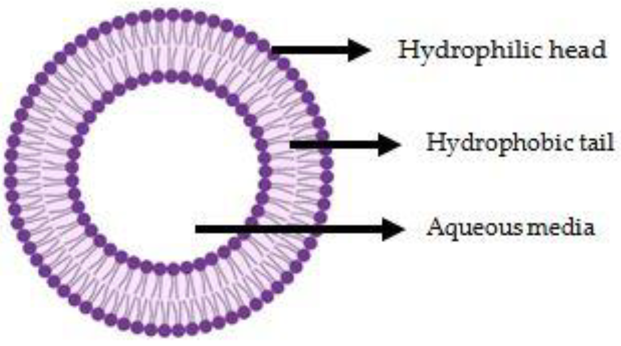
In order to boost the desired properties, such as targeting capability, enhanced selectivity, improved cell internalisation, extended period of exposure, minimised side effects, better therapeutic index, and active targeting, the liposomal delivery system evolves as a convenient and widespread strategic approach [6]. Liposomes became more prominent as they are able to encapsulate both hydrophilic and lipophilic drugs in an aqueous core and lipid bilayer, respectively, thus are appealing for drug loading [7]. The Figure 1 shows structure of conventional liposome.
Liposomes as drug carrier agents are designed to selectively localise therapeutic drugs to sick regions, such as tumours or inflammatory regions. Either passive or active targeting can be used to achieve selective localization [8].
- Active targeting—in active targeting, to increase contact with a targeted cellular membrane, certain targeting ligands are linked to the liposome surface [9]. Through receptor-mediated endocytosis (RME), often referred to as active drug targeting, those ligands bind to specific receptors on the surface of cells and encourage the internalisation of therapeutic compounds that operate on cellular organelles, such as mitochondria, microtubules, the nucleus, etc. [10].
- Passive Targeting—the primary reason liposomes are able to passively target tumour tissues is due to the differing pore diameters of tumour vascular endothelium as compared to the ‘tightened’ structures present within healthy capillaries. An optimal targeting aim would be attained if liposomes were prepared with a size that allows them to extravasate in tumour tissues, simultaneously preventing the carriers from exiting the capillaries in healthy tissue [11].
3. Conjugated Polymeric Liposomes
When the primary -C-C backbone of a compound has alternating sigma and pi bonds, the macromolecule is referred to as a conjugated polymer [12]. The term ‘‘Conjugated Polymeric Liposomes’’ applies to the hybrid carrier known as “polymer-modified liposomes” that have polymers bonded or attached to liposome surfaces. Moreover, the introduction of polymers helps to create liposomes with a decent condition, such as zeta potential, surface characteristics, particle characteristics and membrane flexibility [13].
By retrieving the features of every constituent material, conjugated polymers and liposomes enable the fabrication of surfaces with distinctive properties. Liposomes offer a soft template with a flexible surface chemistry, a prototype for the cell membrane, drug loading, encapsulation and efficient delivery [14]. Table 1 below mentions certain advantages of conjugated polymeric liposomes over conventional ones [15].
Various surface alterations offer various advantages. Liposome surface modifications and functionalization can significantly enhance the treatment of solid tumours and cancer. By modifying these liposomes, novel and effective anticancer drug delivery strategies are made possible. Due to their distinct physiology, treating malignant tumours with traditional therapeutic methods presents a significant challenge [16].
Liposomes can have their modification with polymers via at least one of the following methods: polymers grafted onto the liposome or coating the liposomes with physiosorbed polymers. Furthermore, by combining polymers with more distinct activity, multi-functional liposomes have also been developed [17].
3.1. “Grafted” Polymeric Liposomes
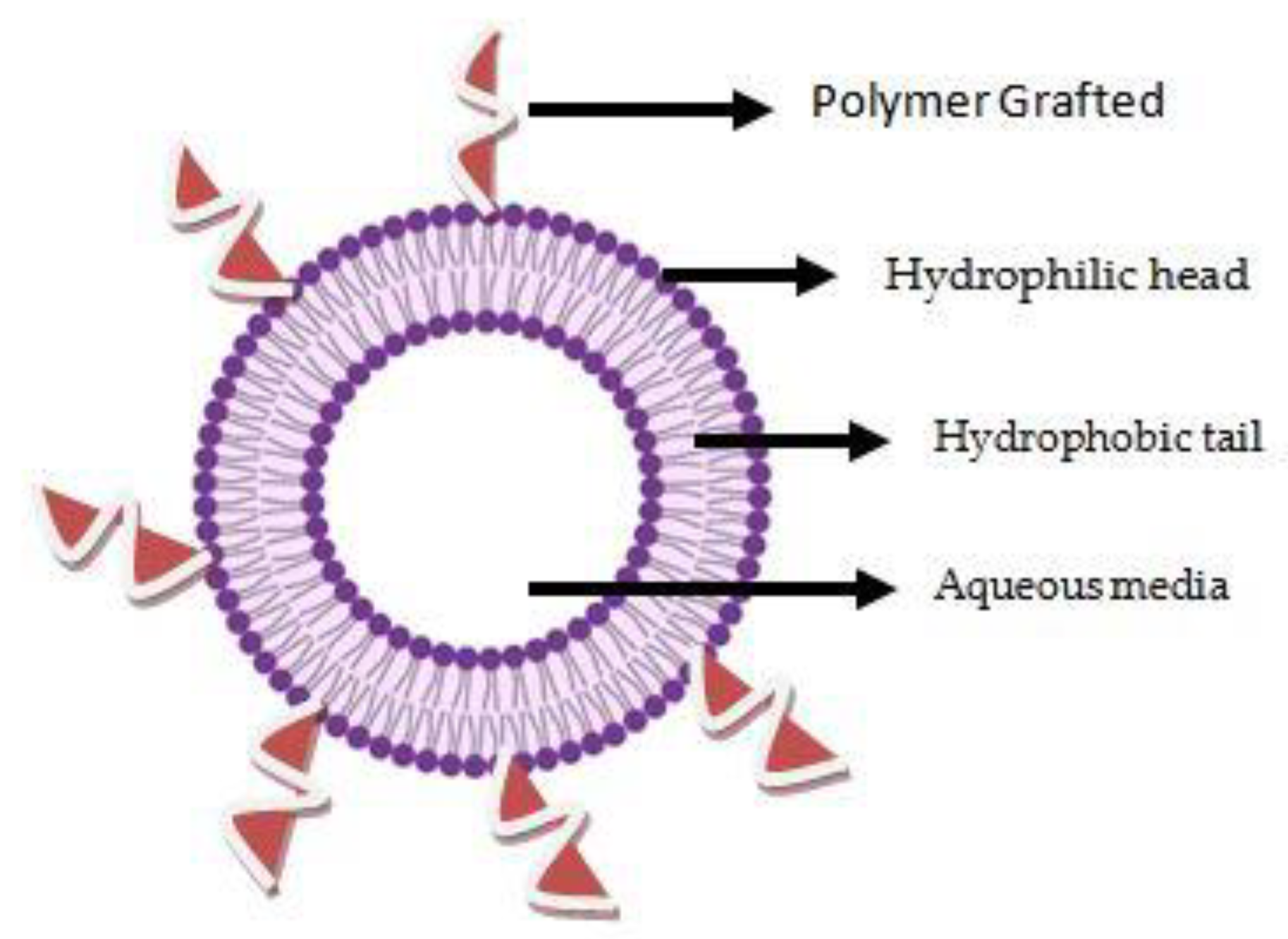
Most often, modifications of polymer chains are added to the surfaces of liposomes via a “grafting” technique [18]. Two concentration zones of polymer lipid content may be identified after the process of polymers grafting on solid substrates. These are distinguished by the grafted polymer chain’s so-called “mushroom” and “brush” forms. At low grafted polymer concentrations, the mushroom regime prevails, while at greater concentrations, the brush regime [19]. The Figure 2 shows structure of grafted polymeric liposome.
3.1.1. PEG (Polyethylene Glycol) Grafted Liposomes
A biocompatible addition is frequently performed with the inert, water-soluble polymer polyethylene glycol (PEG). PEG-polymers have been surface-grafted prevent protein adsorption and adherence to cellular interfaces. The PEG-lipids in liposomes prevent the adsorption of several immune system elements along with interactions between lipoproteins and lipolytic enzymes [20]. A lipid molecule when covalently bonded to a polymer of polyethylene glycol forms the stealth lipid. The addition of these stealth lipids gives the liposome the potential to persist in blood circulation for significantly longer time spans as compared to the behaviour of “conventional” liposomes [19]. Doxil®, which is approved for the treatment of Kaposi’s sarcoma, breast cancer, ovarian cancer, and multiple myeloma, is the most effective example of a PEG grafted liposomal formulation [21].
3.1.2. Zwitter Ion Grafted Liposome
Zwitter ionic materials are characterised by strong dipole moments and highly charged groups, and they also include an equal amount of both cationic and anionic moieties at the same time, preserving overall electrical neutrality and high hydrophilicity [22]. The ability of zwitterionic surface grafting to transfer stealth qualities to a polymer substrate in order to escape the body’s fibrotic and immunological reactions is well known for drug delivery applications. Bioseparations and functionalizing nanomaterials on porous structures in liquid chromatography are further uses of zwitterionic polymer-based surface grafting [23]. Chemotherapeutic (anticancer) agents, DNA, and protein have all been successfully delivered using zwitterionic drug carriers as a biomimicry technique. It should be emphasized that zwitterionic molecules should not only be taken into account as a substitute for PEG, but also because they provide some special qualities that are particularly advantageous for biomedical applications [24].
3.2. “Coated” Polymeric Liposomes
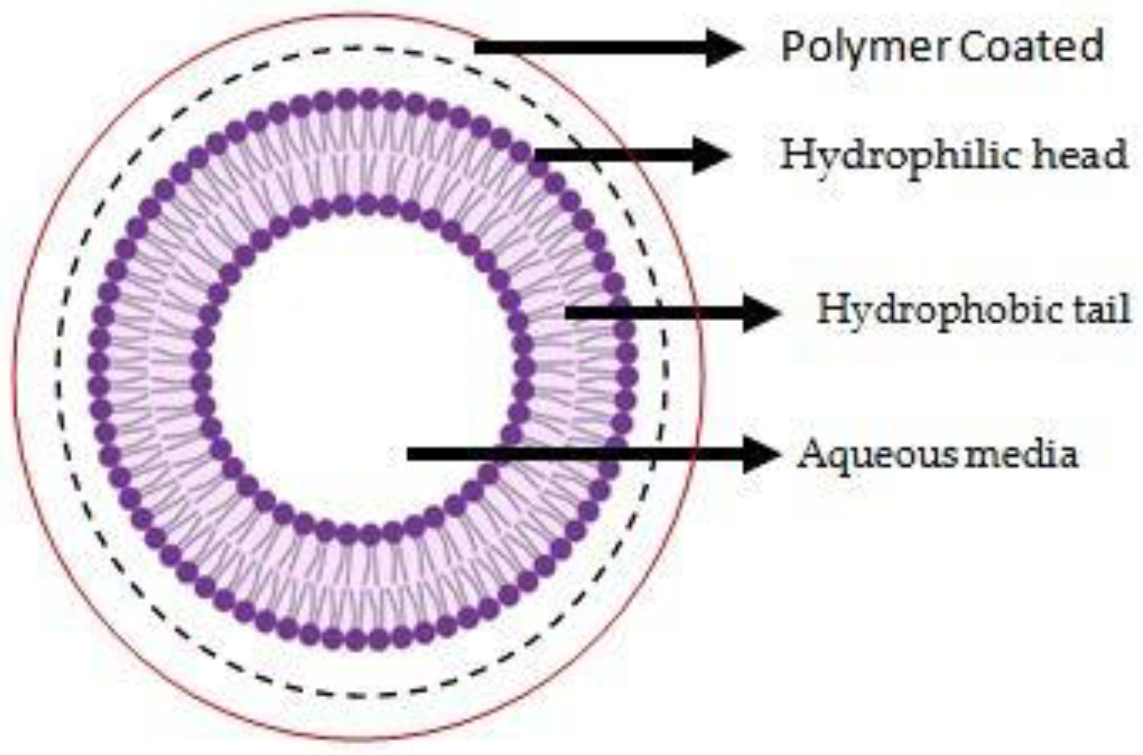
Simply blending the prepared liposomal suspension with a polymer solution produced polymer coated liposomes. The polymer concentration utilised in coating liposomes determined how much their particle size increased [25]. The coating of some hydrophilic and extensible polymers, with physiologically stable and long-circulating liposomes with the objective of drug delivery, has become one of the most well-known and effective techniques [26]. The electrostatic interaction between oppositely charged polyelectrolytes makes the layer by layer (LbL) approach a flexible and adaptable way for fabricating polyelectrolyte multilayer films. It can be used with many different templates, including liposomes [27]. The Figure 3 shows structure of coated polymeric liposome.
4. Application of Conjugated Polymeric Liposomes
4.1. Stimuli Responsive
In the absence of stimuli, the stimuli-responsive polymers should encapsulate medication securely and release them when the stimuli are present. Two types of stimuli can be regarded: either endogenous or exogenous stimuli [28]. Change in the pH, enzyme concentration, and redox gradient are examples of endogenous stimuli that are characteristic of the pathogenic aspects of disease. Exogenous stimuli are those that are purposely applied to the body from the outside, such as heat, light, electromagnetic field, etc. [29]. Thus, stimuli-responsive drug delivery has drawn a lot of attention because of its remarkable ability in adapting to the changed conditions of diseased tissues [30].
4.2. pH Sensitive
The protective effects of additional liposomal formulations with unique environment-controlled release of drug are accessible with pH-sensitive liposomes. There is proof that the alkylated N-isopropylacrylamide (NIPAM) co-polymer makes liposomes pH sensitive [31]. Continually putting pH-sensitive components to liposome dispersion or incorporating pH-sensitive lipids and polymers when creating vesicles can produce pH-sensitive liposomes. Such liposomes are stable at physiological pH but become unstable and develop fusogenic properties in the target tissue’s acidic environment, which effectively releases the aqueous encapsulated substance into the cytoplasm [32].
4.3. Thermosensitive
Due of the regulated and anticipated heating of tumours via external sources of energy, temperature sensitive vesicles, i.e., liposomes, are a highly appealing choice. Using lysolipids and artificial thermo-sensitive polymers(TSPs), temperature-sensitization of liposomes has been demonstrated [33]. The architecture of the polymer should also take into account how the TSPs will be used in the end product, for example, improving cellular activity in tissue regeneration scaffolds necessitates a non-invasive implant. TSPs exhibit either lower-critical solution temperature (LCST, greater than body temperature) or upper-critical solution temperature (UCST) phase behaviours depending on material selection policies and design conditions [34].
5. Conclusions
Using liposomes for drug delivery is a well-established field after a lot of progress. As research advances, methods for avoiding the constraints and drawbacks of conventional liposomes emerge. Thus, polymer modified liposomes have been demonstrated to be effective and convenient vesicles for systematic drug delivery. The requisite physiochemical performance aspects, such as surface chemistry, and functionality, should be rationally developed using polymers and lipids. To conclude, conjugated polymeric liposomes as hybrid drug carriers have a wide range of potential uses. However, further significant studies and efforts need to be undertaken in this arena for advance clinical translation.
Author Contributions
Conception, J.P. and T.G.; writing—original draft preparation, T.G.; writing—review and editing, J.P. and T.G.; resources, S.G.P.; supervision, H.S. and S.A.P. All authors have read and agreed to the published version of the manuscript.
Funding
This review received no external funding.
Institutional Review Board Statement
Not applicable.
Informed Consent Statement
Not applicable.
Data Availability Statement
Not applicable.
Acknowledgments
We would like to convey our obligation to Management and Principal, P.S.G.V.P. Mandal’s College of Pharmacy, Shahada, Dist. Nandurbar.
Conflicts of Interest
The authors declare no conflict of interest.
References
- Daraee, H.; Etemadi, A.; Kouhi, M.; Alimirzalu, S.; Akbarzadeh, A. Application of liposomes in medicine and drug delivery. Artif. Cells Nanomed. Biotechnol. 2016, 44, 381–391. [Google Scholar] [CrossRef] [PubMed]
- Akbarzadeh, A.; Rezaei-Sadabady, R.; Davaran, S.; Joo, S.W.; Zarghami, N.; Hanifehpour, Y.; Samiei, M.; Kouhi, M.; Nejati-Koshki, K. Liposome: Classification, preparation, and applications. Nanoscale Res. Lett. 2013, 8, 102. [Google Scholar] [CrossRef] [PubMed] [Green Version]
- Filipczak, N.; Pan, J.; Yalamarty, S.S.K.; Torchilin, V.P. Recent advancements in liposome technology. Adv. Drug Deliv. Rev. 2020, 156, 4–22. [Google Scholar] [CrossRef] [PubMed]
- Lian, T.; Ho, R.J.Y. Trends and Developments in Liposome Drug Delivery Systems. J. Pharm. Sci. 2001, 90, 667–680. [Google Scholar] [CrossRef]
- Sercombe, L.; Veerati, T.; Moheimani, F.; Wu, S.Y.; Sood, A.K.; Hua, S. Advances and Challenges of Liposome Assisted Drug Delivery. Front. Pharmacol. 2015, 6, 286. [Google Scholar] [CrossRef] [Green Version]
- Bardania, H.; Tarvirdipour, S.; Dorkoosh, F. Liposome-targeted delivery for highly potent drugs. Artif. Cells Nanomed. Biotechnol. 2017, 45, 1478–1489. [Google Scholar] [CrossRef]
- Kim, J.-S. Liposomal drug delivery system. J. Pharm. Investig. 2016, 46, 387–392. [Google Scholar] [CrossRef]
- Barenholz, Y. Liposome application: Problems and prospects. Curr. Opin. Colloid Interface Sci. 2001, 6, 66–77. [Google Scholar] [CrossRef]
- Metselaar, J.; Mastrobattista, E.; Storm, G. Liposomes for Intravenous Drug Targeting: Design and Applications. Mini-Rev. Med. Chem. 2002, 2, 319–329. [Google Scholar] [CrossRef]
- Rahman, M.; Kumar, V.; Beg, S.; Sharma, G.; Katare, O.P.; Anwar, F. Emergence of liposome as targeted magic bullet for inflammatory disorders: Current state of the art. Artif. Cells Nanomed. Biotechnol. 2016, 44, 1597–1608. [Google Scholar] [CrossRef]
- Alavi, M.; Hamidi, M. Passive and active targeting in cancer therapy by liposomes and lipid nanoparticles. Drug Metab. Pers. Ther. 2019, 34, 1–8. [Google Scholar] [CrossRef] [PubMed]
- Romero-García, J.; Ledezma-Pérez, A.; Martínez-Cartagena, M.; Alvarado-Canché, C.; Jiménez-Cárdenas, P.; De-León, A.; Gallardo-Vega, C. Radical addition polymerization: Enzymatic template-free synthesis of conjugated polymers and their nanostructure fabrication. In Methods in Enzymology; Elsevier: Amsterdam, The Netherlands, 2019; Volume 627, pp. 321–337. [Google Scholar] [CrossRef]
- Liu, Y.; Xie, X.; Chen, H.; Hou, X.; He, Y.; Shen, J.; Shi, J.; Feng, N. Advances in next-generation lipid-polymer hybrid nanocarriers with emphasis on polymer-modified functional liposomes and cell-based-biomimetic nanocarriers for active ingredients and fractions from Chinese medicine delivery. Nanomed. Nanotechnol. Biol. Med. 2020, 29, 02237. [Google Scholar] [CrossRef] [PubMed]
- Jephcott, L.; Eslami, M.; Travaglini, L.; Lauto, A.; Mawad, D. A conjugated polymer-liposome complex: A contiguous water-stable, electronic, and optical interface. View 2021, 2, 20200081. [Google Scholar] [CrossRef]
- Brandl, M. Liposomes as drug carriers: A technological approach. In Biotechnology Annual Review; Elsevier: Amsterdam, The Netherlands, 2001; Volume 7, pp. 59–85. [Google Scholar] [CrossRef]
- Milani, D.; Athiyah, U.; Hariyadi, D.M.; Pathak, Y.V. Surface Modifications of Liposomes for Drug Targeting. In Surface Modification of Nanoparticles for Targeted Drug Delivery; Pathak, Y.V., Ed.; Springer International Publishing: Cham, Switzerland, 2019; pp. 207–220. [Google Scholar] [CrossRef]
- Cao, Y.; Dong, X.; Chen, X. Polymer-Modified Liposomes for Drug Delivery: From Fundamentals to Applications. Pharmaceutics 2022, 14, 778. [Google Scholar] [CrossRef]
- Masuda, T.; Shimada, N.; Maruyama, A. Liposome-Surface-Initiated ARGET ATRP: Surface Softness Generated by ‘Grafting from’ Polymerization. Langmuir 2019, 35, 5581–5586. [Google Scholar] [CrossRef]
- Needham, D.; Hristova, K.; McIntosh, T.J.; Dewhirst, M.; Wu, N.; Lasic, D.D. Polymer-Grafted Liposomes: Physical Basis for the ‘Stealth’ Property. J. Liposome Res. 1992, 2, 411–430. [Google Scholar] [CrossRef]
- Marsh, D.; Bartucci, R.; Sportelli, L. Lipid membranes with grafted polymers: Physicochemical aspects. Biochim. Biophys. Acta BBA-Biomembr. 2003, 1615, 33–59. [Google Scholar] [CrossRef]
- Mohamed, M.; Abu Lila, A.S.; Shimizu, T.; Alaaeldin, E.; Hussein, A.; Sarhan, H.A.; Szebeni, J.; Ishida, T. PEGylated liposomes: Immunological responses. Sci. Technol. Adv. Mater. 2019, 20, 710–724. [Google Scholar] [CrossRef] [Green Version]
- Zhou, L.-Y.; Zhu, Y.H.; Wang, X.Y.; Shen, C.; Wei, X.W.; Xu, T.; He, Z.Y. Novel zwitterionic vectors: Multi-functional delivery systems for therapeutic genes and drugs. Comput. Struct. Biotechnol. J. 2020, 18, 1980–1999. [Google Scholar] [CrossRef]
- Erfani, A.; Seaberg, J.; Aichele, C.P.; Ramsey, J.D. Interactions between Biomolecules and Zwitterionic Moieties: A Review. Biomacromolecules 2020, 21, 2557–2573. [Google Scholar] [CrossRef]
- Jin, Q.; Chen, Y.; Wang, Y.; Ji, J. Zwitterionic drug nanocarriers: A biomimetic strategy for drug delivery. Colloids Surf. B Biointerfaces 2014, 124, 80–86. [Google Scholar] [CrossRef] [PubMed]
- Takeuchi, H.; Kojima, H.; Yamamoto, H.; Kawashima, Y. Evaluation of circulation profiles of liposomes coated with hydrophilic polymers having different molecular weights in rats. J. Control. Release 2001, 75, 83–91. [Google Scholar] [CrossRef] [PubMed]
- Torchilin, V.P. How do polymers prolong circulation time of liposomes? J. Liposome Res. 1996, 6, 99–116. [Google Scholar] [CrossRef]
- Fukui, Y.; Fujimoto, K. The Preparation of Sugar Polymer-Coated Nanocapsules by the Layer-by-Layer Deposition on the Liposome. Langmuir 2009, 25, 10020–10025. [Google Scholar] [CrossRef]
- Cheng, W.; Gu, L.; Ren, W.; Liu, Y. Stimuli-responsive polymers for anti-cancer drug delivery. Mater. Sci. Eng. C 2014, 45, 600–608. [Google Scholar] [CrossRef] [PubMed]
- Hatakeyama, H. Recent Advances in Endogenous and Exogenous Stimuli-Responsive Nanocarriers for Drug Delivery and Therapeutics. Chem. Pharm. Bull. 2017, 65, 612–617. [Google Scholar] [CrossRef] [PubMed] [Green Version]
- Chountoulesi, M.; Naziris, N.; Pippa, N.; Pispas, S.; Demetzos, C. Stimuli-responsive nanocarriers for drug delivery. In Nanomaterials for Clinical Applications; Elsevier: Amsterdam, The Netherlands, 2020; pp. 99–121. [Google Scholar] [CrossRef]
- Paliwal, S.R.; Paliwal, R.; Vyas, S.P. A review of mechanistic insight and application of pH-sensitive liposomes in drug delivery. Drug Deliv. 2015, 22, 231–242. [Google Scholar] [CrossRef] [PubMed]
- Liu, X.; Huang, G. Formation strategies, mechanism of intracellular delivery and potential clinical applications of pH-sensitive liposomes. Asian J. Pharm. Sci. 2013, 8, 319–328. [Google Scholar] [CrossRef] [Green Version]
- Ta, T.; Porter, T.M. Thermosensitive liposomes for localized delivery and triggered release of chemotherapy. J. Control. Release 2013, 169, 112–125. [Google Scholar] [CrossRef] [Green Version]
- Zarrintaj, P.; Jouyandeh, M.; Ganjali, M.R.; Hadavand, B.S.; Mozafari, M.; Sheiko, S.S.; Vatankhah-Varnoosfaderani, M.; Gutiérrez, T.J.; Saeb, M.R. Thermo-sensitive polymers in medicine: A review. Eur. Polym. J. 2019, 117, 402–423. [Google Scholar] [CrossRef]
Figure 1.
Conventional liposome.

Figure 2.
Grafted polymeric liposome.

Figure 3.
Coated polymeric liposomes.


Table 1.
Potential advantages of conjugated polymeric liposomes.
| Advantages | |
|---|---|
| Enhanced solubility | |
| Conjugated Polymeric Liposomes | Prolong circulation time |
| Improving immunological response | |
| Surpassing biological barriers | |
| Controlled release of drugs | |
| Greater payload |
Publisher’s Note: MDPI stays neutral with regard to jurisdictional claims in published maps and institutional affiliations. |
© 2022 by the authors. Licensee MDPI, Basel, Switzerland. This article is an open access article distributed under the terms and conditions of the Creative Commons Attribution (CC BY) license (https://creativecommons.org/licenses/by/4.0/).
Share and Cite
MDPI and ACS Style
Patil, J.; Girase, T.; Patil, S.G.; Suryawanshi, H.; Patil, S.A. Conjugated Polymeric Liposomes: A Hybrid Carrier for Contemporary Drug Delivery. Chem. Proc. 2022, 12, 12. https://doi.org/10.3390/ecsoc-26-13640
AMA Style
Patil J, Girase T, Patil SG, Suryawanshi H, Patil SA. Conjugated Polymeric Liposomes: A Hybrid Carrier for Contemporary Drug Delivery. Chemistry Proceedings. 2022; 12(1):12. https://doi.org/10.3390/ecsoc-26-13640
Chicago/Turabian StylePatil, Javesh, Tejasweeni Girase, Sulbha G. Patil, Hemant Suryawanshi, and Sunila A. Patil. 2022. "Conjugated Polymeric Liposomes: A Hybrid Carrier for Contemporary Drug Delivery" Chemistry Proceedings 12, no. 1: 12. https://doi.org/10.3390/ecsoc-26-13640