
Solid-State Electrochemical Energy Storage Based on Soluble Melanin


1
Postgraduate Program in Science and Technology of Materials (POSMAT), School of Sciences, São Paulo State University (UNESP), 17033-360 Bauru, Brazil
2
Department of Physics, School of Sciences, São Paulo State University (UNESP), 17033-360 Bauru, Brazil
*
Author to whom correspondence should be addressed.
Electrochem 2021, 2(2), 264-273; https://doi.org/10.3390/electrochem2020019
Submission received: 14 April 2021
/
Revised: 20 May 2021
/
Accepted: 21 May 2021
/
Published: 25 May 2021
(This article belongs to the Collection Feature Papers in Electrochemistry)
Abstract
:Biocompatible and biodegradable powering materials are appealing systems for biomedical and electronic devices. Melanin is a natural and multifunctional material with redox capability, which is of great interest in electrochemical energy storage functionalities. In our work, we explored the use of soluble melanin derivatives as active materials for symmetric solid-state supercapacitors operating in the dark and under illumination. We observed that our devices were photo-pseudocapacitive. Additionally, under illumination, our best device showed a specific capacitance of 57.7 mFg−1 at a scan rate of 0.01 Vs−1, with a decrease of 53% in resistance compared to that in the dark. Our outcome suggests that soluble melanin is a promising material for solid-state powering elements in wearable and environmentally friendly devices.
1. Introduction
Electronic devices to constantly monitor patients’ health conditions or common citizens will be extremely important in the 21st century [1]. Such devices comprise a sensing element capable of measuring a physiological quantity and converting it into an electronic signal to be transmitted to another apparatus for analysis and interpretation [2]. Both sensing and transmission processes need to be powered by a battery or a supercapacitor [2,3,4,5,6]. Among various materials for electrochemical energy storage applications, melanin and melanin-inspired materials have received increased attention in recent years. The biocompatibility and biodegradability properties of melanin associated with its redox activity have enabled the development of Na+ and Mg2+ batteries [7,8] and supercapacitors [9,10,11,12,13,14] for biomedical and green electronics applications.
Even though several studies use melanin in energy storage applications, most use it to increase the content of nitrogen, oxygen, or sulfur after carbonization and/or calcination [13,14]. Nonetheless, nowadays, few reports in the literature have studied truly melanin-based supercapacitors, and such devices operate in aqueous electrolytes [9,11], implying that it is time to evaluate melanin’s efficiency in the solid state.
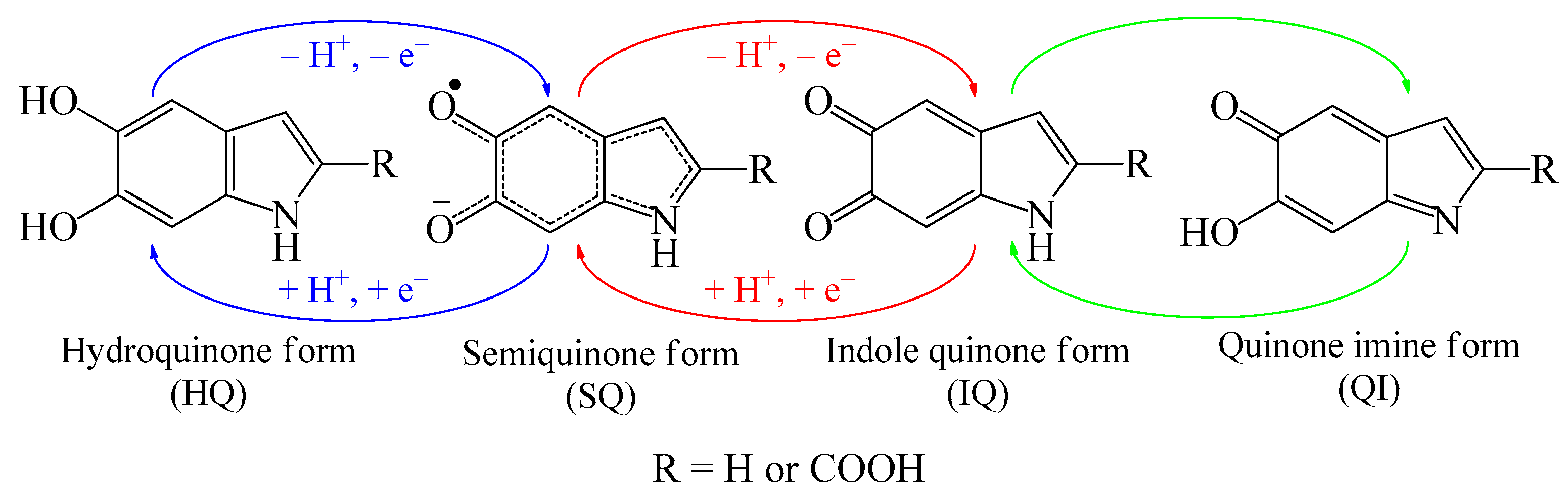
Generally, melanin is described as a system based on 5,6-dihydroxyindole (DHI) and 5,6-dihydroxyindole-2-carboxylic acid (DHICA) in fully reduced (HQ), fully oxidized (IQ), and semi-reduced/semi-oxidized (SQ and QI) states (Figure 1). Up to ten of these monomers self-assemble into 3 to 4 π−π-stacked units of different sizes with an interplanar spacing of 3.7 Å [15,16,17]. This molecular arrangement yields unique physicochemical properties such as biocompatibility [18,19], biodegradability [18], metal-binding affinity [20], a broad-band UV-Vis absorbance [15,21], and a hydration-dependent conductivity [22,23,24,25,26,27].
In this work, we explored two soluble melanin derivatives’ electrochemical energy storage properties with different oxidation states by assembling a solid-state supercapacitor. Additionally, due to the broad-band absorption spectra of melanin, we also evaluated light effects on its energy storage capacity.
2. Materials and Methods
2.1. Melanin Derivatives
Soluble melanin derivatives were synthesized following [21] by dissolving 0.3 g of 3,4-dihydroxyphenyl-DL-alanine (Sigma-Aldrich, 98%, São Paulo, Brazil) in 60 mL of MiliQ water (18 MΩ cm). To increase the solution pH to 8–9, the condition for melanin synthesis, 400 µL of ammonium hydroxide (Synth, 28–30%) was added to the mixture. The synthesis solution was stirred at room temperature (±27 °C) and was oxygenated with an air pump for three days (Mel) or in a 150 mL stainless-steel reactor with 6 atm of internal pressure of industrial oxygen gas for 6 h (Mel-P). Afterward, the solution was filtered using a 3500 MWCO dialysis membrane for four days. The dialysate medium was MiliQ water and it was changed every day. After that, the suspended colloidal particles were dried for two days in an oven at 80 °C. These samples had identical polymerization structures but different DHICA/DHI ratios, i.e., oxidation degrees (Mel < Mel-P) [21].
To confirm the composition of the samples, UV-vis spectroscopy and elemental analysis were performed via X-ray photoelectron spectroscopy. The absorbance spectra and atomic composition (atomic concentration and atomic ratio) are shown in Figure S1 and Table S1, respectively, in Supplementary Materials, and they are compatible with melanin derivatives [21,28]. For more details, see the Supplementary Materials.
2.2. Supercapacitor Assembly and Characterization
Melanin solutions were prepared in ambient conditions by dissolving 30 mg of each sample in 0.5 mL of MiliQ water and by stirring for one hour. Afterward, 1 mL of multi-walled carbon nanotube (MWCNT) solution (0.6 mg/mL in water) was added as a conductive filler and was stirred for another hour. We considered bare melanin solutions (i.e., without MWCNT); however, it presented poor capacitor behaviors (Figure S2) and no further analysis was carried out.
Prior to melanin drop-casting deposition (60 μL; equivalent to a material load of 1.25 mg·cm−2 for Mel and 1.22 mg·cm−2 for Mel-P), the FTO substrates were cleaned with soap (1:1 v%, Extran®:MiliQ water), a sequential sonication (20 min/each) in MiliQ water, acetone, and isopropanol, and were dried with N2. The melanin film was dried for half an hour at 110 °C, under atmospheric conditions, and was allowed to cool down to room temperature (±27 °C). Then, polyvinyl alcohol-phosphoric acid (PVA-H3PO4) gel electrolyte (10% PVA and H3PO4 in H2O) was used to cover the 1 cm2 area of the melanin film and as a spacing layer of a symmetric cell configuration (glass/FTO/Melanin:MWCNT/PVA-H3PO4-gel/Melanin:MWCNT/FTO/glass).
Surface morphological analysis was investigated through scanning electron microscope (SEM) images obtained with a Zeiss EVO/LS15.
The supercapacitor’s electrochemical performance was obtained using a Metrohm Autolab Potentiostat (Autolab PGSTAT302 equipped with a FRA32 impedance module) in a two-electrode cell through cyclic voltammetry and galvanostatic charge/discharge measurements. Electrochemical impedance measurements were conducted within the frequency range of 103 and 10−2 Hz and an offset voltage of 0 V. Impedance spectra were analyzed with ZView-impedance Software (v. 2.8d). The measurements were carried out under dark and light (100 mW/cm2 simulated AM 1.5 G solar irradiation from a calibrated solar simulator, Spectra-Nova) conditions.
The specific capacitance (C, in Fg−1) was calculated using Equation (1), the maximum energy (EM, in mWhg−1) was calculated using Equation (2), and the maximum power (PM, in mWg−1) was calculated using Equation (3) [29],
where Q is the total charge (C), is the active material loading (g), is the scan rate (Vs−1), and and are the negative and positive electrode electrochemical potentials, respectively. is the constant discharge current, is the cutoff potential (the charging up-limited potential), and represents the discharge . ESR (the equivalent series resistance) was calculated by the ratio between the drop voltage () from the galvanostatic charge/discharge spectra and , through the relation .
3. Results and Discussion
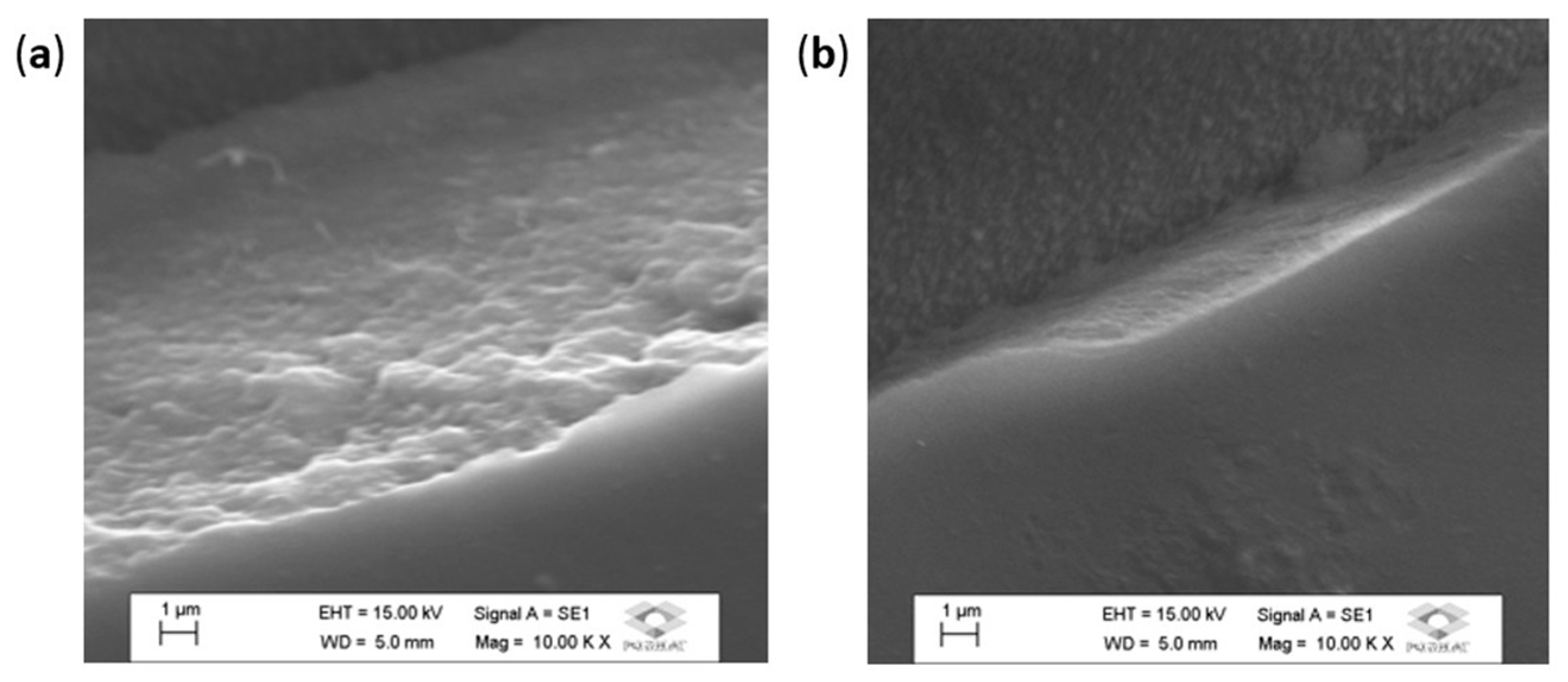
We start with SEM images to give insights into the superficial properties of the films. Mel and Mel-P images are shown in Figure 2. As can be seen, the surface morphological aspects of the films were featureless, smooth, and reasonably homogeneous, independent of the sample, as demonstrated for many melanin films [30,31,32,33,34], implying that any textural effect would be minimal or nonexistent. However, there were significant modifications in the film cross-section. In Figure 2a, the Mel films were composed of large globular structures, whereas Mel-P elongated structures were in an almost lamellar-like assembly (Figure 2b). These differences are related to the different degrees of oxidation mentioned in Section 2.1. Mel-P, a high oxidated sample, should present a higher content of the carboxyl group. Thus, the decrease in the polymerization sites in Mel-P is expected to lead to the formation of a more linear oligomer than Mel [19,34,35]. Based on the morphological aspects of the film, we would expect Mel to a have higher superficial area than Mel-P, which, in turn, would result in a higher interaction of the sample and the electrolyte. In addition, we note that the oxygen and nitrogen content were close in both melanin samples (Table S1), which means that differences in the morphological aspects would be more important than the atomic composition in the devices under study. It is also interesting to mention that, in the absence of MWCNT, such cross-sectional differences were subtle (Figure S3), indicating a possible cause for the poor energy storage effect of pure melanin samples.
The electrochemical behavior of melanin samples was evaluated in a two-electrode system in the dark, followed by an illuminating process. We note here that the same cell was used under both dark and illumination conditions to decrease errors caused by experimental manipulation; in addition, before illumination, the cell was discharged, eliminating any charge accumulated from the previous process. A slightly acidic electrolyte was used to enhance melanin’s proton transport [25,34]. Figure 3a,b show the cyclic voltammetry curves of Mel and Mel-P samples at scan rates ranging from 0.01 to 1.00 Vs−1 in the dark. These curves of both melanin-based devices were quasi-rectangular between 0 and 0.8 V, which is typical of pseudocapacitance [9,11]. In addition, there was no change in the cyclic voltammetry spectra, even at higher scan rates. We note that the spectra shape did not significantly change with illumination, as shown in Figure 3c,d, at a scan rate of 0.01 Vs−1. For other scan rates, see Figure S4. Among these samples, Mel had a tendency to present a higher specific capacitance (57.7 ± 6.3 mFg−1) compared to Mel-P (49.9 ± 5.4 mFg−1) at a scan rate of 0.01 Vs−1 under illumination. These values were 36.7 and 44.5% higher than those in the dark for Mel and Mel-P, respectively. In the same line, we also observed a decrease in specific capacitance with the increase in scan rate (Figure S5), which is compatible with melanin’s pseudocapacitive behavior [9]. Nonetheless, the supercapacitors, operating in an aqueous electrolyte, showed a specific capacitance of 167 Fg−1 [9]. Such a difference is most likely due to the different state of the electrolyte, i.e., wet vs. dry, as it is known that melanin capacitance increases in water [25,26].
The galvanostatic charge/discharge of Mel and Mel-P at a current density of 0.1 mAg−1 were also measured, and the results are shown in Figure 4. Note that our curves could not reach 0.8 V, contrary to the cyclic voltammograms, due to a saturation effect (see Figure S6). This effect is intriguing and will be studied in future work. Figure 4 shows that the galvanostatic charge/discharge curves slightly deviated from the nearly triangular-like shape; however, all samples showed similar charge and discharge behaviors, shapes, and characteristic times. Analyzing the spectra in Figure 4, it is possible to see a small drop in the voltage at the beginning of the discharge, which indicates low equivalent series resistance. Based on Equations (2) and (3), we estimated an increase of 14.1% in energy density and 5.4% in power density for Mel; and increases of 10.3% and 1.5% in energy and power densities, respectively, for Mel-P (Figure 4c,d). A previous DHI/DHICA-melanin supercapacitor operating in aqueous electrolytes showed an energy density of 7.7 μWhg−1 and a power density of 5.6 Wg−1 [11], which indicate that our solid-state system was about three orders of magnitude lower than wet supercapacitors in energy and power densities. The better results of the supercapacitor with the aqueous electrolyte could be related to the lower amount of melanin used in the active layer, which would decrease light scattering and intrinsic charge trap sites and the increased number of ionic charge carriers due to the high humidity [24,25,27].
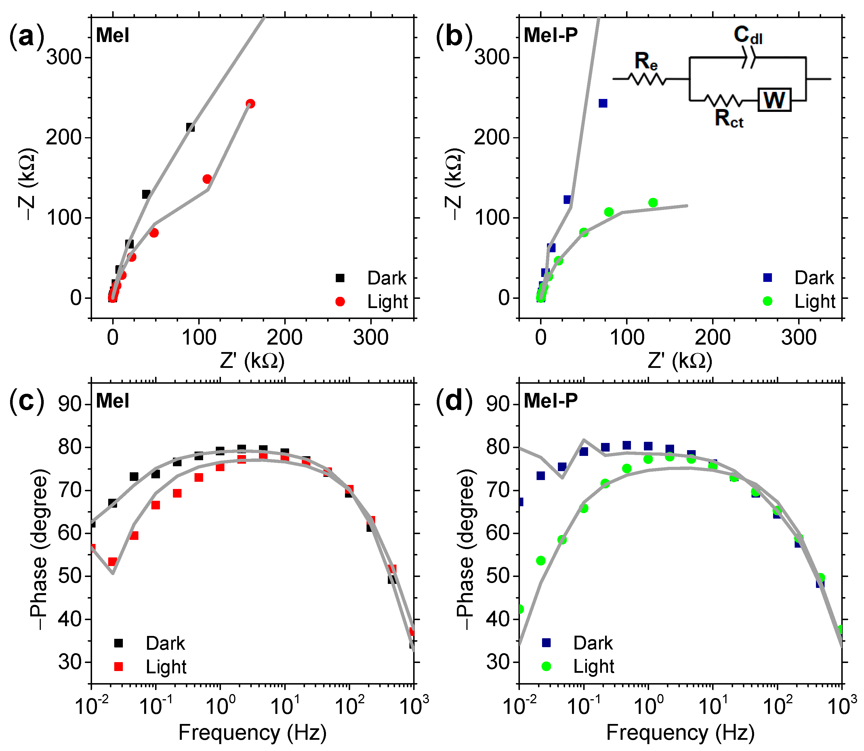
To evaluate the pseudocapacitive behavior of our devices, charge-storage capacity and electrochemical impedance measurements were carried out. Figure 5 shows the Nyquist and phase angle plots and the equivalent circuit (inset in Figure 5b). Re, which represents the electrode and electrolyte resistance, is in series with a charge-transfer resistance Rct, which describes the kinetics of the electron-transfer processes [36,37]. The equivalent circuit also includes a constant phase (Cdl) and Warburg (W) elements to account for the system’s capacitive response and any diffusion process involved, respectively [36,37]. The fitting parameters are shown in Table S2. The constant phase element’s impedance is defined as ZQ = Q−1 (jω)−η, where ω is the angular frequency and η is a constant and fitting parameter [36]. A pure capacitor corresponds to η = 1. We obtained η = 0.89 for Mel and η = 0.87 for Mel-P in the dark, with a reduction of 2.4% when the device was illuminated, implying that the light did not destroy the capacitive behavior. This is further verified by the small changes in the phase angle (Figure 5c,d) and cyclic voltammograms (Figure 3). However, we see a significant drop in the system’s resistance of about 52.8% for Mel and 78.7% for Mel-P under illumination, suggesting an improvement in the conductivity. On the other hand, illumination also decreased the leak resistance by 61.9% for Mel and 98.5% for Mel-P (Figure S7).
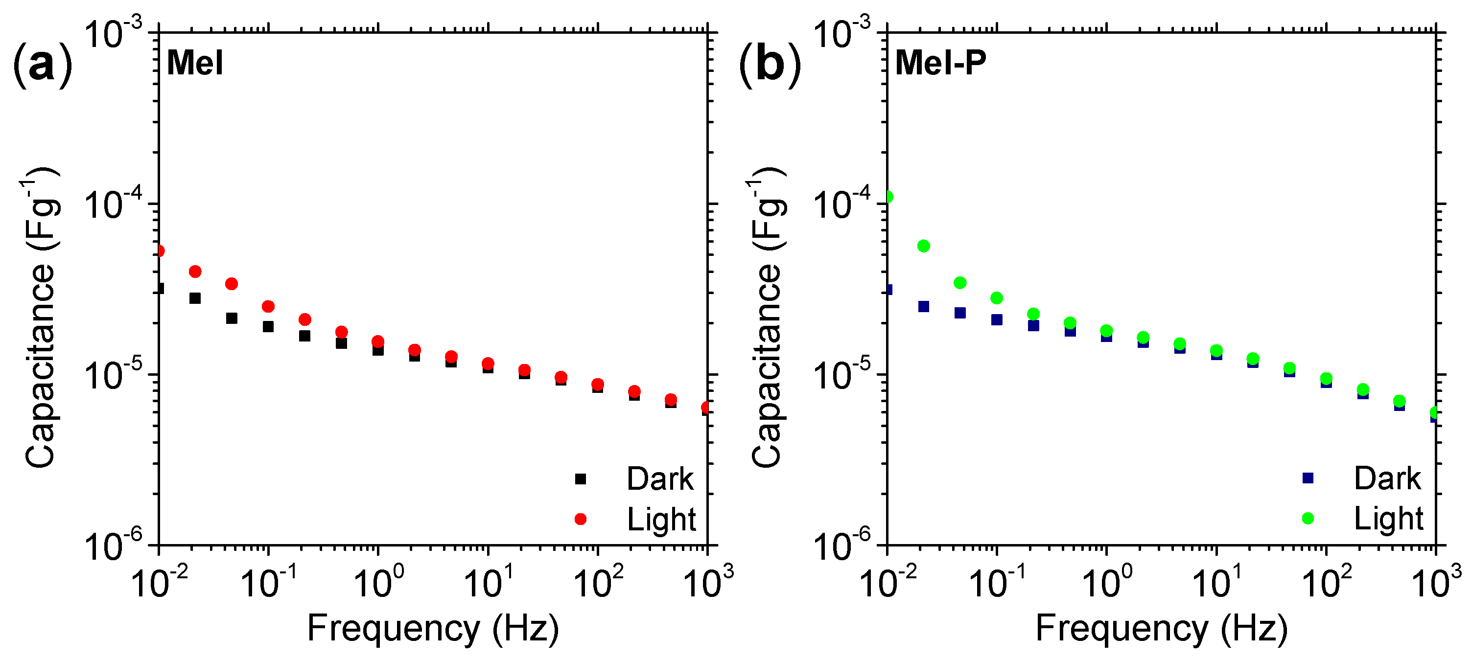
To obtain more information on the device’s intrinsic charge transport, we evaluated the capacitance as a function of frequency. In Figure 6, we observed a capacitance maximum at low frequency. We note that in the dark, both melanin samples had similar frequency dependences. On the other hand, when illuminated, the capacitance intensity increased at frequencies ranging from 10−2 to around 100 Hz, as there were increases of 65.5% for Mel and 250.2% for Mel-P at 10−2 Hz. At lower frequencies, the ions’ transport tended to dominate charge transport, indicating an increase in the number of ions in the system. It is known that light can induce the formation of semiquinone species in melanin [38,39]. Hence, the redox process will donate H+ and electrons to the system (Figure 1) and potentially alter the charge transport mechanisms, as mentioned before. Accordingly, the increase in capacitance would be related to photoinduced charge carriers, as well as changes in the electrochemical processes at the electrolyte/electrode interface [11].
Our results indicate that soluble melanin with a higher density of carboxylic groups (Mel-P) could be an exciting material for solid-state charge storage applications due to its fast synthesis and energy storage similar to Mel. Additionally, light also seems to improve the electronic behavior in the supercapacitor studied, thus being an attractive effect for such applications. At this stage, our solid-state supercapacitor has a lower efficiency than others found in the literature [29,40,41,42,43,44] (none of them being melanin-based devices), which could potentially be related to the melanin’s lower ionic current at a dry state [26]. We believe that optimizing the device could improve the system’s response, whether with other types of electrolytes, a better biocompatible conductive filler, or even different types of melanin. In fact, it has been proposed that S,N-codoped melanin-like carbon spheres significantly increase their specific capacitance in regard to N-doped melanin-like carbon spheres [13], which suggests that soluble sulfonated-melanin derivatives [45,46,47] are attractive candidates for energy storage applications and must be explored in the future. Additionally, this technology can be extrapolated toward flexible substrates, which would be a significant advance for flexible/stretchable wearable devices.
4. Conclusions
The work described here report the use of soluble melanin derivates as organic redox materials to develop solid-state supercapacitors. Our devices demonstrated photo-pseudocapacitive behaviors, with a 57.7 mFg−1 specific capacitance at a scan rate of 0.01 Vs−1 when illuminated, with improved electronic behaviors. We believe that these encouraging results could be improved by further optimizing the electrolyte and melanin derivate-type materials and extrapolating to the development of a flexible device. Our outcome suggests that soluble melanin could be used for solid-state biocompatible powering elements, wearable devices, and even sustainable and environmentally friendly storage technologies.
Supplementary Materials
The following are available online at https://www.mdpi.com/article/10.3390/electrochem2020019/s1: Figure S1. Mel and Mel-P absorbance spectra were obtained with a 7.0 μg·mL−1 solution in water. The absorbance of both derivatives is characterized by a featureless broad-band spectrum that increases toward the ultraviolet. UV–visible measurements were made using a Shimadzu UVmini-1240. Figure S2. Cyclic voltammogram spectra at different scan rates in the dark. Figure S3. SEM images of (a) Mel and (b) Mel-P without MWCNT. Figure S4. Cyclic voltammogram spectra at different scan rates under illumination for (a) Mel and (b) Mel-P. Figure S5. Specific capacitance as a function of scan rate of (a) Mel and (b) Mel-P electrodes. Figure S6. Extrapolation of the galvanostatic charge over time. These curves were obtained by fitting the ex-perimental galvanostatic charge curves in Figure 3a,b (in the main text) with an exponential func-tion. The fitting parameters were then used as fixed specs to estimate the curve as time increases. Figure S7. Estimation of the supercapacitor’s leakage current. The symbols are the experimental data and the lines are the estimated leakage current. Experimental fitting data (in 10−2 to 103 Hz) were considered as initial parameters in impedance software to further extrapolate at frequencies ranging from 10−10 to 106 Hz. Table S1. Atomic composition (atomic concentration %) and atomic ratios of Mel and Mel-P powders. The similarities between experimental and theoretical values indicate that Mel and Mel-P are mixtures of DHI and DHICA species, as expected. Table S2. Fitting parameters used on the equivalent circuit displayed in the inset of Figure 4b.
Author Contributions
Conceptualization, J.V.P. and S.L.F.; Methodology, J.V.P. and S.L.F.; Validation, J.V.P. and S.L.F.; Formal Analysis, J.V.P. and S.L.F.; Investigation, S.L.F.; Resources, J.V.P. and S.L.F.; Data Curation, J.V.P. and S.L.F.; Writing—Original Draft Preparation, J.V.P., S.L.F. and C.F.O.G.; Writing—Review and Editing, J.V.P., S.L.F. and C.F.O.G.; Visualization, J.V.P.; Supervision, C.F.O.G.; Project Administration, C.F.O.G.; Funding Acquisition, C.F.O.G. All authors have read and agreed to the published version of the manuscript.
Funding
This research was funded by São Paulo Research Foundation (FAPESP, grants: 2013/07296-2 and 2015/23000-1).
Institutional Review Board Statement
Not applicable.
Informed Consent Statement
Not applicable.
Data Availability Statement
Data available on request from the authors.
Acknowledgments
We thank M. L. M. Rocco (Institute of Chemistry of Federal University of Rio de Janeiro) for helping obtain the XPS data, and Rafael Aparecido da Silva for his assistance with SEM measurements.
Conflicts of Interest
The authors declare no conflict of interest.
References
- Olson, S. National Academy of Engineering, Grand Challenges for Engineering: Imperatives, Prospects, and Priorities: Summary of a Forum; National Academies Press: Washington, DC, USA, 2016. [Google Scholar]
- Chen, X.; Villa, N.S.; Zhuang, Y.; Chen, L.; Wang, T.; Li, Z.; Kong, T. Stretchable Supercapacitors as Emergent Energy Storage Units for Health Monitoring Bioelectronics. Adv. Energy Mater. 2020, 10, 1902769. [Google Scholar] [CrossRef]
- Ohayon, D.; Inal, S. Organic Bioelectronics: From Functional Materials to Next-Generation Devices and Power Sources. Adv. Mater. 2020, 32, 2001439. [Google Scholar] [CrossRef]
- Li, L.; Wang, L.; Ye, T.; Peng, H.; Zhang, Y. Stretchable Energy Storage Devices Based on Carbon Materials. Small 2021, 2005015. [Google Scholar] [CrossRef] [PubMed]
- Vallem, V.; Sargolzaeiaval, Y.; Ozturk, M.; Lai, Y.C.; Dickey, M.D. Energy Harvesting and Storage with Soft and Stretchable Materials. Adv. Mater. 2021, 33, 2004832. [Google Scholar] [CrossRef]
- Tong, X.; Tian, Z.; Sun, J.; Tung, V.; Kaner, R.B.; Shao, Y. Self-healing flexible/stretchable energy storage devices. Mater. Today 2021, 44, 78–104. [Google Scholar] [CrossRef]
- Kim, Y.J.; Wu, W.; Chun, S.; Whitacre, J.F.; Bettinger, C.J. Biologically derived melanin electrodes in aqueous sodium-ion energy storage devices. Proc. Natl. Acad. Sci. USA 2013, 110, 20912–20917. [Google Scholar] [CrossRef] [Green Version]
- Kim, Y.J.O.; Wu, W.; Chun, S.E.; Whitacre, J.F.; Bettinger, C.J. Catechol-mediated reversible binding of multivalent cations in eumelanin half-cells. Adv. Mater. 2014, 26, 6572–6579. [Google Scholar] [CrossRef]
- Kumar, P.; Di Mauro, E.; Zhang, S.; Pezzella, A.; Soavi, F.; Santato, C.; Cicoira, F. Melanin-based flexible supercapacitors. J. Mater. Chem. C 2016, 4, 9516–9525. [Google Scholar] [CrossRef]
- Cheng, F.; Liu, W.; Zhang, Y.; Wang, H.; Liu, S.; Hao, E.; Zhao, S.; Yang, H. Squid inks-derived nanocarbons with unique “shell@pearls” structure for high performance supercapacitors. J. Power Sources 2017, 354, 116–123. [Google Scholar] [CrossRef]
- Xu, R.; Gouda, A.; Caso, M.F.; Soavi, F.; Santato, C. Melanin: A Greener Route to Enhance Energy Storage under Solar Light. ACS Omega 2019, 4, 12244–12251. [Google Scholar] [CrossRef] [Green Version]
- Ajjan, F.N.; Mecerreyes, D.; Inganäs, O. Enhancing Energy Storage Devices with Biomacromolecules in Hybrid Electrodes. Biotechnol. J. 2019, 14, 1900062. [Google Scholar] [CrossRef]
- Yang, L.; Gu, B.; Chen, Z.; Yue, Y.; Wang, W.; Zhang, H.; Liu, X.; Ren, S.; Yang, W.; Li, Y. Synthetic Biopigment Supercapacitors. ACS Appl. Mater. Interfaces 2019, 11, 30360–30367. [Google Scholar] [CrossRef]
- Yang, L.; Guo, X.; Jin, Z.; Guo, W.; Duan, G.; Liu, X.; Li, Y. Emergence of melanin-inspired supercapacitors. Nano Today 2021, 37, 101075. [Google Scholar] [CrossRef]
- Meredith, P.; Sarna, T. The physical and chemical properties of eumelanin. Pigment Cell Res. 2006, 19, 572–594. [Google Scholar] [CrossRef]
- D’Ischia, M.; Wakamatsu, K.; Napolitano, A.; Briganti, S.; Garcia-Borron, J.-C.; Kovacs, D.; Meredith, P.; Pezzella, A.; Picardo, M.; Sarna, T.; et al. Melanins and melanogenesis: Methods, standards, protocols. Pigment Cell Melanoma Res. 2013, 26, 616–633. [Google Scholar] [CrossRef]
- d’Ischia, M.; Napolitano, A.; Pezzella, A.; Meredith, P.; Buehler, M.J. Melanin biopolymers: Tailoring chemical complexity for materials design. Angew. Chem. Int. Ed. 2020, 59, 11196–11205. [Google Scholar] [CrossRef]
- Bettinger, C.J.; Bruggeman, J.P.; Misra, A.; Borenstein, J.T.; Langer, R. Biocompatibility of biodegradable semiconducting melanin films for nerve tissue engineering. Biomaterials 2009, 30, 3050–3057. [Google Scholar] [CrossRef] [Green Version]
- Piacenti-Silva, M.; Matos, A.A.; Paulin, J.V.; Alavarce, R.A.d.S.; de Oliveira, R.C.; Graeff, C.F. Biocompatibility investigations of synthetic melanin and melanin analogue for application in bioelectronics. Polym. Int. 2016, 65, 1347–1354. [Google Scholar] [CrossRef]
- Di Mauro, E.; Xu, R.; Soliveri, G.; Santato, C. Natural melanin pigments and their interfaces with metal ions and oxides: Emerging concepts and technologies. MRS Commun. 2017, 7, 141–151. [Google Scholar] [CrossRef] [Green Version]
- Bronze-Uhle, E.S.; Paulin, J.V.; Piacenti-Silva, M.; Battocchio, C.; Rocco, M.L.M.; Graeff, C.F.D.O. Melanin synthesis under oxygen pressure. Polym. Int. 2016, 65, 1339–1346. [Google Scholar] [CrossRef]
- Jastrzebska, M.M.; Isotalo, H.; Paloheimo, J.; Stubb, H. Electrical conductivity of synthetic DOPA-melanin polymer for different hydration states and temperatures. J. Biomater. Sci. Polym. Ed. 1995, 7, 577–586. [Google Scholar] [CrossRef]
- Mostert, B.; Powell, B.J.; Gentle, I.R.; Meredith, P. On the origin of electrical conductivity in the bio-electronic material melanin. Appl. Phys. Lett. 2012, 100, 093701. [Google Scholar] [CrossRef]
- Mostert, A.B.; Powell, B.J.; Pratt, F.L.; Hanson, G.R.; Sarna, T.; Gentle, I.R.; Meredith, P. Role of semiconductivity and ion transport in the electrical conduction of melanin. Proc. Natl. Acad. Sci. USA 2012, 109, 8943–8947. [Google Scholar] [CrossRef] [Green Version]
- Wünsche, J.; Deng, Y.; Kumar, P.; Di Mauro, E.; Josberger, E.; Sayago, J.; Pezzella, A.; Soavi, F.; Cicoira, F.; Rolandi, M.; et al. Protonic and Electronic Transport in Hydrated Thin Films of the Pigment Eumelanin. Chem. Mater. 2015, 27, 436–442. [Google Scholar] [CrossRef]
- Sheliakina, M.; Mostert, A.B.; Meredith, P. Decoupling Ionic and Electronic Currents in Melanin. Adv. Funct. Mater. 2018, 28, 1805514. [Google Scholar] [CrossRef] [Green Version]
- Reali, M.; Saini, P.; Santato, C. Electronic and protonic transport in bio-sourced materials: A new perspective on semiconductivity. Mater. Adv. 2021, 2, 15–31. [Google Scholar] [CrossRef]
- Paulin, J.V.; Mcgettrick, J.D.; Graeff, C.F.O.; Mostert, A.B. Melanin system composition analyzed by XPS depth profiling. Surf. Interface 2021, 24, 101053. [Google Scholar] [CrossRef]
- Akbulut, S.; Yilmaz, M.; Raina, S.; Hsu, S.H.; Kang, W.P. Solid-state supercapacitor cell based on 3D nanostructured MnO2/CNT microelectrode array on graphite and H3PO4/PVA electrolyte. Diam. Relat. Mater. 2017, 74, 222–228. [Google Scholar] [CrossRef]
- Bothma, J.P.; de Boor, J.; Divakar, U.; Schwenn, P.E.; Meredith, P. Device-Quality Electrically Conducting Melanin Thin Films. Adv. Mater. 2008, 20, 3539–3542. [Google Scholar] [CrossRef]
- Abbas, M.; D’Amico, F.; Morresi, L.; Pinto, N.; Ficcadenti, M.; Natali, R.; Ottaviano, L.; Passacantando, M.; Cuccioloni, M.; Angeletti, M.; et al. Structural, electrical, electronic and optical properties of melanin films. Eur. Phys. J. E 2009, 28, 285–291. [Google Scholar] [CrossRef]
- Wünsche, J.; Cicoira, F.; Graeff, C.F.O.; Santato, C. Eumelanin thin films: Solution-processing, growth, and charge transport properties. J. Mater. Chem. B 2013, 1, 3836–3842. [Google Scholar] [CrossRef] [PubMed] [Green Version]
- Piacenti-Silva, M.; Fernandes, J.C.; de Figueiredo, N.B.; Congiu, M.; Mulato, M.; Graeff, C.F.d.O. Melanin as an active layer in biosensors. AIP Adv. 2014, 4, 037120. [Google Scholar] [CrossRef]
- Albano, L.G.S.; Di Mauro, E.; Kumar, P.; Cicoira, F.; Graeff, C.F.O.; Santato, C. Novel insights on the physicochemical properties of eumelanins and their DMSO derivatives. Polym. Int. 2016, 65, 1315–1322. [Google Scholar] [CrossRef]
- Panzella, L.; Gentile, G.; D’Errico, G.; Della Vecchia, N.F.; Errico, M.E.; Napolitano, A.; Carfagna, C.; D’Ischia, M. Atypical structural and π-electron features of a melanin polymer that lead to superior free-radical-scavenging properties. Angew. Chemie 2013, 52, 12684–12687. [Google Scholar] [CrossRef]
- Bisquert, J.; Garcia-Belmonte, G.; Bueno, P.; Longo, E.; Bulhões, L.O.S. Impedance of constant phase element (CPE)-blocked diffusion in film electrodes. J. Electroanal. Chem. 1998, 452, 229–234. [Google Scholar] [CrossRef]
- Huggins, R.A. Simple method to determine electronic and ionic components of the conductivity in mixed conductors a review. Ionics 2002, 8, 300–313. [Google Scholar] [CrossRef]
- Sarna, T.; Sealy, R.C. Free radicals from eumelanins: Quantum yields and wavelength dependence. Arch. Biochem. Biophys. 1984, 232, 574–578. [Google Scholar] [CrossRef]
- Mostert, A.B.; Rienecker, S.B.; Noble, C.; Hanson, G.R.; Meredith, P. The photoreactive free radical in eumelanin. Sci. Adv. 2018, 4, eaaq1293. [Google Scholar] [CrossRef] [Green Version]
- Sun, K.; Dong, M.; Feng, E.; Peng, H.; Ma, G.; Zhao, G.; Lei, Z. High performance solid state supercapacitor based on a 2-mercaptopyridine redox-mediated gel polymer. RSC Adv. 2015, 5, 22419–22425. [Google Scholar] [CrossRef]
- Shao, Y.; Li, J.; Li, Y.; Wang, H.; Zhang, Q.; Kaner, R.B. Flexible quasi-solid-state planar micro-supercapacitor based on cellular graphene films. Mater. Horiz. 2017, 4, 1145–1150. [Google Scholar] [CrossRef]
- Kundu, A.; Fisher, T.S. Symmetric All-Solid-State Supercapacitor Operating at 1.5 v Using a Redox-Active Gel Electrolyte. ACS Appl. Energy Mater. 2018, 1, 5800–5809. [Google Scholar] [CrossRef]
- Song, J.; Ma, G.; Qin, F.; Hu, L.; Luo, B.; Liu, T.; Yin, X.; Su, Z.; Zeng, Z.; Jiang, Y.; et al. High-conductivity, flexible and transparent PEDOT: PSS electrodes for high performance semi-transparent supercapacitors. Polymers 2020, 12, 450. [Google Scholar] [CrossRef] [PubMed] [Green Version]
- Huang, Z.; Ji, Z.; Feng, Y.; Wang, P.; Huang, Y. Flexible and stretchable polyaniline supercapacitor with a high rate capability. Polym. Int. 2021, 70, 437–442. [Google Scholar] [CrossRef]
- Dezidério, S.N.; Brunello, C.A.; da Silva, M.I.N.; Cotta, M.A.; Graeff, C.F.O. Thin films of synthetic melanin. J. Non. Cryst. Solids 2004, 338–340, 634–638. [Google Scholar] [CrossRef]
- Piacenti-Silva, M.; Bronze-Uhle, E.S.; Paulin, J.V.; Graeff, C.F.O. Temperature-enhanced synthesis of DMSO-Melanin. J. Mol. Struct. 2014, 1056–1057, 135–140. [Google Scholar] [CrossRef]
- Paulin, J.V.; Veiga, A.G.; Garcia-Basabe, Y.; Rocco, M.L.M.; Graeff, C.F. Structural and optical properties of soluble melanin analogues with enhanced photoluminescence quantum efficiency. Polym. Int. 2018, 67, 550–556. [Google Scholar] [CrossRef]
Figure 1.
Scheme of the different redox forms of melanin building blocks in increased oxidation state. R = H for DHI species and R = COOH for DHICA species. Indolequinone imine and quinone imine forms are tautomers.
Figure 1.
Scheme of the different redox forms of melanin building blocks in increased oxidation state. R = H for DHI species and R = COOH for DHICA species. Indolequinone imine and quinone imine forms are tautomers.

Figure 2.
SEM images of (a) Mel and (b) Mel-P with a 10 k magnification.

Figure 3.
(a) Mel and (b) Mel-P cyclic voltammogram spectra at different scan rates. (c,d) Comparison of the cyclic voltammograms under dark and light conditions at a scan rate of 0.01 Vs−1.
Figure 3.
(a) Mel and (b) Mel-P cyclic voltammogram spectra at different scan rates. (c,d) Comparison of the cyclic voltammograms under dark and light conditions at a scan rate of 0.01 Vs−1.
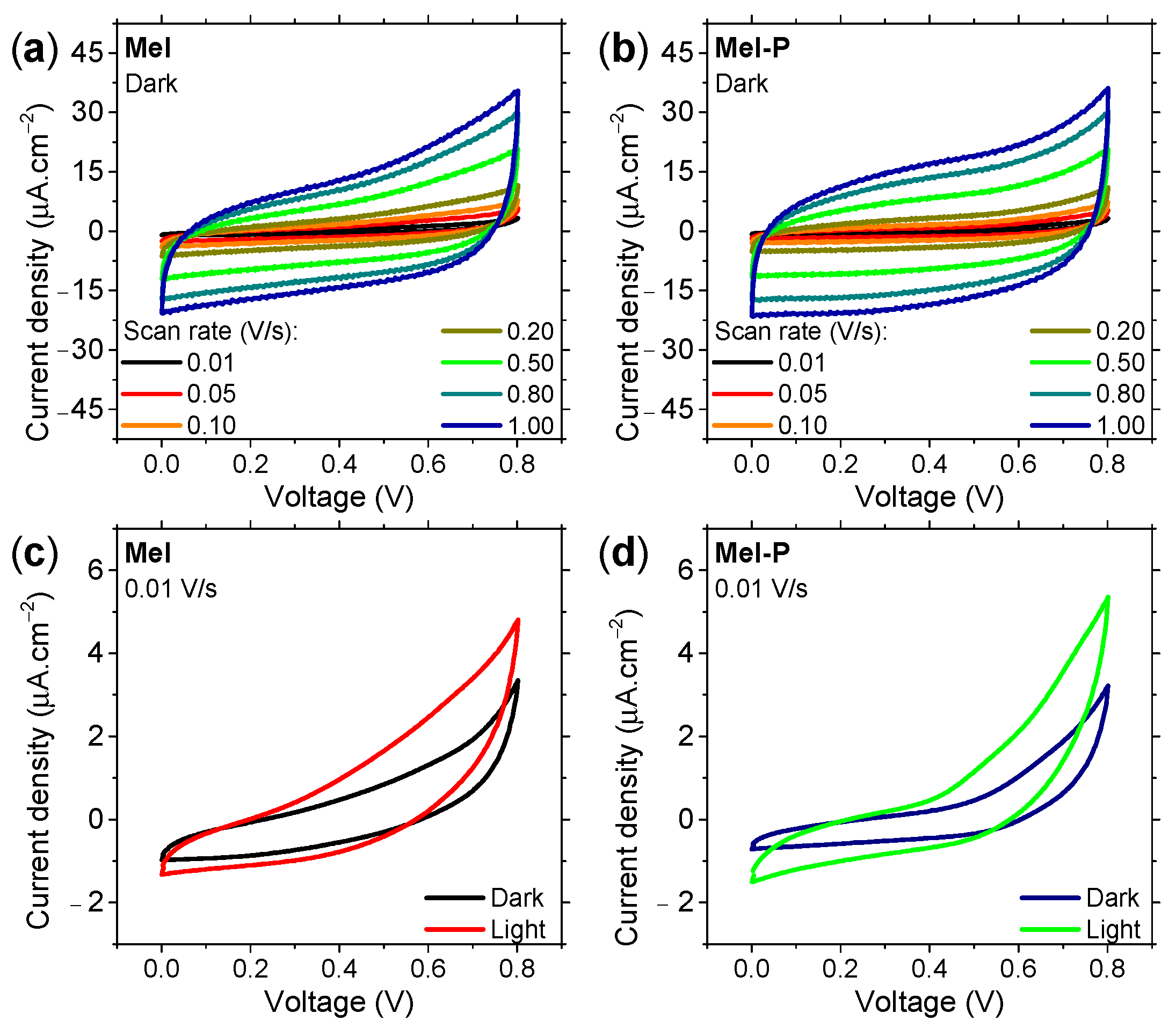
Figure 4.
Galvanostatic charge and discharge curves of (a) Mel and (b) Mel-P supercapacitor in the dark and under illumination of 0.1 mAg−1. In (a), under both dark and light conditions, the curves showed the same overall behavior. (c) Energy density and (d) power density estimated for the supercapacitors under study.

Figure 5.
(a,b) Nyquist plot and (c,d) phase angle as a function of frequency in the dark and under illumination. The inset in (b) corresponds to the equivalent circuit used for spectra fitting.
Figure 5.
(a,b) Nyquist plot and (c,d) phase angle as a function of frequency in the dark and under illumination. The inset in (b) corresponds to the equivalent circuit used for spectra fitting.

Figure 6.
The frequency-dependence capacitance of (a) Mel and (b) Mel-P under dark and light conditions.
Figure 6.
The frequency-dependence capacitance of (a) Mel and (b) Mel-P under dark and light conditions.

Publisher’s Note: MDPI stays neutral with regard to jurisdictional claims in published maps and institutional affiliations. |
© 2021 by the authors. Licensee MDPI, Basel, Switzerland. This article is an open access article distributed under the terms and conditions of the Creative Commons Attribution (CC BY) license (https://creativecommons.org/licenses/by/4.0/).
Share and Cite
MDPI and ACS Style
Paulin, J.V.; Fernandes, S.L.; Graeff, C.F.O. Solid-State Electrochemical Energy Storage Based on Soluble Melanin. Electrochem 2021, 2, 264-273. https://doi.org/10.3390/electrochem2020019
AMA Style
Paulin JV, Fernandes SL, Graeff CFO. Solid-State Electrochemical Energy Storage Based on Soluble Melanin. Electrochem. 2021; 2(2):264-273. https://doi.org/10.3390/electrochem2020019
Chicago/Turabian StylePaulin, João V., Silvia L. Fernandes, and Carlos F. O. Graeff. 2021. "Solid-State Electrochemical Energy Storage Based on Soluble Melanin" Electrochem 2, no. 2: 264-273. https://doi.org/10.3390/electrochem2020019