Cyclotron Production of Unconventional Radionuclides for PET Imaging: the Example of Titanium-45 and Its Applications


1
Nuclear Medicine Department, School of Health, Polytechnic Institute of Porto, 4200-072 Porto, Portugal
2
Health and Environment Research Center (CISA), Polytechnic Institute of Porto, 4200-072 Porto, Portugal
3
Isótopos para Diagnóstico e Terapêutica (IsoPor), S.A., 4445-526 Ermesinde, Portugal
4
Institute of Nuclear Sciences Applied to Health (ICNAS), University of Coimbra, 3000-548 Coimbra, Portugal
5
Coimbra Health School, Polytechnic Institute of Coimbra , 3046-854 Coimbra, Portugal
6
Physics Department, University of Trás-os-Montes e Alto Douro, 5000-801 Vila Real, Portugal
7
Centre for Mechanical Engineering, Materials and Processes (CEMMPRE), University of Coimbra, 3030-788 Coimbra, Portugal
*
Author to whom correspondence should be addressed.
Instruments 2018, 2(2), 8; https://doi.org/10.3390/instruments2020008
Submission received: 2 May 2018
/
Revised: 28 May 2018
/
Accepted: 31 May 2018
/
Published: 3 June 2018
(This article belongs to the Special Issue Instruments and Methods for Cyclotron Produced Radioisotopes)
Abstract
:Positron emitting radionuclides are used to label different compounds, allowing the study of the major biological systems using PET (positron emission tomography) imaging. Although there are several radionuclides suited for PET imaging, routine clinical applications are still based on a restrict group constituted by 18F, 11C, and, more recently, 68Ga. However, with the enlarged availability of low-energy cyclotrons and technical improvements in radionuclide production, the use of unconventional radionuclides is progressively more common. Several examples of unconventional radionuclides for PET imaging are being suggested, and 45Ti could be suggested as a model, due to its interesting properties such as its abundant positron emission (85%), reduced positron energy (β+ endpoint energy = 1040 keV), physical half-life of 3.09 h, and interesting chemical properties. This review aims to introduce the role of cyclotrons in the production of unconventional radionuclides for PET imaging while using 45Ti as an example to explore the potential biomedical applications of those radionuclides in PET imaging.
1. Introduction: Unconventional Radionuclides for PET Imaging
The incorporation and utilization of technological developments in clinical routine is becoming normal in modern practice of medicine. The application of imaging modalities is improving the quality of medical care and procedures that are available in medicine, which is an attitude that enhances the rapid implementation of modern paradigms of evidence-based medicine and science-based medicine. In fact, medical imaging occupies a primary position in clinical decision algorithms. In such context, non-invasive imaging modalities could allow for accurate diagnoses, increase precision in treatment choice/planning, and provide opportunities to follow the evolution of the clinical status of patients [1]. These modalities could also act over wide ranges of time and size scales involved in biological and pathological processes. Multiple imaging techniques are available, including X-ray imaging, computerized tomography (CT), magnetic resonance (MR) imaging, ultrasonography (US), optical imaging using fluorescent molecules (OF), and nuclear medicine (NM) using radioisotopes for different procedures such as planar imaging, single photon emission computed tomography (SPECT) and positron emission tomography (PET) [2].
Nuclear medicine has been an independent medical specialty since 1972, and is defined by the World Health Organization (WHO) as one that “encompasses applications of radioactive materials in diagnosis, treatment, or in medical research, with the exception of the use of sealed radiation sources in radiotherapy” [3]. In practice, nuclear medicine procedures are methods and techniques based in radionuclides. For diagnostic purposes, nuclear medicine uses primarily two types of radionuclides: (i) gamma-photon emitting nuclides (the ones that decay by isomeric transition or electron capture) for planar imaging and SPECT; and (ii) positron emitting nuclides for PET, which will be deeply discussed along this paper.
Radionuclides are used to label very small amounts of a pharmaceutical compound of interest, creating radiopharmaceuticals. According to the decay type of the nuclide component, the radiopharmaceutical can be optimized for a given procedure (SPECT versus PET). This industry of radiopharmaceuticals in general present a significant impact at the economical level. According to recent reports, the global radiopharmaceuticals market in 2017 was evaluated in $5.5 billion USD [4].
There are several factors that enhance the success of the application of a certain radionuclide. Considerations such as the physicochemical properties, availability, cost, and ease of use are some of the most relevant. For instance, according the literature, the usual clinical practice of PET imaging relies just on four radionuclides, namely: carbon-11 (11C), nitrogen-13 (13N), oxygen-15 (15O), and fluorine-18 (18F). All of them are characterized by relatively short physical half-lives [5]. The commercially available radiopharmaceuticals are almost invariably associated with 18F, due to the technical constraints in the production and distribution processes of the other short-lived nuclides. In fact, radiopharmaceuticals labeled with 18F are very interesting and commonly used mainly because of physical properties, such as a period of half-life that allows local (in situ) production and short-medium scale distribution, and the low β+ energy endpoint, which guarantees a good image quality [6].
Recently, a global trend was observed of exploring several new radionuclides for PET imaging that were different than the most classical ones, in order to increase the range of applications of the technology, as well as diversify the radiolabeled compounds. This become possible due to new insights into the production processes, radiolabeling methodologies, and interest in clinical applications of different radionuclides [7]. Examples of these unconventional radionuclides for PET imaging are: Scandium-44 (44Sc), Titanium-45 (45Ti), Cobalt-55 (55Co), Copper-60/61/64 (60/61/64Cu), Bromium-76 (76Br), Rubidium-82 (82Rb), Yttrium-86 (86Y), Technetium-94m (94mTc), Zirconium-89 (89Zr), and Iodine-124 (124I) [8,9]. Recently published papers confirm the investigational trend underlined in this paper [10,11,12]. The correspondent physical properties are presented in Table 1.
As already cited, conventional applications of PET imaging are based on organic elements with short half-lives (minutes to hours). A careful analysis of the radionuclides listed in Table 1 shows a tendency to explore radionuclides with higher physical half-lives and chemical characteristics associated with metallic elements (inorganic elements). In fact, the use of nuclides of inorganic elements has been studied since the discovery of radioactivity, but nuclear properties are principal factors that justify the development of the new compounds, while the availability of radionuclides in high specific activities and inorganic chemistry issues at the radiotracer level are the most frequent challenges [14]. Even so, unconventional radiometals are gradually being introduced, diversifying the available tools and procedures in nuclear medicine, particularly in PET imaging.
2. Cyclotrons and the Production of Unconventional Radionuclides
Today, more than 2700 radionuclides have been produced artificially with particle accelerators [15]. In addition, a few examples are also being obtained in loco using radionuclide generators through radioactive decay.
PET radionuclides, in particular, can be produced in cyclotrons, especially using inducing (p,n) nuclear reactions in the targets of stable isotopes.
Routine cyclotron production processes are made possible by the actual dissemination of these devices. As a matter of fact, by the end of 2005, there were 262 cyclotrons operating in the 39 member states of International Atomic Energy Agency (IAEA); however, it was believed that around 350 cyclotrons were operating in the whole world, according to a database of the agency [16]. Unfortunately, there is no official update of this report, because it is not easy to correctly estimate the number of cyclotrons operating nowadays all over the world.
The commercially available cyclotrons can be classified with respect to the particle type and maximum energy reached, the method of ion production, the technique of beam extraction from the cyclotron (or absence of extraction), the intensity of the accelerated ion beams, and other specific properties or features [17,18]. There are different classifications based on the type and energy of the accelerated particles [17,19]. Independently from the classification used, an important aspect to mention is that near 70% of the cyclotrons disseminated over the world are low-energy cyclotrons (≤20 MeV) [16].
According to empirical and practical evidence, the cyclotrons that typically have been applied worldwide in radionuclide production comprise properties such as: (i) the capability of accelerating negative ions (H−); (ii) beam extraction using stripper foils; (iii) fixed beam energy between 10–18 MeV, or 10–24 MeV mainly if the installation is intended for the production of many radionuclides, large-scale production and/or research purposes; (iv) fixed frequency of the RF generator; (v) two or four dees placed in valleys; (vi) internal ion source(s); (vii) the possibility of adjusting the beam position on the target; (viii) possibility of multi-target irradiations; (ix) compact radiation shielding around the device (“self-shielded” cyclotron); and (x) a high level of automation and simplicity in maintenance.
Despite the existence of the formal classifications, cyclotrons that respect the criteria stated in the last paragraph are normally classified by professionals of the field as “small cyclotrons”, “low-energy cyclotrons” or even as “medical cyclotrons”, and are applied to induce (p,n) reactions, which are typically low-energy processes with a constant onset below 9 MeV [20].
After the careful optimization of technical details such as targetry and irradiation parameters, high production yields of some of the unconventional radionuclides covered in this paper have been obtained in cyclotrons [21,22,23,24,25,26]. Examples of such radionuclides and recommended nuclear reactions are given in Table 2.
As more research studies in radionuclide production are delivered with successful results, which are conjugated with promising results in the preclinical and clinical trials that are currently being developed, these “unconventional” radionuclides are set to play an increasingly important role in nuclear medicine. These considerations highlight the pivotal role that cyclotrons have in solving the availability issues of interesting radionuclides for PET imaging [21].
3. Titanium-45 and Its Applications
Among the different examples that could be used to explore the paradigm of unconventional radionuclides for PET imaging in analysis, 45Ti is an interesting case study.
The nuclide 45Ti has a half-life of 3.09 h, which allows for distribution within a broader region than radiopharmaceuticals based on 18F or other shorter half-life radionuclides. On the other hand, its decay is predominantly based on positron emission (85%), with a maximum positron energy of 1040 keV and an average energy of 439 keV, which assures good image quality in PET images.
Preliminary results that were obtained in the experimental program of our group revealed the production feasibility of 45Ti in low-energy cyclotrons through the use of a 45Sc(p,n)45Ti nuclear reaction, with proton beams higher than 3 MeV. The maximum cross-section values were determined for proton beams with energy in the range between 10–14 MeV, while significant impurities production was found for energies higher than 17 MeV (see Table 3). These results mean that the development of a routine production process is possible, and research on the subject is ongoing [27].
In addition, the chemical properties of Ti allow two different scenarios for developing radiopharmaceuticals: (i) the “direct” labeling of ligands using chelation chemistry and/or simple covalent bounds with ligands of interest; or (ii) radiolabeled nanoparticles (either to label nanoparticles that are already explored in the field of nuclear medicine, or to label titanium oxide nanoparticles that could be relevant for clinical purposes).
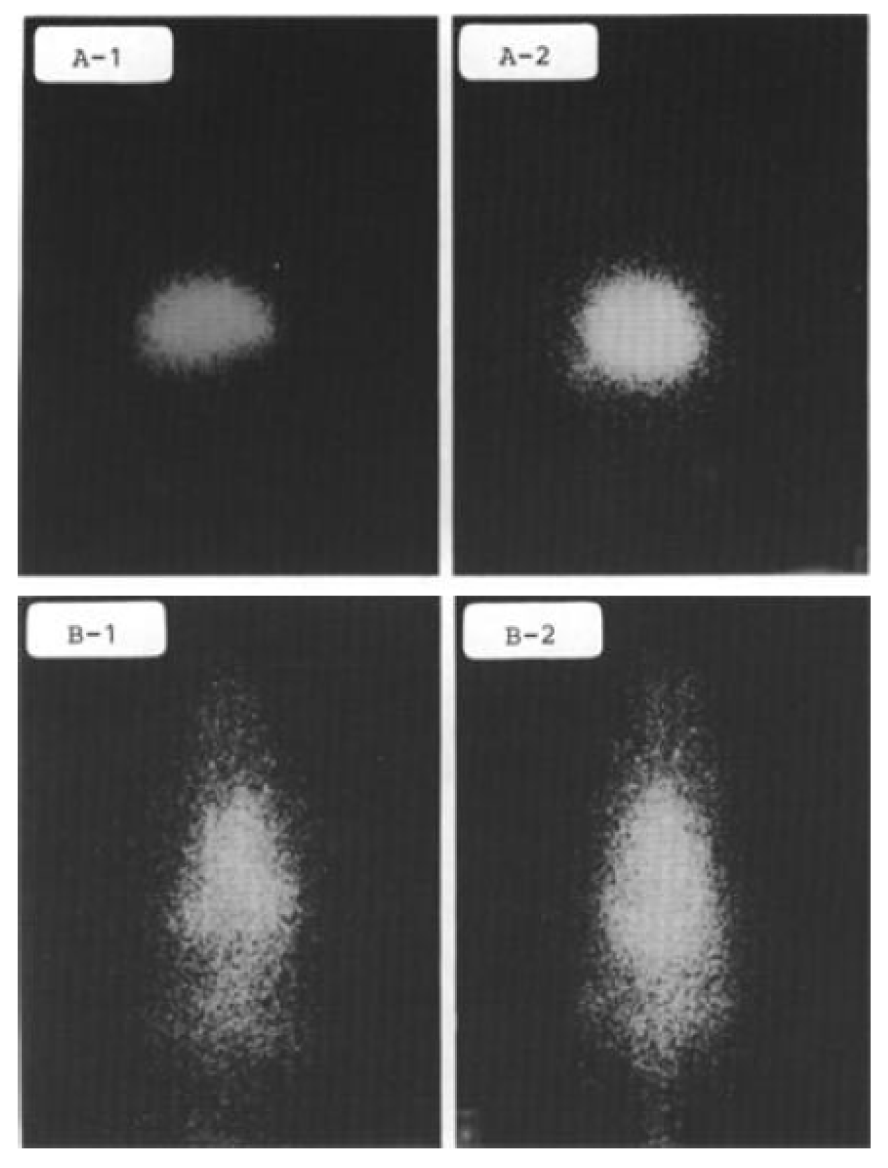
The first reference to a potential application of 45Ti belongs to a Japanese research team, and was published in 1981 by the Cyclotron and Radionuclide Center (CYRIC) of Tokohu University [28]. This group simultaneously evaluated the production, target chemistry (extraction and purification of 45Ti), and radiochemistry (labeling), and conducted some animal experiments. The main conclusion was centered on the potential of 45Ti-compounds to become “a new series of the positron-emitting radiopharmaceutical”, even considering some problems related to the stability of 45TiCl4 [28]. Examples of 45Ti-phytate and 45Ti-DTPA images are shown in Figure 1.
Some years later, the same group reported the use of other 45Ti-labeled compounds, namely 45TiOCl2 and 45TiO-phytate, as potential colloid agents for imaging the reticuloendothelial system. They also indicated 45Ti-DTPA (diethylenetriaminepentaacetic), 45Ti-citrate, and 45Ti-HSA (human serum albumin) as possible agents for estimating blood volume and indicators of the breakdown of the blood–brain barrier [29].
Considering the beginning of the study on the pathway of incorporation of titanium-based anticancer drugs such as budotitane(), 45Ti-budotitane was prepared and incorporated into liposomes to provide optimal tumor targeting [30]. Apart from this work, no further experimental studies were found on biodistribution or imaging using this compound.
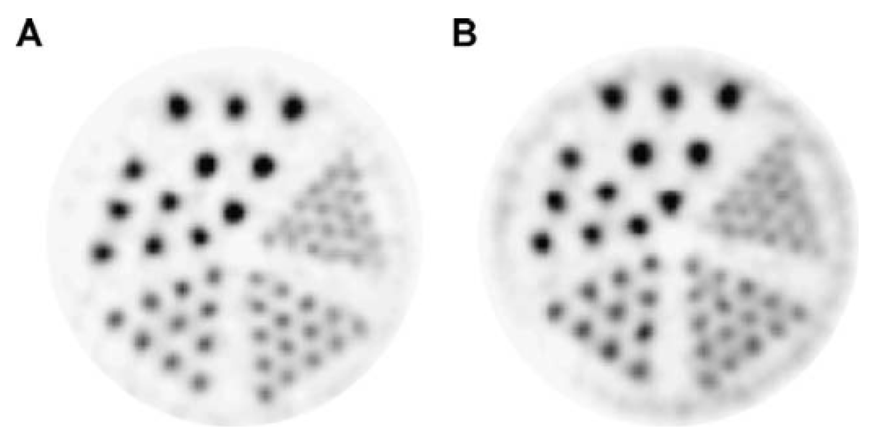
After a gap of some years, Vavere et al. [31] reported an evaluation study of 45Ti as a radionuclide for PET imaging. Despite other considerations, it was reported that the cyclotron production and purification of 45Ti is feasible. The work also demonstrated a clear spatial resolution observed down to a rod diameter of 1.25 mm using a Derenzo phantom, i.e., a resolution comparable with 18F, only with a slight degradation due to the higher energy endpoint and consequent range widening of positrons emitted by 45Ti (see Figure 2) [31].
Another study of Vavere and Welch [32] produced 45Ti and used small animal imaging with 45Ti-transferrin, which provided new insights about the mechanism of action of a new class of cytostatic agents based on titanium complexes. This research performed a direct labeling of apotransferrin with 45Ti and studied the resultant biodistribution. The microPET images provided indications on the increased uptake of 45Ti-transferrin in tumors, with retention up to 24 h, demonstrating that titanium radiopharmaceuticals could be explored as new tools for the in vivo imaging of tumors [31]. This data also supports the use of 45Ti-labeled compounds in theranostics and personalized medicine, taking the ability to select patients for a specific type of treatment into account.
However, Price et al. [33] published a work reporting some problems regarding the purification of 45Ti to be used in the radiolabeling process of transferrin. They reported losses of up to 53% of the 45Ti activity in the waste fractions during the separation process. However, it seems to be a problem with this particular experiment, because other authors have achieved good radiochemical yields in the labeling of proteins such as transferrin or Df-antibody [34].
In general, the reappearance of titanium-based drugs to treat cancer has been met with some success. The several chemical steps that are needed to stabilize these new titanium-based antineoplastics are being developed. In this research, the ligand salan seems to play a special role and lead to a significant investment in the development of these titanium-based drugs [35,36]. However, it is also known that the phase of drug development represents a highly complex, inefficient and costly process that usually takes huge amounts of time and money. Again, nuclear medicine imaging can be decisive, enhancing the efficiency of selecting the candidate drugs that should move forward into clinical trials or be abandoned [37]. The conjugation of the need for more information about drug pharmacokinetics, and the added value of nuclear medicine in the drug development process, can be used strategically; 45Ti labeling of compounds of interest could boost the research on titanium-based drugs. In fact, this conceptual idea is not totally original, because already published data shows the use of 45Ti-salan compounds as translational tools to help these drugs in the passage from fundamental research to clinical applications (example in Figure 3) [38].
However, the concept can be even taken further, if one hypothesizes that 45Ti compounds could act as diagnostic imaging agents of the same cancers that could be hereafter treated with titanium-based drugs. This once again suggests a role for 45Ti in theranostics and personalized medicine.
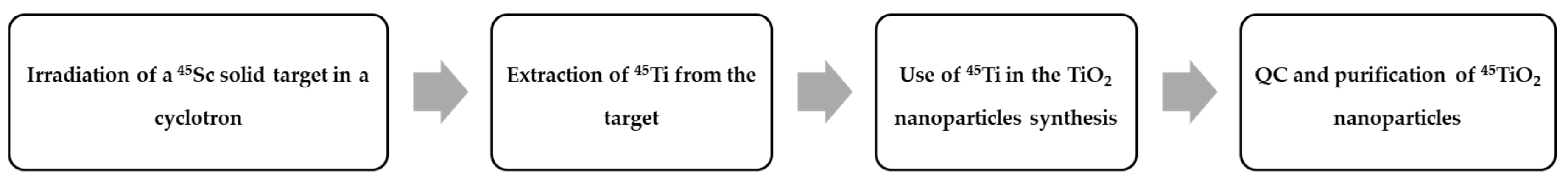
In the paradigm of personalized medicine, nanoparticles are being studied as drug delivery systems, and their application as vectors for radionuclide-based molecular imaging is a powerful tool of growing interest [39]. Indeed, in another context, recent data suggests that functionalized titanium dioxide nanoparticles (TiO2) can be surface-engineered to target cancer cells [40,41,42,43,44]. Thus, the radiolabeling of TiO2 nanoparticles with 45Ti could constitute a tool to provide in vitro biodistribution studies, which can also allow the in vivo monitoring of its biological distribution and the pharmacokinetics of such drug delivery systems. The nanoparticle radiolabeling could be achieved by different methodologies, including activation methods and synthesis based on radioactive precursors [45]. However, apart from other advantages related to the specific activity obtained, synthesis based on radioactive precursors appears to be the simplest and most favorable option, mainly due to the coincidence between the nanoparticle chemical nature (titanium) and the radionuclide that is aimed to be used for radiolabeling (45Ti). Figure 4 presents an overview of the processing steps to radiolabel TiO2 nanoparticles by the method suggested above.
To implement the process presented in Figure 4, once the cyclotron production of 45Ti seems to be feasible, our group looks for optimizing all of the technical aspects. Then, the extraction of 45Ti from the solid target constitutes a formidable challenge due to pharmaceutical requirements and technical issues, such as the specific activity needed. The task will require, for sure, wet chemistry techniques to fulfill the predefined requisites. The use of radioactive precursors for the labeling of TiO2 nanoparticles was already mentioned and briefly justified, but special attention should be given to the reduction of preparation time, because the operation is actually very time-consuming. Finally, 45TiO2 nanoparticles should be purified, filtered by size, and controlled regarding the surface properties for the appropriate targeting of biological processes.
As a final note, our literature review made on 45Ti found only one non-oncological application where labeled cations were used for the investigation of cerebral neurodegeneration [46].
4. Conclusion and Future Perspectives
Over the last decades, significant efforts have been made in the fundamental study, testing, development, and optimization of radionuclide production processes. The achievements have benefited the field of medical imaging and, particularly, radionuclide imaging using nuclear medicine methods and techniques. PET presents some advantages that could be used in accordance with the actual paradigms of medicine, in such a way that truthfully modern and personalized medical care could be conducted on patients. The wider use of low-energy cyclotrons is playing a crucial role in increasing the availability of radionuclides for preclinical or clinical trials, diversifying the set of radionuclides available for biomedical applications.
Several unconventional radionuclides for PET are being suggested to be introduced in clinical practice, because of the diversity of pathophysiological processes that could be studied. 55Co, 60/61/64Cu, 76Br, 82Rb, 86Y, 89Zr or 124I represent examples of this trend. This review focused on the exploration of 45Ti as a reliable candidate for PET imaging, emphasizing again the importance of cyclotrons to provide the production of the radionuclide. The titanium-45 half-life of 3.09 h, together with low energy positrons, were key factors for its selection as an interesting candidate. Potential applications of 45Ti include some possibilities that have been proposed and briefly studied by other groups, such as imaging with 45Ti-transferrin to study titanium-based chemotherapy agents or with 45Ti-salan compounds to be applied in theranostics of cancer. The use of 45TiO2 nanoparticles here suggested is an innovative proposal that should be developed in the near future.
Acknowledgements
The authors would like to thank the support of all the institutions involved in this work, namely School of Health of the Polytechnic Institute of Porto, IsoPor, S.A. and ICNAS.
Conflicts of Interest
The authors declare no conflict of interest.
References
- Acharya, R.; Wasserman, R.; Stevens, J.; Hinojosa, C. Biomedical imaging modalities: A tutorial. Comput. Med. Imaging Graph. 1995, 19, 3–25. [Google Scholar] [CrossRef]
- Cherry, S.R. Multimodality in vivo imaging systems: Twice the power or double the trouble? Annu. Rev. Biomed. Eng. 2006, 8, 35–62. [Google Scholar] [CrossRef] [PubMed]
- World Health Organization. Technical Report Series No 492; World Health Organization: Geneva, Switzerland, 1972; pp. 34–50. [Google Scholar]
- Nuclear Medicine and Radiopharmaceuticals Market 2012–2017. Available online: https://healthmanagement.org/c/imaging/news/nuclear-medicine-and-radiopharmaceuticals-market-2012-2017 (accessed on 20 April 2018).
- Saha, G.B. Diagnostic Uses of Radiopharmaceuticals in Nuclear Medicine. In Fundamentals of Nuclear Pharmacy; Saha, G.B., Ed.; Springer: Berlin, Germany, 2005. [Google Scholar]
- Saha, G.B. Cyclotron and Production of PET Radionuclides. In Basics of PET Imaging: Physics, Chemistry and Regulations; Saha, G.B., Ed.; Springer: New York, NY, USA, 2010; pp. 257–339. [Google Scholar]
- Conti, M.; Eriksson, L. Physics of pure and non-pure positron emitters for PET: A review and a discussion. EJNMMI Phys. 2016, 3, 8. [Google Scholar] [CrossRef] [PubMed]
- Jalilian, A.R. The Application of Unconventional PET Tracers in Nuclear Medicine. Iran. J. Nucl. Med. 2009, 17, 1–11. [Google Scholar]
- Welch, M.J.; Laforest, R.; Lewis, J.S. Production of Non-Standard PET Radionuclides and Application of Radiopharmaceuticals Labeled with these Nuclides. In PET Chemistry: The Driving Force in Molecular Imaging Series; Schubiger, P.A., Lehmann, L., Friebe, M., Eds.; Springer: Berlin, Germany, 2007. [Google Scholar]
- Arasaratnam, P.; Sadreddini1, M.; Yam, Y.; Kansal, V.; Dorbala, S.; di Carli, M.F.; Beanlands, R.S.; Merhige, M.E.; Williams, B.A.; Veledar, E.; et al. Prognostic value of vasodilator response using rubidium-82 positron emission tomography myocardial perfusion imaging in patients with coronary artery disease. Eur. J. Nucl. Med. Mol. Imaging 2018, 45, 538–548. [Google Scholar] [CrossRef] [PubMed]
- Rossi, S.; Toschi, L.; Castello, A.; Grizzi, F.; Mansi, L.; Lopci, E. Clinical characteristics of patient selection and imaging predictors of outcome in solid tumors treated with checkpoint-inhibitors. Eur. J. Nucl. Med. Mol. Imaging 2017, 44, 2310–2325. [Google Scholar] [CrossRef] [PubMed]
- England, C.G.; Jiang, D.; Ehlerding, E.B.; Rekoske, B.T.; Ellison, P.A.; Hernandez, R.; Barnhart, T.E.; McNeel, D.G.; Huang, P.; Cai, W. 89Zr-labeled nivolumab for imaging of T-cell infiltration in a humanized murine model of lung cancer. Eur. J. Nucl. Med. Mol. Imaging 2018, 45, 110–120. [Google Scholar] [CrossRef] [PubMed]
- Chu, S.Y.F.; Ekström, L.P.; Firestone, R.B. WWW Table of Radioactive Isotopes, database version 1999-02-28. Available online: http://nucleardata.nuclear.lu.se/nucleardata/toi/ (accessed on 23 April 2018).
- Carrol, V.; Demoin, D.W.; Hoffman, T.J.; Jurisson, S.S. Inorganic chemistry in nuclear imaging and radiotherapy: Current and future directions. Radiochim. Acta 2012, 100, 653–667. [Google Scholar] [CrossRef] [PubMed]
- Saha, G.B. Production of Radionuclides, 6th ed.; Springer: Berlin, Germany, 2010. [Google Scholar]
- International Atomic Energy Agency (IAEA). Directory of Cyclotrons used for Radionuclide Production in Member States—2006 Update; IAEA: Vienna, Austria, 2006. [Google Scholar]
- International Atomic Energy Agency (IAEA). Technical Report Series No465—Cyclotron Produced Radionuclides: Principles and Practice; IAEA: Vienna, Austria, 2008; pp. 59–72. [Google Scholar]
- Papash, A.I.; Alenitskii, Y.G. Commercial Cyclotrons. Part I: Commercial Cyclotrons in the Energy Range 10–30 MeV for Isotope Production. Phys. Part. Nucl. 2008, 39, 597–631. [Google Scholar] [CrossRef]
- Qaim, S.M. Nuclear data relevant to the production and application of diagnostic radionuclides. Radiochim. Acta 2001, 89, 223–232. [Google Scholar] [CrossRef]
- Jensen, M. Particle accelerators for PET radionuclides. Nucl. Med. Rev. 2012, 15, C9–C12. [Google Scholar]
- Holland, J.; Williamson, M.; Lewis, J. Unconventional Nuclides for Radiopharmaceuticals. Mol. Imaging 2010, 9, 1–20. [Google Scholar] [CrossRef] [PubMed]
- Sadeghi, M.; Enferadi, M.; Aref, M.; Jafari, H. Nuclear data for the cyclotron production of 66Ga, 86Y, 76Br, 64Cu and 43Sc. Nucleonika 2010, 55, 293–302. [Google Scholar]
- Link, J.; Krohn, K.A.; O’Hara, M.J. A simple thick target for production of 89Zr in an 11 MeV cyclotron. Appl. Radiat. Isot. 2017, 122, 211–214. [Google Scholar] [CrossRef] [PubMed]
- Qaim, S. Production of High Purity 94mTc for Positron Emission Tomographic Studies. Nucl. Med. Biol. 2000, 27, 323–328. [Google Scholar] [CrossRef]
- Asabella, A.; Cascini, G.; Altini, C.; Paparella, D.; Notaristefano, A.; Rubini, G. The Copper Radioisotopes: A systematic review with special interest to 64Cu. Biomed. Res. Int. 2014, 2014, 786463. [Google Scholar]
- Schmitz, J. The production of [124I]iodine and [86Y]yttrium. Eur. J. Nucl. Med. Mol. Imaging 2011, 38 (Suppl. 1), S4–S9. [Google Scholar] [CrossRef] [PubMed]
- Costa, P.; Metello, L.F.; do Carmo, S.J.C.; Alves, F.; Duarte Naia, M. Titanium-45 as an innovative radionuclide for PET imaging: From cyclotron production to potential biomedical applications. In Proceedings of the 29th Annual Congress of the European Association of Nuclear Medicine 2016, Barcelona, Spain, 15–19 October 2016. Oral Presentation 447. [Google Scholar]
- Ishiwata, K.; Ido, T.; Monma, M.; Murakami, M.; Fukuda, H.; Yamada, K.; Endo, S.; Yoshioka, H.; Sato, T.; Matsuzawa, T. Preparation and Medical Application of 45Ti; CYRIC Annual Report; CYRIC: Tohoku, Japan, 1981. [Google Scholar]
- Ishiwata, K.; Ido, T.; Monma, M.; Murakami, M.; Fukuda, H.; Kameyama, M.; Yamada, K.; Endo, S.; Yoshioka, H.; Sato, T.; et al. Potential radiopharmaceuticals labeled with titanium-45. Int. J. Radiat. Appl. Instrum. Part A Appl. Radiat. Isot. 1991, 42, 707–712. [Google Scholar] [CrossRef]
- Waterhouse, R.N.; Mattner, F.; Najdovski, L.; Collier, T.L.; Fallon, J. Synthesis and Characterisation of [111In]-Liposome Encapsulated [45Ti]-Budotitane. In Proceedings of the Eleventh International Symposium on Radiopharmaceutical Chemistry, Vancouver, BC, Canada, 13–17 August 1995. [Google Scholar]
- Vavere, A.L.; Laforest, R.; Welch, M.J. Production, processing and small animal PET imaging of titanium-45. Nucl. Med. Biol. 2005, 32, 117–122. [Google Scholar] [CrossRef] [PubMed]
- Vavere, A.L.; Welch, M.J. Preparation, biodistribution, and small animal PET of 45Ti-transferrin. J. Nucl. Med. 2005, 46, 683–690. [Google Scholar] [PubMed]
- Price, R.I.; Sheil, R.W.; Scharli, R.K.; Chan, S.; Gibbons, P.; Jeffery, C.; Morandeau, L. Titanium-45 as a Candidate for PET Imaging: Production, Processing & Applications. In Proceedings of the 15th International Workshop on Targetry and Target Chemistry, Prague, Czech Republic, 18–21 August 2015. [Google Scholar]
- Siikanen, J.; Hong, H.; Valdovinos, H.; Hernandez, R.; Zhang, Y.; Barnhart, T.; Cai, W.; Nickles, R. Production, separation and labeling of 45Ti. In Proceedings of the SNMMI Annual Meeting, Vancouver, BC, Canada, 8–12 June 2013. [Google Scholar]
- Tshuva, E.Y.; Ashenhurst, J.A. Cytotoxic Titanium(IV) Complexes: Renaissance. Eur. J. Inorg. Chem. 2009, 2009, 2203–2218. [Google Scholar] [CrossRef]
- Immel, T.A.; Groth, U.; Huhn, T. Cytotoxic Titanium Salan Complexes: Surprising Interaction of Salan and Alkoxy Ligands. Chem. Eur. J. 2010, 16, 2775–2789. [Google Scholar] [CrossRef] [PubMed]
- Cunha, L.; Szigeti, K.; Mathé, D.; Metello, L.F. The role of molecular imaging in modern drug development. Drug Discov. Today 2014, 19, 936–948. [Google Scholar] [CrossRef] [PubMed] [Green Version]
- Severin, G.W.; Nielsen, C.H.; Jensen, A.I.; Fonslet, J.; Kjær, A.; Zhuravlev, F. Bringing Radiotracing to Titanium-Based Antineoplastics: Solid Phase Radiosynthesis, PET and ex Vivo Evaluation of Antitumor Agent [45Ti](salan)Ti(dipic). J. Med. Chem. 2015, 58, 7591–7595. [Google Scholar] [CrossRef] [PubMed] [Green Version]
- Assadi, M.; Afrasiabi, K.; Nabipour, I.; Seyedabadi, M. Nanotechnology and nuclear medicine; research and preclinical applications. Hell. J. Nucl. Med. 2011, 14, 149–159. [Google Scholar] [PubMed]
- Zarytova, V.F.; Zinov’ev, V.V.; Ismagilov, Z.R.; Levina, A.S.; Repkova, M.N.; Shikina, N.V.; Evdokimov, A.A.; Belanov, E.F.; Balakhnin, S.M.; Serova, O.A.; et al. An Examination of the Ability of Titanium Dioxide Nanoparticles and Its Conjugates with Oligonucleotides to Penetrate into Eucariotis Cells. Nanotechnol. Russia 2009, 4, 732–735. [Google Scholar] [CrossRef]
- Stefanou, E.; Evangelou, A.; Falaras, P. Effects of UV-irradiated titania nanoparticles on cell proliferation, cancer metastasis and promotion. Catal. Today 2010, 151, 58–63. [Google Scholar] [CrossRef]
- El-Said, K.S.; Ali, E.M.; Kanehira, K.; Taniguchi, A. Molecular mechanism of DNA damage induced by titanium dioxide nanoparticles in toll-like receptor 3 or 4 expressing human hepatocarcinoma cell lines. J. Nanobiotechnol. 2014, 12, 48. [Google Scholar] [CrossRef] [PubMed] [Green Version]
- Mund, R.; Panda, N.; Nimesh, S.; Biswas, A. Novel titanium oxide nanoparticles for effective delivery of paclitaxel to human breast cancer cells. J. Nanopart. Res. 2014, 16, 2739. [Google Scholar] [CrossRef]
- Chen, Y.; Wan, Y.; Wang, Y.; Zhang, H.; Jiao, Z. Anticancer efficacy enhancement and attenuation of side effects of doxorubicin with titanium dioxide nanoparticles. Int. J. Nanomed. 2011, 6, 2321–2326. [Google Scholar] [Green Version]
- Gibson, N.; Holzwarth, U.; Abbas, K.; Simonelli, F.; Kozempel, J.; Cydzik, I.; Cotogno, G.; Bulgheroni, A.; Gilliland, D.; Ponti, J.; et al. Radiolabelling of engineered nanoparticles for in vitro and in vivo tracing applications using cyclotron accelerators. Arch. Toxicol. 2011, 85, 751–773. [Google Scholar] [CrossRef] [PubMed]
- Salber, D.; Manuvelpillai, J.; Spahn, I.; Klein, S.; Uhlenbruck, F.; Palm, C.; Matusch, A.; Becker, S.; Langen, K.J.; Coenen, H.H. 45Ti-cations as potential PET-tracers for cerebral neurodegeneration. In Proceedings of the International Symposium on Technetium and Other Radiometals in Chemistry and Medicine, Bressanone, Italy, 8–11 September 2010. [Google Scholar]
Figure 1.
Whole body images of 45Ti-phytate (A) and 45Ti-DTPA (diethylenetriaminepentaacetic) (B) acquired 10 minutes (A1 and B1) and 60 minutes (A2 and B2) after injection in rats. Images published by Ishiwata et al. in 1981 [28].
Figure 1.
Whole body images of 45Ti-phytate (A) and 45Ti-DTPA (diethylenetriaminepentaacetic) (B) acquired 10 minutes (A1 and B1) and 60 minutes (A2 and B2) after injection in rats. Images published by Ishiwata et al. in 1981 [28].

Figure 2.
Comparison of image quality of 18F (A) and 45Ti (B) using a Derenzo phantom. Images published by Vavere et al. [31].
Figure 2.
Comparison of image quality of 18F (A) and 45Ti (B) using a Derenzo phantom. Images published by Vavere et al. [31].

Figure 3.
Example of acquired PET/computerized tomography (CT) images of a 45Ti-compound in mice. Images published by Severin et al. [38].
Figure 3.
Example of acquired PET/computerized tomography (CT) images of a 45Ti-compound in mice. Images published by Severin et al. [38].
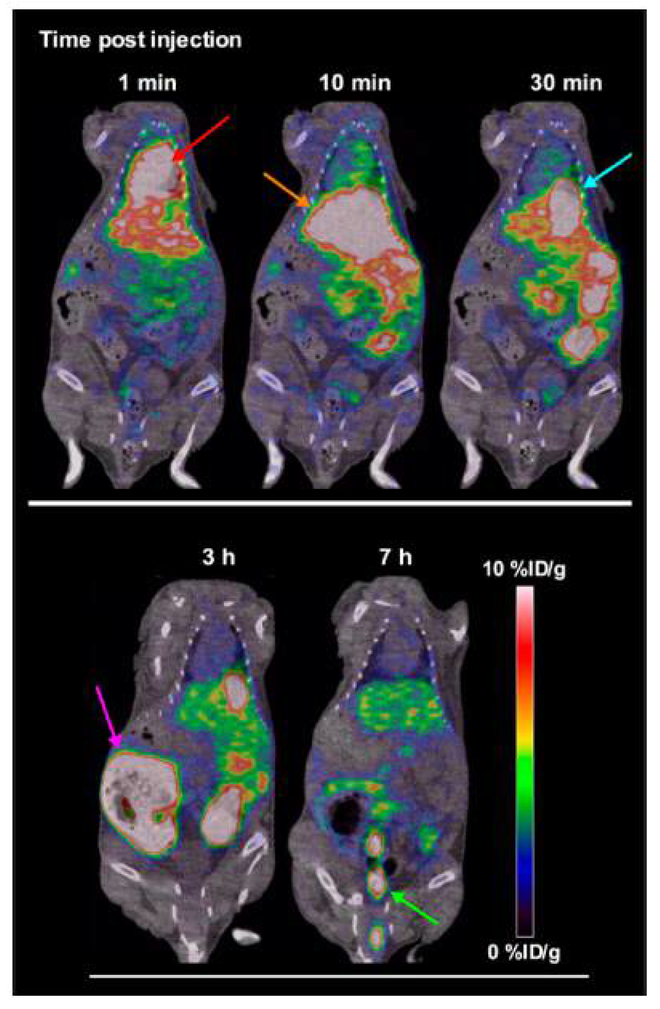
Figure 4.
Possible approach to obtain 45TiO2 nanoparticles using the cyclotron irradiation of a solid target of 45Sc.
Figure 4.
Possible approach to obtain 45TiO2 nanoparticles using the cyclotron irradiation of a solid target of 45Sc.

Table 1.
Some unconventional positron emission tomography (PET) radionuclides and corresponding physical properties [13].
Table 1.
Some unconventional positron emission tomography (PET) radionuclides and corresponding physical properties [13].
| Radionuclide | Half-Life | β+ Endpoint Energy (MeV) |
|---|---|---|
| 44Sc | 3.927 h | 1.47 |
| 45Ti | 3.1 h | 1.04 |
| 55Co | 17.53 h | 1.50 |
| 60Cu | 23.7 min | 3.77 |
| 61Cu | 3.333 h | 1.21 |
| 64Cu | 12.700 h | 0.653 |
| 76Br | 16.2 h | 3.94 |
| 82Rb | 1.273 min | 3.15 |
| 86Y | 14.7 h | 3.14 |
| 89Zr | 3.3 day | 0.902 |
| 94mTc | 52.0 min | 2.44 |
| 124I | 4.176 day | 2.14 |
Table 2.
Examples of cyclotron-produced unconventional PET radionuclides.
| Radionuclide | Most Common Nuclear Reaction |
|---|---|
| 44Sc | 44Ca(p,n)44Sc |
| 45Ti | 45Sc(p,n)45Ti |
| 55Co | 58Ni(p,α)55Co |
| 60Cu | 60Ni(p,n)60Cu |
| 61Cu | 61Ni(p,n)61Cu |
| 64Cu | 64Ni(p,n)64Cu |
| 76Br | 76Se(p,n)76Br |
| 86Y | 86Sr(p,n)86Y |
| 89Zr | 89Y(p,n)89Zr |
| 94mTc | 94Mo(p,n)94mTc |
| 124I | 124Te(p,n)124I |
Table 3.
Preliminary results of experimental cross-section values for 45Sc(p,n)45Ti nuclear reaction.
Table 3.
Preliminary results of experimental cross-section values for 45Sc(p,n)45Ti nuclear reaction.
| Mean Beam Energy (MeV) | Cross-Section (mbarn) | Uncertainty (mbarn) |
|---|---|---|
| 16.1 ± 1.0 | 1.2 × 10² | ±0.4 × 10² |
| 15.3 ± 1.0 | 2.1 × 10² | ±0.7 × 10² |
| 12.4 ± 0.9 | 4.4 × 10² | ±1.4 × 10² |
| 8.9 ± 0.8 | 3.4 × 10² | ±1.0 × 10² |
| 4.9 ± 0.7 | 1.2 × 10² | ±0.4 × 10² |
| 3.4 ± 0.7 | 0.8 × 101 | ±0.3 × 101 |
© 2018 by the authors. Licensee MDPI, Basel, Switzerland. This article is an open access article distributed under the terms and conditions of the Creative Commons Attribution (CC BY) license (http://creativecommons.org/licenses/by/4.0/).
Share and Cite
MDPI and ACS Style
Costa, P.; Metello, L.F.; Alves, F.; Duarte Naia, M. Cyclotron Production of Unconventional Radionuclides for PET Imaging: the Example of Titanium-45 and Its Applications. Instruments 2018, 2, 8. https://doi.org/10.3390/instruments2020008
AMA Style
Costa P, Metello LF, Alves F, Duarte Naia M. Cyclotron Production of Unconventional Radionuclides for PET Imaging: the Example of Titanium-45 and Its Applications. Instruments. 2018; 2(2):8. https://doi.org/10.3390/instruments2020008
Chicago/Turabian StyleCosta, Pedro, Luís F. Metello, Francisco Alves, and M. Duarte Naia. 2018. "Cyclotron Production of Unconventional Radionuclides for PET Imaging: the Example of Titanium-45 and Its Applications" Instruments 2, no. 2: 8. https://doi.org/10.3390/instruments2020008