


Abstract
Pompe disease, Krabbe disease, and mucopolysaccharidosis type I were added to the newborn screen in Indiana in 2020. Clinical and laboratory teams collaborated to determine optimal strategies for first- and second-tier testing on newborn screen prior to referral to the metabolism clinic. Our metabolism clinic has worked with neurology, cardiology, and hematology/oncology departments to determine and execute best initial work-up, therapeutic, and ongoing surveillance approaches to manage patients diagnosed with one of these lysosomal disorders on newborn screen. In the first five years of this screening, 27 patients have been diagnosed with Pompe disease (three infantile-onset, 24 late-onset), 13 patients with Krabbe disease (one early infantile, 12 late-onset), and five patients with mucopolysaccharidosis type I (three severe phenotype, one with attenuated phenotype, one with possible attenuated disease). Our experience in caring for these individuals has helped to further refine second-tier testing and our clinical evaluation and management of these patients.
1. Introduction
Newborn screening for lysosomal disorders (LSDs) has evolved over the last 20 years. Principles of newborn screening (NBS) prioritize conditions with effective treatments, reliable methods of diagnosis, and serious health implications if not diagnosed and managed in a timely manner. As treatments for LSDs have become more numerous and effective over the years, the importance of screening for these conditions is increasingly recognized. Pompe disease, mucopolysaccharidosis type I (MPSI), and Infantile Krabbe were added to the Recommended Uniform Screening Panel (RUSP) over several years, with Pompe receiving approval from the Secretary of Health and Human Services in March of 2015, MPSI in February of 2016, and Infantile Krabbe in July of 2024. Following the efforts of advocacy groups, the Indiana state legislature voted in 2019 to add Krabbe disease to Indiana’s newborn screening panel. Given that Pompe and MPSI were already on the RUSP at that time, we advocated for the inclusion of all three of these conditions in Indiana Newborn Screening. The Indiana Department of Health, Indiana Newborn Screening Lab, and Indiana University Medical Genetics teams met regularly and designed our screening and follow-up algorithms together with primary goals of identifying all at-risk infants, reducing the risk of false positive results, expediting care for those with severe infantile disease, all while remaining cost-effective. A month-long pilot program for all three conditions began in June 2020, with full screening starting 1 July 2020.
2. Materials and Methods
2.1. Laboratory Evaluation
There were two first-tier screening options. The Baebies SEEKER, FDA-approved in February 2017, was a digital microfluidic system. It is a closed system that has the capability to screen for Pompe, MPSI, Fabry, and Gaucher. However, Krabbe screening would need to be done differently. The Revvity NeoLSD was FDA-approved in September of 2018, using a tandem mass spectrometry platform. This kit included reagents used to screen for Pompe, MPSI, Krabbe, Gaucher, Fabry and Niemann–Pick A/B. Both methods measured the affected enzymes, with reduced levels indicating the possibility of disease. However, enzyme levels could also be lower due to a heterozygous state, pseudodeficiencies, and poor sample quality. We reached out to states using both platforms, looking at cost, our ability to bring on the technology, and assay performance, including the number of samples reflexed to second-tier testing. We ultimately chose to go with the Revvity NeoLSD assay because it allowed us to screen for Pompe, MPSI, and Krabbe using a multiplex approach from a dried blood spot (DBS) punch of 3.2 mm diameter. This method also offered more flexibility in adding additional reagents to screen for other LSDs not included with either system. This was an important factor since many additional disorders are being evaluated as potential RUSP candidate conditions, including mucopolysaccharidosis type II (MPSII).
Once the first-tier testing approach was determined, we looked at second-tier testing and developed the overall follow-up algorithm. At the request of clinical genetics providers, second-tier testing was included to reduce the number of false positive results for infants with heterozygous variants or pseudodeficiency, shorten the time to definitive diagnosis so we can promptly begin treatment in those with true infantile-onset disease, and limit cases referred to the clinical care teams to those with a high likelihood of disease. We use the same initial NBS DBS used for all other screenings and send a portion of that sample to a referral lab for additional testing. Different reference labs were considered using cost, turnaround time, and testing confidence to guide the advisory group. The care team weighed in on minimum testing they would need, and the timeline needed for prompt treatment and intervention. Each disorder was evaluated separately as those timelines vary with each lysosomal condition. Initial algorithms established were as follows. Pompe second-tier testing includes acid alpha-glucosidase (GAA) enzyme activity and (Creatine/Creatinine)/GAA measurements by tandem mass spectrometry [1,2]. All positive results for these values automatically reflex to GAA (HGNC:4065) sequencing. MPSI second-tier testing included quantitative glycosaminoglycan (GAG) measurements (heparan, dermatan, and keratan sulfate) via liquid chromatography–tandem mass spectrometry. Krabbe second-tier testing included psychosine measurement via liquid chromatography–tandem mass spectrometry along with testing for the common 30 kb deletion. We chose to include molecular testing for Pompe and Krabbe disease in the initial screening process because treatment for infantile forms of both conditions should begin by 4 weeks of age for optimal outcomes. No results are reported until second-tier testing is completed. This allows the LSD screen to be reported without any additional follow-up needed for any normal second-tier result. Any abnormal second-tier result is reported as abnormal on the newborn screen and referred for clinical evaluation and further diagnostic testing.
As part of any screening process, we continuously look at ways to improve positive predictive value, as false-positive screen results cause anxiety for the families, increase cost for the program, and burden the follow-up teams. With that in mind, we have made the following changes. Second-tier testing for MPSI now begins with the measurement of an endogenous biomarker specific to MPSI. If that is positive, measurement of GAGs is performed. Initial second-tier testing for Krabbe is now only psychosine measurement. If psychosine level is greater than 10 nmol/L, the sample is reflexed to sequencing for the common 30 kb deletion. We have also increased the cutoff for abnormal psychosine to be greater than 2.0 nmol/L. Prior to 2025, we were using Revvity Omics, and their cutoff was 1.5 nmol/L. Many of our abnormal Krabbe results had psychosine levels between 1.5 and 2.0 nmol/L; all of these infants were confirmed to be unaffected (see Results). Case data and literature pointed to 2.0 nmol/L being a better threshold. We now use Mayo Clinical Laboratories, and their reported reference range is 2.0 nmol/L.
2.2. Clinical Evaluation and Management
We established an algorithm for follow-up of infants with abnormal second-tier screens. This has involved multidisciplinary cooperation with providers from neurology, hematology/oncology, cardiology, and additional specialists. We meet periodically to review and improve the process. Initial clinical evaluation focuses on molecular confirmation of diagnosis for those conditions in which full gene sequencing is not part of the NBS algorithm. Infants with borderline screens may meet with the genetic counselor first to provide testing and counseling, then a metabolic physician if positive. All infants with positive genetic testing or highly suspicious second-tier biochemical testing will meet with both a metabolic physician and genetic counselor for an initial visit. Differentiation of early- versus late-onset disease is established through review of genotype with the established literature and databases, as well as additional evaluations such as imaging or biochemical labs for more novel variants.
2.2.1. Pompe Disease
For Pompe disease, we typically receive genetic test results when the patient is 2–3 weeks old for those with abnormal second-tier testing. We review GAA variants in the established database to correlate with phenotype [3]. A creatine kinase (CK) level, echocardiogram, ECG, and urine hex4 level (if a sample can be collected) are obtained at or before our first visit for infants with possible infantile-onset disease, and less urgently for those with late-onset disease. As urgent echocardiography is routinely available at our center for these patients, other cardiac screening mechanisms such as chest X-ray have not been necessary. Timely administration of therapy is essential in infants confirmed to have infantile-onset Pompe disease (IOPD), as starting treatment at <4 weeks of age improves outcomes [4]. We start infants with IOPD on alglucosidase alfa 40 mg/kg weekly and have always successfully gotten this approved through insurance appeals by providing evidence that increased dosing improves survival [5]. Inpatient admission to initiate IOPD treatment may be necessary if the patient has any health or logistical issues that could interfere with timely administration of enzyme replacement therapy (ERT) +/− immune tolerance induction (ITI). Infants with late-onset Pompe disease (LOPD) are monitored for symptoms, and those with intermediate results for whom second-tier results did not automatically trigger GAA gene sequencing but are still concerning for possible LOPD meet with a genetic counselor to discuss the need for sequencing.
Patients with Pompe disease are followed in our multidisciplinary Pompe clinic, which is staffed by a geneticist, neurologist with expertise in neuromuscular disorders, a pediatric physical therapist, and a genetic counselor, plus coordination with a pediatric cardiologist with cardiomyopathy specialty training for same-day appointments when needed. Neurology evaluations for infants and younger children have a strong focus on monitoring growth and progress towards achievement of developmental milestones in areas such as gross and fine motor skills, as well as communication. Discussion with caregivers, and patients if age-appropriate, includes addressing feeding/swallowing issues, activity level/endurance, musculoskeletal complaints (stiffness, cramps, spasms, strength), ambulation and any other functional concerns. The neurologic physical examination evaluates muscle tone, strength of major muscle groups, joint mobility for spasticity or contractures, deep tendon reflexes, coordination, gait analysis and scoliosis surveillance. Notable physical exam findings in patients with Pompe disease can include hypotonia and (proximal greater than distal) muscle weakness, which may present as difficulty climbing stairs, rising from a chair, or Gower’s sign when rising from the floor.
Cardiac involvement in IOPD has been well documented with a well-defined clinical trajectory. Patients develop cardiac hypertrophy, arrhythmias, and cardiac dysfunction secondary to glycogen deposits in the myocardium and conduction system [6,7]. Heart failure and cardiac arrhythmias can lead to or contribute to early mortality in these patients. Cardiac involvement is rare in non-classical Pompe disease but can be severe [8]. Prior to the advent of ERT, options for treatment of cardiac involvement were limited and not effective. ERT, however, has shown to be effective in reversing (either partially or fully) cardiac hypertrophy and arrhythmias in most patients [9,10,11].
Since the addition of Pompe disease to NBS, we have integrated cardiac evaluations as soon as possible, noting the effectiveness of ERT on long-term cardiac health. During a patient’s initial visit after an NBS positive for Pompe, they undergo an echocardiogram, ECG, and clinical visit with a pediatric cardiologist with cardiomyopathy specialty training. These serve as baseline evaluations, family education, and possible guides for our geneticists to inform them of the type of Pompe disease a patient has (infantile, non-classic infantile, late-onset) as they await further genetic and biochemical testing results. We are specifically looking for evidence of cardiac hypertrophy or cardiac dysfunction by echocardiogram and any evidence of increased voltages (surrogate for potential cardiac hypertrophy), pre-excitation (shortened PR interval), or increased QT dispersion (difference from the longest to shortest QT) on ECG. If a patient demonstrated any ECG or echocardiogram findings concerning for early cardiac involvement and before confirmatory testing results, ERT would likely be initiated without waiting for confirmatory molecular test results. Given the rapidity of newborn screening results and patient triaging to our clinic, finding pre-existing cardiac involvement will be unlikely.
Patients identified as infantile-onset will then get serial testing/evaluations with an echocardiogram and ECG at baseline (goal within 10 days of a positive newborn screen), 3 months, 6 months and 1 year, then yearly to ensure the effectiveness of ERT. They may get additional testing if new symptoms or clinical concerns arise, including blood work to evaluate their BNP/NTproBNP, ambulatory Holter monitoring, or additional imaging. Patients with non-classic infantile Pompe will receive similar testing at baseline, 6 months and 1 year, and then every 1–2 years thereafter. Data on long-term cardiac involvement in LOPD is less clear. However, it has been rarely reported, with potential for more of an arrhythmia phenotype [12]. We have therefore opted to have patients identified as late-onset get cardiac surveillance at baseline, 1 year and then every 3 years as we await more data to inform that practice.
2.2.2. Krabbe Disease
Psychosine levels greater than 10 nM in DBS are highly concerning for infantile Krabbe disease (IKD) and prompt us to immediately alert our hematopoietic stem cell transplant (HSCT) specialists. Our process for patients with this degree of elevation involves scheduling a visit with a geneticist, genetic counselor, and stem cell team within 1–2 business days, then admission to the stem cell transplant unit if desired by parents to expedite pre-transplant workup and expedited GALC (HGNC:4115) sequencing to confirm the diagnosis. While we have not performed a transplant for IKD thus far, our calculations indicate that we would be able to do so within 30 days of age, assuming that there are no complications in finding a matched donor. Infants with intermediate psychosine results are seen by a genetic counselor for GALC sequencing and counseling. Those with positive or inconclusive results are then seen by a metabolic physician. Most of these infants have inconclusive results, with variants of uncertain significance on sequencing. Repeating galactosylceramidase (GALC) enzyme level and psychosine level on whole blood sample for indeterminate results has been helpful, as some are in the reference range and thus reassuring, though some families are still left with inconclusive results, which caused significant parental anxiety for patients who are unlikely to develop late-onset disease. Patients with follow-up testing consistent with or concerning for late-onset Krabbe disease (LOKD) are followed by our neurology clinic. For the purposes of this paper, LOKD includes both juvenile Krabbe disease and adult Krabbe disease. We have not had any patients with concern for late-infantile Krabbe disease.
Patients with Krabbe disease are followed in our pediatric neurology clinic to identify early signs concerning for disease progression in order to provide prompt treatment as indicated, with the goal of preserving and maintaining strength and function in these patients. Neurological evaluation focuses on identifying stiffness and/or spasticity, weakness, ambulatory difficulties, changes in gait, vision or hearing loss, and seizures. Brain MRI is obtained urgently in any patient noted to have early signs or symptoms of disease. For those without clinical signs or symptoms and at high risk of onset of late-infantile or early juvenile Krabbe disease, we recommend brain MRI within 2 months of diagnosis with repeat surveillance MRI every 4 months for the first year, then every 6 months until age 3 years. In individuals without development of clinical signs or symptoms of Krabbe disease by age 3 years, MRI is spaced to annually through age 12 years and then every 2–5 years until adulthood. For those at risk of later juvenile or adult-onset Krabbe disease and without any clinical signs or symptoms of Krabbe disease, we recommend baseline brain MRI at 12–18 months of age with repeat MRI every 2–5 years until adulthood. EEG should be obtained if there are concerns for seizure activity [13].
2.2.3. Mucopolysaccharidosis Type I
For MPSI, most of the second-tier results that we receive have mildly elevated GAGs noted to be more suspicious for pseudodeficiency, and our genetic counselor orders IDUA (HGNC:5391) sequencing and counsels the family. When infants have second-tier testing with higher GAGs noted to be in the range of infants affected by MPSI, the physician and genetic counselor see the patient together. We order IDUA sequencing, echocardiogram, and skeletal survey X-rays, and we refer to the NBS hearing evaluation to differentiate between classic and attenuated disease. All infants whom we have diagnosed with classic Hurler syndrome have had at least one abnormality on this initial testing, such as a failed newborn hearing screen, early changes in vertebral bone morphology, or subtle cardiac valve thickening. Infants whose phenotype fits with classic Hurler syndrome are referred to our stem cell transplant team and/or outside clinical trial sites per parental preference. Those with pathogenic variants and no evidence of early systemic disease are diagnosed with attenuated disease and started on laronidase enzyme replacement therapy, and treatment is deferred for infants with high GAGs per NBS second tier test on DBS and variants of uncertain significance, though we recommend that all of these infants follow up in our clinic for surveillance for the first few years of life.
In addition to following in the metabolism clinic, patients with MPSI are followed in our pediatric cardiology clinic. MPSI-related cardiac disease is characterized by a high involvement of thickening of the aortic valve, mitral valve, or both, leading to varying degrees of valvar regurgitation [14]. Valvopathy can become so severe that it may lead to heart failure symptoms necessitating valve replacement surgery. Further, some patients may develop cardiomyopathy, with both hypertrophic and dilated cardiomyopathy having been described [15,16,17]. ERT has been shown to stabilize, attenuate or fully reverse cardiac involvement, both cardiomyopathy and valve disease [14,17]. Similarly, there has been cardiac improvement in patients who have received bone marrow or stem cell transplants [18,19].
Severe forms of MPSI appear to show cardiac involvement in more mid to late infancy, but neonatal/early infancy presentations have been reported. Advanced therapies such as ERT and HSCT are more effective at reducing cardiac involvement the earlier they are instituted. Therefore, we have opted to screen these patients closely in the first year of life for the development of cardiomyopathy, cardiac hypertrophy, and valvopathy with a baseline clinical visit with a pediatric cardiologist with cardiomyopathy specialty training, echocardiogram, and ECG during initial intake in our multidisciplinary genetics clinic, then again at 3, 6, and 9 months, 1 year and tailored follow-up thereafter based on their cardiac status [20].
2.2.4. Hematopoietic Stem Cell Transplant
For patients diagnosed with disorders amenable to HSCT, early engagement of the stem cell transplant team is crucial to achieving optimal outcomes. For both MPSI and infantile Krabbe disease, early HSCT offers the best potential for neurologic preservation and improved outcomes. Thus, timely coordination is essential. Once referred, our pre-transplant evaluation includes neuroimaging (e.g., brain/spine MRI to assess for early white matter changes); baseline neurodevelopmental assessment; echocardiogram; abdominal ultrasound; infectious disease screening; HLA typing; organ function labs; and consultations with metabolic genetics, neurology, cardiology, audiology/ENT, pulmonology, and ophthalmology as needed. We also engage with social work and psychology early to support family understanding and readiness, especially given the complexity and urgency of transplant decisions in the neonatal period.
For MPSI, genotype and enzyme level are integrated with clinical features and expert metabolic input to determine transplant candidacy. Infants with MPSI with severe phenotype are prioritized for transplant, while attenuated forms are monitored or managed with ERT alone. For Krabbe disease, timing is more nuanced. In presumed early-infantile disease, we aim to proceed to HSCT urgently, often within the first 30 days of life, pending donor identification.
Post-HSCT, the stem cell transplant team remains involved in monitoring engraftment and immune reconstitution, ongoing coordination with metabolic and neurodevelopmental specialists, and adjusting supportive care based on evolving neurocognitive and somatic outcomes.
3. Results
Between 1 July 2020 and 30 June 2025, a total of 397,255 newborns were screened, and 145 screens were flagged for review by the metabolism department—76 concerning for Pompe disease, 44 concerning for Krabbe disease, and 45 concerning for MPSI.
3.1. Pompe Disease
Of the 76 positive first-tier screens reported for Pompe disease, there were 50 positive second-tier screens, and 27 individuals with positive sequencing. Of note, screens are only referred to our clinic for review if second-tier has low GAA enzyme activity. The 26 individuals with positive first-tier testing but negative second-tier testing that were reported to us had low GAA enzyme activity but a normal (Creatine/Creatinine)/GAA ratio.
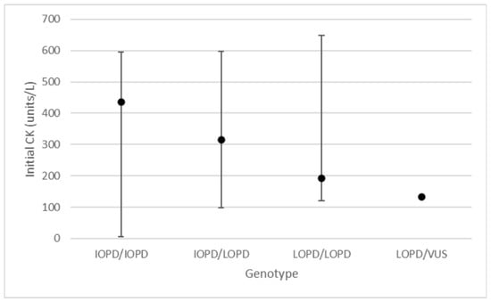
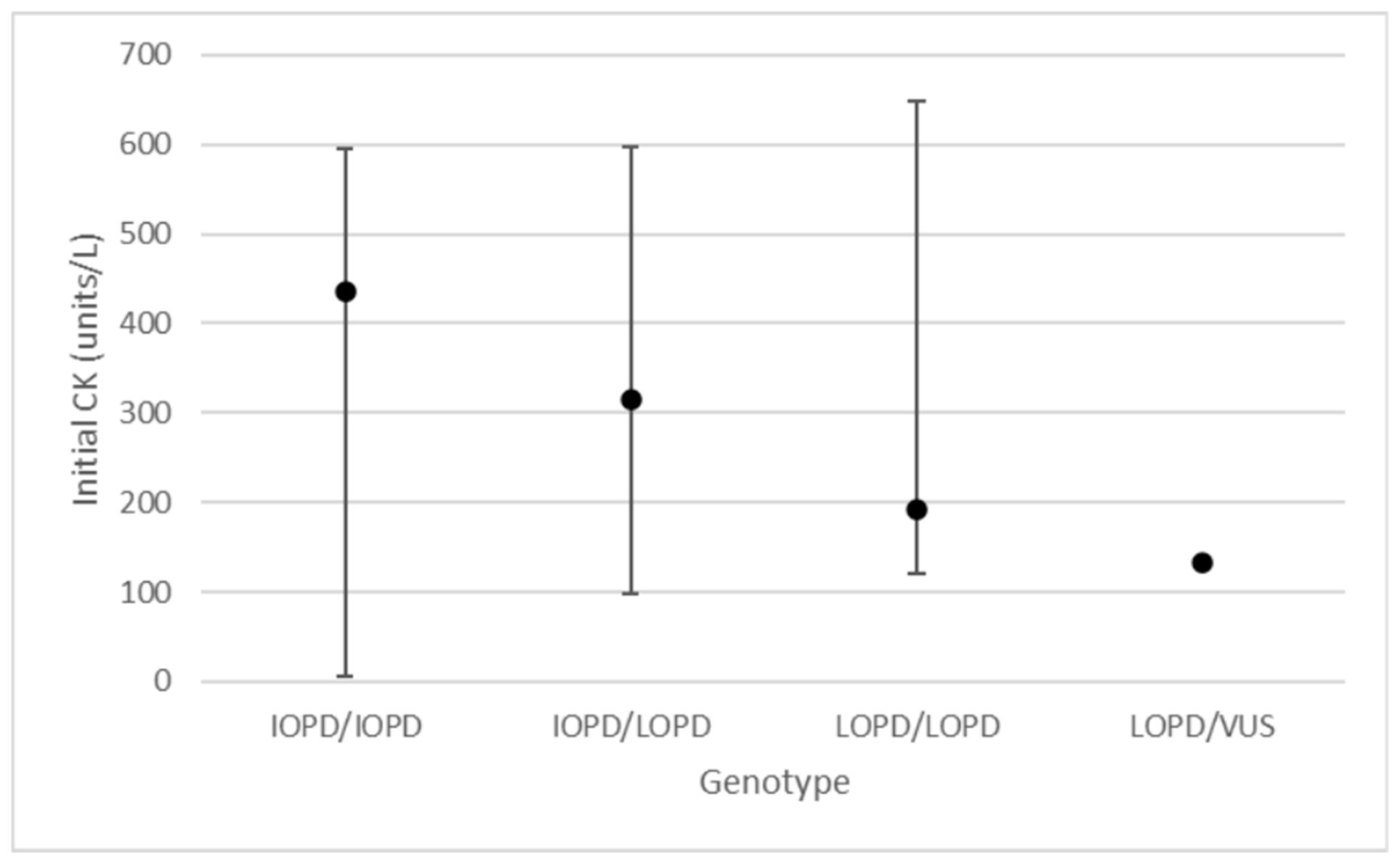
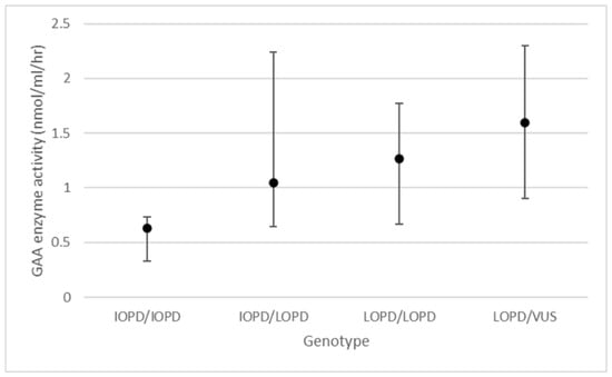
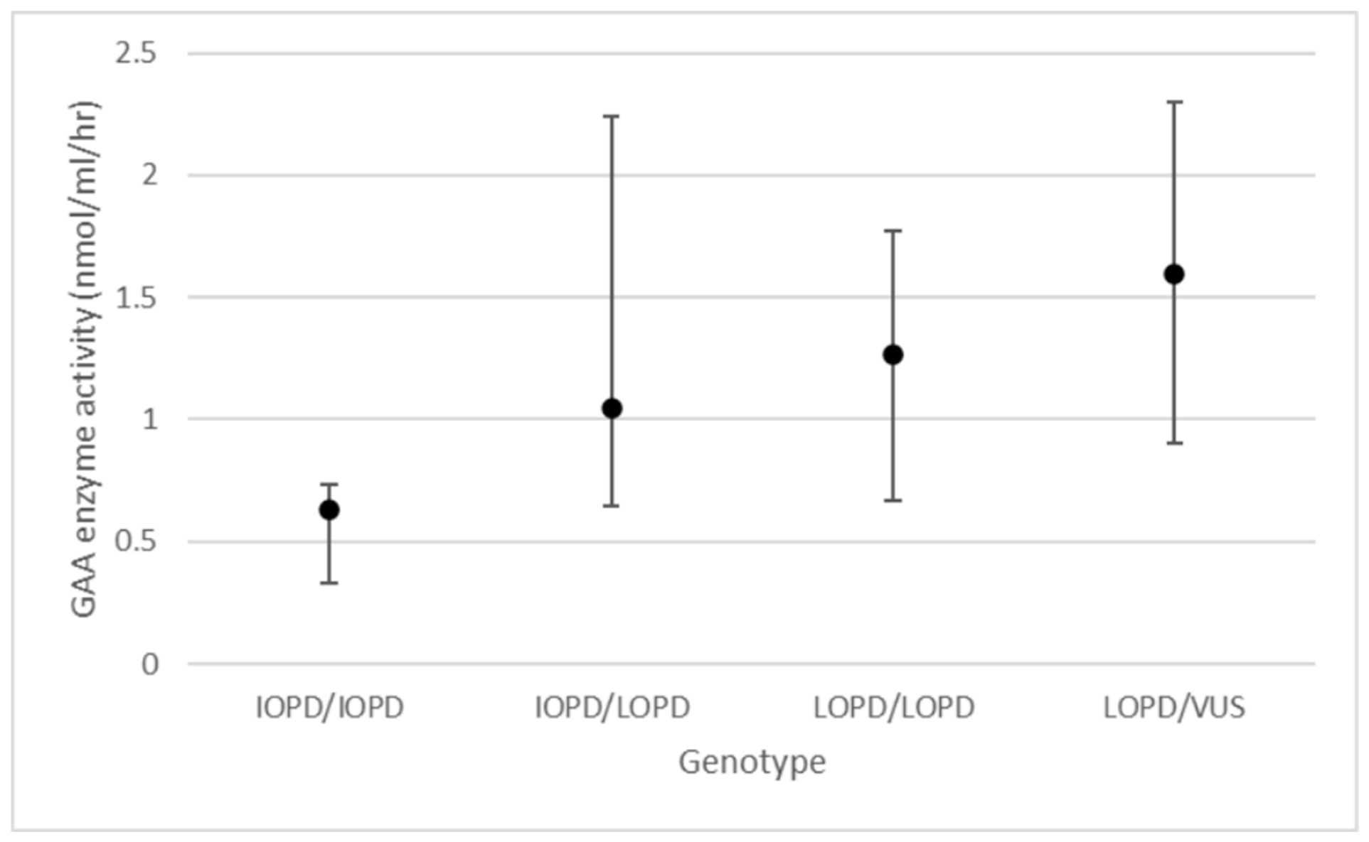
Of the 76 screens that were reported, 27 have Pompe disease (3 IOPD and 24 LOPD), seven are heterozygous for a pathogenic GAA variant, two are heterozygous for a GAA variant of uncertain significance, 11 have a pseudodeficiency, 27 had negative second-tier testing and/or negative sequencing, and two were lost to follow-up prior to completion of initial workup (positive predictive value (PPV) for IOPD or LOPD = 35%). Of those with pseudodeficiency, seven have one pseudodeficiency allele, and four have two pseudodeficiency alleles; for those with two pseudodeficiency alleles, phase was not able to be determined as parental studies were not sent. For patients with LOPD, mean GAA enzyme activity in NBS DBS was 1.21 nmol/mL/h (SD = 0.47, ref > 6.49 nmol/mL/h) with a minimum of 0.5 nmol/mL/h and a maximum of 2.3 nmol/mL/h; mean (Creatine/Creatinine)/GAA ratio was 8.0 (SD = 3.3, ref < 0.91) with a minimum of 3.5 and a maximum of 16.4; and mean initial CK level was 240 units/L (SD = 176, ref 20–160 units/L) with a minimum of 70 units/L and a maximum of 597 units/L (Figure 1 and Figure 2). For patients with IOPD, mean GAA enzyme activity was 0.63 nmol/mL/h (SD = 0.21) with a minimum of 0.4 nmol/mL/h and a maximum of 0.8 nmol/mL/h; mean (Creatine/Creatinine)/GAA ratio was 8.7 (SD = 1.1) with a minimum of 7.4 and a maximum of 9.5; and mean initial CK level was 436 units/L (SD = 305) with a minimum of 96 units/L and a maximum of 685 units/L.
Figure 1.
Mean initial creatine kinase (CK) level with minimum and maximum based on genotype. IOPD = variant causative of infantile-onset Pompe disease; LOPD = variant causative of late-onset Pompe disease; VUS = variant of uncertain significance.
Figure 2.
Mean acid alpha-glucosidase (GAA) enzyme activity with minimum and maximum based on genotype. IOPD = variant causative of infantile-onset Pompe disease; LOPD = variant causative of late-onset Pompe disease; VUS = variant of uncertain significance.
We have met our goal of starting all patients with IOPD on ERT within four weeks of life, including ITI for CRIM-negative patients, by working closely with our pharmacy, infusion center, and manufacturer. Of the three patients with IOPD, one started ERT (with ITI) at 21 days, one started ERT at 25 days, and one transferred to a center closer to home for ongoing management prior to initiation of ERT. Alglucosidase alfa was used initially at a dose of 40 mg/kg weekly. This is currently the only drug FDA-approved to treat IOPD, and we were concerned that attempting to acquire a different medication off-label drug could delay treatment. Avalglucosidase alfa is increasingly being prescribed for IOPD due to promising clinical trial results [21]. We use shared decision-making with families about whether they would like to change to avalglucosidase alfa once they are established on treatment. One patient did switch, primarily due to increasing CK and urine hex4 levels concerning for advancing disease, and biomarkers normalized on avalglucosidase alfa at a dose of 40 mg/kg weekly.
Of the 24 patients with LOPD, three are currently on ERT. One LOPD patient started ERT at 21 days old due to an echocardiogram with possible borderline left ventricular hypertrophy, significant hypotonia, and initial GAA sequencing with three variants, including two variants associated with IOPD and one associated with LOPD, without phase information at that time. Parental testing ultimately came back indicating that the two IOPD variants are in cis with each other and in trans with the LOPD variant, conferring a diagnosis of LOPD. One LOPD patient started ERT at 7 months of age due to diminished axial tone. Finally, one LOPD patient started ERT at 34 months of age after being found to have a slightly wide-based and unsteady gait and being noted to use his hands to push up from the floor (not a full Gower sign). Mean peak CK for these individuals with LOPD who started ERT was 408 units/L, with a minimum of 97 units/L and a maximum of 588 units/L. Of note, the individual with LOPD with early cardiac findings who started ERT at 21 days of age was found to have three GAA variants, two of which were previously reported to compose a complex allele in cis causative for IOPD in trans with an additional LOPD variant. The additional two individuals with LOPD who were started on ERT are both compound heterozygotes for one variant causative of IOPD and one variant associated with LOPD. Nineteen patients with LOPD have been asymptomatic and continue to be monitored clinically. Of these asymptomatic individuals, five are compound heterozygotes for one variant causative of IOPD and one variant associated with LOPD, 12 have two variants associated with LOPD, and two have one variant associated with LOPD and a suspicious variant of uncertain significance. The remaining two individuals with LOPD have transferred care to centers closer to their homes; one of these individuals had two variants associated with LOPD and the other had one variant associated with LOPD and a suspicious variant of uncertain significance.
3.2. Krabbe Disease
44 positive first-tier screens were reported for Krabbe disease. There were 19 individuals with elevated psychosine on second-tier screening based on a cutoff of 2.0 nM and 34 individuals with elevated psychosine on second-tier screening based on a cutoff at the time of reporting. Those individuals with normal psychosine on second-tier screening were reported to our clinic for review due to a heterozygous exon 11–14 deletion seen on second-tier testing. Thirteen individuals had positive sequencing.
Of the 44 screens that were reported, 13 have confirmed or suspected Krabbe disease (1 IKD and 12 LOKD), 16 are heterozygous for a pathogenic GALC variant, one is heterozygous for a variant of uncertain significance that has been seen in individuals with Krabbe disease (c.334A>G discussed further below) and an additional more uncertain VUS, 11 have pseudodeficiency, three had negative second tier testing and/or negative sequencing, and one has ongoing workup underway to determine phase of variants to determine whether they are affected or heterozygous (PPV for IKD or LOKD = 30%). Of the 12 individuals with confirmed or suspected LOKD, three are homozygous for pathogenic GALC variants, six have one pathogenic variant and one copy of the c.334A>G variant previously reported in affected individuals, one has one pathogenic variant and one copy of the c.956A>G variant previously reported in affected individuals, one individual is homozygous for the c.334A>G variant, and one individual is homozygous for the c.956A>G variant [22,23,24,25,26]. Of the 11 individuals with pseudodeficiency, five had three pseudodeficiency alleles, four had two pseudodeficiency alleles, and one had two pseudodeficiency alleles; for those with two pseudodeficiency alleles, phase could not be determined as parental studies were not sent.
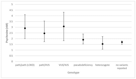
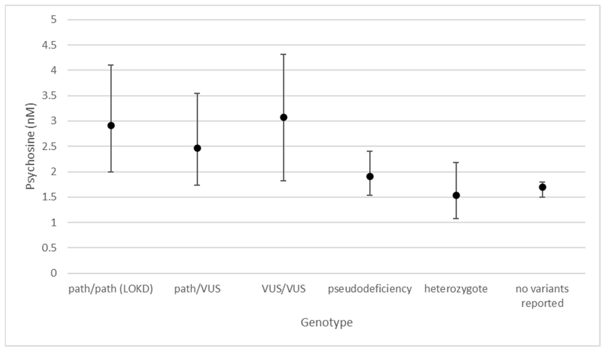
The patient with IKD had a psychosine on NBS DBS of 49 nM (ref < 2 nM). For individuals with LOKD, mean psychosine was 2.61 nM (SD = 0.90) with a minimum of 1.73 nM and a maximum of 4.32 nM. For individuals with pseudodeficiency alleles in GALC, mean psychosine was 1.91 nM (SD = 0.27) with a minimum of 1.54 nM and a maximum of 2.4 nM (Figure 3). For heterozygotes, five had undetectable psychosine levels (<1.08 nM or <1.50 nM, depending on the assay used). Of the heterozygotes with detectable psychosine levels, mean psychosine was 1.64 nM (SD = 0.31) with a maximum of 2.18 nM. All patients with molecular testing concerning for LOKD continue to be monitored clinically. While several have mild developmental delays, none have been overtly symptomatic or had abnormal brain MRIs, so none have been referred for HSCT evaluation.
Figure 3.
Mean psychosine level with minimum and maximum based on genotype. For individuals with a psychosine level below the detection limit of the assay, the lowest detectable limit was used for calculation of the mean and minimum. Path/path (LOKD) = homozygous or compound heterozygous for pathogenic variant(s) with diagnosis of late-onset Krabbe disease; path/VUS = compound heterozygous for one pathogenic variant and one variant of uncertain significance; VUS/VUS = homozygous or compound heterozygous for variants of uncertain significance.
3.3. Mucopolysaccharidosis Type I
Of the 45 screens reported to our clinic for review, all had positive second-tier screens, and five had positive sequencing. Five have MPSI (three severe phenotype, one with attenuated phenotype, one with possible attenuated disease), 28 have pseudodeficiency, five are heterozygous for one pathogenic IDUA variant, two had negative second-tier testing and/or negative sequencing, and five were lost to follow-up prior to completion of molecular testing (PPV = 11%). All of those lost to follow-up had GAG levels in NBS DBS suggestive of pseudodeficiency. Of the 28 individuals with pseudodeficiency, eight had one pseudodeficiency allele, two were homozygous for two pseudodeficiency alleles, and two were compound heterozygous for two pseudodeficiency alleles; for those who were compound heterozygous, phase cannot be determined as parental studies were not sent. For patients with MPSI, dermatan sulfate in NBS DBS ranged from 2036 nmol/L to >2400 nmol/L (ref ≤ 200 nmol/L), heparan sulfate in NBS DBS ranged from 241 nmol/L to 519 nmol/L (ref ≤ 96 nmol/L), and alpha-L-iduronidase (IDUA) enzyme activity in leukocytes ranged from 0 nmol/h/mL to 0.6 nmol/h/mL (ref 6–71.4 nmol/h/mg).
Of the three patients with a severe phenotype, two received ERT and then HSCT at our center, and the third was managed at an outside center with ERT and gene therapy clinical trial. Of the two patients with an attenuated phenotype, one started ERT with laronsidase at 4 months of age. This individual has had no signs or symptoms of MPSI and has had a normal brain MRI. The other individual with concern for attenuated MPSI has one variant of uncertain significance and one pathogenic variant, borderline whole blood enzyme activity, and normal auxiliary studies, so for now is being monitored clinically.
Most common reasons patients were lost to follow-up include difficulty contacting family to pursue further workup or schedule follow-up and patients moving out of state.
4. Discussion
One of our significant challenges has been determining appropriate follow-up protocols for infants with uncertain results or potential risk for late-onset disease for each condition. Many more infants have been identified than we anticipated, and while there are some published guidelines, we are finding that these patients are much more subtle and unique than one can gain from a guideline [27,28]. Our goal is to stratify risk as much as possible so that our surveillance is not missing infants who are more likely to present with earlier symptom onset while not overly burdening those unlikely to have disease until adulthood. For example, all our patients with LOPD who required treatment early in childhood have been compound heterozygous for one late-onset variant and one infantile-onset variant, and their CK levels were persistently above 500 units/L (ref 20–160 units/L) for several months before overt symptoms were noticeable. Thus, we schedule closer follow-up for infants with the above characteristics, whereas others with a less concerning genotype may only require annual follow-up long term.
Comprehensive care of these patients with LSDs would not be possible without the ongoing collaboration between the metabolism, neurology, cardiology, and hematology/oncology teams. Input from these various specialties, along with sequencing information, allows us to make timely determinations of patient phenotype, the primary driver of therapeutic decision-making. We work to streamline these visits with multiple specialists by coordinating same-day visits whenever possible, as many families travel long distances to be seen. We have also developed a multidisciplinary Pompe disease clinic in which patients are seen by a metabolic specialist, neurologist, and physical therapist during a single office visit to consolidate care for families.
Our clinical team has found that regular attendance at national and international meetings pertaining to lysosomal disorders has been instrumental in caring for these patients. Ongoing communication with providers from other states screening for these conditions has helped us continue to refine our management algorithm. We hope that by continuing to collaborate with providers from other states, we can continue to refine our management strategy for later-onset conditions, including LOPD, which were not diagnosed pre-symptomatically prior to their addition to NBS.
The addition of Pompe disease, Krabbe disease, and MPSI to NBS in Indiana has allowed for early identification and treatment with the goal of improving long-term outcomes for these patients. In addition to minimizing unnecessary distress to families, the implementation of second-tier screening tests for all three of these disorders has allowed us to optimize resource utilization by focusing additional work-up and clinical evaluation on patients at higher risk of having true underlying disease. Additionally, we have been able to refine the reporting cutoff for psychosine levels to help us better identify individuals at high risk of having true disease rather than those with pseudodeficiency or heterozygotes.
Author Contributions
Conceptualization: M.G., B.H.G., K.S. and M.M.; Data curation: M.G. and C.S.; Formal analysis: M.G.; Methodology: all authors; Writing—original draft: M.G., A.C., M.K., B.L., J.J.P. and M.M.; Writing—review and editing: all authors. All authors have read and agreed to the published version of the manuscript.
Funding
This research received no external funding.
Institutional Review Board Statement
This study was reviewed and determined to be exempt by the Indiana University IRB.
Informed Consent Statement
Informed consent was not required as many of the patients included are not actively followed in our clinic and therefore do not have up-to-date contact information available to our health system. Additionally, given the relatively small sample size, we thought it was imperative to include all patients with flagged NBS regardless of whether they were actively followed and had up-to-date contact information.
Data Availability Statement
Complete dataset is unavailable due to patient privacy. Select data available upon request.
Acknowledgments
Special thanks to all of the clinical staff who provide support for these patients and the families who entrust us with their children’s care.
Conflicts of Interest
The authors declare no conflicts of interest.
Abbreviations
The following abbreviations are used in this manuscript:
| LSD | Lysosomal disorder |
| NBS | Newborn screening |
| MPSI | Mucopolysaccharidosis type I |
| RUSP | Recommended uniform screening panel |
| DBS | Dried blood spot |
| MPSII | Mucopolysaccharidosis type II |
| GAA | Acid alpha-glucosidase |
| CK | Creatine kin |
| GAG | Glycosaminoglycans |
| IOPD | Infantile-onset Pompe disease |
| ERT | Enzyme replacement therapy |
| ITI | Immune tolerance induction |
| LOPD | Late-onset Pompe disease |
| IKD | Infantile Krabbe disease |
| HSCT | Hematopoetic stem cell transplant |
| GALC | Galactosylceramidase |
| LOKD | Late-onset Krabbe disease |
| IDUA | Alpha-L-iduronidase |
| PPV | Positive predictive value |
References
- Turgeon, C.; Magera, M.J.; Allard, P.; Tortorelli, S.; Gavrilov, D.; Oglesbee, D.; Raymond, K.; Rinaldo, P.; Matern, D. Combined newborn screening for succinylacetone, amino acids, and acylcarnitines in dried blood spots. Clin. Chem. 2008, 54, 657–664. [Google Scholar] [CrossRef] [Scilit] [PubMed]
- Tortorelli, S.; Turgeon, C.T.; Gavrilov, D.K.; Oglesbee, D.; Raymond, K.M.; Rinaldo, P.; Matern, D. Simultaneous Testing for 6 Lysosomal Storage Disorders and X-Adrenoleukodystrophy in Dried Blood Spots by Tandem Mass Spectrometry. Clin. Chem. 2016, 62, 1248–1254. [Google Scholar] [CrossRef] [Scilit] [PubMed]
- de Faria, D.O.S.; Groen, S.L.M.; Hoogeveen-Westerveld, M.; Nino, M.Y.; van der Ploeg, A.T.; Bergsma, A.J.; Pim Pijnappel, W.W.M. Update of the Pompe variant database for the prediction of clinical phenotypes: Novel disease-associated variants, common sequence variants, and results from newborn screening. Hum. Mutat. 2021, 42, 119–134. [Google Scholar] [CrossRef] [Scilit] [PubMed] [PubMed Central]
- Desai, A.K.; Rodriguez-Rassi, E.; Parikh, S.; Russo, R.S.; Kronn, D.; Hamm, J.A.; Chang, I.J.; Ortiz, D.; McPheron, M.; Lydigsen, H.; et al. Early initiation of enzyme replacement therapy as facilitated by newborn screening improves health outcomes among patients with infantile-onset Pompe disease. Genet. Med. Open 2025, 4, 103478. [Google Scholar] [CrossRef] [Scilit] [PubMed] [PubMed Central]
- Kishnani, P.S.; Kronn, D.; Suwazono, S.; Broomfield, A.; Llerena, J.; Al-Hassnan, Z.N.; Batista, J.L.; Wilson, K.M.; Periquet, M.; Daba, N.; et al. Higher dose alglucosidase alfa is associated with improved overall survival in infantile-onset Pompe disease (IOPD): Data from the Pompe Registry. Orphanet J. Rare Dis. 2023, 18, 381. [Google Scholar] [CrossRef] [Scilit] [PubMed] [PubMed Central]
- Hahn, A.; Schänzer, A. Long-term outcome and unmet needs in infantile-onset Pompe disease. Ann. Transl. Med. 2019, 7, 283. [Google Scholar] [CrossRef] [Scilit] [PubMed] [PubMed Central]
- van der Ploeg, A.T.; Reuser, A.J. Pompe’s disease. Lancet 2008, 372, 1342–1353. [Google Scholar] [CrossRef] [Scilit] [PubMed]
- van Kooten, H.A.; Roelen, C.H.A.; Brusse, E.; van der Beek, N.A.M.E.; Michels, M.; van der Ploeg, A.T.; Wagenmakers, M.A.E.M.; van Doorn, P.A. Cardiovascular disease in non-classic Pompe disease: A systematic review. Neuromuscul. Disord. 2021, 31, 79–90. [Google Scholar] [CrossRef] [Scilit] [PubMed]
- Chen, L.R.; Chen, C.A.; Chiu, S.N.; Chien, Y.H.; Lee, N.C.; Lin, M.T.; Hwu, W.L.; Wang, J.K.; Wu, M.H. Reversal of cardiac dysfunction after enzyme replacement in patients with infantile-onset Pompe disease. J. Pediatr. 2009, 155, 271–275.e2. [Google Scholar] [CrossRef] [Scilit] [PubMed]
- McDowell, R.; Li, J.S.; Benjamin, D.K., Jr.; Morgan, C.; Becker, A.; Kishnani, P.S.; Kanter, R.J. Arrhythmias in patients receiving enzyme replacement therapy for infantile Pompe disease. Genet. Med. 2008, 10, 758–762. [Google Scholar] [CrossRef] [Scilit] [PubMed] [PubMed Central]
- Parent, J.J.; Schamberger, M. Resolution of severe cardiomyopathy in infantile Pompe disease. Cardiol. Young 2015, 25, 146–148. [Google Scholar] [CrossRef] [Scilit] [PubMed]
- Palladino, A.; Passamano, L.; Scutifero, M.; Morra, S.; Picillo, E.; Papa, A.A.; Nigro, G.; Politano, L. Risk of Cardiac Arrhythmias in Patients with Late-Onset Pompe Disease—Results from a Long Follow-Up in a Group of 12 Patients and Review of Literature. Cardiogenetics 2024, 14, 38–50. [Google Scholar] [CrossRef] [Scilit]
- Thompson-Stone, R.; Ream, M.A.; Gelb, M.; Matern, D.; Orsini, J.J.; Levy, P.A.; Rubin, J.P.; Wenger, D.A.; Burton, B.K.; Escolar, M.L.; et al. Consensus recommendations for the classification and long-term follow up of infants who screen positive for Krabbe Disease. Mol. Genet. Metab. 2021, 134, 53–59. [Google Scholar] [CrossRef] [Scilit] [PubMed]
- Guffon, N.; Pettazzoni, M.; Pangaud, N.; Reynes, N.; Feuillet, E.L.P.; Journeau, P.; Fouilhoux, A. Clinical outcomes of exclusive enzyme therapy (laronidase) in a cohort of patients with mucopolysaccharidosis type I. Orphanet J. Rare Dis. 2026, 21, 11. [Google Scholar] [CrossRef] [Scilit] [PubMed]
- Miselli, F.; Brambilla, A.; Calabri, G.B.; Favilli, S.; Sanvito, M.C.; Ragni, L.; Torcetta, F.; Rossi, K.; Donati, M.A.; Procopio, E. Neonatal heart failure and noncompaction/dilated cardiomyopathy from mucopolysaccharidosis. First description in literature. Mol. Genet. Metab. Rep. 2021, 26, 100714. [Google Scholar] [CrossRef] [Scilit] [PubMed] [PubMed Central]
- Pillai, N.R.; Ahmed, A.; Vanyo, T.; Whitley, C.B. Early Neonatal Cardiac Phenotype in Hurler Syndrome: Case Report and Literature Review. Genes 2022, 13, 1293. [Google Scholar] [CrossRef] [Scilit] [PubMed]
- Braunlin, E.A.; Berry, J.M.; Whitley, C.B. Cardiac findings after enzyme replacement therapy for mucopolysaccharidosis type I. Am. J. Cardiol. 2006, 98, 416–418. [Google Scholar] [CrossRef] [Scilit] [PubMed]
- Viñallonga, X.; Sanz, N.; Balaguer, A.; Miro, L.; Ortega, J.J.; Casaldaliga, J. Hypertrophic cardiomyopathy in mucopolysaccharidoses: Regression after bone marrow transplantation. Pediatr. Cardiol. 1992, 13, 107–109. [Google Scholar] [CrossRef] [Scilit] [PubMed]
- Wiseman, D.H.; Mercer, J.; Tylee, K.; Malaiya, N.; Bonney, D.K.; Jones, S.A.; Wraith, J.E.; Wynn, R.F. Management of mucopolysaccharidosis type IH (Hurler’s syndrome) presenting in infancy with severe dilated cardiomyopathy: A single institution’s experience. J. Inherit. Metab. Dis. 2013, 36, 263–270. [Google Scholar] [CrossRef] [Scilit] [PubMed]
- Braunlin, E.A.; Harmatz, P.R.; Scarpa, M.; Furlanetto, B.; Kampmann, C.; Loehr, J.P.; Ponder, K.P.; Roberts, W.C.; Rosenfeld, H.M.; Giugliani, R. Cardiac disease in patients with mucopolysaccharidosis: Presentation, diagnosis and management. J. Inherit. Metab. Dis. 2011, 34, 1183–1197. [Google Scholar] [CrossRef] [Scilit] [PubMed] [PubMed Central]
- Kronn, D.; Davison, J.; Broomfield, A.; Brassier, A.; Labarthe, F.; Hahn, S.H.; Kumada, S.; Ohki, H.; Prakalapakorn, S.G.; Wilson, C.; et al. The Mini-COMET Clinical Trial: Safety Efficacy of Avalglucosidase Alfa after 97 Weeks of Treatment in Children with Infantile-Onset Pompe Disease Previously Treated with Alglucosidase Alfa. J. Pediatr. 2025, 285, 114664. [Google Scholar] [CrossRef] [Scilit] [PubMed]
- Nashabat, M.; Al-Khenaizan, S.; Alfadhel, M. Report of a Case that Expands the Phenotype of Infantile Krabbe Disease. Am. J. Case Rep. 2019, 20, 643–646. [Google Scholar] [CrossRef] [Scilit] [PubMed] [PubMed Central]
- Shao, Y.H.; Choquet, K.; La Piana, R.; Tétreault, M.; Dicaire, M.J.; Care4Rare Canada Consortium; Boycott, K.M.; Majewski, J.; Brais, B. Mutations in GALC cause late-onset Krabbe disease with predominant cerebellar ataxia. Neurogenetics 2016, 17, 137–141. [Google Scholar] [CrossRef] [Scilit] [PubMed]
- Puckett, R.L.; Orsini, J.J.; Pastores, G.M.; Wang, R.Y.; Chang, R.; Saavedra-Matiz, C.A.; Torres, P.A.; Zeng, B.; Caggana, M.; Lorey, F.; et al. Krabbe disease: Clinical, biochemical and molecular information on six new patients and successful retrospective diagnosis using stored newborn screening cards. Mol. Genet. Metab. 2012, 105, 126–131. [Google Scholar] [CrossRef] [Scilit] [PubMed]
- Bascou, N.A.; Beltran-Quintero, M.L.; Escolar, M.L. Pathogenic Variants in GALC Gene Correlate with Late Onset Krabbe Disease and Vision Loss: Case Series and Review of Literature. Front. Neurol. 2020, 11, 563724. [Google Scholar] [CrossRef] [Scilit] [PubMed] [PubMed Central]
- Fu, L.; Inui, K.; Nishigaki, T.; Tatsumi, N.; Tsukamoto, H.; Kokubu, C.; Muramatsu, T.; Okada, S. Molecular heterogeneity of Krabbe disease. J. Inherit. Metab. Dis. 1999, 22, 155–162. [Google Scholar] [CrossRef] [Scilit] [PubMed]
- Kwon, J.M.; Matern, D.; Kurtzberg, J.; Wrabetz, L.; Gelb, M.H.; Wenger, D.A.; Ficicioglu, C.; Waldman, A.T.; Burton, B.K.; Hopkins, P.V.; et al. Consensus guidelines for newborn screening, diagnosis and treatment of infantile Krabbe disease. Orphanet J. Rare Dis. 2018, 13, 30. [Google Scholar] [CrossRef] [Scilit] [PubMed] [PubMed Central]
- Kronn, D.F.; Day-Salvatore, D.; Hwu, W.L.; Jones, S.A.; Nakamura, K.; Okuyama, T.; Swoboda, K.J.; Kishnani, P.S.; Pompe Disease Newborn Screening Working Group. Management of Confirmed Newborn-Screened Patients with Pompe Disease Across the Disease Spectrum. Pediatrics 2017, 140, S24–S45. [Google Scholar] [CrossRef] [Scilit] [PubMed]
Disclaimer/Publisher’s Note: The statements, opinions and data contained in all publications are solely those of the individual author(s) and contributor(s) and not of MDPI and/or the editor(s). MDPI and/or the editor(s) disclaim responsibility for any injury to people or property resulting from any ideas, methods, instructions or products referred to in the content. |
© 2026 by the authors. Published by MDPI on behalf of the International Society for Neonatal Screening. Licensee MDPI, Basel, Switzerland. This article is an open access article distributed under the terms and conditions of the Creative Commons Attribution (CC BY) license.