


Simple Summary
Computed tomography pulmonary angiography is the standard test for suspected pulmonary embolism, a potentially life-threatening blood clot in the lung arteries. Everyday scans can still be spoiled by breathing or heart motion, weak contrast filling, and uncertainty in the smallest lung arteries, and reporting practices vary. Photon-counting detector CT is a new scanner generation that images the whole chest in under one second, fast enough that patients can breathe freely, while recording spectral information that can be converted into iodine distribution maps—an approximate picture of where blood reaches the lungs rather than a true blood-flow measurement—without extra radiation or contrast. This article assembles a practical one-stop protocol covering patient selection, scanning, contrast injection of only 25–40 mL, image reconstruction, and structured reporting aligned with the new PE-RADS framework. It summarizes the supporting evidence, including lower radiation dose and contrast use, and explains where caution is still needed. Whether these technical gains translate into more accurate diagnosis of small (subsegmental) clots remains unproven, and the protocol requires local validation.
Abstract
Background/Objectives: Computed tomography pulmonary angiography (CTPA) is the first-line test for suspected acute pulmonary embolism (PE), yet respiratory and cardiac motion artifacts, non-diagnostic opacification, subsegmental interpretive uncertainty, contrast medium and radiation dose burdens, and historically variable reporting persist. Photon-counting detector CT (PCCT), together with the newly released Pulmonary Embolism Reporting and Data System (PE-RADS) v2026, creates the opportunity for a standardized one-stop examination. This technical note consolidates the PCCT-CTPA evidence base into a practical acquisition, reconstruction, and reporting protocol. Methods: The protocol was developed as a narrative, literature-informed synthesis of the PCCT-CTPA evidence base, identified by searching PubMed/MEDLINE, Scopus, and Web of Science from January 2021 (first clinical PCCT installation) to July 2026; sources included randomized and comparative clinical studies, phantom experiments, and a 2026 systematic review and meta-analysis pooling 12 studies and 950 patients. This technical note is not itself a systematic review, and pooled effect sizes are quoted from that existing meta-analysis rather than newly derived. Results: The protocol specifies a sub-second (<1 s), free-breathing, dual-source high-pitch acquisition in QuantumPlus multi-energy mode, 25–40 mL of contrast medium as evidence-based example regimens rather than fixed volumes, routine reading on 50–55 keV virtual monoenergetic images (VMIs) with 40 keV bolus salvage and 90–120 keV streak suppression, iodine distribution (perfused-blood-volume surrogate) maps from the same scan at no additional dose, an optional ultra-high-resolution (UHR) problem-solving tier that replaces, rather than adds to, the standard acquisition in selected patients, and reporting aligned with PE-RADS v2026 categories, designations, and accessory findings. Effective doses of 1.3–1.4 mSv have been reported for low-dose PCCT protocols (including the free-breathing ultra-high-pitch protocol) under specified conditions; 89% of routine PCCT examinations were entirely artifact-free versus 28.6% on energy-integrating detector CT (EID-CT); and iodine maps diagnosed chronic thromboembolism with sensitivity 0.94 and specificity 0.84 at 1.3 mSv. Reported dose and contrast savings are protocol-dependent and did not retain meta-analytic significance after conservative adjustment. Conclusions: PCCT enables an evidence-supported, PE-RADS-ready one-stop CTPA protocol delivering morphology and iodine-distribution (perfused-blood-volume surrogate) maps in every patient. It complements rather than replaces conventional CTPA, and local validation of dose, contrast use, and reporting outcomes is advised. Whether improved peripheral visibility translates into improved diagnostic accuracy for subsegmental PE remains unproven.
1. Introduction
Photon-counting detector CT (PCCT) has rapidly matured into a clinically evaluated platform for pulmonary vascular imaging: comparative clinical studies, now pooled in a 2026 systematic review and meta-analysis, document reduced radiation dose and iodine load [1,2,3], the Pulmonary Embolism Reporting and Data System (PE-RADS) v2026 has just standardized reporting [3], and iodine distribution mapping extends the same acquisition to chronic thromboembolic disease [4]. Computed tomography pulmonary angiography (CTPA) is the established first-line imaging test for suspected acute pulmonary embolism (PE), a position reaffirmed by the 2019 ESC guidelines and the 2026 multisociety AHA/ACC guideline [5,6]. It is fast, widely available, and accurate for central and segmental thrombus, yet several real-world weaknesses persist. Respiratory and cardiac motion artifacts degrade the peripheral pulmonary arteries and can both mask and mimic filling defects; suboptimal bolus timing, poor venous access, and high cardiac output still produce examinations with non-diagnostic opacification; and the subsegmental arteries remain the zone of greatest reader uncertainty. In an expert re-review of CTPA examinations initially reported as PE-positive, 25.9% of interpretations were discordant, and 59.4% of subsegmental calls were overturned, with breathing motion the most common cause of misdiagnosis [7]. Such overcalls expose patients to anticoagulation without benefit and have fueled a long-running debate about overdiagnosis. Contrast medium and radiation dose remain additional concerns, particularly in young patients, in pregnancy, in renal impairment, and in the growing population undergoing repeat imaging. Finally, reporting has historically been variable: the 2025 clinical consensus statement of the ESC Working Group on Pulmonary Circulation and Right Ventricular Function, issued jointly with the Fleischner Society, was the first coordinated attempt to standardize how CTPA is performed and reported [8].
That standardization effort culminated in July 2026 with the Pulmonary Embolism Reporting and Data System (PE-RADS) v2026, developed by the American College of Radiology together with the American College of Emergency Physicians, the American College of Chest Physicians, the PERT Consortium, the Society of Thoracic Radiology, the Society for Cardiovascular Angiography and Interventions, and the Society of Interventional Radiology [3]. PE-RADS grades examinations on a 0–4 ordinal scale according to the most proximal thrombus (0, none; 1, subsegmental; 2, segmental; 3, lobar/interlobar; 4, main/right/left), adds the designations N (non-diagnostic: the central pulmonary arteries cannot be evaluated and PE cannot be excluded), L (used only in the form 0L: the central arteries are adequately evaluated and show no acute PE, but segmental or subsegmental assessment is limited), and E (a qualitative category for filling defects not consistent with acute embolus, such as chronic thromboembolism, in situ thrombus, tumor, or cement or air embolism), and records right ventricular enlargement (RV+) and right heart thrombus (T+) as accessory findings [3]. This framework raises the technical bar for acquisition: distinguishing category 1 from category 2, and avoiding the N and L designations altogether, requires consistently diagnostic opacification and motion-free depiction of the subsegmental tree in every patient.
Photon-counting detector CT (PCCT) is the most credible technical answer to these requirements. The first clinical system, introduced in 2021, combines direct-conversion cadmium telluride detectors with dual-source geometry, offering higher spatial resolution, elimination of electronic noise, improved iodine contrast and, critically, multi-energy data acquisition at full temporal resolution and at high helical pitch [9,10]. Three years of thoracic clinical implementation have generated a growing CTPA literature [10], and a 2026 systematic review and meta-analysis now pools 12 studies comprising 950 patients, documenting significant reductions in radiation dose and iodine load with preserved image quality relative to energy-integrating detector (EID) CT [1].
What the literature lacks is consolidation. The published evidence is fragmented across protocol variants that differ in pitch, tube voltage, monoenergetic reconstruction energy, contrast regimen, and detector mode, and no study or review assembles a complete acquisition, reconstruction, and reporting pathway. In particular, no existing work links PCCT acquisition and reconstruction choices to the newly released PE-RADS categories. This technical note consolidates the validated literature into a practical one-stop protocol (a sub-second, free-breathing acquisition that delivers morphology and iodine-distribution maps in every patient), specifies reconstruction task sets aligned with PE-RADS reporting, and states dose management, contrast management, and limitations explicitly. The incremental contribution relative to the existing literature is threefold: whereas the 2026 meta-analysis answers whether PCCT outperforms energy-integrating detector CT [1] and recent thoracic PCCT reviews describe the state of the art [10,11], the present technical note is, to our knowledge, the first to integrate the fragmented evidence into a directly implementable acquisition–reconstruction–reporting pathway; it is the first to map acquisition and reconstruction choices onto the PE-RADS v2026 categories; and it explicitly distinguishes directly evaluated protocol combinations from recommendations synthesized across studies or reflecting the authors’ practical judgment (Section 2; Supplementary Table S1).
2. Evidence Identification and Protocol Synthesis
The protocol presented in this technical note was developed as a narrative, literature-informed synthesis of the published PCCT-CTPA evidence base. We searched PubMed/MEDLINE, Scopus, and Web of Science for the period from January 2021 (installation of the first clinical photon-counting CT system) to July 2026, using combinations of the terms “photon-counting”, “photon-counting detector”, “PCD-CT”, and “PCCT” with “pulmonary angiography”, “CTPA”, “pulmonary embolism”, “lung perfusion”, and “iodine map”. The reference lists of the 2026 systematic review and meta-analysis [1] and of recent thoracic PCCT reviews [10,11] were hand-searched for additional studies, and guideline and consensus statements were purposively added where they define the clinical or reporting context [3,5,6,8,12].
Eligible sources were peer-reviewed original studies (clinical, phantom, or technical evaluations) and systematic reviews reporting acquisition, reconstruction, contrast-medium, radiation-dose, or diagnostic outcomes of PCCT-CTPA or closely related PCCT thoracic vascular imaging. Conference abstracts, non-English publications, and single case reports were excluded. Two authors (Y.C. and H.Z.) independently screened titles and abstracts, and disagreements were resolved by discussion.
Evidence was synthesized narratively. Where several studies addressed the same protocol element, we report the range of published parameters and give the greatest weight to the study whose design most closely resembles routine practice. Recommendations that combine findings across studies, or that reflect the authors’ practical judgment, are identified as such in the text and in Supplementary Table S1, which maps each principal recommendation to its source study or studies, population and setting, scanner configuration and acquisition approach, and endpoint evaluated, and classifies the evidence type accordingly. These labels describe the source of each recommendation; they do not grade evidence certainty, and no label implies that the complete integrated workflow has been validated.
This is a narrative, literature-informed synthesis, not a systematic review; no PRISMA flow diagram is provided and no new meta-analysis was performed. Pooled effect sizes quoted in this article are taken from the existing 2026 systematic review and meta-analysis [1]. The complete one-stop workflow, as an integrated protocol, has not been prospectively validated; it is presented as a proposed framework requiring local validation rather than as an already validated complete examination.
3. Technical Background and Rationale
3.1. Photon-Counting Detector Technology and the Dual-Source PCCT Platform
Conventional CT detectors are energy-integrating: a scintillator converts x-rays to visible light, and the resulting electrical signal is proportional to the total deposited energy, so that low-energy photons, which carry most of the iodine contrast information, are under-weighted, and reflective septa between pixels limit how small detector elements can be made [13,14]. Photon-counting detectors replace this two-step process with direct conversion: a high voltage across a thin (1.4–2 mm) cadmium telluride (CdTe) layer converts each absorbed x-ray into a nanosecond voltage pulse whose height is proportional to the photon energy [14]. Because no optical septa are required, detector pixels can be far smaller than in EIDs: on the first clinical system the subpixels measure 0.151 × 0.176 mm2 projected at the isocenter [9,14]. Pulses are counted only when they exceed a low-energy threshold of typically 20–25 keV, which sits above the electronic noise floor; electronic noise, which degrades EID images at low flux, is thereby eliminated [13,14]. Because every photon above the threshold is counted with equal weight, low-energy photons around the iodine K-edge (33 keV) contribute fully, yielding a higher iodine contrast-to-noise ratio than EID-CT at identical tube voltage [14].
The platform underlying all clinical PCCT-CTPA evidence to date is the dual-source NAEOTOM Alpha (Siemens Healthineers AG, Forchheim, Germany). Two CdTe detector arrays (50 cm and 36 cm fields of view) combined with a 0.25 s gantry rotation provide 66 ms temporal resolution, and the second measurement system enables helical pitch values up to 3.2 [9]. In the standard mode, 2 × 2 subpixels are combined into macro-pixels, giving 144 × 0.4 mm collimation with spectrally resolved readout (QuantumPlus multi-energy mode); tin-prefiltered spectra (QuantumSn) are additionally available for dose-efficient non-spectral acquisitions [9,14]. Because multi-energy separation occurs within a single detector at a single tube potential, the system supports a standardized acquisition strategy at 120 or 140 kVp whose primary output is virtual monoenergetic images at a keV level tailored to the clinical question, making iodine contrast largely independent of acquisition conditions and enabling standardized, comparable CT numbers [14,15]. Three engineering caveats deserve mention: charge sharing at pixel boundaries and K-escape fluorescence cause photons to be counted more than once at erroneously low energies, limiting spectral separation; and pulse pile-up distorts the recorded spectrum at very high flux, although it does not play a role at realistic clinical CT flux rates [13,14].
All published PCCT-CTPA clinical evidence currently derives from this single-vendor dual-source CdTe platform (NAEOTOM Alpha, Siemens Healthineers). The terms QuantumPlus, CARE Dose4D, CARE keV, QIR, and IQ level used below are specific to this platform. Throughout this article we therefore separate general PCCT principles—simultaneous high-pitch and multi-energy acquisition, the small-pixel effect, and iodine amplification on low-keV virtual monoenergetic images—from vendor-specific settings; transfer of the latter to other PCCT implementations requires renewed validation (Section 7). Dual-source geometry describes scanner capability; it does not imply dual-source acquisition in every study, and the acquisition mode actually used is therefore specified when individual protocols are discussed.
3.2. High-Pitch Acquisition with Unrestricted Spectral Capability
The defining differentiator of dual-source PCCT for CTPA is a combination that no EID system can reproduce: simultaneous high-pitch and multi-energy acquisition. On dual-source EID-CT, dual-energy imaging requires the two tubes to operate at different tube potentials, which precludes using the second measurement system for pitch; dual-energy acquisitions are therefore restricted to routine pitch and 125 ms temporal resolution [9,16]. A phantom experiment with moving iodine-filled tubes and simulated intravascular thrombus confirmed that PCCT in high-pitch multi-energy mode yields iodine concentration and thrombus-occlusion estimates that remain stable under motion, whereas EID multi-energy estimates deteriorate significantly [16].
The clinical consequences have been demonstrated in three complementary studies. When a large academic center replaced EID-CT with PCCT for routine CTPA using single-source spectral acquisition on a dual-source PCCT scanner, spectral imaging became available to all patients rather than the 66.5% who had received dual-energy protocols, mean acquisition time fell from 4.0 s to 0.9 s, and 89% of PCCT examinations were entirely free of motion artifacts versus 28.6% of breath-held EID examinations, at a radiation dose reduced by approximately 48% [2]. Ultra-high-pitch (pitch 3.2) free-breathing PCCT-CTPA with 40 mL of contrast medium produced excellent or good image quality in 96.1% of examinations versus 50.9% for EID-CTPA, with no non-diagnostic PCCT study against three (5.9%) on EID-CT, a lower effective dose (1.33 versus 1.80 mSv), and iodine maps that only the high-pitch PCCT acquisition could provide [17]. A three-arm comparison against high-pitch conventional CT and routine-pitch conventional dual-energy CT showed the lowest dose for high-pitch PCCT (CTDIvol 8.1 ± 2.5 mGy versus 9.6 ± 6.8 and 16.2 ± 8.5 mGy), higher CT numbers and CNR, better subjective pulmonary artery enhancement, and fewer motion artifacts than routine-pitch dual-energy CT at every anatomic level [18]. Together, these studies support a one-stop protocol combining rapid acquisition with spectral reconstructions; the routine-replacement study used single-source spectral imaging [2], whereas the dedicated high-pitch studies used dual-source acquisition [17,18].
3.3. Virtual Monoenergetic Imaging, Iodine Quantification, and Material Decomposition
Because spectral data are acquired by default, virtual monoenergetic images (VMIs) can be reconstructed retrospectively across a wide energy range (approximately 40–190 keV) without any change to the scan itself [14]. Low-keV VMIs exploit the steep rise of iodine attenuation toward its K-edge and are the principal reconstruction lever in CTPA. Quantitatively, 40 keV provides the strongest vascular signal: main pulmonary artery attenuation of approximately 1053 HU [19] and up to 1405 ± 535 HU [20], with the highest signal-to-noise and contrast-to-noise ratios (49.3 ± 22.2 and 48.2 ± 22.1) [20]. Subjectively, however, PE visibility has been rated best at 50 keV, because 40 keV images carry higher noise and more beam-hardening artifacts (reported in 28% of patients) [19]; 40–60 keV reconstructions otherwise maintain near-perfect subjective quality down to the subsegmental arteries [20]. In routine individualized protocols at a fixed 120 kV, 55 keV VMIs have emerged as a practical default [21], whereas readers working with mixed-bolus, fixed-delay injection protocols prefer 40 keV reconstructions, which also minimized non-diagnostic (<350 HU) injections in that setting [22].
Material decomposition underpins the quantitative outputs. Polychromatic images reconstructed from all quanta above the lowest threshold (T3D images) are equivalent to conventional single-energy CT at the same tube voltage and serve as the anatomic reference [14]; iodine distribution maps (hereafter “iodine maps”)—a surrogate of regional perfused blood volume rather than true perfusion, i.e., blood flow per unit time [8,23]—and virtual non-contrast images derive from the same decomposition. The reliability of these outputs is supported quantitatively: a 2-year longitudinal study found VMI CT numbers stable (relative-error variation of 0.11–0.30% at 70 keV) with iodine-density errors improving to 0.03 mg/mL in dual-source mode [24], and a phantom comparison showed iodine-image noise 39–41% lower on dual-source PCCT than on dual-source EID-CT, with quantification accuracy within 5% even at half dose [25]. Noise in the inherently noisy low-keV and high-resolution reconstructions is controlled by quantum iterative reconstruction (QIR), which at strengths 1–4 reduces noise magnitude while preserving noise texture [9,14]. The iodine-CNR advantage also translates directly into contrast-sparing: a 25% contrast-volume reduction with 50 keV VMIs proved non-inferior to full-dose EID-CT angiography [26], and a dedicated PCCT-CTPA protocol maintained good-to-excellent image quality with only 25 mL of contrast medium [27]. Low-keV VMI is therefore the enabling mechanism for the reduced-contrast strategies specified below.
3.4. Ultra-High-Resolution Mode and the Subsegmental Question
Reading out the detector subpixels individually yields the ultra-high-resolution (UHR) mode with 120 × 0.2 mm collimation. Technical evaluation measured a limiting in-plane resolution of 125 µm and a modulation-transfer-function cutoff of 40 lp/cm in this mode, approximately twice the sharpness of EID-CT (18 lp/cm) and the highest spatial resolution reported for a clinical CT system [9]. Realizing this resolution in the lung requires matched reconstruction: sharp kernels (Bl56–Bl64), a 1024 × 1024 matrix, thin sections, and mandatory iterative noise reduction, with optimization studies converging on QIR strengths 2–4 for low-dose UHR imaging and the sharpest kernel at intermediate slice thickness providing the best balance of bronchial, fissural, and vascular sharpness [28,29,30]. The traditional objection that higher resolution must be bought with dose is mitigated by the small-pixel effect: UHR acquisitions reconstructed to standard resolution exploit the finer detector sampling to reduce image noise, creating a dose-reduction potential rather than a dose penalty [31].
Applied to CTPA, UHR acquisition has produced the strongest evidence to date on the subsegmental question. In a comparison against EID-CTPA, UHR-PCD-CTPA yielded higher median pulmonary trunk attenuation (623.3 HU at the lower-dose UHR setting versus 397.2 HU), significantly better peripheral artery visibility and overall image quality, and a last visible arterial branch lying markedly closer to the pleura (median minimal distance 11–15 mm across readers versus 22–31 mm) [32]. Readers reported the two highest diagnostic-confidence scores in 92.1–100% of UHR cases versus 66.6–73.3% of EID cases, at a CTDIvol of 3.0 ± 1.0 mGy versus 4.7 ± 2.7 mGy for the lower-dose UHR protocol [32]. These gains must be stated precisely: what has been demonstrated is improved peripheral vessel visibility and reader confidence, not improved diagnostic accuracy for subsegmental PE against an independent reference standard; the UHR investigators state this explicitly [32], and the 2026 meta-analysis confirms that no accuracy-level evidence for subsegmental PE detection yet exists [1]. UHR acquisition therefore complements rather than replaces careful interpretation, and it positions—but does not yet prove—PCCT as the platform on which the subsegmental overdiagnosis problem may eventually be addressed.
4. Protocol Description
4.1. Patient Selection and Indications
The standard indication for photon-counting detector CT (PCCT) pulmonary angiography is unchanged from conventional practice: suspected acute pulmonary embolism (PE) in patients for whom current guidelines and the 2025 multisociety consensus statement identify CT pulmonary angiography (CTPA) as the first-line imaging test [5,6,8]. Within that population, PCCT is particularly advantageous in five situations. First, emergency and high-throughput pathways, in which rapid spectral PCCT acquisition can cover the chest in less than one second: the routine-replacement study reported 0.9 ± 0.1 s using single-source spectral imaging [2], and dedicated dual-source high-pitch protocols provide another directly evaluated sub-second approach [17], shortening room time and reducing respiratory and cardiac motion artifacts. Second, patients unable to hold their breath (dyspneic, obtunded, or ventilated) because the free-breathing pitch-3.2 protocol produced no non-diagnostic examination in its prospective validation, against 5.9% non-diagnostic EID-CTPA studies [17]. Third, patients in whom contrast sparing is a priority—such as those with impaired renal function—for whom 25–35 mL protocols maintained diagnostic image quality in the cited studies [27,33]; as those studies enrolled general CTPA populations, these regimens are contrast-sparing options rather than protocols specifically validated in renal impairment. Fourth, suspected chronic thromboembolic disease, in which iodine maps from the same acquisition diagnosed chronic thromboembolism with a sensitivity of 0.94 and specificity of 0.84 in an 83-patient pilot study against the clinical reference diagnosis [4], and with a sensitivity of 0.90 and reader-specific specificities of 0.81 and 0.88 in a 26-patient comparison against perfusion SPECT/CT [34]; they represent a promising complementary approach, and a potential alternative to scintigraphy, in the initial work-up of chronic thromboembolic disease, pending further validation [4,34]. Fifth, concern for isolated subsegmental PE or an inconclusive prior CTPA, where the ultra-high-resolution (UHR) mode improves peripheral pulmonary artery visibility and reader diagnostic confidence beyond EID-CTPA; whether this translates into improved diagnostic accuracy for subsegmental PE against a reference standard is unproven, and UHR is therefore a problem-solving option, not a routine default [32]. These five applications are not supported by evidence of equal weight: the motion-artifact and opacification benefits rest on randomized and large comparative cohorts [2,17,18], the contrast-sparing regimens on smaller prospective studies [21,27,33], the chronic thromboembolic application on pilot cohorts [4,34], and the subsegmental UHR application on visibility and confidence endpoints only [32].
Three boundaries define appropriate use. First, UHR acquisition (120 × 0.2 mm collimation) carries a dose penalty at full image-quality settings (CTDIvol 6.7 ± 2.4 mGy at image-quality (IQ) level 50); it should therefore be reserved as a problem-solving tier or run at the reduced IQ-25 setting (3.0 ± 1.0 mGy), which remained below the 4.7 ± 2.7 mGy of the EID comparator [32]. UHR is an acquisition mode that replaces, rather than adds to, the standard acquisition in the selected patient (Section 4.3). Second, where PCCT is not available, modern EID-CT CTPA remains an acceptable examination: pooled analysis found no significant SNR/CNR difference between detector types, and an indicated study should never be delayed to await a PCCT slot [1]. Third, none of the advantages above outweighs timely diagnosis in the hemodynamically unstable patient, for whom the nearest adequate scanner remains the correct choice.
4.2. Patient Preparation
Intravenous access is an 18–20 G cannula in the right antecubital vein; right-sided injection avoids a dense contrast column crossing the brachiocephalic confluence and reduces perivenous streak artifact over the right upper lobe arteries (a residual artifact is further managed by high-keV reconstruction, Section 4.5). Renal function (estimated glomerular filtration rate, eGFR) should be checked before contrast dosing, because it determines whether the standard 35–40 mL regimen or the 25 mL ultra-low-volume variant applies (Section 4.4). Patients are positioned supine with both arms elevated above the head. No electrocardiographic gating is required for routine CTPA. Breath-hold training in inspiration remains good practice, but with the pitch-3.2 free-breathing protocol it is optional rather than mandatory, a deliberate departure from conventional CTPA workflow [17]. In pregnant patients, CTPA is performed when clinically indicated following the dose-optimized principles validated by the OPTICA study, with the shared decision-making discussion, including the limited role of lead shielding, documented accordingly [35]. No PCCT-CTPA pregnancy study exists; the pregnancy pathway is an extrapolation from conventional-CT benchmarks (OPTICA), not direct PCCT evidence. No beta-blockade, nitroglycerin, or other premedication is required.
4.3. Data Acquisition
The default acquisition is a dual-source high-pitch helical scan in QuantumPlus multi-energy mode, which, uniquely among current platforms, combines pitch values up to 3.2 with complete spectral information in a single pass [16,17]; a dual-source QuantumPlus acquisition at pitch 2.4, as evaluated by Stammen and colleagues [21], provides another directly evaluated configuration. Recommended parameters are summarized in Table 1. Tube voltage is 120 kV in QuantumPlus mode [21,27], with 140 kV reserved for larger patients and for the free-breathing ultra-high-pitch variant, as used in its validating study [17]. Automatic exposure control (CARE Dose4D, Siemens Healthineers) is always active, referenced to an IQ level (BQ/IQ 50 for routine high-pitch work [17,27] and the task-based CARE keV “vascular” setting at IQ 70 for the pitch-2.4 QuantumPlus acquisition [21]). Because IQ values are vendor-specific and not transferable between scanner generations, sites should audit local dose indices rather than adopt them blindly. The scan range covers the lung apices to the diaphragm; published PCCT protocols scan craniocaudally at full inspiration [20], a direction we retain because it places the apical, motion-prone segments at the start of the sub-second pass. Contrast timing uses bolus tracking with the region of interest in the main pulmonary artery, a 100 HU trigger threshold, and a ~5 s post-threshold delay [27,32]; a fixed-delay mixed-bolus scheme is the robustness-optimized alternative detailed in Section 4.4 [22]. Directly evaluated high-pitch protocols provide sub-second acquisition [17,27,36]; the routine-replacement study likewise reported a mean acquisition time of 0.9 ± 0.1 s using single-source spectral imaging [2].
Table 1.
Recommended scan parameters for one-stop PCCT CTPA (synthesized from [2,17,18,21,27,32,33,36,37]).
An optional second tier is the UHR acquisition mode (120 × 0.2 mm collimation, pitch 3.2, 140 kV) for dedicated subsegmental or chronic thromboembolic work-up. UHR is an acquisition mode, not a retrospective reconstruction option: when the UHR tier is selected, it replaces the standard acquisition in that patient (a single acquisition, not an additional scan), and standard-acquisition data cannot subsequently be reconstructed at UHR resolution; only VMI and iodine maps are retrospectively reconstructable from the spectral data. In the evaluating study, the reduced IQ-25 setting increased peripheral visibility and diagnostic confidence at a CTDIvol of 3.0 ± 1.0 mGy, below that study’s EID-CTPA comparator (4.7 ± 2.7 mGy) [32]; the UHR tier therefore adds no second acquisition and, at IQ-25, did not add radiation exposure relative to the evaluated conventional protocol, whereas the full IQ-50 setting (6.7 ± 2.4 mGy) exceeded EID-CTPA dose and should be reserved for selected cases [32]. These dose comparisons are specific to the evaluated configurations and should not be generalized. Because accuracy-level evidence for subsegmental PE is lacking, UHR is a problem-solving option, not a routine default.
The reconstruction task sets corresponding to these acquisition tiers are detailed in Section 4.5 (Table 2).
4.4. Contrast Injection Protocol
The default regimen is 35–40 mL of a 300–370 mgI/mL agent at 3–4 mL/s, followed by an equal-volume saline chaser at the same flow rate [17,33]. This volume is a deliberate departure from the 50–80 mL loads of EID-era CTPA: on PCCT, 35 mL achieved the highest subjective image quality in a stepwise reduction study, with pulmonary trunk attenuation (321 ± 92 HU) statistically indistinguishable from 45 and 60 mL regimens [33]. For patients in whom contrast sparing is a priority (for example, impaired renal function), an ultra-low protocol of 25 mL of a 350 mgI/mL agent at 2.5 mL/s (iodine delivery rate 875 mg/s) maintained good-to-excellent quality in 93.8% of examinations when read at low-keV virtual monoenergetic images (VMIs) [27]; because that study enrolled an unselected suspected-PE population, this regimen is a contrast-sparing option rather than one specifically validated in renal impairment. Two structured alternatives deserve mention. First, total-body-weight-adapted dosing with a fixed 8 s injection duration, timed by a 2–3 s test bolus with the scan delay set to the time-to-peak in the pulmonary trunk plus 7 s, reduced the total iodine load by 26.7% versus individualized EID-CT protocols [21]. Second, the robustness-optimized approach mixes contrast and saline (50/50 or 35/35 mL) and applies a fixed scan delay; combined with 40 keV reconstruction, this eliminated non-diagnostic (<350 HU) injections entirely and is attractive for high-throughput emergency operation [22]. Whatever the regimen, low-keV VMI is the built-in salvage path for a suboptimal bolus, restoring diagnostic enhancement and frequently obviating repeat injection [10,22]. The practical quality target is pulmonary trunk attenuation well above 300 HU at the 50–55 keV reading energy; published PCCT protocols comfortably exceed this (321–347 HU at 35–60 mL on polychromatic images [33], rising to 568–623 HU in UHR series [32]).
Contrast prescribing is more completely described by concentration, flow rate, total iodine load (TIL), and iodine delivery rate (IDR) than by volume alone [38]; Table 3 summarizes the published PCCT-CTPA and reference CTA regimens on these parameters, distinguishing values reported directly in the source publications from values calculated from the reported parameters, and indicating where the source publications did not report a value. In conventional EID-CT CTPA, individualized protocols deliver an IDR of approximately 0.8–1.9 gI/s (mean 1.2 ± 0.4 gI/s in the EID control arm of the comparative study of Stammen and colleagues [21]). The iodine amplification of low-keV VMI makes lower-IDR and lower-flow-rate regimens feasible on PCCT: aortic CTA at 3 mL/s (IDR 1.11 gI/s) proved non-inferior to 4 mL/s [26]; a randomized trial on dual-layer spectral detector CT maintained diagnostic CTPA at reduced contrast doses [39]; and high-pitch EID-CT precedents showed that diagnostic CTPA is achievable at very low contrast volumes [40,41]. Lower flow rates are particularly valuable in patients with poor venous access-notably oncologic patients, in whom the prevalence of PE exceeds that of the general population-and contrast-enhanced chest CT on PCCT has already been shown feasible at reduced dose and contrast in an oncologic cohort [42]. The 35–40 mL and 25 mL regimens above are therefore evidence-based example protocols, not fixed standards: volume, concentration, flow rate, and timing should be individualized to patient body size, cardiac output, venous access, and scanner configuration.
Table 2.
Recommended reconstruction sets by diagnostic task (synthesized from [4,19,20,21,22,27,28,29,30,32,43,44]).
Table 3.
Published contrast-medium regimens for PCCT-CTPA/CTA.
4.5. Image Reconstruction and Post-Processing
VMIs, not the polychromatic (T3D) series, are the default diagnostic output, a further departure from EID-era practice. For routine PE reading, 50–55 keV is recommended: subjective PE visibility peaked at 50 keV (Likert 4.7/5) in the Zurich series [19], 40–60 keV reconstructions are qualitatively interchangeable with excellent subsegmental quality (4.9/5) in an optimization study [20], and 55 keV is the level proposed by the vendor’s task-based CARE keV “vascular” setting [21]. A 40 keV series should additionally be generated for problem-solving and bolus salvage: it yields the highest attenuation and SNR/CNR quantitatively (main pulmonary artery 1405 ± 535 HU; SNR 49.3 ± 22.2) [20], was preferred by readers for vascular assessment (4.7 versus 3.6 at 67 keV), and eliminated non-diagnostic injections [22], at the cost of more frequent beam-hardening artifacts (28% of examinations) [19]. Conversely, high-keV VMIs (90–120 keV) suppress superior vena cava streak artifacts (110–120 keV was rated most diagnostically valuable for the perivenous mediastinum [43]) and should be available whenever a dense SVC column obscures the right upper lobe arteries. Quantum iterative reconstruction (QIR) at strength 3–4 is applied throughout [21,27,29].
Routine vascular series use a medium-soft vascular kernel (Bv36–Bv48; Bv40 as the default) at 0.4–1.0 mm slice thickness [21,27,32]. Lung parenchyma is reconstructed with sharp Bl56–Bl64 kernels at 0.4–1.0 mm [29,30]. When UHR data have been acquired (an acquisition-mode choice made prospectively, Section 4.3), reconstructions use a 1024 × 1024 matrix with 0.2–0.4 mm sections at 0.15–0.3 mm increments; among sharp kernels, Bl64 at 0.4 mm gave the best balance of bronchovascular sharpness and nodule conspicuity in optimization studies [28,29,30]. Iodine maps (perfused-blood-volume surrogate maps) are generated from the same spectral data in every examination, the defining feature of the one-stop protocol, and provide iodine-based perfusion assessment with image quality superior to dual-energy EID maps [44]; in suspected chronic thromboembolic disease they are read alongside the angiographic series as the primary functional output [4]. Virtual non-contrast images can be derived from the same dataset when a pre-contrast reference is needed [45]. Interpretation proceeds on a dedicated workstation using multiplanar reformation, thin-slab maximum intensity projection along the vessel axes, and cinematic volume rendering for communication, following established thoracic PCCT practice [11]. Table 2 summarizes the recommended reconstruction sets by diagnostic task.
5. Radiation and Contrast Dose Considerations
Across the published PCCT-CTPA literature, effective doses range from approximately 1.2 to 4.0 mSv depending on protocol, and the dose advantage over energy-integrating detector CT (EID-CT) is real but highly protocol-dependent; the published values, summarized in Table 4, should be read with that distinction in mind: they are reported results under specified conditions, not universal expected targets. The largest reduction was reported in the first dedicated comparison by Pannenbecker and colleagues, in which a high-pitch PCD protocol with 25 mL of contrast medium achieved an effective dose of 1.4 mSv versus 3.3 mSv for dual-energy EID-CTPA with 50 mL, a reduction of approximately 58% [27]. In the same group’s subsequent prospective randomized trial (n = 117), high-pitch PCD-CTPA lowered the volume CT dose index (CTDIvol) by 13% (3.9 vs. 4.5 mGy) and the dose–length product (DLP) to 123.5 versus 157.0 mGy·cm [36]. An ultra-high-pitch (pitch 3.2) free-breathing protocol at 140 kV reduced the effective dose by 26% (1.33 ± 0.47 vs. 1.80 ± 0.82 mSv) while eliminating non-diagnostic examinations [17]. When PCD-CT replaced EID-CT in the daily routine of a high-volume center using single-source spectral acquisition on a dual-source PCCT scanner, mean CTDIvol and DLP fell by 46.3% and 47.7%, respectively (an approximately 48% dose reduction with spectral imaging available to every patient [2]) and the companion perfusion study documented an approximately 49% lower DLP (172.6 vs. 339.4 mGy·cm) for PCD-CT lung perfusion imaging [44]. More conservative estimates come from settings in which the EID comparator was already optimized: a three-arm comparison found CTDIvol values of 8.1 ± 2.5 mGy for high-pitch PCD-CT versus 9.6 ± 6.8 mGy for high-pitch conventional and 16.2 ± 8.5 mGy for routine-pitch dual-energy CT [18], and a study using total-body-weight-adapted contrast and individualized kV on both platforms still found a 24.4% lower CTDIvol (3.4 ± 1.1 vs. 4.5 ± 2.7 mGy) with PCD-CT [21].
Table 4.
Radiation dose and contrast load reported by published PCCT-CTPA studies.
Contrast load reductions parallel the dose reductions and share the same mechanistic basis: low-keV virtual monoenergetic imaging (VMI) amplifies iodine attenuation [19,20], and the elimination of electronic noise preserves signal-to-noise ratio at reduced tube output and iodine delivery rates [26]. Contrast volume was halved from 50 to 25 mL without loss of diagnostic quality [27]; diagnostic CTPA was maintained at 35 mL in a stepwise reduction study [33]; a total-body-weight-adapted protocol reduced the total iodine load by 26.7% (10.2 ± 1.4 vs. 13.9 ± 4.1 gI) [21]; and a mixed-bolus strategy of 35 mL contrast with 35 mL saline at fixed scan delay, read at 40 keV, eliminated non-diagnostic injections [22]. These regimens are compared parameter-by-parameter in Table 3 (Section 4.4).
Ultra-high-resolution (UHR) acquisition requires explicit qualification. At an image-quality index of 50, UHR-PCD-CTPA carried a higher CTDIvol than EID-CTPA (6.7 ± 2.4 vs. 4.7 ± 2.7 mGy; effective dose 4.0 ± 1.4 mSv), whereas the same UHR acquisition at an image-quality index of 25 was dose-neutral to dose-saving (3.0 ± 1.0 mGy) while still outperforming EID-CTPA in peripheral artery visibility and diagnostic confidence [32]. Complementary phantom and clinical work on the “small pixel effect” indicates that UHR acquisitions reconstructed to standard resolution can in themselves reduce dose, because the smaller detector pixels of the UHR mode improve geometric efficiency [31]. UHR is therefore not intrinsically a high-dose mode, but its dose depends on the selected quality level.
Finally, a note of meta-analytic honesty is warranted. The 2026 systematic review and meta-analysis by Kato and colleagues (12 studies, 950 patients) found pooled mean differences favoring PCD-CT of −2.85 mGy for CTDIvol and −5.62 gI for total iodine load under standard random-effects models, but neither difference remained significant under Hartung–Knapp adjustment (p = 0.113 and p = 0.208, respectively), reflecting pronounced protocol heterogeneity across studies [1]. The defensible wording is therefore that PCCT-CTPA offers a potential for radiation and contrast dose reduction, conditional on protocol discipline, rather than an automatic dividend of the detector technology.
Three practical recommendations follow. (1) For dose, high-pitch or ultra-high-pitch acquisition with low-keV VMI reconstruction is supported by the large reductions in the dedicated high-pitch studies (58% effective dose [27]; 26% effective dose [17]); however, the 46–48% CTDIvol/DLP reductions in the routine-replacement study were achieved with single-source spectral imaging [2], and the dual-source QuantumPlus acquisition at pitch 2.4 also reduced CTDIvol by 24.4% [21]. Dose savings should therefore be attributed to the complete acquisition and reconstruction protocol rather than to pitch category alone. (2) IQ50-level UHR acquisition should be reserved for problem-solving (suspected subsegmental or chronic occlusive disease in which peripheral visibility is the limiting factor) because it is the one configuration in Table 4 that exceeds EID-CT dose; where UHR is indicated routinely, IQ25 delivers its visibility benefit at below-EID dose [31,32]. (3) Sites introducing the protocol should audit their own CTDIvol and DLP distributions against Table 4 during the first months of clinical rollout, because the protocol heterogeneity underlying the meta-analytic caveat [1] means that local acquisition choices—not the detector alone—determine whether the published dose reductions are actually realized.
6. Clinical Applications and Reporting: Toward PE-RADS-Ready CTPA
6.1. Acute PE Detection and Standardized Reporting Under PE-RADS v2026
In July 2026 the American College of Radiology, jointly with the American College of Emergency Physicians, the American College of Chest Physicians (CHEST), the Pulmonary Embolism Response Team Consortium, the Society of Thoracic Radiology, the Society for Cardiovascular Angiography and Interventions, and the Society of Interventional Radiology, published the Pulmonary Embolism Reporting and Data System (PE-RADS) v2026, the first standardized lexicon and structured reporting framework for acute PE on CT and MR angiography [3]. PE-RADS grades examinations 0–4 by the anatomic location of the most proximal thrombus: 0, no PE; 1, subsegmental; 2, segmental; 3, lobar or interlobar; and 4, central (main, right, or left pulmonary artery). Three further designations qualify the study itself: N (non-diagnostic: the central pulmonary arteries cannot be evaluated and PE cannot be excluded); L, used only in the form “0L” (the central pulmonary arteries are adequately evaluated and show no acute PE, but segmental or subsegmental assessment is limited); and E, a qualitative category for pulmonary arterial filling defects not consistent with acute embolus (chronic thromboembolism, in situ thrombus, tumor, cement or air embolism, and similar). Two accessory findings describe cardiovascular status: RV+ (right ventricular enlargement, RV/LV diameter ratio ≥1.0) and T+ (visualized right-heart thrombus). Management tiers are linked to category, from consideration of an alternative diagnosis in PE-RADS 0, through anticoagulation and lower-extremity venous assessment in isolated subsegmental disease, to formal risk stratification and specialist consultation in categories 3–4, in particular when RV+ or T+ is present.
The present protocol maps directly onto this framework (Figure 1); Table 5 summarizes the formal PE-RADS categories and definitions together with the authors’ proposed PCCT acquisition and reconstruction implications, which are not part of the PE-RADS framework itself. First, the N and L designations exist because conventional CTPA fails in predictable ways: respiratory and cardiac motion, transient interruption of contrast, and poor arterial opacification render studies indeterminate. Rapid spectral acquisition and 40–55 keV virtual monoenergetic imaging (VMI), which amplifies iodine attenuation even from reduced or imperfectly timed boluses, address both failure modes: single-source spectral imaging in routine PCCT practice yielded a mean acquisition time of 0.9 s and 89% artifact-free examinations versus 28.6% on dual-source dual-energy EID-CT [2]; a dual-source free-breathing ultra-high-pitch protocol produced no non-diagnostic scans versus 5.9% on EID-CT [17]; and low-keV VMI improved the robustness of vascular opacification [22]. Second, category 1 is where reporting has historically been least reliable: expert re-review overturned 59.4% of initially reported subsegmental and 26.8% of segmental PE diagnoses, with breathing-motion artifact the most common cause [7]. Ultra-high-resolution (UHR) acquisition brings peripheral pulmonary arteries within 11–15 mm of the pleura, versus 22–31 mm on EID-CT, with higher reader confidence [32]; whether this visibility improves subsegmental accuracy against a reference standard remains unproven, and isolated PE-RADS 1 calls should still be read with the caution that the 2019 ESC guideline demands [5]. Third, iodine maps corroborate suspected emboli with matched perfusion defects; the dual-energy precedent showed a small but real incremental benefit (27 additional PEs in 1144 examinations, 77.8% subsegmental), tempered by the caveat that most iodine-map defects were band-like artifacts and that maps show iodine, not true perfusion [46]. Representative anonymized examples of the acquisition tiers and reconstruction outputs are provided in Figure 2.
Figure 1.
One-stop PCCT-CTPA workflow. A single sub-second high-pitch acquisition in multi-energy mode provides morphology and iodine-distribution (perfused-blood-volume surrogate) maps in every patient; VMI and iodine maps are retrospectively reconstructable, whereas the optional UHR tier must be prospectively selected and replaces the standard acquisition in selected patients. Structured reporting follows PE-RADS v2026 categories [3] with the authors’ proposed PCCT-specific additions; pregnancy/pediatric pathways are extrapolations without direct PCCT-CTPA evidence.
Table 5.
PE-RADS v2026 categories and definitions (from [3]) and the authors’ proposed PCCT acquisition/reconstruction implications (right column; not part of the PE-RADS framework).
Figure 2.
Representative PCCT-CTPA images. All panels are previously acquired, fully anonymized routine clinical examinations from the authors’ institution and are shown solely for illustration; no panel has been reproduced or adapted from any previous publication. Panels (a,b) show the same patient; panels (c,d) show different patients, and the PCCT–EID juxtaposition is illustrative only, not a controlled or matched comparison. (a) Free-breathing ultra-high-pitch PCCT-CTPA (pitch 3.2, 140 kV; 40 mL contrast medium; effective dose ~1.3 mSv): axial 50 keV virtual monoenergetic image showing motion-free opacification to the subsegmental level. (b) Iodine distribution (perfused-blood-volume surrogate) map of the same patient from the same acquisition, showing a matched perfusion defect. (c) Ultra-high-resolution PCCT-CTPA (120 × 0.2 mm collimation; 0.4 mm, Bl64) demonstrating peripheral arterial visibility. (d) Routine clinical conventional EID-CTPA shown for orientation.
Right ventricular assessment and clot-burden quantification differ in reporting status. Assessment of right ventricular enlargement by the RV/LV diameter ratio on transverse sections is an integral part of PE-RADS reporting, because it operationalizes the RV+ accessory finding (RV/LV diameter ratio ≥1.0) [3]; it is also the CT parameter with the strongest prognostic evidence in acute PE, a ratio ≥1.0 carrying an approximately 2.5-fold risk of all-cause and a five-fold risk of PE-related mortality [47]. Obstruction scores (Qanadli [48], Mastora [49]), endorsed as optional complements by the 2025 multisociety consensus on performing and reporting CTA for suspected acute PE [8], quantify clot burden for severity communication and follow-up comparison, although they are time-consuming and, notably, were not independently predictive of mortality in meta-analysis [47]; we therefore suggest reporting them selectively rather than routinely.
6.2. One-Stop Morphology and Perfusion: Chronic Thromboembolic Disease
Because the photon-counting detector records spectral information in every acquisition, the same CTPA scan that answers the acute question also yields iodine distribution (perfused-blood-volume surrogate) maps at no additional dose or contrast: iodine-based perfusion assessment is available for 100% of PCCT patients, versus roughly two-thirds on EID platforms in routine practice [2]. The dedicated PCCT perfusion acquisition images the chest in 0.93 s with an approximately 49% lower dose–length product than dual-source dual-energy CT, with markedly fewer cardiac-motion artifacts [44]. This capability inherits a decade of dual-energy evidence: iodine maps incrementally detect occlusive segmental and subsegmental emboli [46], perfusion-defect scores track obstruction scores and RV/LV ratios [45], and relative perfusion-defect volume is an independent prognostic marker (each standard-deviation increase roughly doubling the odds of 30-day adverse events) even in patients without a visible embolus [50].
The strongest one-stop use case is chronic thromboembolic disease. The 2022 ESC/ERS pulmonary hypertension guideline places the ventilation/perfusion (V/Q) scan at the entry of the chronic thromboembolic pulmonary hypertension (CTEPH) work-up and CTPA within the diagnostic algorithm, and it explicitly recommends enhanced recognition of chronic thromboembolic signs at the time of acute PE imaging [12]. Expert-read CTPA already detects CTEPH with pooled sensitivity of 0.98 and specificity of 0.99, and perfusion-change evaluation with sensitivity of 0.99 [51]. PCCT iodine maps now approach this from the functional side: in a pilot study of 83 patients they diagnosed chronic pulmonary thromboembolism with sensitivity 0.94 and specificity 0.84 at a mean effective dose of 1.3 mSv, missing no confirmed CTEPH or chronic thromboembolic disease case [4]; against perfusion SPECT/CT as the comparison modality they achieved a sensitivity of 0.90 with reader-specific specificities of 0.81 and 0.88 against the clinical reference diagnosis, at approximately one-fifth the dose (1.19 vs. 6.34 mSv), with perfusion-defect extent correlating with mean pulmonary artery pressure (r = 0.49) and pulmonary vascular resistance (r = 0.60) [34]; fully automated lobar quantification correlates with V/Q-SPECT at r = 0.72 for whole-lung perfusion [52]; and against a V/Q reference, PCCT perfusion mapping reached lobar accuracy of 0.85 with the first quantitative iodine thresholds (normally perfused lung 2.1 ± 0.29 vs. 0.40 ± 0.18 mg/mL in defect lobes) [53]. Based on present evidence, PCCT iodine maps are therefore a promising complementary approach and a potential alternative to scintigraphy in the initial work-up of chronic thromboembolic disease, requiring further validation rather than a current replacement for V/Q imaging. Morphologically, UHR reconstructions depict the webs, bands, and wall-adherent thrombi of chronic disease and, uniquely, the parenchymal correlates of microvasculopathy: ill-defined micronodules, lobular ground-glass, and haziness concentrated in hypoperfused zones [54]. Thrombus characterization remains investigational: the largest dual-energy study found lower attenuation and iodine density in CTEPH than in acute thrombi (iodine density 0.5 vs. 1.2 mg/mL), but earlier work reported the opposite, so we treat iodine-based acute-versus-chronic discrimination as hypothesis-generating rather than reportable [55]. In practice, when PE-RADS E findings (e.g., chronic thrombus) are suspected, the same acquisition already contains the morphology, the perfusion map and, where findings consolidate, a one-stop referral package for the CTEPH multidisciplinary team.
6.3. Special Populations, Artifacts, and Workflow
Three populations merit explicit protocol positions. In pregnancy, the OPTICA study defines the current benchmark: a maternal effective dose of 1.4 mSv and fetal dose of 0.1 mGy with a safely negative examination [35]. No PCCT-CTPA pregnancy study exists; the recommendation below is an extrapolation from conventional-CT benchmarks (OPTICA), not direct evidence. The platform’s demonstrated 1.3–1.4 mSv low-dose CTPA protocols (including the free-breathing ultra-high-pitch protocol) with reduced contrast nonetheless make PCCT a rational choice when available, with the shortened acquisition additionally minimizing breath-hold demands [17,27]. In children, PCCT chest imaging is feasible at sub-millisievert doses (0.19 ± 0.07 mSv) [56], but pediatric PE evidence is extrapolation only; no pediatric PCCT CTPA cohort has been published. For patients with renal impairment, in whom iodine-load reduction is clinically important, the 25 mL contrast protocol preserved good-to-excellent quality in 93.8% of examinations at 1.4 mSv, roughly halving both iodine load and dose relative to dual-energy EID-CTPA [27]; because that study enrolled an unselected suspected-PE population, these data support the protocol as a contrast-sparing option for such patients rather than as a regimen specifically validated in renal impairment.
Artifact management is where daily reporting quality is won. Transient interruption of contrast and flow artifacts produce ill-defined, smoke-like pseudo-defects that mimic embolus; true emboli have sharp margins and lower attenuation. Sub-second acquisition removes most respiratory-motion contributions, and high-keV VMI suppresses streak artifact from dense superior vena cava contrast [43]. When doubt persists, quantitative spectral analysis discriminates beam-hardening pseudo-defects from true embolus with an AUC up to 0.963 using 100 keV attenuation [57]. Operationally, the emergency workflow is deliberately simple: a sub-second free-breathing scan without breath-hold coaching, after which spectral data remain permanently available for retrospective reconstruction of VMI and iodine maps as the clinical question evolves; UHR imaging, by contrast, must be prospectively selected at the time of acquisition and cannot be reconstructed retrospectively from a standard acquisition [11,17]. As one editorial framed the routine-replacement experience, radiologists no longer have to choose between image quality and motion artifacts [58]. Wider diffusion of this one-stop protocol will ultimately depend less on physics than on economics and availability: current PCCT systems carry acquisition costs near US$5 million, and health-technology assessment has flagged uncertain training and integration costs [59], realities that should temper, but not obscure, the reporting gains described above.
7. Limitations and Pitfalls
The evidence base underpinning this protocol, though internally consistent, is narrow. All published PCCT-CTPA evidence derives from a single-vendor dual-source CdTe platform (NAEOTOM Alpha, Siemens Healthineers), that vendor having been first on the PCCT scene for approximately five years, and several primary studies derive from a small number of institutions with overlapping investigator groups; the lung-perfusion literature in particular rests on single-center cohorts of only 26–83 patients. Other PCCT implementations are emerging-CZT-based systems (Canon; a Philips clinical prototype [60]), deep-silicon detectors (a GE prototype [61]), and systems from other manufacturers reported to have received regulatory clearance [62]—but no non-Siemens PCCT-CTPA clinical study has been published. Whether the present findings are reproduced or improved on other PCCT implementations (different detector materials, spectral separation, and pitch capability) remains to be shown, and the protocol parameters given here are not directly transferable. The 2026 meta-analysis of Kato et al. illustrates the fragility of pooled estimates: the radiation-dose and iodine-load reductions that reach statistical significance under a standard random-effects model lose significance after Hartung–Knapp adjustment, largely because of heterogeneity among the contributing acquisition protocols [1]. Reported dose and contrast savings should therefore be read as potential and protocol-dependent rather than guaranteed, and the protocol presented here is a starting point for local validation rather than a finished product.
Improved visibility must not be confused with improved diagnosis. Ultra-high-resolution CTPA demonstrably increases peripheral pulmonary artery visibility and reader confidence [32], but no photon-counting study has yet shown improved diagnostic accuracy for subsegmental PE against an independent reference standard; the authors of the pivotal visibility study state this explicitly. The gap matters because the clinical stakes of detecting ever-smaller emboli are unresolved: CTPA overdiagnosis is well documented [7], and isolated subsegmental PE managed without anticoagulation still carried a 3.1% risk of recurrent venous thromboembolism at 90 days in carefully selected patients [63]. Sharper images enlarge rather than settle the management dilemma of PE-RADS 1 findings.
Spectral reconstructions have their own failure modes. Low-keV virtual monoenergetic images amplify iodine signal and artifact alike: in one photon-counting CTPA series, beam-hardening artifacts were most frequent at 40 keV, affecting 28% of examinations [19], so very-low-keV series should always be read alongside a higher-keV or conventional reconstruction. Iodine maps display a first-pass attenuation surrogate, not true perfusion; in dual-energy data, 69.5% of map defects were band-like artifacts [46], and photon-counting maps inherit many of the same hemodynamic and motion-related pitfalls. Thrombus iodine characterization is contradictory across the literature [55], and substitution of virtual non-contrast images for true non-contrast acquisitions remains incompletely validated in the chest; neither should drive management in isolation.
Ultra-high-resolution mode also carries physical trade-offs. The 120 × 0.2 mm collimation halves z-coverage to approximately 24 mm per rotation, and UHR dose is image-quality-level dependent: CTDIvol was 6.7 mGy at IQ50 (above the energy-integrating detector comparator) versus 3.0 mGy at IQ25 [32]. Routine UHR CTPA should therefore use a low image-quality-level setting. The 0.2 mm datasets further multiply reconstruction time, storage, and network load, a non-trivial burden in an emergency workflow.
Three positioning caveats remain. First, PE-RADS v2026 is itself unvalidated (no inter-reader reliability or outcome studies exist yet [3]), so the hypothesis that photon-counting acquisition quality reduces non-diagnostic (N) and limited (L) designations is plausible but unproven. Second, no PCCT-CTPA pregnancy or pediatric PE study exists; recommendations for these populations are extrapolations from conventional-CT pregnancy benchmarks (OPTICA) and pediatric photon-counting chest experience [35,56], and gadolinium K-edge or multi-contrast imaging is not yet clinically available on first-generation systems. Third, acquisition cost (roughly US$5 million per PCCT system versus US$1.0–1.7 million for conventional CT [59]) restricts availability; fortunately, the protocol’s philosophy of high-pitch acquisition, low-energy reading, and weight-based contrast partially transfers to modern dual-source EID systems. Photon-counting CTPA thus complements rather than replaces conventional CTPA where PCCT is unavailable, and local audit of dose, contrast use, and PE-RADS N/L designation rates is recommended before and after adoption.
8. Conclusions
Photon-counting detector CT converts CTPA from a purely anatomic test into a one-stop examination that delivers morphology and iodine-distribution (perfused-blood-volume surrogate) maps in every patient. Anchored in randomized, comparative, phantom, and meta-analytic evidence, the protocol presented here (a sub-second, free-breathing, dual-source high-pitch acquisition in QuantumPlus multi-energy mode, 25–40 mL of contrast medium as evidence-based example regimens, routine reading at 50–55 keV with 40 keV salvage and high-keV streak suppression, and iodine distribution (perfused-blood-volume surrogate) maps generated from the same data) builds on directly evaluated single-source spectral and dual-source high-pitch configurations that shortened acquisition and reduced motion artifacts [2,17], with effective doses of 1.3–1.4 mSv reported for low-dose protocols under specified conditions [17,27]; the integrated workflow itself, as a complete protocol, has not been prospectively validated. The ultra-high-resolution tier extends visible pulmonary arteries to within 11–15 mm of the pleura and raises reader confidence at below-EID dose when run at a reduced image-quality level, making it a credible problem-solving tool (an acquisition mode that replaces the standard acquisition in selected patients) for subsegmental and chronic occlusive disease; in suspected chronic thromboembolism, iodine maps approach the accuracy of expert-read CTPA and scintigraphy at roughly 1.3 mSv. Alignment with PE-RADS v2026 gives this acquisition quality a structured reporting destination and offers a plausible route to fewer non-diagnostic and limited examinations. The caveats are equally clear: dose and contrast savings are protocol-dependent rather than automatic, improved peripheral visibility is not yet proven diagnostic accuracy for subsegmental PE, PE-RADS itself awaits validation, and pregnancy and pediatric PCCT-CTPA remain unstudied. Where photon-counting systems are unavailable, modern conventional CTPA remains an excellent examination, and the protocol’s philosophy transfers partially to it. PCCT-CTPA therefore complements rather than replaces conventional CTPA, and sites adopting this protocol should audit local dose, contrast use, and PE-RADS N/L designation rates as part of validation.
Supplementary Materials
The following supporting information can be downloaded at: https://www.mdpi.com/article/10.3390/tomography12110158/s1, Table S1: Evidence-to-recommendation mapping for the one-stop PCCT-CTPA protocol. Each principal recommendation is mapped to its source study or studies, population and setting, scanner configuration and acquisition approach, and endpoint evaluated; the evidence type classifies whether the protocol combination was directly evaluated, synthesized across studies, or reflects the authors’ practical judgment.
Author Contributions
Conceptualization, Y.C.; methodology, Y.C.; validation, H.Z.; writing—original draft preparation, Y.C. and H.Z.; writing—review and editing, H.Z.; supervision, H.Z. All authors have read and agreed to the published version of the manuscript.
Funding
This research received no external funding.
Institutional Review Board Statement
This technical note is a synthesis of previously published studies and does not report new research involving human subjects. The images shown in Figure 2 were acquired during routine clinical examinations at the authors’ institution and were fully anonymized before use. In accordance with applicable national regulations in China, Measures for Ethical Review of Life Sciences and Medical Research Involving Human Subjects, article 32 of this regulation states that ethical review may be waived for research using anonymized data that involves no sensitive personal information and causes no harm to human subjects; therefore, ethical review and approval were waived for this study.
Informed Consent Statement
No new patient data were collected for this work. The images shown in Figure 2 are fully anonymized and cannot be traced back to any individual; therefore, the requirement for individual informed consent was waived for this study.
Data Availability Statement
No new data were created or analyzed in this study. Data sharing is not applicable to this article. The “Data Acquisition” section (Section 4.3) describes scanner acquisition parameters of the proposed protocol as reported in the cited literature; it does not involve the collection or analysis of new patient data. The anonymized images shown in Figure 2 originate from routine clinical examinations at the authors’ institution; they are not deposited in a public repository because of patient-privacy regulations but are available from the corresponding author on reasonable request, subject to institutional approval. No other data sharing applies to this article.
Conflicts of Interest
The authors declare no conflicts of interest.
Abbreviations
| Abbreviation | Definition |
| ACC | American College of Cardiology |
| AHA | American Heart Association |
| AUC | Area under the receiver operating characteristic curve |
| CdTe | Cadmium telluride |
| CHEST | American College of Chest Physicians |
| CNR | Contrast-to-noise ratio |
| CTEPH | Chronic thromboembolic pulmonary hypertension |
| CTPA | Computed tomography pulmonary angiography |
| CTDIvol | Volume CT dose index |
| DLP | Dose–length product |
| eGFR | Estimated glomerular filtration rate |
| EID-CT | Energy-integrating detector computed tomography |
| ERS | European Respiratory Society |
| ESC | European Society of Cardiology |
| HU | Hounsfield units |
| IDR | Iodine delivery rate |
| IQ | Image quality (level) |
| MR | Magnetic resonance |
| PBV | Perfused blood volume |
| PCCT/PCD-CT | Photon-counting detector computed tomography |
| PE | Pulmonary embolism |
| PE-RADS | Pulmonary Embolism Reporting and Data System |
| PERT | Pulmonary Embolism Response Team |
| PRISMA | Preferred Reporting Items for Systematic Reviews and Meta-Analyses |
| QIR | Quantum iterative reconstruction |
| ROI | Region of interest |
| RV/LV | Right ventricular/left ventricular (diameter ratio) |
| SNR | Signal-to-noise ratio |
| SPECT | Single-photon emission computed tomography |
| SVC | Superior vena cava |
| T3D | Polychromatic image reconstructed from all quanta above the lowest energy threshold |
| TBW | Total body weight |
| TIL | Total iodine load |
| UHR | Ultra-high-resolution |
| VMI | Virtual monoenergetic image/imaging |
| V/Q | Ventilation/perfusion (scintigraphy) |
References
- Kato, S.; Azuma, M.; Horita, N.; Kinoshita, M.; Iwahashi, T.; Yasuda, N.; Sawamura, S.; Ishiwata, Y.; Utsunomiya, D. Photon-counting detector CT for CT pulmonary angiography: A systematic review and meta-analysis. Eur. Radiol. 2026, 195, 112557. [Google Scholar] [CrossRef] [Scilit] [PubMed]
- Remy-Jardin, M.; Oufriche, I.; Guiffault, L.; Duhamel, A.; Flohr, T.; Schmidt, B.; Remy, J. Diagnosis of acute pulmonary embolism: When photon-counting-detector CT replaces energy-integrating-detector CT in daily routine. Eur. Radiol. 2024, 34, 6544–6555. [Google Scholar] [CrossRef] [Scilit] [PubMed]
- Koweek, L.M.; Agarwal, P.; Brosnahan, S.B.; Bhalla, S.; Bishay, V.; Cronin, P.; François, C.J.; Giri, J.; Litmanovich, D.E.; Leung, A.N.; et al. Pulmonary Embolism Reporting and Data System (PE-RADS) v2026 for the Diagnosis of Acute Pulmonary Embolism on CT and MR Angiography. Radiology 2026, 320, e252721. [Google Scholar] [CrossRef] [Scilit] [PubMed]
- Kerber, B.; Flohr, T.; Ulrich, S.; Lichtblau, M.; Frauenfelder, T.; Franckenberg, S. Photon-Counting CT Iodine Maps for Diagnosing Chronic Pulmonary Thromboembolism: A Pilot Study. Investig. Radiol. 2025, 60, 328–333. [Google Scholar] [CrossRef] [Scilit] [PubMed]
- Konstantinides, S.V.; Meyer, G.; Becattini, C.; Bueno, H.; Geersing, G.-J.; Harjola, V.-P.; Huisman, M.V.; Humbert, M.; Jennings, C.S.; Jiménez, D.; et al. 2019 ESC Guidelines for the diagnosis and management of acute pulmonary embolism developed in collaboration with the European Respiratory Society (ERS). Eur. Heart J. 2020, 41, 543–603. [Google Scholar] [CrossRef] [Scilit] [PubMed]
- Creager, M.A.; Barnes, G.D.; Giri, J.; Mukherjee, D.; Jones, W.S.; Burnett, A.E.; Carman, T.; Casanegra, A.I.; Castellucci, L.A.; Clark, S.M.; et al. 2026 AHA/ACC/ACCP/ACEP/CHEST/SCAI/SHM/SIR/SVM/SVN Guideline for the Evaluation and Management of Acute Pulmonary Embolism in Adults: A Report of the American College of Cardiology/American Heart Association Joint Committee on Clinical Practice Guidelines. Circulation 2026, 153, e977–e1051. [Google Scholar] [CrossRef] [Scilit] [PubMed]
- Hutchinson, B.D.; Navin, P.; Marom, E.M.; Truong, M.T.; Bruzzi, J.F. Overdiagnosis of Pulmonary Embolism by Pulmonary CT Angiography. AJR Am. J. Roentgenol. 2015, 205, 271–277. [Google Scholar] [CrossRef] [Scilit] [PubMed]
- Klok, F.A.; Barco, S.; Bertoletti, L.; Bhalla, S.; Dubois, S.; Le Gal, G.; Haramati, L.B.; van Hooidonk, E.; Humbert, M.; Konstantinides, S.V.; et al. Optimal Approach to Performing and Reporting Computed Tomography Angiography for Suspected Acute Pulmonary Embolism: A Clinical Consensus Statement of the ESC Working Group on Pulmonary Circulation & Right Ventricular Function, the Fleischner Society, the ACVC and the EACVI of the ESC. Radiology 2025, 315, e243833. [Google Scholar] [CrossRef] [Scilit] [PubMed]
- Rajendran, K.; Petersilka, M.; Henning, A.; Shanblatt, E.R.; Schmidt, B.; Flohr, T.G.; Ferrero, A.; Baffour, F.; Diehn, F.E.; Yu, L.; et al. First Clinical Photon-counting Detector CT System: Technical Evaluation. Radiology 2022, 303, 130–138. [Google Scholar] [CrossRef] [Scilit] [PubMed]
- Remy-Jardin, M.; Flohr, T.; Remy, J. Thoracic applications of photon-counting CT: Where are we after 3 years of clinical implementation? Br. J. Radiol. 2025, 98, 1813–1820. [Google Scholar] [CrossRef] [Scilit] [PubMed]
- Fletcher, J.G.; Inoue, A.; Bratt, A.; Horst, K.K.; Koo, C.W.; Rajiah, P.S.; Baffour, F.I.; Ko, J.P.; Remy-Jardin, M.; McCollough, C.H.; et al. Photon-counting CT in thoracic imaging: Early clinical evidence and incorporation into clinical practice. Radiology 2024, 310, e231986. [Google Scholar] [CrossRef] [Scilit] [PubMed]
- Humbert, M.; Kovacs, G.; Hoeper, M.M.; Badagliacca, R.; Berger, R.M.F.; Brida, M.; Carlsen, J.; Coats, A.J.S.; Escribano-Subias, P.; Ferrari, P.; et al. 2022 ESC/ERS Guidelines for the diagnosis and treatment of pulmonary hypertension. Eur. Heart J. 2022, 43, 3618–3731. [Google Scholar] [CrossRef] [Scilit] [PubMed]
- Willemink, M.J.; Persson, M.; Pourmorteza, A.; Pelc, N.J.; Fleischmann, D. Photon-counting CT: Technical Principles and Clinical Prospects. Radiology 2018, 289, 293–312. [Google Scholar] [CrossRef] [Scilit] [PubMed]
- Flohr, T.; Schmidt, B. Technical Basics and Clinical Benefits of Photon-Counting CT. Investig. Radiol. 2023, 58, 441–450. [Google Scholar] [CrossRef] [Scilit] [PubMed]
- McCollough, C.H.; Rajendran, K.; Leng, S. Standardization and Quantitative Imaging with Photon-Counting Detector CT. Investig. Radiol. 2023, 58, 451–458. [Google Scholar] [CrossRef] [Scilit] [PubMed]
- Mihailovic, J.M.; Bruesewitz, M.R.; Swicklik, J.R.; Yalon, M.; Rajiah, P.S.; Fletcher, J.G.; McCollough, C.H.; Yu, L. Simultaneous high-pitch multi-energy CT pulmonary angiography using a dual-source photon-counting-detector CT: A phantom experiment. J. Appl. Clin. Med. Phys. 2024, 25, e14496. [Google Scholar] [CrossRef] [Scilit] [PubMed]
- Pannenbecker, P.; Heidenreich, J.F.; Huflage, H.; Gruschwitz, P.; Patzer, T.S.; Weng, A.M.; Grunz, J.P.; Kunz, A.S.; Bley, T.A.; Petritsch, B. The Best of Both Worlds: Ultra-high-pitch Pulmonary Angiography with Free-Breathing Technique by Means of Photon-Counting Detector CT for Diagnosis of Acute Pulmonary Embolism. Acad. Radiol. 2024, 31, 5280–5288. [Google Scholar] [CrossRef] [Scilit] [PubMed]
- Yalon, M.; Hoodeshenas, S.; Chan, A.; Horst, K.K.; Crum, I.; Thorne, J.E.; Lee, Y.S.; Yu, L.; McCollough, C.H.; Fletcher, J.G.; et al. Improved Pulmonary Artery Evaluation Using High-Pitch Photon-Counting CT Compared to High-Pitch Conventional or Routine-Pitch Conventional Dual-Energy CT. J. Comput. Assist. Tomogr. 2024, 48, 897–905. [Google Scholar] [CrossRef] [Scilit] [PubMed]
- Yalynska, T.; Polacin, M.; Frauenfelder, T.; Martini, K. Impact of Photon Counting Detector CT Derived Virtual Monoenergetic Images on the Diagnosis of Pulmonary Embolism. Diagnostics 2022, 12, 2715. [Google Scholar] [CrossRef] [Scilit] [PubMed]
- Chamberlin, J.H.; Toth, A.; Hinen, S.; O’Doherty, J.; Baruah, D.; Maisuria, D.; McGuire, A.; Knight, H.; Schoepf, U.J.; Munden, R.F.; et al. Optimisation of virtual monoenergetic reconstructions for the diagnosis of pulmonary embolism using photon-counting detector computed tomography angiography. Pol. J. Radiol. 2024, 89, e63–e69. [Google Scholar] [CrossRef] [Scilit] [PubMed]
- Stammen, L.; Hoeijmakers, E.J.I.; Flohr, T.G.; Gietema, H.A.; Vandewall, J.; Wildberger, J.E.; Jeukens, C.R.L.P.N.; Martens, B. Reducing contrast media dosage for pulmonary embolism CTPA in PCD-CT: A comparative study of EID-CT and PCD-CT in the era of individualized protocolling. Eur. Radiol. 2026, 36, 3206–3215. [Google Scholar] [CrossRef] [Scilit] [PubMed]
- Schwartz, F.R.; Khanungwanitkul, K.; Czajkowski, B.; Duraisamy, S.; Jhala, K.; Hammer, M.M.; Sodickson, A.D. Optimizing photon-counting CT pulmonary angiography for robustness, combining mixed bolus, fixed scan delay and low keV. Eur. J. Radiol. 2026, 195, 112540. [Google Scholar] [CrossRef] [Scilit]
- Fuld, M.K.; Halaweish, A.F.; Haynes, S.E.; Divekar, A.A.; Guo, J.; Hoffman, E.A. Pulmonary perfused blood volume with dual-energy CT as surrogate for pulmonary perfusion assessed with dynamic multidetector CT. Radiology 2013, 267, 747–756. [Google Scholar] [CrossRef] [Scilit] [PubMed]
- Liu, L.P.; Pasyar, P.; Liu, F.; Cao, Q.; Sandvold, O.F.; Rybertt, M.V.; Sahbaee, P.; Shinohara, R.T.; Litt, H.I.; Noël, P.B. Assessing the stability of photon-counting CT: Insights from a 2-year longitudinal study. Eur. Radiol. 2025, 35, 3721–3728. [Google Scholar] [CrossRef] [Scilit] [PubMed]
- Winfree, T.; Treb, K.; McCollough, C.; Leng, S. Spectral performance for iodine quantification of a dual-source, dual-kV photon counting detector CT. Med. Phys. 2025, 52, 2824–2831. [Google Scholar] [CrossRef] [Scilit] [PubMed]
- Higashigaito, K.; Mergen, V.; Eberhard, M.; Jungblut, L.; Hebeisen, M.; Rätzer, S.; Zanini, B.; Kobe, A.; Martini, K.; Euler, A.; et al. CT Angiography of the Aorta Using Photon-counting Detector CT with Reduced Contrast Media Volume. Radiol. Cardiothorac. Imaging 2023, 5, e220140. [Google Scholar] [CrossRef] [Scilit] [PubMed]
- Pannenbecker, P.; Huflage, H.; Grunz, J.P.; Gruschwitz, P.; Patzer, T.S.; Weng, A.M.; Heidenreich, J.F.; Bley, T.A.; Petritsch, B. Photon-counting CT for diagnosis of acute pulmonary embolism: Potential for contrast medium and radiation dose reduction. Eur. Radiol. 2023, 33, 7830–7839. [Google Scholar] [CrossRef] [Scilit] [PubMed]
- Sartoretti, T.; Racine, D.; Mergen, V.; Jungblut, L.; Monnin, P.; Flohr, T.G.; Martini, K.; Frauenfelder, T.; Alkadhi, H.; Euler, A. Quantum Iterative Reconstruction for Low-Dose Ultra-High-Resolution Photon-Counting Detector CT of the Lung. Diagnostics 2022, 12, 522. [Google Scholar] [CrossRef] [Scilit] [PubMed]
- Graafen, D.; Halfmann, M.C.; Emrich, T.; Yang, Y.; Kreuter, M.; Düber, C.; Kloeckner, R.; Müller, L.; Jorg, T. Optimization of the reconstruction settings for low-dose ultra-high-resolution photon-counting detector CT of the lungs. Diagnostics 2023, 13, 3522. [Google Scholar] [CrossRef] [Scilit] [PubMed]
- Milos, R.I.; Röhrich, S.; Prayer, F.; Strassl, A.; Beer, L.; Heidinger, B.H.; Weber, M.; Watzenboeck, M.L.; Kifjak, D.; Tamandl, D.; et al. Ultrahigh-resolution photon-counting detector CT of the lungs: Association of reconstruction kernel and slice thickness with image quality. AJR Am. J. Roentgenol. 2023, 220, 672–680. [Google Scholar] [CrossRef] [Scilit] [PubMed]
- Fix Martinez, M.; Klein, L.; Maier, J.; Rotkopf, L.T.; Schlemmer, H.P.; Schönberg, S.O.; Kachelrieß, M.; Sawall, S. Potential radiation dose reduction in clinical photon-counting CT by the small pixel effect: Ultra-high resolution (UHR) acquisitions reconstructed to standard resolution. Eur. Radiol. 2024, 34, 4484–4491. [Google Scholar] [CrossRef] [Scilit] [PubMed]
- Pannenbecker, P.; Rüth, C.; Grunz, J.P.; Kollmann, A.; Gruschwitz, P.; Heidenreich, J.F.; Kunz, A.S.; Bley, T.A.; Huflage, H. Increasing pulmonary artery visibility and diagnostic confidence with ultra-high resolution photon-counting detector CT pulmonary angiography. Eur. Radiol. 2026, 36, 7174–7185. [Google Scholar] [CrossRef] [Scilit] [PubMed]
- Saeed, S.; Niehoff, J.H.; Boriesosdick, J.; Michael, A.; Woeltjen, M.M.; Surov, A.; Moenninghoff, C.; Borggrefe, J.; Kroeger, J.R. Minimizing Contrast Media Dose in CT Pulmonary Angiography with Clinical Photon Counting Using High Pitch Technique. Acad. Radiol. 2024, 31, 686–692. [Google Scholar] [CrossRef] [Scilit] [PubMed]
- Kerber, B.; Hüllner, M.; Maurer, A.; Flohr, T.; Ulrich, S.; Lichtblau, M.; Frauenfelder, T.; Franckenberg, S. Photon-Counting Detector CT Iodine Maps Versus SPECT/CT: Advancing Lung Perfusion Imaging in Chronic Thromboembolic Pulmonary Hypertension. Investig. Radiol. 2025, 60, 569–576. [Google Scholar] [CrossRef] [Scilit] [PubMed]
- Gillespie, C.D.; Yates, A.; Hughes, M.; Ewins, K.; McMahon, G.; Hynes, J.; Murphy, M.C.; Galligan, M.; Vencken, S.; Alih, E.; et al. Validating the safety of low-dose CTPA in pregnancy: Results from the OPTICA (Optimised CT Pulmonary Angiography in Pregnancy) Study. Eur. Radiol. 2024, 34, 4864–4873. [Google Scholar] [CrossRef] [Scilit] [PubMed]
- Pannenbecker, P.; Heidenreich, J.F.; Grunz, J.P.; Huflage, H.; Gruschwitz, P.; Patzer, T.S.; Feldle, P.; Bley, T.A.; Petritsch, B. Image Quality and Radiation Dose of CTPA With Iodine Maps: A Prospective Randomized Study of High-Pitch Mode Photon-Counting Detector CT Versus Energy-Integrating Detector CT. AJR Am. J. Roentgenol. 2024, 222, e2330154. [Google Scholar] [CrossRef] [Scilit] [PubMed]
- Chinese Society of Radiology; Chinese Journal of Radiology. Photon-Counting CT Clinical Application Collaborative Group. Expert consensus on clinical application of photon-counting CT. Chin. J. Radiol. 2025, 59, 364–383. [Google Scholar] [CrossRef]
- Bae, K.T. Intravenous contrast medium administration and scan timing at CT: Considerations and approaches. Radiology 2010, 256, 32–61. [Google Scholar] [CrossRef] [Scilit] [PubMed]
- Kristiansen, C.H.; Thomas, O.; Nyquist, A.B.; Sanderud, A.; Boavida, J.; Geitung, J.T.; Tran, T.T.; Lauritzen, P.M. A randomised controlled trial comparing three clinical administration strategies in spectral detector CT pulmonary angiography with low contrast medium dose. Eur. Radiol. 2025, 35, 4649–4660. [Google Scholar] [CrossRef] [Scilit] [PubMed]
- Schönfeld, T.; Seitz, P.; Krieghoff, C.; Ponorac, S.; Wötzel, A.; Olthoff, S.; Schaudt, S.; Steglich, J.; Gutberlet, M.; Gohmann, R.F. High-pitch CT pulmonary angiography (CTPA) with ultra-low contrast medium volume for the detection of pulmonary embolism: A comparison with standard CTPA. Eur. Radiol. 2024, 34, 1921–1931. [Google Scholar] [CrossRef] [Scilit] [PubMed]
- Alobeidi, H.; Alshamari, M.; Widell, J.; Eriksson, T.; Lidén, M. Minimizing contrast media dose in CT pulmonary angiography with high-pitch technique. Br. J. Radiol. 2020, 93, 20190995. [Google Scholar] [CrossRef] [Scilit] [PubMed]
- Hagen, F.; Walder, L.; Fritz, J.; Gutjahr, R.; Schmidt, B.; Faby, S.; Bamberg, F.; Schoenberg, S.; Nikolaou, K.; Horger, M. Image quality and radiation dose of contrast-enhanced chest-CT acquired on a clinical photon-counting detector CT vs. second-generation dual-source CT in an oncologic cohort: Preliminary results. Tomography 2022, 8, 1466–1476. [Google Scholar] [CrossRef] [Scilit] [PubMed]
- Thater, G.; Frerichs, I.; Büttner, S.; Schoenberg, S.O.; Froelich, M.; Ayx, I. Reduction of Streak Artifacts in the Superior Vena Cava for Better Visualization of Mediastinal Structures Through Virtual Monoenergetic Reconstructions Using a Photon-counting Detector Computed Tomography. J. Thorac. Imaging 2025, 40, e0822. [Google Scholar] [CrossRef] [Scilit] [PubMed]
- Remy-Jardin, M.; Guiffault, L.; Oufriche, I.; Duhamel, A.; Flohr, T.; Schmidt, B.; Remy, J. Image quality of lung perfusion with photon-counting-detector CT: Comparison with dual-source, dual-energy CT. Eur. Radiol. 2024, 34, 7831–7844. [Google Scholar] [CrossRef] [Scilit] [PubMed]
- Hong, Y.J.; Shim, J.; Lee, S.M.; Im, D.J.; Hur, J. Dual-Energy CT for Pulmonary Embolism: Current and Evolving Clinical Applications. Korean J. Radiol. 2021, 22, 1555–1568. [Google Scholar] [CrossRef] [Scilit] [PubMed]
- Weidman, E.K.; Plodkowski, A.J.; Halpenny, D.F.; Hayes, S.A.; Perez-Johnston, R.; Zheng, J.; Moskowitz, C.; Ginsberg, M.S. Dual-Energy CT Angiography for Detection of Pulmonary Emboli: Incremental Benefit of Iodine Maps. Radiology 2018, 289, 546–553. [Google Scholar] [CrossRef] [Scilit] [PubMed]
- Meinel, F.G.; Nance, J.W., Jr.; Schoepf, U.J.; Hoffmann, V.S.; Thierfelder, K.M.; Costello, P.; Goldhaber, S.Z.; Bamberg, F. Predictive Value of Computed Tomography in Acute Pulmonary Embolism: Systematic Review and Meta-analysis. Am. J. Med. 2015, 128, 747–759.e2. [Google Scholar] [CrossRef] [Scilit] [PubMed]
- Qanadli, S.D.; El Hajjam, M.; Vieillard-Baron, A.; Joseph, T.; Mesurolle, B.; Oliva, V.L.; Barré, O.; Bruckert, F.; Dubourg; O. Lacombe, P. New CT index to quantify arterial obstruction in pulmonary embolism: Comparison with angiographic index and echocardiography. AJR Am. J. Roentgenol. 2001, 176, 1415–1420. [Google Scholar] [CrossRef] [Scilit] [PubMed]
- Mastora, I.; Remy-Jardin, M.; Masson, P.; Galland, E.; Delannoy, V.; Bauchart, J.J.; Remy, J. Severity of acute pulmonary embolism: Evaluation of a new spiral CT angiographic score in correlation with echocardiographic data. Eur. Radiol. 2003, 13, 29–35. [Google Scholar] [CrossRef] [Scilit] [PubMed]
- Foldyna, B.; Zangeneh, F.A.; Wagner, M.; Doktorov, K.; Basmagi, S.; Matveeva, A.; Denecke, T.; Gohmann, R.; Gutberlet, M.; Lehmkuhl, L. Pulmonary perfusion defect volume on dual-energy CT: Prognostic marker of adverse events in patients with suspected pulmonary embolism. Int. J. Cardiovasc. Imaging 2023, 39, 1333–1341. [Google Scholar] [CrossRef] [Scilit] [PubMed]
- Lambert, L.; Michalek, P.; Burgetova, A. The diagnostic performance of CT pulmonary angiography in the detection of chronic thromboembolic pulmonary hypertension-systematic review and meta-analysis. Eur. Radiol. 2022, 32, 7927–7935. [Google Scholar] [CrossRef] [Scilit] [PubMed]
- Moeskes, M.M.V.; Derlin, T.; Hunkemöller, A.; Schäfer-Prokop, C.; Kornemann, N.; Eckstein, J.W.; Meyer, B.; Vogel-Claussen, J.; Wacker, F.K.; Shin, H.-O. Photon-counting CT vs V/Q SPECT for lobar perfusion quantification in chronic thromboembolic pulmonary hypertension. Eur. Radiol. 2026, 36, 7139–7151. [Google Scholar] [CrossRef] [Scilit] [PubMed]
- Ogbonna, A.; Chamberlin, J.H.; Baruah, D.; Rodriguez, C.L.; Mencken, I.; Burt, J.R.; Elojeimy, S.; Kabakus, I.M. Comparative accuracy of photon-counting and dual energy CT pulmonary angiography perfusion mapping using V/Q scans as reference standard. Emerg. Radiol. 2026, 33, 743–753. [Google Scholar] [CrossRef] [Scilit] [PubMed]
- Remy-Jardin, M.; Duhamel, A.; Delobelle, M.; Bervar, J.F.; Flohr, T.; Remy, J. Lung microvasculopathy in chronic thromboembolic pulmonary hypertension: High-resolution findings with photon-counting detector CT in 29 patients. Eur. Radiol. 2025, 35, 6369–6381. [Google Scholar] [CrossRef] [Scilit] [PubMed]
- Gertz, R.J.; Lennartz, S.; Kaya, K.; Wawer Matos Reimer, R.P.; Pennig, L.; Kottlors, J.; Kröger, J.R.; Gietzen, C.H.; Große Hokamp, N.; Rosenkranz, S.; et al. Dual-layer dual-energy CT characterization of thrombus composition in acute pulmonary embolism and chronic thromboembolic pulmonary hypertension. Int. J. Cardiovasc. Imaging 2025, 41, 303–314. [Google Scholar] [CrossRef] [Scilit] [PubMed]
- Tsiflikas, I.; Thater, G.; Ayx, I.; Weiss, J.; Schaefer, J.; Stein, T.; Schoenberg, S.O.; Weis, M. Low dose pediatric chest computed tomography on a photon counting detector system—Initial clinical experience. Pediatr. Radiol. 2023, 53, 1057–1062. [Google Scholar] [CrossRef] [Scilit] [PubMed]
- Wang, Q.; Wang, H.; Lin, C.; Huang, X.; Chen, S.; Zhang, J.; Pei, J.; Zhang, Z.; Zhou, J. Differentiating pulmonary embolism from contrast accumulation-induced beam hardening artifacts using dual-energy CT quantitative parameters. Quant. Imaging Med. Surg. 2025, 15, 5593–5603. [Google Scholar] [CrossRef] [Scilit] [PubMed]
- Andersen, M.B. Photon-counting CT for pulmonary embolisms-when radiologists don’t have to choose between image quality or motion artifacts. Eur. Radiol. 2024, 34, 7829–7830. [Google Scholar] [CrossRef] [Scilit] [PubMed]
- Lachance, C.; Horton, J. Photon-Counting CT: High Resolution, Less Radiation (CADTH Horizon Scan); Canadian Agency for Drugs and Technologies in Health: Ottawa, ON, Canada, 2024. Available online: https://www.ncbi.nlm.nih.gov/books/NBK602525/ (accessed on 4 August 2026).
- Si-Mohamed, S.A.; Boccalini, S.; Villien, M.; Yagil, Y.; Erhard, K.; Boussel, L.; Douek, P.C. First experience with a whole-body spectral photon-counting CT clinical prototype. Investig. Radiol. 2023, 58, 459–471. [Google Scholar] [CrossRef] [Scilit] [PubMed]
- Almqvist, H.; Crotty, D.; Nyrén, S.; Yu, J.; Arnberg-Sandor, F.; Brismar, T.; Tovatt, C.; Linder, H.; Dagotto, J.; Fredenberg, E.; et al. Initial clinical images from a second-generation prototype silicon-based photon-counting computed tomography system. Acad. Radiol. 2024, 31, 572–581. [Google Scholar] [CrossRef] [Scilit] [PubMed]
- Schwartz, F.R.; Sodickson, A.D.; Pickhardt, P.J.; Sahani, D.V.; Lev, M.H.; Gupta, R. Photon-Counting CT: Technology, Current and Potential Future Clinical Applications, and Overview of Approved Systems and Those in Various Stages of Research and Development. Radiology 2025, 314, e240662. [Google Scholar] [CrossRef] [Scilit] [PubMed]
- Le Gal, G.; Kovacs, M.J.; Bertoletti, L.; Couturaud, F.; Dennie, C.; Hirsch, A.M.; Huisman, M.V.; Klok, F.A.; Kraaijpoel, N.; Mallick, R.; et al. Risk for Recurrent Venous Thromboembolism in Patients with Subsegmental Pulmonary Embolism Managed Without Anticoagulation: A Multicenter Prospective Cohort Study. Ann. Intern. Med. 2022, 175, 29–35. [Google Scholar] [CrossRef] [Scilit] [PubMed]
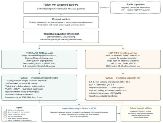
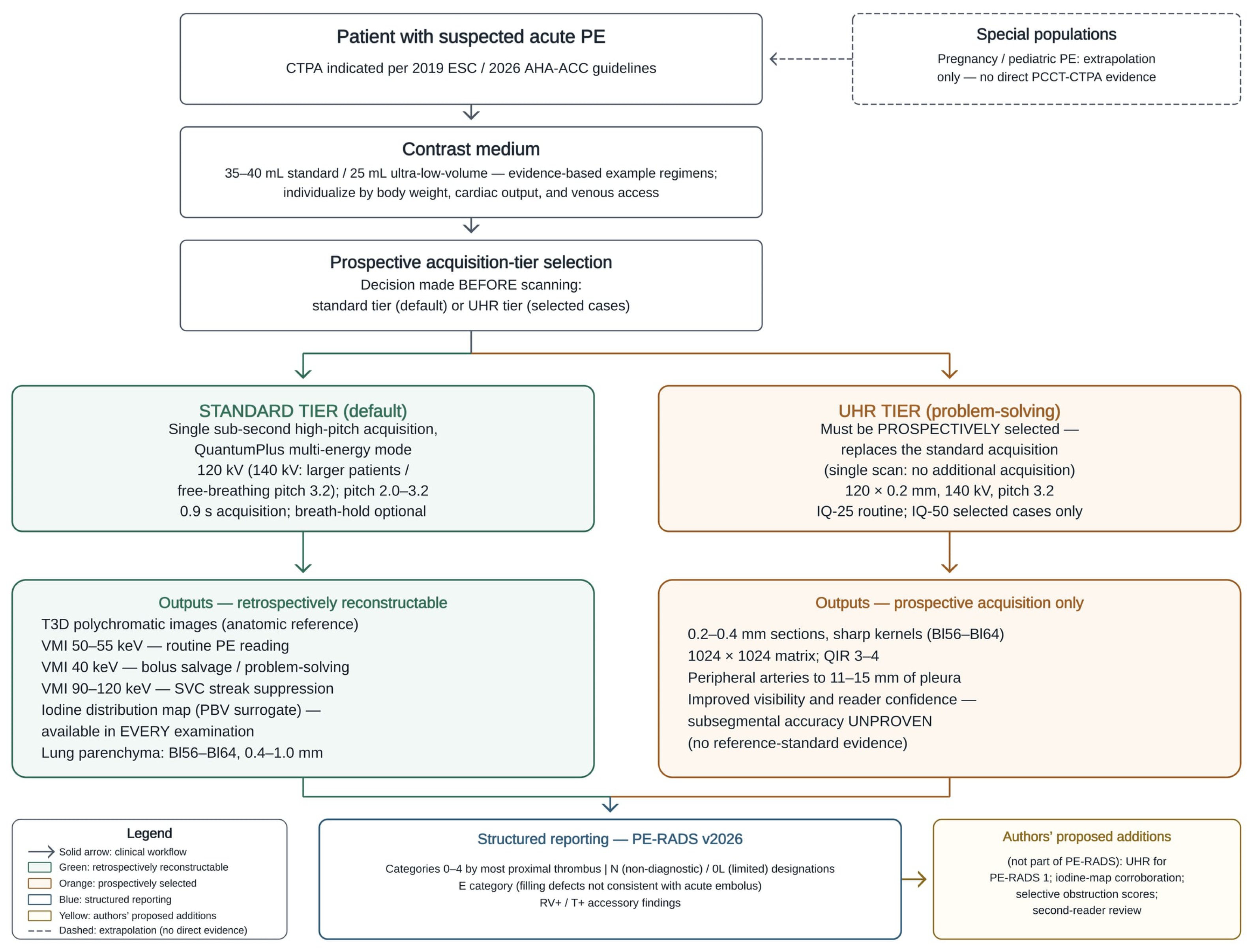
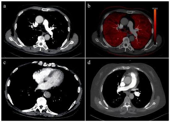
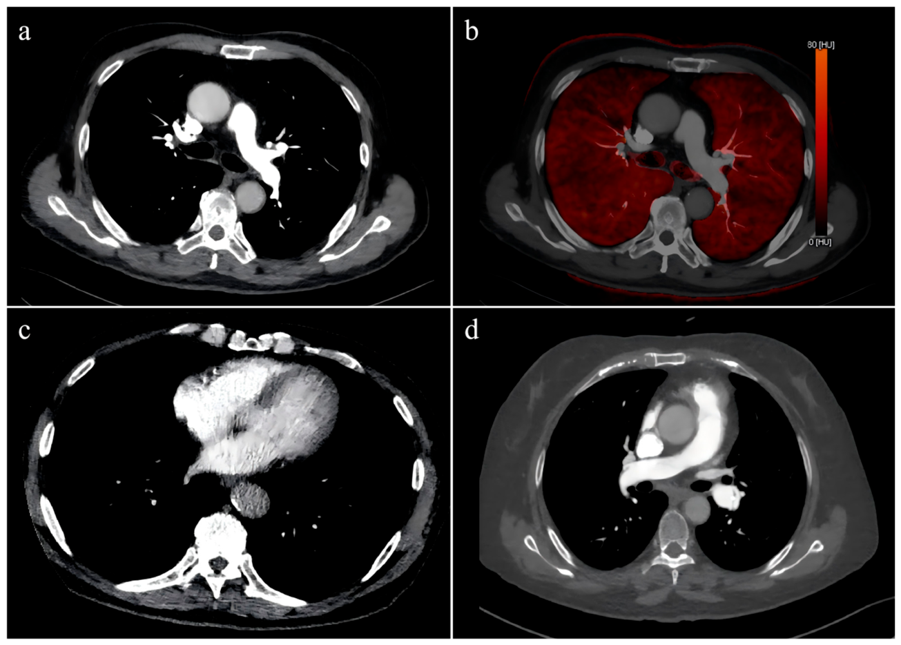

Disclaimer/Publisher’s Note: The statements, opinions and data contained in all publications are solely those of the individual author(s) and contributor(s) and not of MDPI and/or the editor(s). MDPI and/or the editor(s) disclaim responsibility for any injury to people or property resulting from any ideas, methods, instructions or products referred to in the content. |
© 2026 by the authors. Licensee MDPI, Basel, Switzerland. This article is an open access article distributed under the terms and conditions of the Creative Commons Attribution (CC BY) license.