
Substituted Poly(Vinylphosphonate) Coatings of Magnetite Nanoparticles and Clusters
by

Alexander Bunge
1,*, 
Cristian Leoștean
1,
Teodora Radu
1,
Septimiu Cassian Tripon
2,3,
Gheorghe Borodi
1 and
Rodica Turcu
1,*
1
Physics of Nanostructured Systems Department, National Institute for Research and Development of Isotopic and Molecular Technologies, 400293 Cluj-Napoca, Romania
2
Electron Microscopy Center “Prof. C. Craciun”, Faculty of Biology & Geology, “Babes-Bolyai” University, 5-7 Clinicilor St., 400006 Cluj-Napoca, Romania
3
Integrated Laboratory of Electron Microscopy, National Institute for Research and Development of Isotopic and Molecular Technologies, 400293 Cluj-Napoca, Romania
*
Authors to whom correspondence should be addressed.
Magnetochemistry 2022, 8(8), 79; https://doi.org/10.3390/magnetochemistry8080079
Submission received: 5 July 2022
/
Revised: 19 July 2022
/
Accepted: 23 July 2022
/
Published: 27 July 2022
(This article belongs to the Special Issue Advances in Magnetic Soft Materials: Synthesis, Characterization and Applications)
Abstract
:Magnetite nanoparticles and clusters of nanoparticles have been of Increasing scientific interest in the past decades. In order to prepare nanoparticles and clusters that are stable in suspension, different coatings have been used. Phosphates and phosphonates are a preferred anchoring group for the coating of magnetite nanomaterials. However, poly(vinylphosphonates) have rarely been used as a coating agent for any nanoparticles. Here, poly(methylvinylphosphonate) and other substituted polyvinylphosphonates are described as new coatings for magnetite nanoparticles and clusters. They show great stability in aqueous suspension. This is also the first time phosphonate-coated magnetite clusters have been synthesized in a one-pot polyol reaction. The coated magnetite nanoparticles and clusters have been characterized by TEM, EDX, FTIR, magnetization measurement, XRD as well as XPS. It has been shown that substituted vinylphosphonates can be easily synthesized in one-step procedures and as a polymeric coating can imbue important properties such as stability in suspension, tight binding to the particle surface, the ability to be further functionalized or to tightly adsorb metal ions. For the synthesis of magnetite clusters the cluster formation, polymerization and coating are done in a one-pot reaction and the resulting magnetite clusters show a higher amount of phosphonate coating than with a three-step procedure including a ligand exchange.
1. Introduction
Magnetite nanoparticles (MNP) are a field of research that has been increasingly growing, especially in the last two decades [1]. They offer unique properties thanks to their size [2], like a high reactivity because of a large surface-to-volume ratio, as well as superparamagnetism. Magnetite is the most commonly used material for the synthesis of magnetic nanostructures thanks to its high saturation magnetization (the highest of all single metal oxides) as well as relative chemical stability (compared to nanoparticles made of metals such as Fe, Co, Ni). These properties lead to magnetite nanoparticles finding applications in diverse fields such as catalysis [3], data storage [4], magnetic separation [5], wastewater purification/depollution [6], magnetorheologic fluids [7], and biomedicine [8,9].
Depending on the application and the properties required by it, magnetite nanoparticles can be synthesized by multiple routes [10], some of the most common ones being co-precipitation [11], hydrothermal synthesis [12], thermal decomposition [13], polyol reaction [14], microemulsion [15], or combustion [16].
For some applications, single nanoparticles are not the best option to use. This is especially the case when larger sizes (>~20 nm) are required while keeping the particles superparamagnetic or having a high surface. Magnetite clusters are ideal for these applications because they consist of smaller, individual particles which are superparamagnetic and keep this property even when clustered together [17,18,19,20,21]. Furthermore, depending on the synthesis procedure the space between the single particles allows them to retain a very high specific surface compared to single particles of the same size [22]. In addition, synergistic effects in the particle-particle interactions also increase the saturation magnetization of the obtained material [23,24,25,26]. Magnetite clusters can be principally synthesized in two ways [17,18,21], the first being a two-step process, in which the magnetite particles synthesized in the first step are clustered together in the second step. This is often done by a mini-emulsion process [27]. A single-step process combines nanoparticle formation with clustering in the same step; this is normally done in a polyol process [14]. The polyol reaction is a reduction of metal salts by polyols such as ethylene glycol at elevated temperatures and has been usually used to prepare metal nanoparticles [28]. For iron salts, the reduction generally proceeds only to magnetite and is normally (but not always [23]) done under solvothermal conditions. Many different coating agents such as polyacrylate [29], poly(vinylpyrrolidone) [30], acetate [31], oleate [32], ethylenediaminotetraacetate (EDTA) [33], polyethylene glycol (PEG) [34], citrate [35,36] or others have been used, and the properties such as cluster- and particle size can be controlled by varying reaction conditions such as solvent composition [37,38]. However, neither phosphates nor phosphonates have been described yet as a coating agent in the polyol synthesis of magnetite clusters.
Nanoparticle coatings play an important role in synthesis for their future applications [39,40], influencing parameters such as stability in suspension [41], hydrophobic/hydrophilic behavior [42], functionality for pollutant removal [43], catalytic activity [44], activity for biomedical applications [45,46,47], or future functionalization [48]. The coatings are normally supposed to stay anchored on the particle surface, as otherwise, they would lose much or all of their functionality, but this is not always easy [29]. Ideally, the anchoring should be done covalently [49], but especially for bare iron oxides, it can commonly only be done ionically or by complexation, which forms weaker bonds [50]. One strategy to overcome this is by using multiple functional groups in the same coating molecule, such as citrate or polyacrylate which offer multiple carboxylate groups to anchor to the particle surface [51]. Another strategy is to use functional groups as anchors that bind very tightly, such as thiol groups for silver- and gold nanoparticles [52]. For magnetite, phosphate and phosphonate groups fulfil this role very well [53,54,55]. It has been shown that phosphate groups bind better to the magnetite surface than sulfonate—or carboxylate groups [56,57,58,59], and traditionally only weakly complexing coating agents such as polyethylene glycol have shown improved binding capabilities by being functionalized with phosphate- or phosphonate groups [60,61]. Further, thanks to the negative surface charge of polyanionic groups the stability in the aqueous suspension of nanoparticles coated with them tends to be higher than the stability of nanoparticles coated with nonionic/monoionic coatings [62].
There have been attempts to coat magnetite nanoparticles with polymers containing one or multiple phosphonate or phosphate groups [63,64,65], but one of the most simple classes of polymers containing phosphonate groups, poly(vinylphosphic acids), has been mostly [66] overlooked. Not even other types of nanoparticles coated with poly(vinylphosphonic acids) have been described much in the literature, and if so, only poly(vinlyphosphonic acid) has been used either as homopolymer [67] or in the form of a copolymer [68]. There can be several reasons for that, but probably the price (EUR 424 per gram at Aldrich for simple poly(vinylphosphonic acid)) and the commercial unavailability of substituted poly(vinylphosphonic acids) plays some role. By adding extra functionality to phosphonate containing polymers [69] multifunctional stable coatings for nanoparticles could be created.
This lack of use of poly(vinylphosphonates) as a coating is unfortunate, as the potential benefits of large stability of the coating due to multiple strongly binding functional groups, as well as the stability in aqueous suspension due to electrostatic repulsion are obvious. This article aims to show that substituted poly(vinylphosphonic acids) can be synthesized cheaply and in large quantities, and used as a coating for both magnetite nanoparticles as well as magnetite clusters. Poly(vinylphosphonate) coated magnetite clusters have been synthesized for the first time in a one-pot reaction, and their properties are characterized by TEM, EDX, FTIR, XRD, magnetization measurements as well as XPS.
2. Materials and Methods
2.1. Chemicals
Chemicals were obtained from Sigma-Aldrich or Alfa Aesar in analytical or pure grade quality and were used without further purification.
2.2. Syntheses
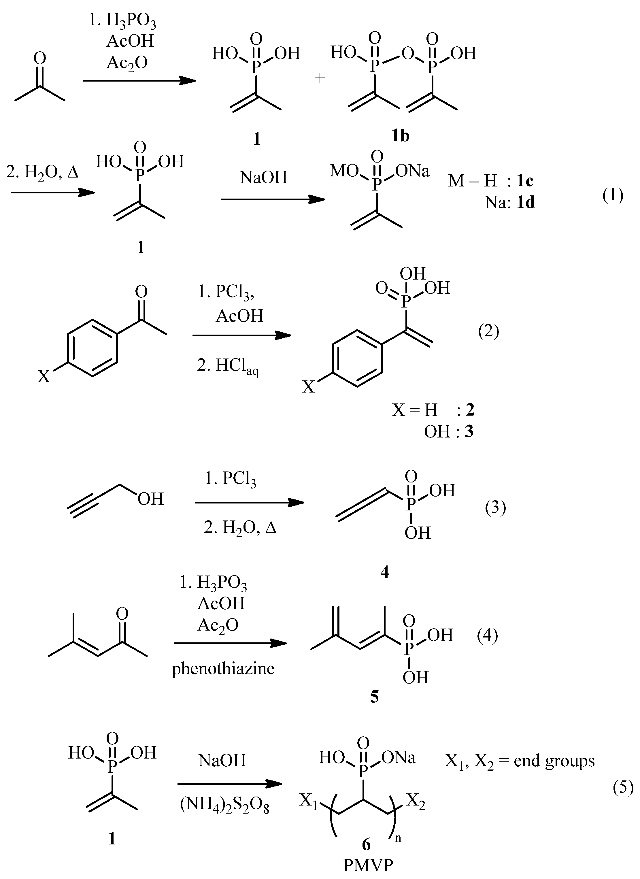
Methylvinylphosphonic acid (MVPA 1):
To phosphorous acid (41 g, 0.5 mol) in a 250 mL two-necked flask fitted with a thermometer was added acetic anhydride (100 g, 0.8 mol) and acetic acid (40 g, 0.67 mol) and stirred until everything dissolved. Afterward, acetone (35 g, 0.6 mol) was added dropwise, keeping the temperature below 40 °C (cooling with a water bath when necessary). After addition, the temperature was raised to 55 °C for 1 h, then to 100 °C for another hour. The flask was placed in a distillation setup and acetic acid was distilled off at 150 °C oil bath temperature. When nothing further distilled off at ambient pressure, a vacuum (~20 mbar) was applied and the bath temperature was raised to 170 °C until nothing more distilled off. The remaining viscous dark brown oil is a mixture of product 1 and its anhydride 1b (ratio ca. 2:8 by 31P NMR). To hydrolyze it, water (54 mL) was added and the mixture refluxed for 2 h to obtain a 50% aq. solution of methylvinylphosphonic acid 1.
The sodium salts 1c and 1d could be obtained by adding either 1 or 2 equivalents of 50% NaOH solution, precipitating with acetone, filtering and drying the precipitate.
1H-NMR (D2O, 500 MHz) [70]: δ[ppm] = 5.56–5.41 (m, 2H, C=CH2), 1.67 (dd, 3H, J1 = 14.3 Hz, J2 = 0.7 Hz, CH3). 13C-NMR (D2O, 125 MHz): δ[ppm] = 136.1 (d, J = 169.6 Hz, C-PO3H2), 127.9 (d, J = 9.9 Hz, C=CH2), 17.8 (d, J = 13.2 Hz, CH3). 31P-NMR (D2O, 202 MHz): δ[ppm] = 18.25.
The 31P-NMR signals (ppm) are 9.38 for the diphosphonic acid 1b, 12.84 for the monosodium phosphonate 1c (compare with [71] for the monobenzylammonium salt; 7.07 for the corresponding diphosphonate) and 14.71 for the disodiumphosphonate 1d (7.03 for the corresponding diphosphonate).
Poly(Methylvinylphosphonate) (PMVP 6):
Methylvinylphosphonic acid 1 (~50%, 20 g) was heated with NaOH (3.276 g) to 70 °C for 24 h and portionwise 2 g ammonium persulfate was added over this time while stirring. Finally, 19.945 g of solution was obtained.
PMVP Coated Magnetite Nanoparticles 7:
A solution of FeCl3·6H2O (3.028 g, 8 mmol) and FeCl2·4H2O (1.084 g, 4 mmol) in water (16 mL) was made and degassed by bubbling Ar gas through it. Ammonia solution (40 mL, 25%) was degassed in a 100 mL 3-necked flask fitted with a thermometer and condenser, then heated to 80 °C. The iron chloride solution was quickly added and the reaction stirred for 3 h at the same temperature, then 7.156 g of the poly(methylvinylphosphonate) solution 6 was added and the mixture stirred for another 3 h. After magnetic separation, the sample was washed with water (3×). The sample was precipitated by adding 1:1 acetone and magnetically separating overnight, then suspended in water (17.5 mL) to obtain a suspension of 1.046 g/mL density and 73.4 g/L concentration. An aliquot was dried in an oven (60 °C) to obtain samples for characterization.
One Pot Solvothermal Reaction to Get PMVP Coated Magnetite Clusters 12:
FeCl3·6H2O (1.085 g), NaOAc (2.88 g) and ethylene glycol (EG, 40 mL) were stirred for 10 min and then heated to reflux. The mixture turned darker slowly, and when it was completely black at 2 h, disodium methylvinylphosphonate 1d (300 mg) was added. The mixture was refluxed for another 6 h, cooled down, washed with EtOH (2×), water (3×) and magnetically separated, then dried in an oven (60 °C) to obtain 345 mg of a brown powder.
2.3. Characterization
Magnetic characterization of the samples at room temperature was performed using a Vibrating Sample Magnetometer (VSM) Cryogenic.
The size and shape of most of the nanostructures were examined by scanning transmission electron microscopy (STEM) with a Hitachi HD2700 equipped with a cold field emission gun, Dual EDX System (X-Max N100TLE Silicon Drift Detector (SDD)) from Oxford Instruments. For the analysis, a suspension of the samples was sonicated (<10 s) with a UP100H ultrasound finger and deposited by the droplet method on a 400-mesh copper grid coated with a thin carbon layer. For both types of analysis, the nominal operating tension was 200 kV. For samples 7, 10 and 11, a 1010 JEOL transmission electron microscope was used instead. The size of the nanoparticles and clusters was determined using the ImageJ software from 26–302 particles/clusters.
A SPECS XPS spectrometer equipped with an Al/Mg dual-anode X-ray source, a PHOIBOS 150 2D CCD hemispherical energy analyzer, and a multichanneltron detector with vacuum maintained at 1 × 10⁻⁹ Torr was used to record the XPS spectra. The Al Kα X-ray source (1486.6 eV) was operated at 200 W. The XPS survey spectra were recorded at 30 eV pass energy and 0.5 eV/step. The high-resolution spectra for the individual elements (Fe, C, O, P, Er, Eu) were recorded by accumulating 10 scans at 30 eV pass energy and 0.1 eV/step. Data analysis and curve fitting was performed using CasaXPS software with a Gaussian-Lorentzian product function and a nonlinear Shirley background subtraction. Peak shifts due to any apparent charging were normalized with the C1s peak set to 284.8 eV. The high-resolution spectra were partly deconvoluted into the components in order to determine the particular bond types present on the sample surface.
Fourier transform infrared (FTIR) spectra of samples 8, 14 and 16 were recorded using a JASCO FTIR 4600A spectrophotometer with ATR-PRO-ONE accessory, CO2-, H2O-, ATR- and baseline-corrected as well as normalized for better visibility of the bands. FTIR of all other samples was done with KBr pellets using a JASCO FTIR 610 spectrophotometer. The spectra were baseline-corrected for better visibility of the bands.
The NMR experiments were performed at 500 MHz for 1H, 125 MHz for 13C and 202 MHz for 31P, on a Bruker Avance III spectrometer. The NMR spectra were recorded in solution and all chemical shifts were measured relative to the respective solvent signal.
X-ray powder diffraction (XRPD) measurements were performed with a Bruker D8 Advance X-ray diffractometer, with a Ge (111) monochromator for Cu-Kα1 radiation (λ = 1.5406 Å) having the source power of 40 kV and 40 mA, at room temperature and LynxEye position-sensitive detector. Crystallite sizes were determined using the Scherrer equation [72]. Additional reference from supporting information: [73,74].
3. Results and Discussion
3.1. Synthesis and Polymerization of Vinylphosphonates
There exist multiple different routes to prepare vinylphosphonic acids [75,76,77]. For 1-methylvinylphosphonic acid 1, several different procedures exist starting from cheap acetone by reacting it with a trivalent phosphorous compound and subsequent dehydration. We decided to use a method employing phosphorous acid [78] (Scheme 1, (1)) instead of phosphorous trichloride [70] as it is less corrosive and does not produce HCl as byproduct. The initial product was a mixture of 1 and its anhydride 1b. The anhydride 1b (diphosphonic acid) could be hydrolyzed to 1 by heating an aqueous solution of the mixture. This aqueous solution was then used for further synthesis. Water-free methylvinylphosphonate could be obtained in the form of its monosodium or disodium salt (1c or 1d) by adding sodium hydroxide and precipitating the salt with acetone. The different salts, diphosphonates as well as the free acid were easily distinguished by 31P-NMR.
Another often described vinylphosphonic acid is 2-phenyldivinylphosphonic acid 2 [79,80,81]. It was synthesized according to Gulyukina et al. [82] from acetophenone with PCl3, (2) and aside from a relatively long crystallization time was easy to obtain.
Since other substituted phenylvinylphosphonic acids had been synthesized by the same method, it was then attempted to prepare 2-(p-propargyloxyphenyl)vinylphos- phonic acid from the respective acetophenone by the same method, however, under the reaction conditions a polymerization occurred, and the attempt was aborted. It was possible to synthesize 2-(p-hydroxyphenyl)phosphonic acid 3 from p-hydroxyacetophenone (2), however, the product did not crystallize and had to be obtained as an impure salt (the main impurity being sodium chloride) by evaporation of the neutralized solution.
Vinylphosphonic acids can also be prepared in different ways. According to Macomber et al. [83] allenylphosphonic acid 4 was prepared by a rearrangement from propargyl alcohol (3). It should be noted here that at least the allyloxyphosphonic dichloride has been reported as explosive [84], so care should be taken and intermediates not isolated.
Like allenylphosphonic acid 4, the phosphonic acid 5 obtained by reaction of mesityl oxide with phosphorous trichloride possesses two double bonds. The compound was synthesized according to [85,86] in impure form (4), as described there. The presence of a second double bond in 4 and 5 might make it possible to obtain coatings that have additional functionality, for example by the thiolene reaction.
The polymerization of 2-methylvinylphosphonic acid 1 was done in water using ammonium persulfate as an initiator (5). The polymerization went sluggishly, so more initiator was added over time. Even after 24 h, the polymerization was not complete, as could be seen from discrete peaks still appearing in the 31P-NMR spectrum. Since for nanoparticle synthesis the coating agent is added in excess anyway, and a polymer containing multiple phosphonate binding groups [65] binds stronger to the magnetite surface than a monomer containing only one phosphonate group, poly(2-methylvinylphosphonic acid) 6 was used further in this way.
3.2. Synthesis and Characterization of Magnetite Nanoparticles
For the synthesis of magnetite nanoparticles, the coprecipitation method was used. One of the advantages of the coprecipitation method for the synthesis of magnetite nanoparticles is that the synthesis procedure can be easily adapted to obtain either bare MNP or MNP with a specific coating, as the reaction is done in a one-pot two-step way, with the second step consisting of the addition of the ligand for coating. Thus poly(2-methylvinylphosphonate) coated MNP 7 was synthesized according to a well-known procedure (Scheme 2, (6)) [87], replacing the coating agent with the previously obtained polymer 6. For comparison, bare MNP 8 was also synthesized (7). The obtained MNP 7 were well-dispersible in water, and the suspension was stable enough to behave as a magnetic fluid when moved with a magnet (as opposed to just the MNP moving without the water), and even after years, a portion of the MNP 7 was still suspended in water.
In order to demonstrate the usefulness of these particles, Gd-, Tb-, Er- and Eu-ions were then adsorbed on the surface to obtain rare earth-PMVP-magnetite composite nanoparticles 9 (8). Rare earth ions have multiple uses in biomedicine [88], for example they can serve as a contrast agent for MRI, and as a fluorescent probe. In ionic form, rare earth metals are normally toxic, and thus to be used in the human body they have to be enclosed in a well-binding complex. Phosphates and phosphonates however bind also very strongly to rare earth ions, and thus leaching does not occur [89] even without complexation.
The synthesis of MNP coated with PMVP by coprecipitation 7 was done successfully. TEM (Figure 1) proved that the magnetic nanoparticles successfully formed. The nanoparticles are small, generally about 10 nm (Table 1), though some larger particles can also be found, making the sample polydisperse. The synthesized bare MNP 8 show a similar morphology, which means that the addition of PMVP only caused a coating, not a reforming of the MNP. For the MNP 9 with adsorbed rare earth metals, the morphology is the same as for 8 and 7. For 9a and 9b, with Gd and Er, respectively, EDX measurements were also performed (Figure S1). Besides iron and oxygen, both samples showed significant amounts of phosphorus and gadolinium/erbium as well, proof that the coating is anchored well to the surface of the nanoparticles and that adsorption of the rare earth ions has taken place.
FTIR spectroscopy of MNP 7, 8 and 9 (Figure 1 and Figure S2) show bands at ca. 550–570 cm−1, but none at or slightly higher than 635 cm−1 which is proof that magnetite and not maghemite has formed [90,91]. A broad band at 3000–3500 cm−1 stems from ν(O-H), partly from hydroxygroups bound to the magnetite surface, and (for 7 and 9) possibly from phosphonate groups. In 7 and 9 there are also bands at 2919 and 2854 cm−1 from ν(C-H) of PMVP; however, there is no ν(C-H) band at >3000 cm−1, which means that no methylvinylphosphonate monomers have attached to the MNP. A band at ca. 1150–1000 cm−1 points at ν(P=O) from the phosphonate groups. These bands signify the successful coating of the MNP with PMVP. The spectra of 7, 9a, 9b, 9c and 9d show no great differences, which means that the attachment of rare earth ions underwent without changes in the structure of the coating. It is likely that the phosphonate groups not already attached to the magnetite surface existed at least partially in anionic form (with ammonium or sodium as counterions), and these counterions were simply exchanged for the rare earth ions. It also means that there is no visible difference in the interaction of the different rare earth metal ions with the phosphonate coating.
Magnetization measurements (Figure 1 and Figure S3) of MNP 7, 8 and 9 showed that all samples were superparamagnetic, with saturation magnetizations of 74 (8), 63.7 (7), 63.7 (9a), 62.9 (9b), 63.2 (9c) and 63.8 (9d) emu/g. The comparatively large decrease of saturation magnetization between 8 and 7 is additional proof that the coating of MNP with PMVP has been successful. Between 7 and 9 the saturation magnetizations do not decrease as much—many phosphonate groups are likely bound to iron ions, and the rare earth ions do not have many free phosphonate groups to bind with. Hence, the amount of rare earth metal ions that are bound to the MNP by PMVP is likely relatively low.
XRD was measured for samples 7 and 8 (Figure S8a), and show typical peaks for spinel ferrites. Under the conditions used here, XRD by itself cannot distinguish between magnetite and maghemite, but taking into account the results from FTIR, magnetization and XPS measurements it is clear that the nanoparticles are magnetite.
The size distribution was determined both by TEM as well as XRD using the Scherrer equation (Table 1). The sizes determined by TEM do not differ notably, as was to be expected. Interesting is that the crystallite sizes determined from XRD patterns of 7 (11.3) and 8 (26.9 nm) do differ by a large amount, while the sizes by TEM (9.8/12.0 nm) do not differ that much. For an explanation it helps to keep in mind that the samples are polydisperse (which stems from the synthesis method, coprecipitation) and that while from TEM you can determine a number average of sizes (by measuring the diameter of the particles), from the Scherrer equation one would actually get the root (mean-4th-power divided by mean-square) of sizes [92]. The latter value takes into account far more the outliers with large sizes. From these results, one can see that by coating the magnetite nanoparticles with PMVP, their size decreases slightly, while at the same time also making them less polydisperse.
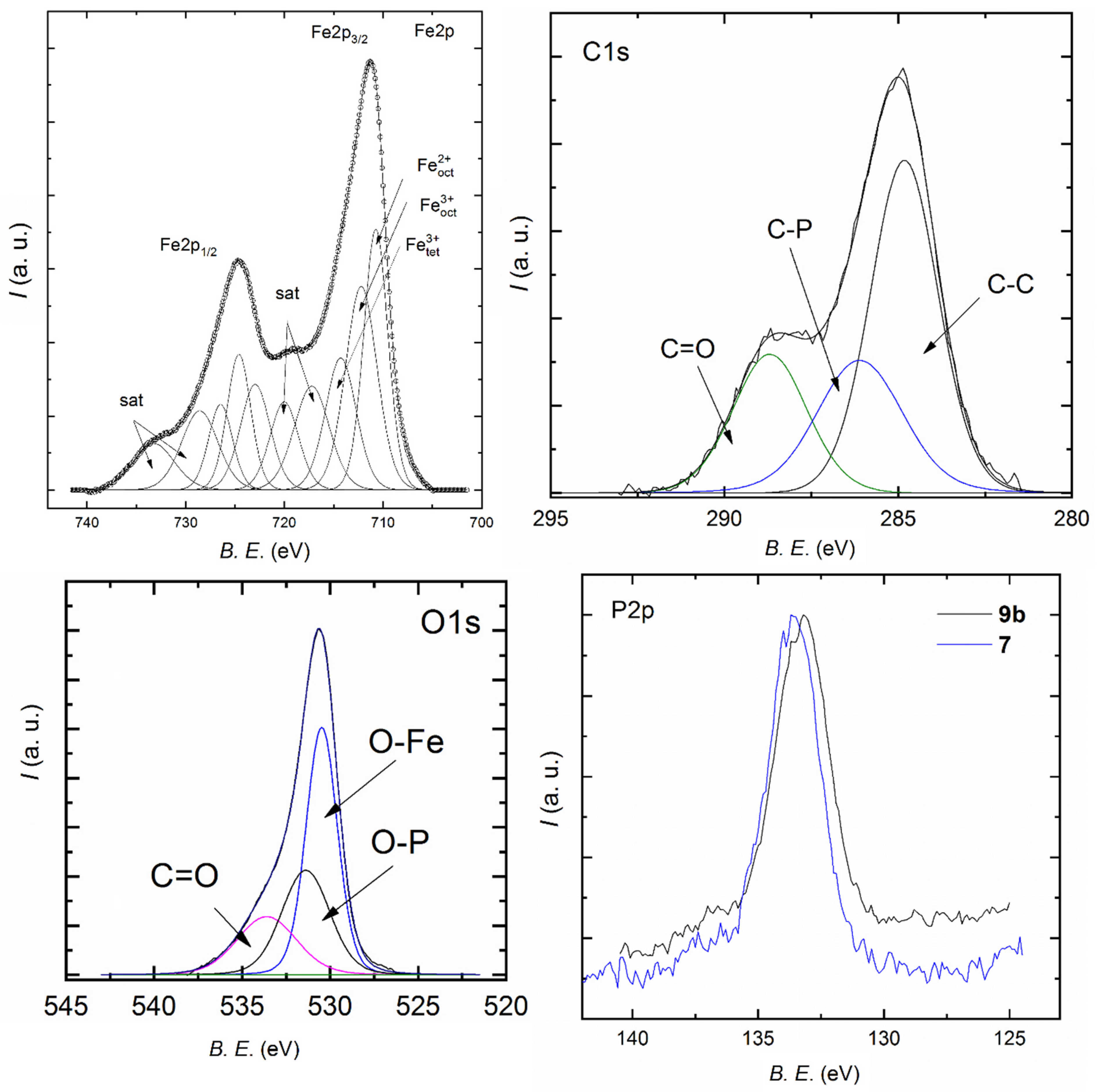
The MNP 7, 9b and 9d were also analyzed by XPS. The Fe2p core-level spectra of MNP (in Figure 2 is shown by way of example the Fe2p spectrum of 7) consists of the Fe2p1/2 (724.7 eV) and Fe2p3/2 (711.3 eV) contributions, along with their satellites. It can be further deconvoluted into contributions of tetrahedral and octahedral Fe3+, as well as octahedral Fe2+, according to the magnetite crystal structure (inverse spinel [93]). Using these peak areas, a ratio of Fe3+/Fe2+ can be calculated. In bulk magnetite, this ratio should be 2; however, in our samples, the ratio determined was 2.2. This is not surprising, as XPS is a surface analysis method and a slight deviation of the ideal ratio for surface ions is to be expected and in fact, has been found before for MNP synthesized by co-precipitation [87]. The C1s spectra can be deconvoluted into three components, a C–C component (284.8 eV), a C-P component at 286.2 eV as well as a component at 288.7 eV that represents carbonyl or carboxyl-C and is most likely contamination. The ratio of C–C/C–P component is 1.9, which is close to the expected ratio of 2 for PMVP. For the O1s core-level spectra (Figure 2 and Figure S4) three components can be identified, an O-Fe/O-metal contribution at 530.5 eV, an O-P contribution at 531.4 eV and a C=O contribution at 533.7 eV, which represents the same contamination found in the C1s spectra. The ratio O-Me/O-P changes from 1.5 (7) to 1.7 (9b) and 1.7 (9d). This is also proof that rare earth metal ions bound to the phosphate groups, increasing the relative amount of O-Me bonds. Interesting are the P2p core-level spectra. The peak maximum changes from 133.6 eV (7) to 133.1 eV (9b). The P2p peaks contain contributions of both P2p1/2 and P2p3/2, which likely contain contributions of multiple components according to the binding of the respective phosphonate group and are difficult to deconvolute. Unfortunately, there is little literature data comparing P2p spectra peak shifts of phosphonates according to binding state; however, for phosphates and phosphoric acid, which are similar, such a study has been done [94]. It has been found that a gradual replacement of hydrogen in phosphoric acid with sodium in phosphates causes the P2p peak to shift to lower energy. Furthermore, in the survey spectra (Figure S4) it is possible to see that 7 contains a small amount of sodium, which could not be washed away during synthesis; however, neither 9b nor 9d contain any sodium anymore. Together, these results demonstrate how the phosphonate binding situation changes during the synthesis of 9: PMVP is partially bound to iron in the MNP; however, there are groups that did not bind to the particles and are partially neutralized with sodium ions. On adsorption of rare earth metal (RE) ions, both sodium and protons are exchanged with RE ions. Since the pH during the reaction does not change or is maybe even more acidic due to the RE salts, this means that the RE ions bind very strongly to the phosphonate groups because they were able to replace hydrogen despite the neutral or low pH.
The Er4d core-level spectrum is shown in Figure S4, along with the P2s peak. The maximum of the Er4d5/2 peak is 169.4 eV. Unfortunately, there are not enough data in the literature to draw any conclusions, other than that Er is indeed bound to the MNP 9b. However, for the Eu3d core level spectra (Figure S4), Mercier et al. [95] collected data on the spectra of different Eu compounds. Comparing the value of the Eu3d5/2 peak (1135.5 eV) with these literature data, while explicit data for phosphonate salts of Eu are not given, it can be concluded that Eu is likely bound to phosphonate, and not, for example, precipitated as an oxide. This is important if the MNP is further used for medical applications, where RE metals could dissolve and show toxicity if they are not tightly bound to the MNP.
In conclusion, XPS proved that the MNP formed and consist of magnetite, that PMVP has coated them in 7 as well as 9, and that the RE ions are adsorbed and bound tightly to PMVP in compounds 9.
3.3. Synthesis and Characterization of Magnetite Clusters
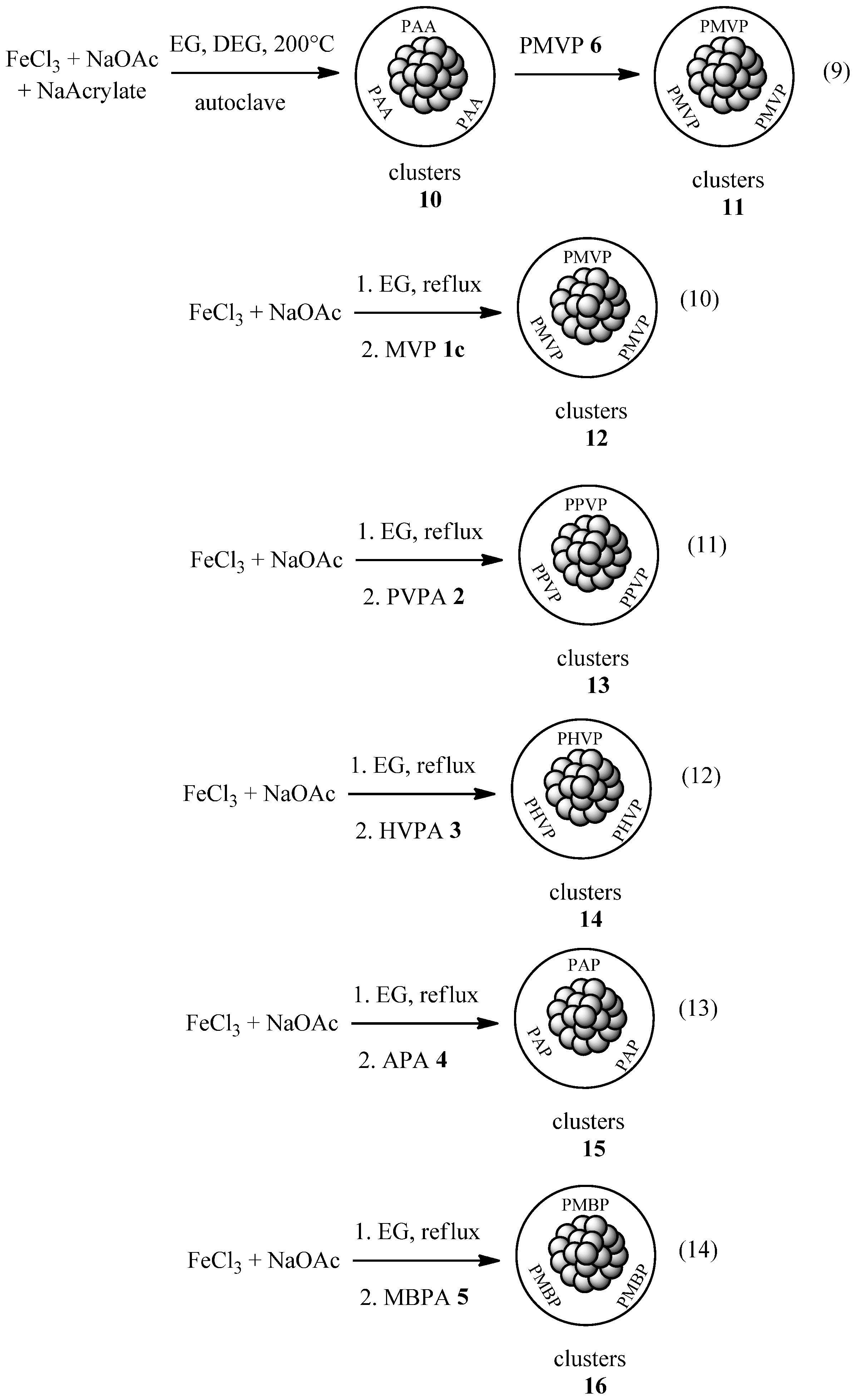
Magnetite clusters can be easily prepared in a polyol process and coated with a variety of organic ligands in a one-pot reaction (see introduction). It was first tried to replace the coating agent sodium acrylate [32] in a solvothermal process with PMVP, but this did not obtain magnetic materials. Only a brown nonmagnetic precipitate formed. Different ligands (free MVPA or sodium or disodium salts) and even other phosphates like phosphoric acid-2-hydroxyethyl methacrylate ester did not produce any magnetic product. It is likely that phosphates and phosphonates interact too strongly with the iron ions [96] so the normal mechanism of magnetite formation [32] in the polyol reaction cannot occur. One synthesis of phosphonate-coated MNP prepared hydrothermally in a one-step process has been described so far [97], but the reaction conditions are too different to be applied here. The only literature reference to solvothermally (in a polyol process) prepared magnetite nanoparticles coated with any phosphates or phosphonates described a two-step process, where the initially formed magnetite clusters were later coated with triphosphate by ligand exchange [98]. We attempted a similar reaction as well (9), and thus synthesized first magnetite clusters 10 with only acetate as a coating agent, which was then replaced in a second step by PMVP to obtain 11 (Scheme 3, first reaction).
A solvothermal process is however not the only way to do a polyol reaction, and indeed the polyol reaction [28] (for other metals) was first described as a reaction under normal pressure at elevated temperature or reflux. Furthermore, magnetite clusters have been prepared this way before [23]. It was thus decided to prepare PMVP-coated magnetite clusters under refluxing conditions, and only when the clusters have formed to add the phosphonate (Scheme 3). It was also decided that an initial polymerization is not necessary, as for example acrylic acid can polymerize under hydrothermal conditions without a catalyst [99], and even polyacrylate-coated magnetite clusters have been prepared from sodium acrylate before [32,37]. Thus PMVP coated magnetite clusters 12 were prepared by refluxing a mixture of iron chloride and sodium acetate in ethylene glycol (10), and when the clusters had formed (visible by the color change to black) MVPA (in the form of its disodium salt) was added. A magnetic product was formed successfully, which was easily dispersible in water.
It was then attempted to extend the reaction to other vinylphosphonates, and thus magnetite clusters 13 using 2-phenylvinylphosphonic acid (PVPA), 14 using 2-(p-hydroxyphenyl)vinylphosphonic acid (HVPA), 15 using allenylphosphonic acid (APA, 4) and 16 using 4-methylbuta-1,3-dien-2-ylphosphonic acid (MBPA) 5 were prepared (11–14). Magnetic material was formed in each reaction, but interestingly, the poly(2-phenylvinylphosphonate) (PPVP) coated clusters 13, which were synthesized for their potential hydrophobicity, were not too well dispersible neither in toluene nor water (but better in water than toluene). Poly(2-(p-hydroxyphenyl)vinylphosphonate) (PHVP) coated clusters 14 were, on the other hand, easily dispersible in water, as were poly(allenylphosphonate) (PAP) coated magnetite clusters 15. When synthesizing poly(4-methylbuta-1,3-dien-2-ylphosphonate) (PMBP) coated magnetic clusters 16, besides the clusters a hydrogel formed, which was not or hardly containing magnetic material and could be separated manually.
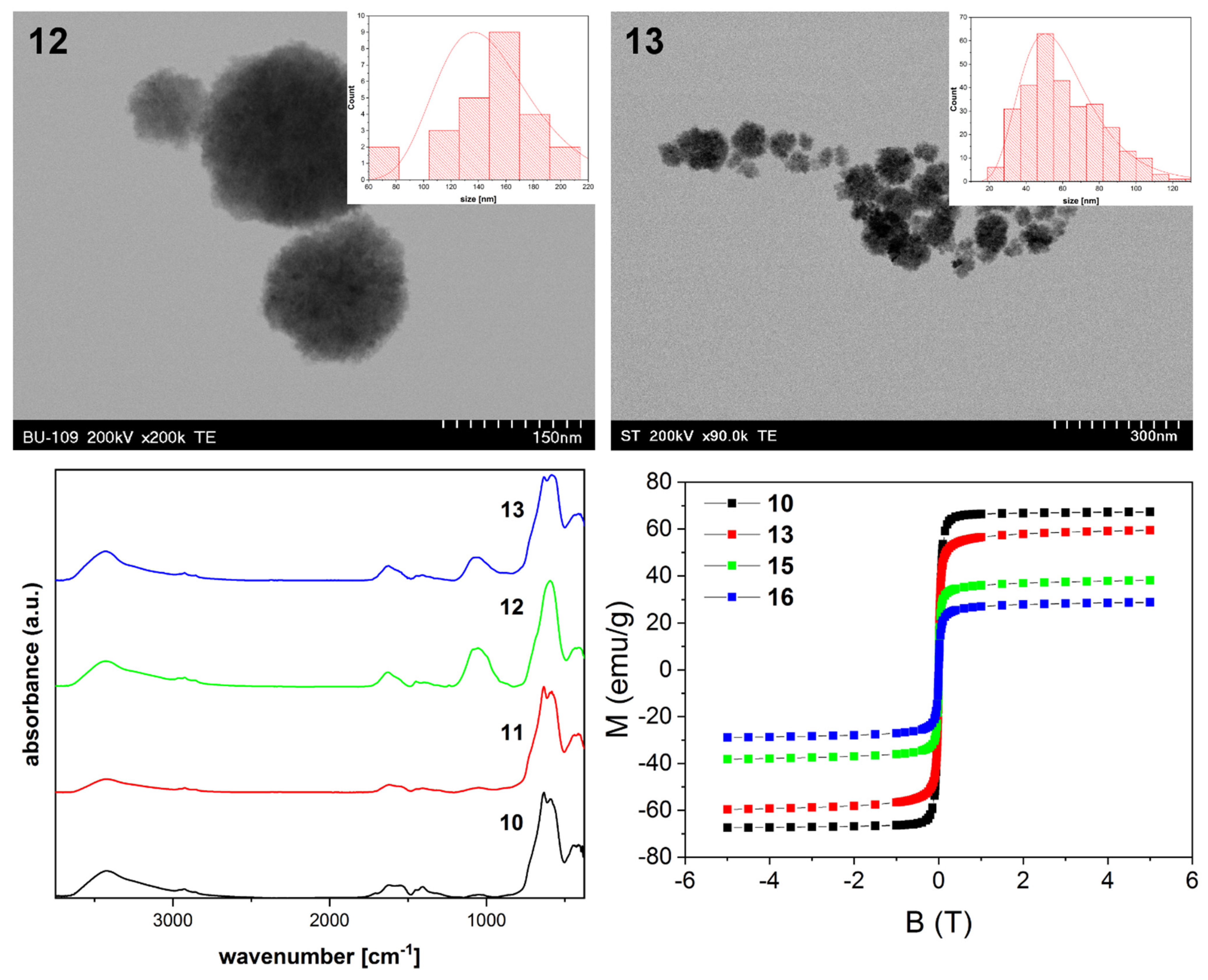
TEM and EDX (Figure 3, Figures S5 and S6) of the samples showed that magnetite clusters formed in each case. For the solvothermally produced samples 10 (300 nm) and 11 (299 nm) as well as the PMVP coated clusters 12 (149 nm) they were larger, whereas for the other samples they were less than 100 nm. EDX (Figure S6) of all clusters (except 10) showed the presence of phosphorus, which indicates that the synthesis of polyvinylphosphonate-coated magnetite clusters has been accomplished successfully. In the case of 11, the phosphorous content was relatively low, whereas for 15 it was quite high. Clusters 15 and 16 are formed; however, it appears that they are connected by the polymer.
FTIR analysis (Figure 3 and Figure S7) of the samples showed several differences. First, clusters 10, without phosphonate coating, showed bands at ca. 585–625 cm−1, indicating magnetite, a large band at 3000–3500 cm−1 (ν(O–H) of the magnetite surface, remaining ethylene glycol and polyacrylate), bands at 2850, 2915 and 2969 (ν(C-Halkyl)), 1632 and 1446 (symmetric and asymmetric ν(C=O) of carboxylate from acetate and polyacrylate) as well as a band around 1000 cm−1 (ν(C–O) of ethylene glycol) [29]. Sample 11, which should be coated with PMVP, showed almost no differences, except maybe slight changes in the band at around 1050 cm−1, which would stem from ν(P=O) of PMVP. Together with the low P content of this sample found by EDX, this means that the coating with PMVP is not very efficient in this method, as the sample seems to be only sparsely coated with the polymer. In contrast, clusters 12 show a much larger band around 1000 cm−1, indicating a much better coating with PMVP in this case. Furthermore, 13 and 14 also show such a large band, which is larger for 14 than for 13. These bands are even larger for 15 and 16, rating in intensity even higher than their magnetite band at 585–625 cm−1. On the other hand, neither 11, 12, 15 nor 16 show any ν(C–H) band above 3000 cm−1. For 11 and 12 this means that only polymer and no MVPA are attached to the magnetite clusters. For 15 and 16 this also means that in addition to only polymers attaching to the magnetic clusters, both of the double bonds of the monomers must have reacted and thus a large amount of crosslinking must have taken place. This correlates well with the fact that hydrogel visible by eye was found during the workup of sample 16, and the appearance of the clusters of both 15 and 16 being connected in TEM. From the analysis, one can conclude that gel-like crosslinked polymers were formed for both 15 and 16, but due to structural differences in sample 15 a part was soluble in water and washed away, while in 16 a part was insoluble and could be detected besides the magnetic clusters. In both cases a large part of the crosslinked polymer remained attached to the clusters, causing large bands assigned to the polymers to appear in the IR spectra.
The here-discussed results can also be verified by magnetization measurements (Figure 3): Magnetization curves were measured from non-phosphonate coated magnetite clusters 10, PPVP coated 13, PAP coated 15 and PMBP coated 16. All samples showed superparamagnetic behavior, which is one of the advantages of preparing multicore magnetite clusters instead of single-core magnetite particles of the same size. Compared to 10 (Ms = 67.9 emu/g), the saturation magnetization of 13 is lower (60 emu/g), indicating a larger amount of nonmagnetic polymer and thus a better coating. The saturation magnetization for samples 15 and 16 is even lower (38.7 and 29.1 emu/g, respectively). The effects that cluster size [29] or particle size (for particles larger than 10 nm, as is the case here) can have on saturation magnetization are not enough to explain this result. Thus these values confirm a larger amount of coating because larger amounts of crosslinked gel are attached to these samples.
XRD was done for selected samples (Figure S8b) and proved that the samples had a spinel ferrite structure. Among pure iron oxides both magnetite and maghemite have a spinel structure. Together with the other characterization (especially FTIR) as well as previous observations [29] it can however be concluded that the clusters consist of magnetite. Interestingly in sample 16, one peak exists at 2θ = 11°. It does not belong to the spinel pattern, and since these clusters contain a lot of the polymer, it is possible that the peak stems from the crystallized polymer instead.
Cluster size was determined using TEM results, while crystallite size was determined from XRD patterns via the Scherrer equation (Table 1). Samples 10 and 11 have a comparable size, which was to be expected as a ligand exchange should not change the size notably. Interesting on the other hand is that both cluster as well as crystallite size varies somewhat between clusters 12–16. It was expected that in the one pot reaction the clusters already formed fully before adding the phosphonate component (magnetite should definitely have formed as could be seen from the black color of the reaction mixture); however, it seems that while the particles were already somewhat formed [29], cluster formation was not yet finished and even the crystallites were still in the aggregation stage. The large size difference of cluster 12 compared to the others is likely because the sodium salt of MVPA was used in this reaction, and the pH can drastically influence the resulting clusters [23].
4. Conclusions
In this work, it was shown that substituted vinylphosphonic acids can be easily synthesized by different routes and polymerized to produce coatings for magnetite nanoparticles. Poly(methylvinylphosphonate) can be used to coat magnetite nanoparticles synthesized by coprecipitation and causes the particles to be very stable in aqueous suspension, as well as have the potential to bind rare earth metal ions for functionalization. These rare earth metal ions are adsorbed strongly on the surface of the MNP. Polymeric phosphonate-coated magnetite clusters were synthesized for the first time in a one-pot reaction—in this case, the monomeric phosphonates can be used, and the polymerization happens during the reaction. The coating with polymeric phosphonates is much stronger than in the two-step procedure that synthesizes the clusters first and then affects a ligand exchange. Several monomers have been used for coating, with different properties of the resulting phosphonate-coated magnetite clusters. For future applications, PHVP coated clusters could be further derivatized by linking different molecules to the phenol group, imbuing additional functionality, whereas with some change to the reaction conditions MBPA might be used to synthesize a magnetic hydrogel in a one-pot reaction. It is the hope of the authors to have shown the usefulness of substituted poly(vinylphosphonic acids) as a coating for magnetite nanomaterials and to encourage other scientists to begin using them as well.
Supplementary Materials
The following supporting information can be downloaded at: https://www.mdpi.com/article/10.3390/magnetochemistry8080079/s1. Additional synthesis procedures, TEM, EDX, FTIR, XRD and magnetization data. Figure S1: TEM (top) with size distribution and EDX (bottom) of MNP 9a and 9b; peaks of Au are from the sample holder, Si is an artifact of the device, and Os a misinterpretation of the evaluation software; Figure S2: FTIR spectra of MNP 9a, 9b and 9c; Figure S3: Magnetization of MNP 9a, 9b, 9c and 9d; Figure S4: XPS survey spectra of 7, 9a and 9b (top left); O1s spectra of 9b and 9d (top right); Er4d and P2s spectra of 9b (bottom left); Eu3d spectrum of 9d (bottom right); Figure S5: TEM results of magnetite clusters 10 (scalebar 200 nm), 11 (scalebar 500 nm), 15, 16 and 14 and their size distributions; Figure S6: EDX of magnetite clusters 11, 12, 13, 14, 15 and 16; Figure S7: FTIR spectra of clusters 14, 15 and 16; Figure S8: XRD patterns of samples (a): 7 and 8; (b): 11, 12, 13 and 16. The values in brackets represent the planes representing the specific reflection, the reflections are the same as magnetite (JCPDS file No. 19-0629).
Author Contributions
Conceptualization, A.B. and R.T.; methodology, A.B.; investigation, A.B., C.L., T.R., G.B. and S.C.T.; writing—original draft preparation, A.B.; writing—review and editing, A.B., C.L., T.R., S.C.T. and R.T.; visualization, A.B., C.L., T.R., G.B. and S.C.T.; supervision, R.T.; project administration, R.T.; funding acquisition, R.T. All authors have read and agreed to the published version of the manuscript.
Funding
This study was supported by the National Authority for Scientific Research and Innovation Romania (ANCS), Project PN16-30 02 02 and the Romanian Ministry of Research and Innovation, Project PN-19-35-02-03.
Data Availability Statement
Not applicable.
Acknowledgments
The authors would like to thank Sorina Ciupe for measuring most of the FTIR spectra, Adrian Pîrnău for measuring NMR, and Alin Sebastian Porav and Lucian Barbu-Tudoran for some of the TEM data.
Conflicts of Interest
The authors declare no conflict of interest. The funders had no role in the design of the study; in the collection, analyses, or interpretation of data; in the writing of the manuscript, or in the decision to publish the results.
References
- Laurent, S.; Forge, D.; Port, M.; Roch, A.; Robic, C.; Elst, L.V.; Muller, R.N. Magnetic iron oxide nanoparticles: Synthesis, stabilization, vectorization, physicochemical characterizations, and biological applications. Chem. Rev. 2008, 108, 2064–2110. [Google Scholar] [CrossRef] [PubMed]
- Roco, M.C. Nanoparticles and nanotechnology research. J. Nanopart. Res. 1999, 1, 1–6. [Google Scholar] [CrossRef]
- Mrowczynski, R.; Nan, A.; Liebscher, J. Magnetic nanoparticle-supported organocatalysts—An efficient way of recycling and reuse. RSC Adv. 2014, 4, 5927–5952. [Google Scholar] [CrossRef]
- Terris, B.D.; Thomson, T. Nanofabricated and self-assembled magnetic structures as data storage media. J. Phys. D Appl. Phys. 2005, 38, R199–R222. [Google Scholar] [CrossRef]
- Wang, Z.; Liu, C.T.; Wei, W.Z. Industry applications of magnetic separation based on nanoparticles: A review. Int. J. Appl. Electromagn. Mech. 2019, 60, 281–297. [Google Scholar] [CrossRef]
- Gupta, N.; Pant, P.; Gupta, C.; Goel, P.; Jain, A.; Anand, S.; Pundir, A. Engineered magnetic nanoparticles as efficient sorbents for wastewater treatment: A review. Mater. Res. Innov. 2018, 22, 434–450. [Google Scholar] [CrossRef]
- Kumar, J.S.; Paul, P.S.; Raghunathan, G.; Alex, D.G. A review of challenges and solutions in the preparation and use of magnetorheological fluids. Int. J. Mech. Mater. Eng. 2019, 14, 13. [Google Scholar] [CrossRef]
- Lee, H.; Shin, T.H.; Cheon, J.; Weissleder, R. Recent Developments in Magnetic Diagnostic Systems. Chem. Rev. 2015, 115, 10690–10724. [Google Scholar] [CrossRef] [Green Version]
- Lee, N.; Yoo, D.; Ling, D.; Cho, M.H.; Hyeon, T.; Cheon, J. Iron Oxide Based Nanoparticles for Multimodal Imaging and Magnetoresponsive Therapy. Chem. Rev. 2015, 115, 10637–10689. [Google Scholar] [CrossRef]
- Yusoff, A.H.M.; Salimi, M.N.; Jamlos, M.F. A review: Synthetic strategy control of magnetite nanoparticles production. Adv. Nano Res. 2018, 6, 1–19. [Google Scholar] [CrossRef]
- Nan, A.; Bunge, A.; Turcu, R. Hybride Magnetic Nanostructure Based on Amino Acids Functionalized Polypyrrole. In AIP Conference Proceedings; AIP Publishing LLC: Melville, NY, USA, 2015; Volume 1700, p. 060007. [Google Scholar] [CrossRef]
- Chircov, C.; Matei, M.F.; Neacsu, I.A.; Vasile, B.S.; Oprea, O.C.; Croitoru, A.M.; Trusca, R.D.; Andronescu, E.; Sorescu, I.; Barbuceanu, F. Iron Oxide-Silica Core-Shell Nanoparticles Functionalized with Essential Oils for Antimicrobial Therapies. Antibiotics 2021, 10, 1138. [Google Scholar] [CrossRef] [PubMed]
- Craciunescu, I.; Palade, P.; Iacob, N.; Ispas, G.M.; Stanciu, A.E.; Kuncser, V.; Turcu, R.P. High-Performance Functionalized Magnetic Nanoparticles with Tailored Sizes and Shapes for Localized Hyperthermia Applications. J. Phys. Chem. C 2021, 125, 11132–11146. [Google Scholar] [CrossRef]
- Rafienia, M.; Bigham, A.; Hassanzadeh-Tabrizi, S. Solvothermal synthesis of magnetic spinel ferrites. J. Med. Signals Sens. 2018, 8, 108–118. [Google Scholar] [CrossRef] [PubMed] [Green Version]
- Singh, P.; Upadhyay, C. Fine Tuning of Size and Morphology of Magnetite Nanoparticles Synthesized by Microemulsion. In AIP Conference Proceedings; AIP Publishing LLC: Melville, NY, USA, 2018; Volume 1953, p. 030051. [Google Scholar] [CrossRef]
- Pop, D.; Buzatu, R.; Moaca, E.A.; Watz, C.G.; Pinzaru, S.C.; Tudoran, L.B.; Nekvapil, F.; Avram, S.; Dehelean, C.A.; Cretu, M.O.; et al. Development and Characterization of Fe3O4@Carbon Nanoparticles and Their Biological Screening Related to Oral Administration. Materials 2021, 14, 3556. [Google Scholar] [CrossRef]
- Lu, Z.D.; Yin, Y.D. Colloidal nanoparticle clusters: Functional materials by design. Chem. Soc. Rev. 2012, 41, 6874–6887. [Google Scholar] [CrossRef] [PubMed]
- Guo, J.; Yang, W.L.; Wang, C.C. Magnetic Colloidal Supraparticles: Design, Fabrication and Biomedical Applications. Adv. Mater. 2013, 25, 5196–5214. [Google Scholar] [CrossRef]
- Boles, M.A.; Engel, M.; Talapin, D.V. Self-Assembly of Colloidal Nanocrystals: From Intricate Structures to Functional Materials. Chem. Rev. 2016, 116, 11220–11289. [Google Scholar] [CrossRef]
- Zhou, Z.J.; Tian, R.; Wang, Z.Y.; Yang, Z.; Liu, Y.J.; Liu, G.; Wang, R.F.; Gao, J.H.; Song, J.B.; Nie, L.M.; et al. Artificial local magnetic field inhomogeneity enhances T-2 relaxivity. Nat. Commun. 2017, 8, 15468. [Google Scholar] [CrossRef] [Green Version]
- Kostopoulou, A.; Lappas, A. Colloidal magnetic nanocrystal clusters: Variable length-scale interaction mechanisms, synergetic functionalities and technological advantages. Nanotech. Rev. 2015, 4, 595–624. [Google Scholar] [CrossRef]
- Li, Z.H.; Ma, Y.R.; Qi, L.M. Formation of nickel-doped magnetite hollow nanospheres with high specific surface area and superior removal capability for organic molecules. Nanotechnology 2016, 27, 485601. [Google Scholar] [CrossRef]
- Ge, J.P.; Hu, Y.X.; Biasini, M.; Beyermann, W.P.; Yin, Y.D. Superparamagnetic magnetite colloidal nanocrystal clusters. Angew. Chem. Int. Ed. 2007, 46, 4342–4345. [Google Scholar] [CrossRef] [PubMed]
- Kostopoulou, A.; Tsiaoussis, I.; Lappas, A. Magnetic iron oxide nanoclusters with tunable optical response. Photonics Nanostruct. Fundam. Appl. 2011, 9, 201–206. [Google Scholar] [CrossRef]
- Liu, Y.; Cui, T.T.; Li, Y.N.; Zhao, Y.T.; Ye, Y.C.; Wu, W.H.; Tong, G.X. Effects of crystal size and sphere diameter on static magnetic and electromagnetic properties of monodisperse Fe3O4 microspheres. Mater. Chem. Phys. 2016, 173, 152–160. [Google Scholar] [CrossRef]
- Wang, W.T.; Tang, B.T.; Wu, S.L.; Gao, Z.M.; Ju, B.Z.; Teng, X.X.; Zhang, S.F. Controllable 5-sulfosalicylic acid assisted solvothermal synthesis of monodispersed superparamagnetic Fe3O4 nanoclusters with tunable size. J. Magn. Magn. Mater. 2017, 423, 111–117. [Google Scholar] [CrossRef]
- Weiss, C.K.; Landfester, K. Miniemulsion Polymerization as a Means to Encapsulate Organic and Inorganic Materials. Adv. Polym. Sci. 2010, 233, 185–236. [Google Scholar] [CrossRef]
- Fiévet, F.; Brayner, R. The Polyol Process. In Nanomaterials: A Danger or a Promise? A Chemical and Biological Perspective; Brayner, R., Fiévet, F., Coradin, T., Eds.; Springer: London, UK, 2013; pp. 1–25. [Google Scholar]
- Bunge, A.; Porav, A.S.; Borodi, G.; Radu, T.; Pirnau, A.; Berghian-Grosan, C.; Turcu, R. Correlation between synthesis parameters and properties of magnetite clusters prepared by solvothermal polyol method. J. Mater. Sci. 2019, 54, 2853–2875. [Google Scholar] [CrossRef]
- Xuan, S.H.; Wang, F.; Wang, Y.X.J.; Yu, J.C.; Leung, K.C.F. Facile synthesis of size-controllable monodispersed ferrite nanospheres. J. Mater. Chem. 2010, 20, 5086–5094. [Google Scholar] [CrossRef]
- Jean, M.; Nachbaur, V.; Le Breton, J.M. Synthesis and characterization of magnetite powders obtained by the solvothermal method: Influence of the Fe3+ concentration. J. Alloys Compd. 2012, 513, 425–429. [Google Scholar] [CrossRef]
- Susan-Resiga, D.; Socoliuc, V.; Bunge, A.; Turcu, R.; Vekas, L. From high colloidal stability ferrofluids to magnetorheological fluids: Tuning the flow behavior by magnetite nanoclusters. Smart Mater. Struct 2019, 28, 115014. [Google Scholar] [CrossRef]
- Petran, A.; Radu, T.; Nan, A.; Olteanu, D.; Filip, A.; Clichici, S.; Baldea, I.; Suciu, M.; Turcu, R. Synthesis, characterization, and cytotoxicity evaluation of high-magnetization multifunctional nanoclusters. J. Nanopart. Res. 2016, 19, 10. [Google Scholar] [CrossRef]
- Deng, H.; Li, X.L.; Peng, Q.; Wang, X.; Chen, J.P.; Li, Y.D. Monodisperse magnetic single-crystal ferrite microspheres. Angew. Chem. Int. Ed. 2005, 44, 2782–2785. [Google Scholar] [CrossRef] [PubMed]
- Liu, J.; Sun, Z.K.; Deng, Y.H.; Zou, Y.; Li, C.Y.; Guo, X.H.; Xiong, L.Q.; Gao, Y.; Li, F.Y.; Zhao, D.Y. Highly Water-Dispersible Biocompatible Magnetite Particles with Low Cytotoxicity Stabilized by Citrate Groups. Angew. Chem. Int. Ed. 2009, 48, 5875–5879. [Google Scholar] [CrossRef] [PubMed]
- Shen, L.H.; Bao, J.F.; Wang, D.; Wang, Y.X.; Chen, Z.W.; Ren, L.; Zhou, X.; Ke, X.B.; Chen, M.; Yang, A.Q. One-step synthesis of monodisperse, water-soluble ultra-small Fe3O4 nanoparticles for potential bio-application. Nanoscale 2013, 5, 2133–2141. [Google Scholar] [CrossRef] [PubMed]
- Xuan, S.H.; Wang, Y.X.J.; Yu, J.C.; Leung, K.C.F. Tuning the Grain Size and Particle Size of Superparamagnetic Fe3O4 Microparticles. Chem. Mater. 2009, 21, 5079–5087. [Google Scholar] [CrossRef]
- Wang, W.T.; Tang, B.T.; Ju, B.Z.; Zhang, S.F. Size-controlled synthesis of water-dispersible superparamagnetic Fe3O4 nanoclusters and their magnetic responsiveness. RSC Adv. 2015, 5, 75292–75299. [Google Scholar] [CrossRef]
- Heuer-Jungemann, A.; Feliu, N.; Bakaimi, I.; Hamaly, M.; Alkilany, A.; Chakraborty, I.; Masood, A.; Casula, M.F.; Kostopoulou, A.; Oh, E.; et al. The Role of Ligands in the Chemical Synthesis and Applications of Inorganic Nanoparticles. Chem. Rev. 2019, 119, 4819–4880. [Google Scholar] [CrossRef] [Green Version]
- Popescu, R.C.; Andronescu, E.; Vasile, B.S. Recent Advances in Magnetite Nanoparticle Functionalization for Nanomedicine. Nanomaterials 2019, 9, 1791. [Google Scholar] [CrossRef] [Green Version]
- Cartwright, A.; Jackson, K.; Morgan, C.; Anderson, A.; Britt, D.W. A Review of Metal and Metal-Oxide Nanoparticle Coating Technologies to Inhibit Agglomeration and Increase Bioactivity for Agricultural Applications. Agronomy 2020, 10, 1018. [Google Scholar] [CrossRef]
- Almahfood, M.; Bai, B. The synergistic effects of nanoparticle-surfactant nanofluids in EOR applications. J. Pet. Sci. Eng. 2018, 171, 196–210. [Google Scholar] [CrossRef]
- Kefeni, K.K.; Mamba, B.B.; Msagati, T.A.M. Application of spinel ferrite nanoparticles in water and wastewater treatment: A review. Sep. Purif. Technol. 2017, 188, 399–422. [Google Scholar] [CrossRef]
- Rossi, L.M.; Costa, N.J.S.; Silva, F.P.; Wojcieszak, R. Magnetic nanomaterials in catalysis: Advanced catalysts for magnetic separation and beyond. Green Chem. 2014, 16, 2906–2933. [Google Scholar] [CrossRef]
- Schubert, J.; Chanana, M. Coating Matters: Review on Colloidal Stability of Nanoparticles with Biocompatible Coatings in Biological Media, Living Cells and Organisms. Curr. Med. Chem. 2018, 25, 4553–4586. [Google Scholar] [CrossRef] [PubMed]
- Chakraborty, A.; Boer, J.C.; Selomulya, C.; Plebanski, M. Amino Acid Functionalized Inorganic Nanoparticles as Cutting-Edge Therapeutic and Diagnostic Agents. Bioconjug. Chem. 2018, 29, 657–671. [Google Scholar] [CrossRef] [PubMed]
- Kostevsek, N. A Review on the Optimal Design of Magnetic Nanoparticle-Based T-2 MRI Contrast Agents. Magnetochemistry 2020, 6, 11. [Google Scholar] [CrossRef] [Green Version]
- Bunge, A.; Magerusan, L.; Morjan, I.; Turcu, R.; Borodi, G.; Liebscher, J. Diazonium salt-mediated synthesis of new amino, hydroxy, propargyl, and maleinimido-containing superparamagnetic Fe@C nanoparticles as platforms for linking bio-entities or organocatalytic moieties. J. Nanopart. Res. 2015, 17, 379. [Google Scholar] [CrossRef]
- Sulek, F.; Drofenik, M.; Habulin, M.; Knez, Z. Surface functionalization of silica-coated magnetic nanoparticles for covalent attachment of cholesterol oxidase. J. Magn. Magn. Mater. 2010, 322, 179–185. [Google Scholar] [CrossRef]
- Kallay, N.; Matijevic, E. Adsorption at Solid-Solution Interfaces. 1. Interpretation of Surface Complexation of Oxalic and Citric Acids with Hematite. Langmuir 1985, 1, 195–201. [Google Scholar] [CrossRef]
- Fresnais, J.; Yan, M.; Courtois, J.; Bostelmann, T.; Bee, A.; Berret, J.F. Poly(acrylic acid)-coated iron oxide nanoparticles: Quantitative evaluation of the coating properties and applications for the removal of a pollutant dye. J. Colloid Interface Sci. 2013, 395, 24–30. [Google Scholar] [CrossRef] [Green Version]
- Xue, Y.R.; Li, X.; Li, H.B.; Zhang, W.K. Quantifying thiol-gold interactions towards the efficient strength control. Nat. Commun. 2014, 5, 4348. [Google Scholar] [CrossRef] [Green Version]
- Miles, W.C.; Huffstetler, P.P.; Goff, J.D.; Chen, A.Y.; Riffle, J.S.; Davis, R.M. Design of Stable Polyether-Magnetite Complexes in Aqueous Media: Effects of the Anchor Group, Molecular Weight, and Chain Density. Langmuir 2011, 27, 5456–5463. [Google Scholar] [CrossRef]
- Sahoo, Y.; Pizem, H.; Fried, T.; Golodnitsky, D.; Burstein, L.; Sukenik, C.N.; Markovich, G. Alkyl phosphonate/phosphate coating on magnetite nanoparticles: A comparison with fatty acids. Langmuir 2001, 17, 7907–7911. [Google Scholar] [CrossRef]
- Demin, A.M.; Mekhaev, A.V.; Esin, A.A.; Kuznetsov, D.K.; Zelenovskiy, P.S.; Shur, V.Y.; Krasnov, V.P. Immobilization of PMIDA on Fe3O4 magnetic nanoparticles surface: Mechanism of bonding. Appl. Surf. Sci. 2018, 440, 1196–1203. [Google Scholar] [CrossRef]
- Muthukumaran, T.; Philip, J. Effect of phosphate and oleic acid capping on structure, magnetic properties and thermal stability of iron oxide nanoparticles. J. Alloys Compd. 2016, 689, 959–968. [Google Scholar] [CrossRef]
- Yee, C.; Kataby, G.; Ulman, A.; Prozorov, T.; White, H.; King, A.; Rafailovich, M.; Sokolov, J.; Gedanken, A. Self-assembled monolayers of alkanesulfonic and -phosphonic acids on amorphous iron oxide nanoparticles. Langmuir 1999, 15, 7111–7115. [Google Scholar] [CrossRef]
- Portet, D.; Denizot, B.; Rump, E.; Lejeune, J.J.; Jallet, P. Nonpolymeric coatings of iron oxide colloids for biological use as magnetic resonance imaging contrast agents. J. Colloid Interface Sci. 2001, 238, 37–42. [Google Scholar] [CrossRef] [PubMed]
- Shafi, K.V.P.M.; Ulman, A.; Yan, X.Z.; Yang, N.L.; Estournes, C.; White, H.; Rafailovich, M. Sonochemical synthesis of functionalized amorphous iron oxide nanoparticles. Langmuir 2001, 17, 5093–5097. [Google Scholar] [CrossRef]
- Goff, J.D.; Huffstetler, P.P.; Miles, W.C.; Pothayee, N.; Reinholz, C.M.; Ball, S.; Davis, R.M.; Riffle, J.S. Novel Phosphonate-Functional Poly(ethylene oxide)-Magnetite Nanoparticles Form Stable Colloidal Dispersions in Phosphate-Buffered Saline. Chem. Mater. 2009, 21, 4784–4795. [Google Scholar] [CrossRef]
- Oleksa, V.; Bernatova, I.; Patsula, V.; Liskova, S.; Balis, P.; Radosinska, J.; Micurova, A.; Kluknavsky, M.; Jasenovec, T.; Radosinska, D.; et al. Poly(ethylene glycol)-Alendronate-Coated Magnetite Nanoparticles Do Not Alter Cardiovascular Functions and Red Blood Cells’ Properties in Hypertensive Rats. Nanomaterials 2021, 11, 1238. [Google Scholar] [CrossRef]
- Tombácz, E.; Tóth, I.Y.; Nesztor, D.; Illés, E.; Hajdú, A.; Szekeres, M.; Vékás, L. Adsorption of organic acids on magnetite nanoparticles, pH-dependent colloidal stability and salt tolerance. Colloids Surf. A Physicochem. Eng. Asp. 2013, 435, 91–96. [Google Scholar] [CrossRef] [Green Version]
- Carroll, M.R.J.; Huffstetler, P.P.; Miles, W.C.; Goff, J.D.; Davis, R.M.; Riffle, J.S.; House, M.J.; Woodward, R.C.; St Pierre, T.G. The effect of polymer coatings on proton transverse relaxivities of aqueous suspensions of magnetic nanoparticles. Nanotechnology 2011, 22, 325702. [Google Scholar] [CrossRef]
- Pothayee, N.; Balasubramaniam, S.; Davis, R.M.; Riffle, J.S.; Carroll, M.R.J.; Woodward, R.C.; Pierre, T.G.S. Synthesis of ‘ready-to-adsorb’ polymeric nanoshells for magnetic iron oxide nanoparticles via atom transfer radical polymerization. Polymer 2011, 52, 1356–1366. [Google Scholar] [CrossRef]
- Baldim, V.; Bia, N.; Graillot, A.; Loubat, C.; Berret, J.F. Monophosphonic versus Multiphosphonic Acid Based PEGylated Polymers for Functionalization and Stabilization of Metal (Ce, Fe, Ti, Al) Oxide Nanoparticles in Biological Media. Adv. Mater. Interfaces 2019, 6, 1801814. [Google Scholar] [CrossRef]
- Golas, P.L.; Louie, S.; Lowry, G.V.; Matyjaszewski, K.; Tilton, R.D. Comparative Study of Polymeric Stabilizers for Magnetite Nanoparticles Using ATRP. Langmuir 2010, 26, 16890–16900. [Google Scholar] [CrossRef]
- Chen, Y.; He, L.; Chen, Z.Y.; Zhao, L.R.; Liang, J.Y.; Liu, G.J. Under-oil superhydrophilic TiO2/poly(sodium vinylphosphonate) nanocomposite for the separation of water from oil. Sep. Purif. Technol. 2020, 251, 117397. [Google Scholar] [CrossRef]
- Zheng, Z.Q.; Mounsamy, M.; Lauth-de Viguerie, N.; Coppel, Y.; Harrisson, S.; Destarac, M.; Mingotaud, C.; Kahn, M.L.; Marty, J.D. Luminescent zinc oxide nanoparticles: From stabilization to slow digestion depending on the nature of polymer coating. Polym. Chem. 2019, 10, 145–154. [Google Scholar] [CrossRef]
- Dolan, C.; Naysmith, B.; Hinkley, S.F.R.; Sims, I.M.; Brimble, M.A.; Williams, D.E.; Jin, J.Y. Synthesis of Novel Triazole-Containing Phosphonate Polymers. Aust. J. Chem. 2015, 68, 680–686. [Google Scholar] [CrossRef]
- Bal’tser, A.E.; Zaitsev, D.A.; Ivanova, T.V.; Babenko, T.G.; Barskova, E.N. Addition of morpholine and pyrrolidine to isopropenylphosphonic acid in situ. Russ. J. Org. Chem. 2013, 49, 627–628. [Google Scholar] [CrossRef]
- Gubnitskaya, E.S.; Peresypina, L.P. Esters Of N-Protected β-Aminoethylphosphonic Acid. J. Gen. Chem. USSR 1989, 59, 492–499. [Google Scholar]
- Scherrer, P. Bestimmung der Größe und der inneren Struktur von Kolloidteilchen mittels Röntgenstrahlen. Nachr. Ges. Wiss. Goettingen Math. Phys. Kl. 1918, 2, 98–100. [Google Scholar]
- Kenyon, G.L.; Westheimer, F.H. The Stereochemistry of Unsaturated Phosphonic Acids1. J. Am. Chem. Soc. 1966, 88, 3557–3561. [Google Scholar] [CrossRef]
- Dong, K.W.; Wang, Z.; Ding, K.L. Rh(I)-Catalyzed Enantioselective Hydrogenation of alpha-Substituted Ethenylphosphonic Acids. J. Am. Chem. Soc. 2012, 134, 12474–12477. [Google Scholar] [CrossRef]
- Kosolapoff, G.M. The Synthesis of Phosphonic and Phosphinic Acids. Org. React. 2011, 6, 273–338. [Google Scholar] [CrossRef]
- Macarie, L.; Ilia, G. Poly(vinylphosphonic acid) and its derivatives. Prog. Polym. Sci. 2010, 35, 1078–1092. [Google Scholar] [CrossRef]
- David, G.; Negrell-Guirao, C.; Iftene, F.; Boutevin, B.; Chougrani, K. Recent progress on phosphonate vinyl monomers and polymers therefore obtained by radical (co)polymerization. Polym. Chem. 2012, 3, 265–274. [Google Scholar] [CrossRef]
- Dabdoub, A.M. Methods for Synthesizing Phosphonic Compounds and Compounds Thereof. U.S. Patent US2007287858A1, 3 May 2007. [Google Scholar]
- Li, C.Y.; Saga, Y.; Onozawa, S.Y.; Kobayashi, S.; Sato, K.; Fukaya, N.; Han, L.B. Wet and Dry Processes for the Selective Transformation of Phosphonates to Phosphonic Acids Catalyzed by Bronsted Acids. J. Org. Chem. 2020, 85, 14411–14419. [Google Scholar] [CrossRef]
- Bondarenko, G.N.; Ganina, O.G.; Sharma, R.K.; Beletskaya, I.P. Catalytic activity of Pd catalysts on different supports in hydrogenation of 1-phenylethenylphosphonic acid. Russ. Chem. B 2014, 63, 1856–1859. [Google Scholar] [CrossRef]
- Zupancic, B.; Mohar, B.; Stephan, M. Impact on Hydrogenation Catalytic Cycle of the R Groups’ Cyclic Feature in “R-SMS-Phos”. Org. Lett. 2010, 12, 3022–3025. [Google Scholar] [CrossRef]
- Gulyukina, N.S.; Dolgina, T.M.; Bondarenko, G.N.; Beletskaya, I.P.; Bondarenko, N.A.; Henry, J.C.; Lavergne, D.; Ratovelomanana-Vidal, V.; Genet, J.P. Synthesis of biologically active 1-arylethylphosphonates. Russ. J. Org. Chem. 2002, 38, 573–587. [Google Scholar] [CrossRef]
- Macomber, R.S.; Kennedy, E.R. Phosphorus-containing products from the reaction of propargyl alcohols with phosphorus trihalides. 4. Alkyl substituent effects on oxaphospholene formation. J. Org. Chem. 1976, 41, 3191–3197. [Google Scholar] [CrossRef]
- Pirrung, M.C.; Fallon, L.; Lever, D.C.; Shuey, S.W. Inverse phosphotriester DNA synthesis using photochemically-removable dimethoxybenzoin phosphate protecting groups. J. Org. Chem. 1996, 61, 2129–2136. [Google Scholar] [CrossRef]
- Liu, Z.; De Campo, F.; Guy, L.; Wilson, D.J.; Jost, P. New Coupling Agents for Elastomer Compositions. World Patent WO2012146198 (A1), 11 November 2012. [Google Scholar]
- Liu, Z.; De Campo, F. Method for Preparing Conjugated Diene Phosphonate Compounds. World Patent WO2011050533 (A1), 30 October 2009. [Google Scholar]
- Circu, M.; Nan, A.; Borodi, G.; Liebscher, J.; Turcu, R. Refinement of Magnetite Nanoparticles by Coating with Organic Stabilizers. Nanomaterials 2016, 6, 228. [Google Scholar] [CrossRef] [PubMed] [Green Version]
- Ascenzi, P.; Bettinelli, M.; Boffi, A.; Botta, M.; De Simone, G.; Luchinat, C.; Marengo, E.; Mei, H.; Aime, S. Rare earth elements (REE) in biology and medicine. Rend. Lincei. Sci. Fis. E Nat. 2020, 31, 821–833. [Google Scholar] [CrossRef]
- Nieciecka, D.; Rekorajska, A.; Cichy, D.; Konska, P.; Zuk, M.; Krysinski, P. Synthesis and Characterization of Magnetic Drug Carriers Modified with Tb3+ Ions. Nanomaterials 2022, 12, 795. [Google Scholar] [CrossRef]
- Gotić, M.; Koščec, G.; Musić, S. Study of the reduction and reoxidation of substoichiometric magnetite. J. Mol. Struct. 2009, 924–926, 347–354. [Google Scholar] [CrossRef]
- Hu, L.; Percheron, A.; Chaumont, D.; Brachais, C.-H. Microwave-assisted one-step hydrothermal synthesis of pure iron oxide nanoparticles: Magnetite, maghemite and hematite. J. Sol. Gel. Sci. Technol. 2011, 60, 198. [Google Scholar] [CrossRef]
- Langford, J.I.; Wilson, A.J.C. Scherrer after sixty years: A survey and some new results in the determination of crystallite size. J. Appl. Crystallogr. 1978, 11, 102–113. [Google Scholar] [CrossRef]
- Fleet, M.E. The Structure of Magnetite. Acta Cryst. B 1981, 37, 917–920. [Google Scholar] [CrossRef]
- Fluck, E.; Weber, D. P2p-Bindungsenergien in Phosphor(III)—Verbindungen, Phosphoniumsalzen und Sauerstoffsäuren des Phosphors. Z. Naturforsch. B 1974, 29b, 603–607. [Google Scholar] [CrossRef]
- Mercier, F.; Alliot, C.; Bion, L.; Thromat, N.; Toulhoat, P. XPS study of Eu(III) coordination compounds: Core levels binding energies in solid mixed-oxo-compounds EumXxOy. J. Electron. Spectrosc. 2006, 150, 21–26. [Google Scholar] [CrossRef]
- Sudakar, C.; Subbanna, G.N.; Kutty, T.R.N. Effect of anions on the phase stability of gamma-FeOOH nanoparticles and the magnetic properties of gamma-ferric oxide derived from lepidocrocite. J. Phys. Chem. Solids 2003, 64, 2337–2349. [Google Scholar] [CrossRef]
- Thomas, G.; Demoisson, F.; Boudon, J.; Millot, N. Efficient functionalization of magnetite nanoparticles with phosphonate using a one-step continuous hydrothermal process. Dalton Trans. 2016, 45, 10821–10829. [Google Scholar] [CrossRef] [PubMed]
- Wan, J.Q.; Yuan, R.T.; Zhang, C.Y.; Wu, N.; Yan, F.Y.; Yu, S.S.; Chen, K.Z. Stable and Biocompatible Colloidal Dispersions of Superparamagnetic Iron Oxide Nanoparticles with Minimum Aggregation for Biomedical Applications. J. Phys. Chem. C 2016, 120, 23799–23806. [Google Scholar] [CrossRef]
- Kinoshita, K.; Takano, Y.; Ohkouchi, N.; Deguchi, S. Free-Radical Polymerization of Acrylic Acid under Extreme Reaction Conditions Mimicking Deep-Sea Hydrothermal Vents. ACS Omega 2017, 2, 2765–2769. [Google Scholar] [CrossRef] [PubMed] [Green Version]
Scheme 1.
Synthesis of substituted vinylphosphonates and PMVP 6.
Scheme 2.
Synthesis of magnetite nanoparticles.
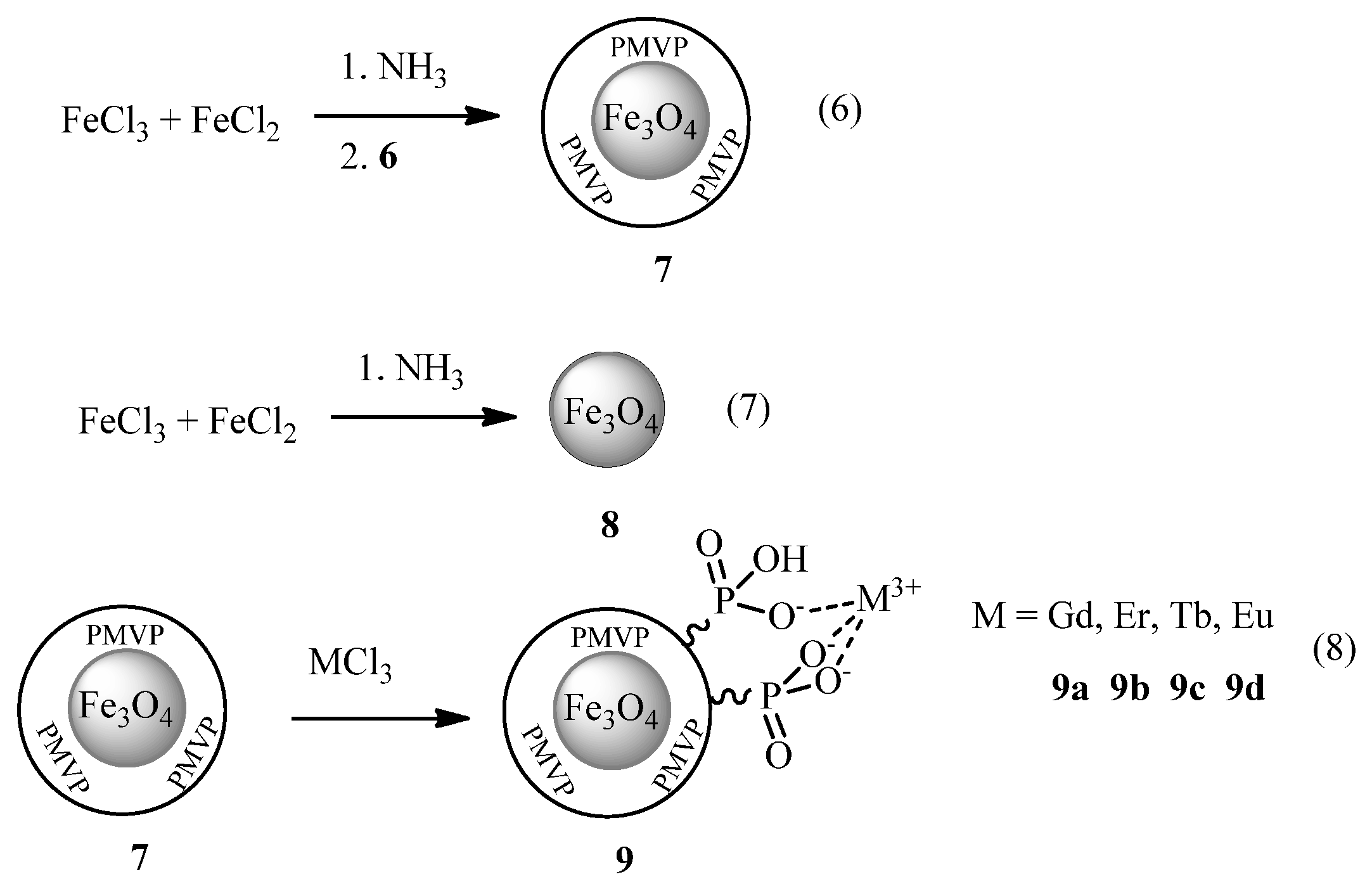
Figure 1.
TEM of magnetite nanoparticles 8 ((a), top left) and PMVP coated magnetite nanoparticles 7 ((b), top right)—scalebars 100 nm, inset are the size distributions; FTIR spectra of 8, 7 and 9a ((c), bottom left); magnetization curves of 8 and 7 ((d), bottom right).
Figure 1.
TEM of magnetite nanoparticles 8 ((a), top left) and PMVP coated magnetite nanoparticles 7 ((b), top right)—scalebars 100 nm, inset are the size distributions; FTIR spectra of 8, 7 and 9a ((c), bottom left); magnetization curves of 8 and 7 ((d), bottom right).
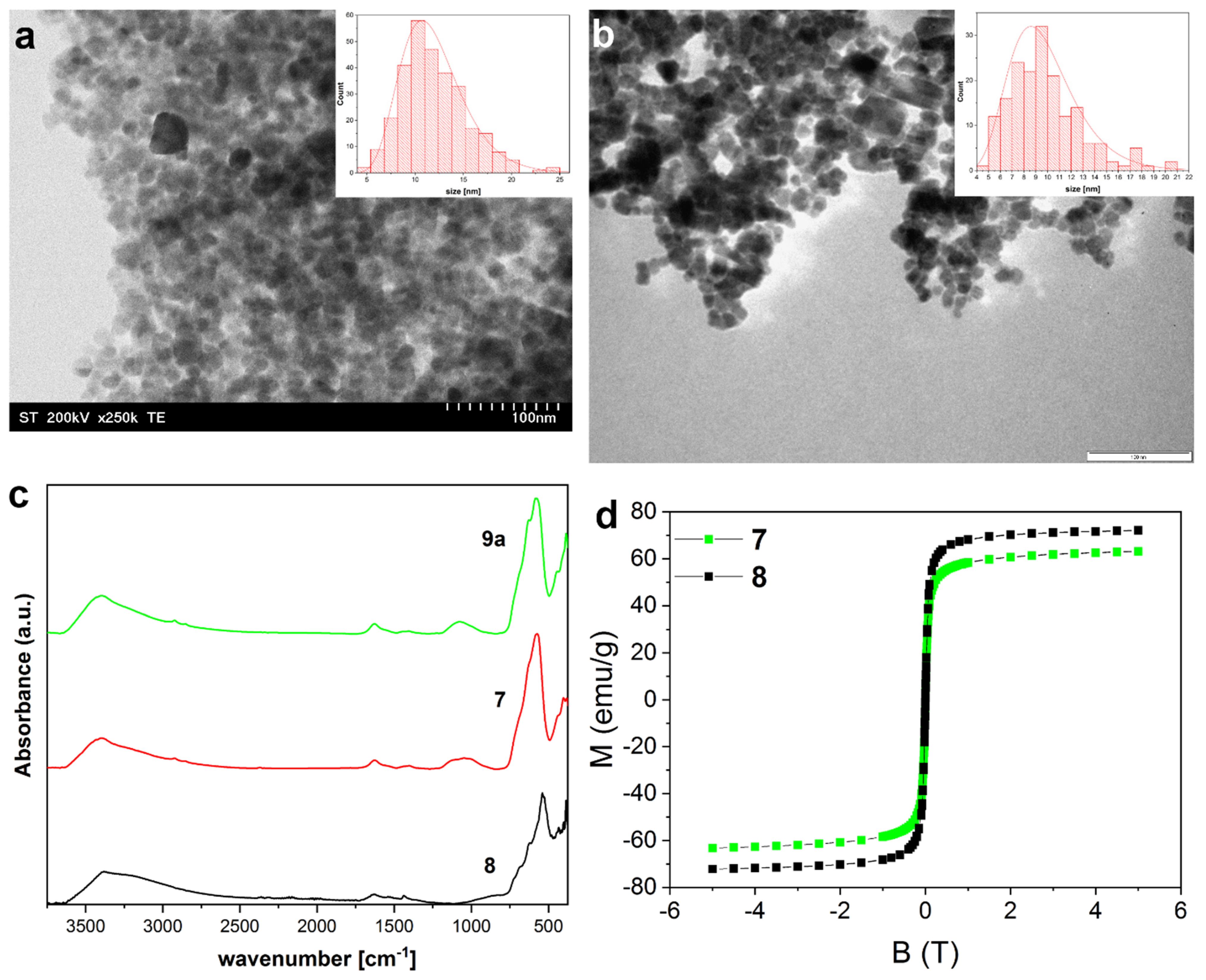
Figure 2.
XPS core level spectra (Fe2p, C1s, O1s) of 7 and P2p of 7 and 9b.
Scheme 3.
Synthesis of magnetite clusters.

Figure 3.
TEM of magnetite clusters 12 (top left) and 13 (top right) with size distribution; FTIR of clusters 10, 11, 12 and 13 (bottom left); magnetization of clusters 10, 13, 15 and 16 (bottom right).
Figure 3.
TEM of magnetite clusters 12 (top left) and 13 (top right) with size distribution; FTIR of clusters 10, 11, 12 and 13 (bottom left); magnetization of clusters 10, 13, 15 and 16 (bottom right).


Table 1.
Average size of the particles and clusters as measured from TEM, and crystallite size as determined by the Scherrer equation (XRD).
Table 1.
Average size of the particles and clusters as measured from TEM, and crystallite size as determined by the Scherrer equation (XRD).
| Entry | Size (TEM) [nm] | Crystallite Size (XRD) [nm] |
|---|---|---|
| 7 | 9.8 ± 3 | 11.3 |
| 8 | 12.0 ± 3.4 | 26.9 |
| 9a | 10.4 ± 2.9 | |
| 9b | 12.5 ± 3.4 | |
| 10 | 300 ± 102 | |
| 11 | 299 ± 79 | 13.7 |
| 12 | 149 ± 32 | 19.4 |
| 13 | 61 ± 21 | 10.9 |
| 14 | 41 ± 9 | |
| 15 | 52 ± 11 | |
| 16 | 59 ± 12 | 19.0 |
Publisher’s Note: MDPI stays neutral with regard to jurisdictional claims in published maps and institutional affiliations. |
© 2022 by the authors. Licensee MDPI, Basel, Switzerland. This article is an open access article distributed under the terms and conditions of the Creative Commons Attribution (CC BY) license (https://creativecommons.org/licenses/by/4.0/).
Share and Cite
MDPI and ACS Style
Bunge, A.; Leoștean, C.; Radu, T.; Tripon, S.C.; Borodi, G.; Turcu, R. Substituted Poly(Vinylphosphonate) Coatings of Magnetite Nanoparticles and Clusters. Magnetochemistry 2022, 8, 79. https://doi.org/10.3390/magnetochemistry8080079
AMA Style
Bunge A, Leoștean C, Radu T, Tripon SC, Borodi G, Turcu R. Substituted Poly(Vinylphosphonate) Coatings of Magnetite Nanoparticles and Clusters. Magnetochemistry. 2022; 8(8):79. https://doi.org/10.3390/magnetochemistry8080079
Chicago/Turabian StyleBunge, Alexander, Cristian Leoștean, Teodora Radu, Septimiu Cassian Tripon, Gheorghe Borodi, and Rodica Turcu. 2022. "Substituted Poly(Vinylphosphonate) Coatings of Magnetite Nanoparticles and Clusters" Magnetochemistry 8, no. 8: 79. https://doi.org/10.3390/magnetochemistry8080079
Note that from the first issue of 2016, this journal uses article numbers instead of page numbers. See further details here.