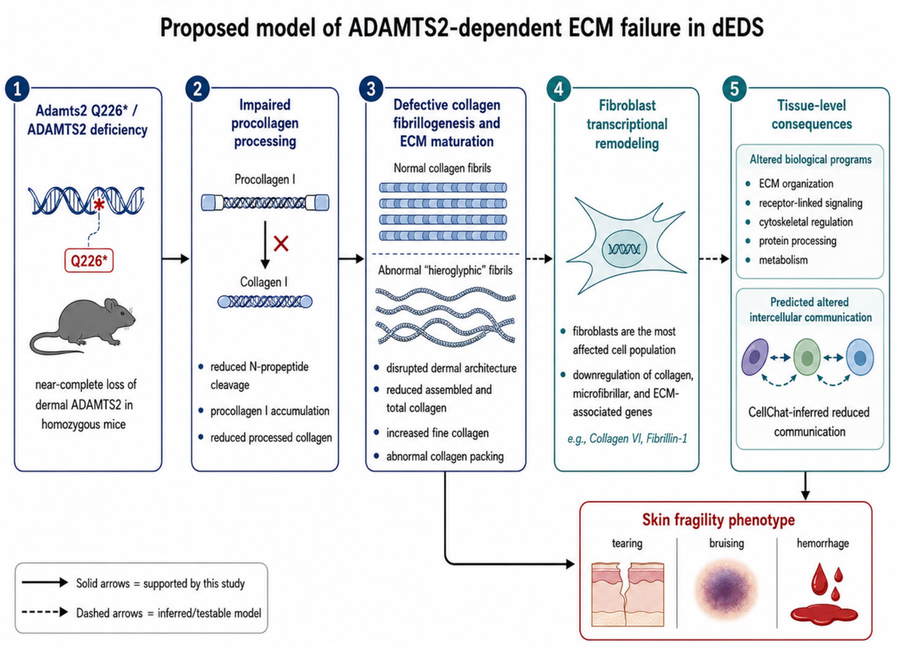

An Adamts2 Knock-In Model of Dermatosparaxis Ehlers–Danlos Syndrome Reveals Defective Collagen Maturation
 , , , , , , , , ,
Abstract
1. Introduction
2. Materials and Methods
2.1. Ethics Statement
2.2. Adamts2 Expression
2.3. Generation of Adamts2Q226* Mouse Model
2.4. Tissue Collection and Processing
2.5. Immunohistochemistry
2.6. Western Analyses
2.7. Transmission Electron Microscopy (TEM)
2.8. Quantitative Fibrosis Profiling
2.9. Single-Nucleus RNA Sequencing (snRNA-Seq) from FFPE Skin
2.10. snRNA-Seq Data Processing and Analysis
3. Results
3.1. Adamts2 Is Broadly Expressed in Developing and Adult Connective Tissue Compartments
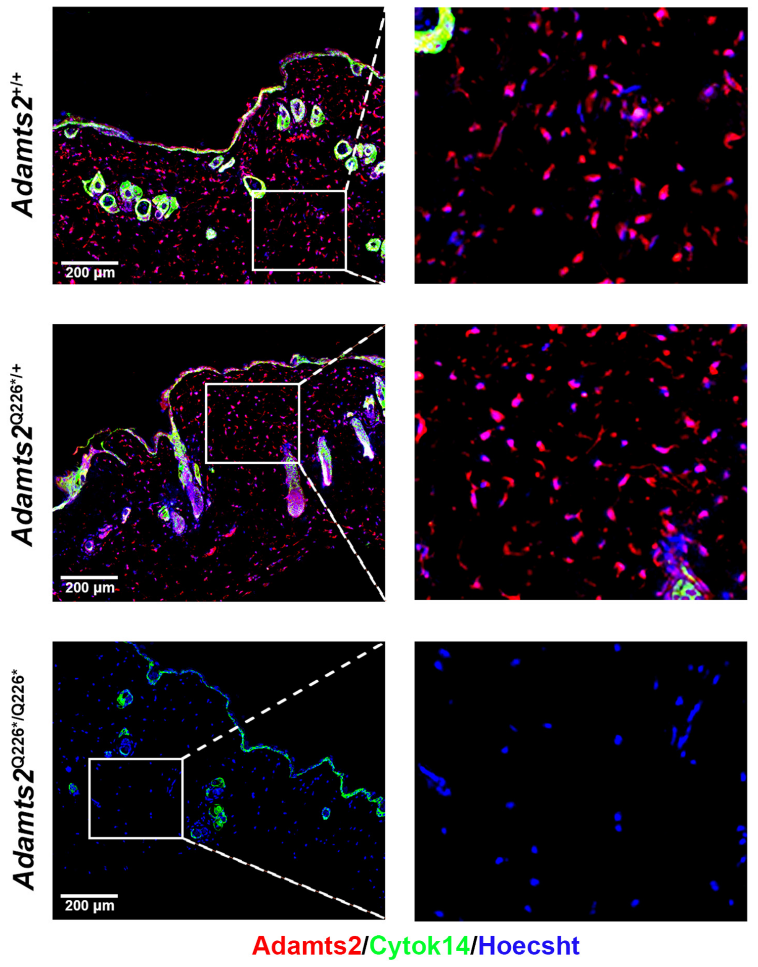
3.2. Gross Phenotype Observations and Loss of Function Validation of Adamts2Q226* In Vivo
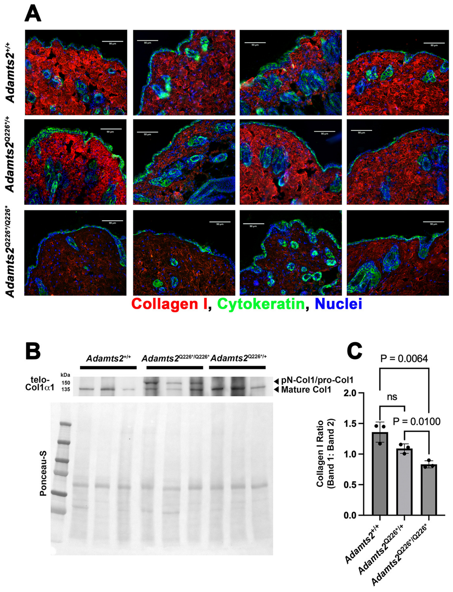
3.3. Collagen I Deposition and Processing Are Impaired in Mutants
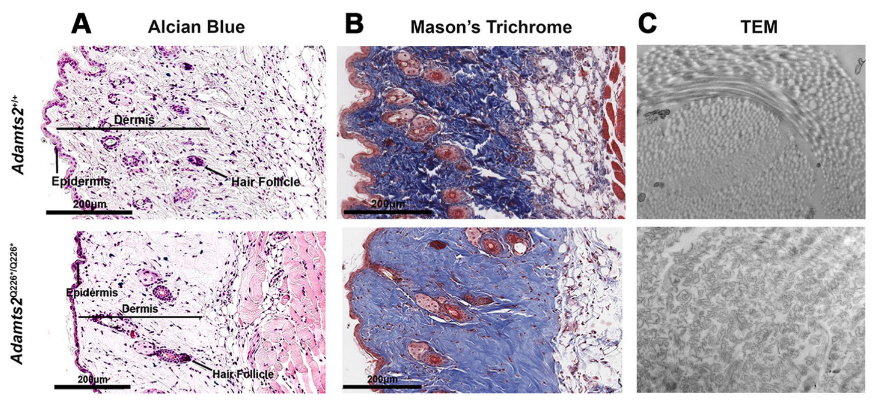
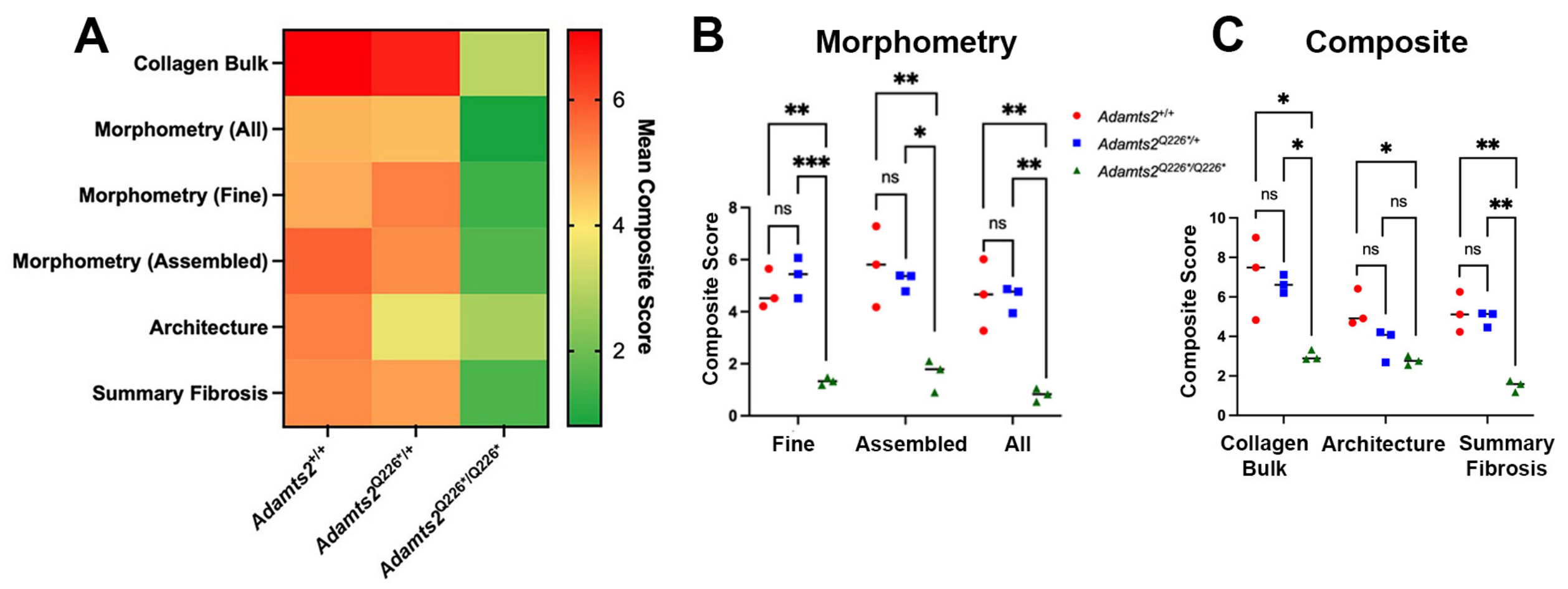
3.4. Digital Pathology Identifies Adamts2-Dependent Alterations in Tissue Architecture
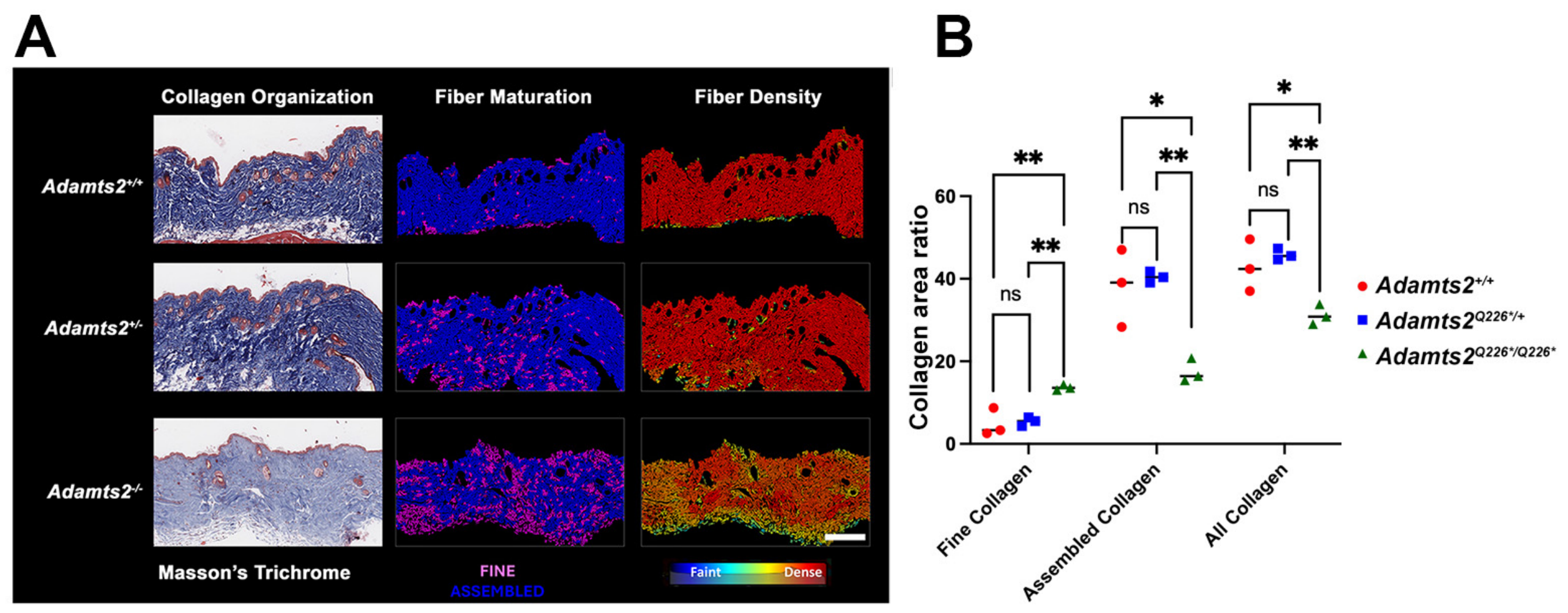
3.5. ADAMTS2 Deficiency Disrupts Dermal Collagen Bulk, Maturation, and Architecture
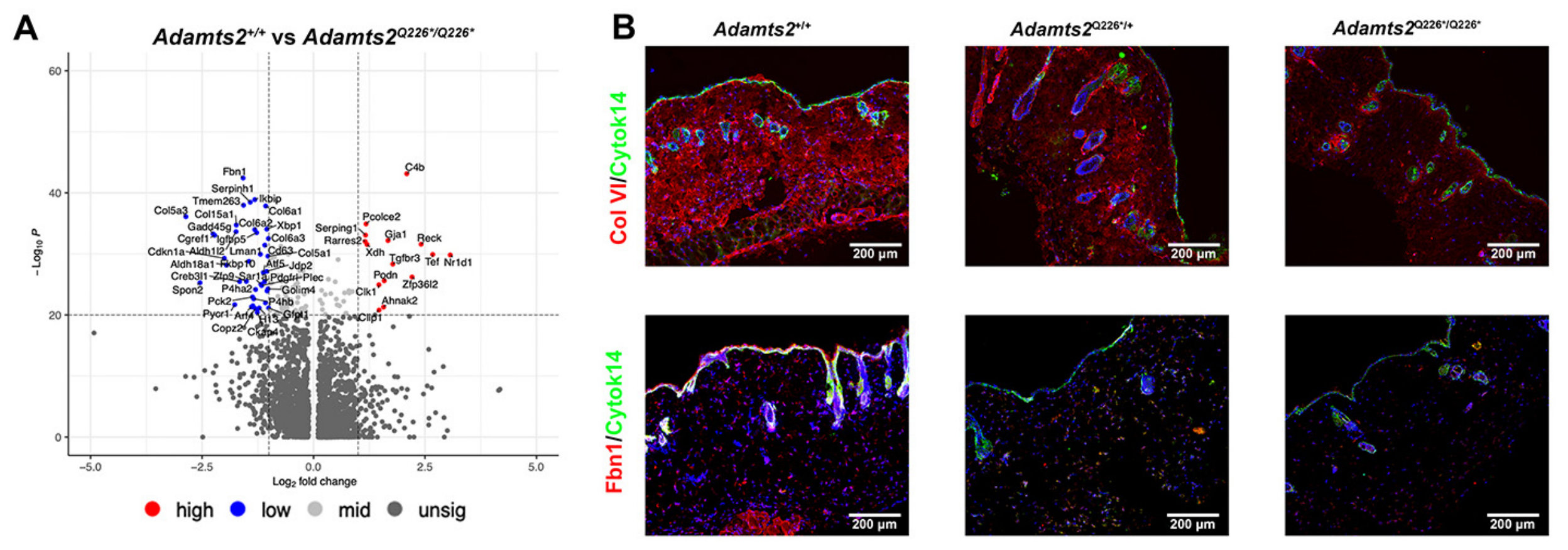
3.6. Single-Nuclei Transcriptomics Reveal Fibroblast-Enriched ECM Disruption
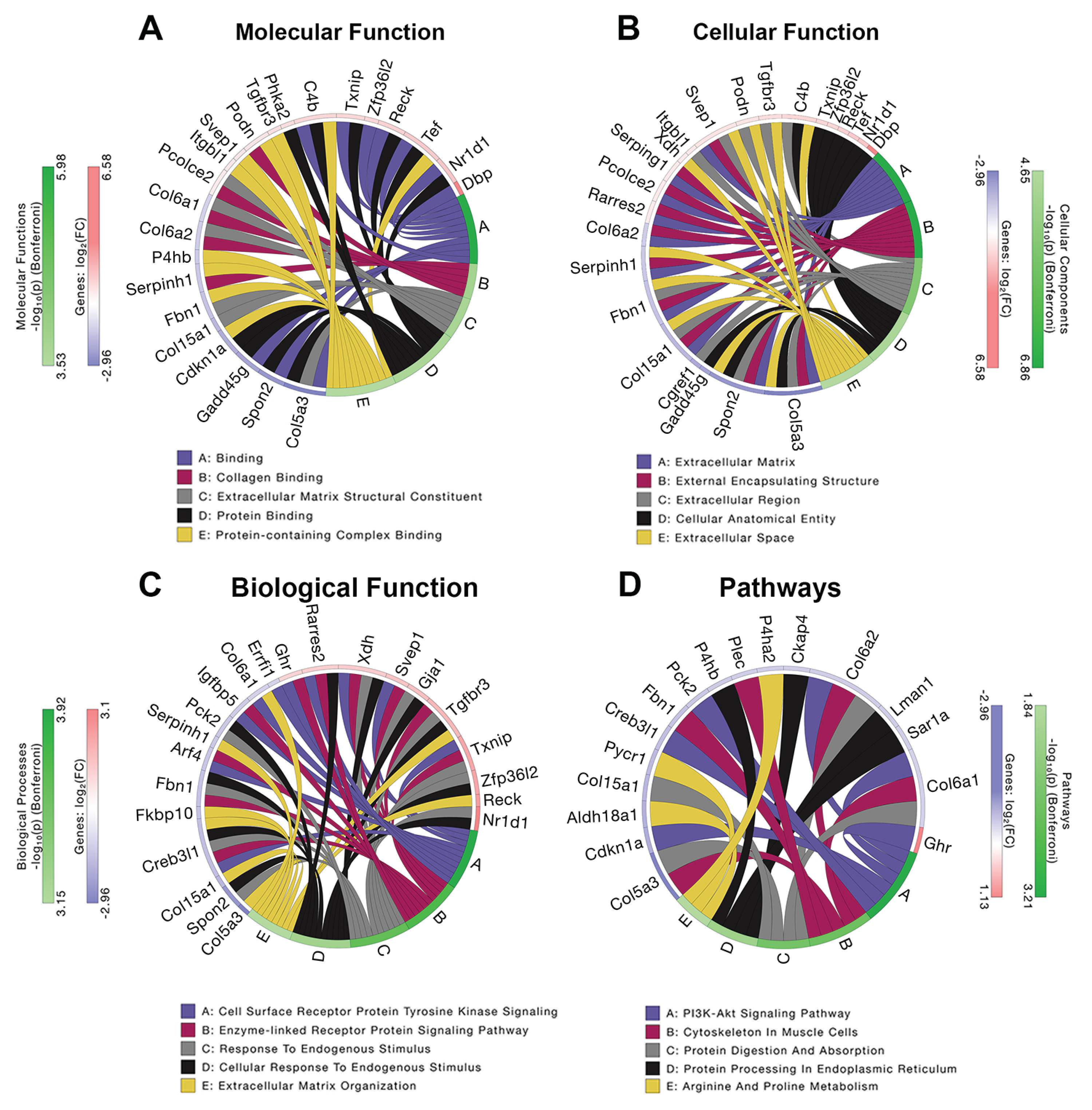
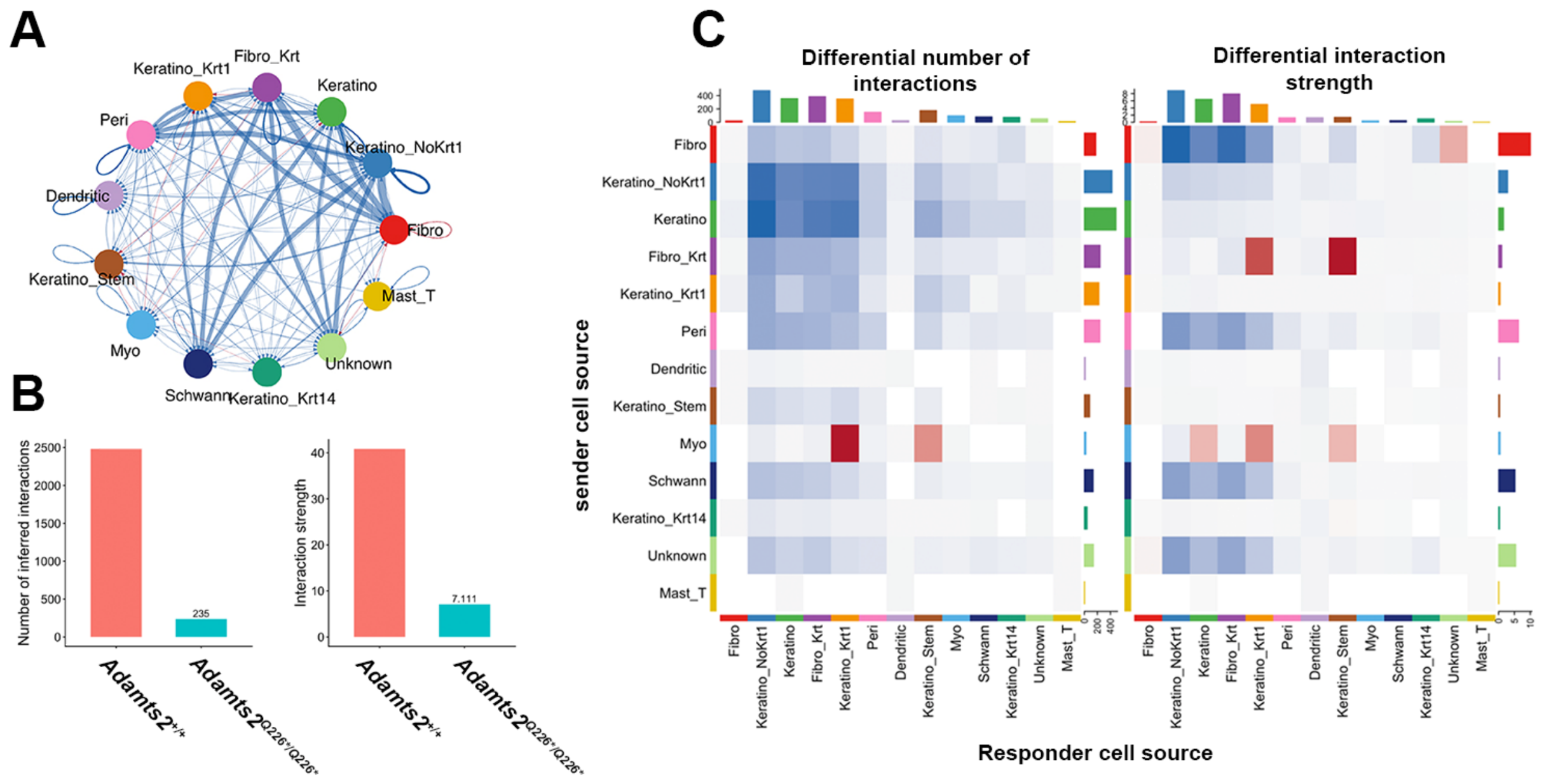
3.7. Functional Enrichment and CellChat Analyses Identify Coordinated Disruption of ECM Programs and Intercellular Communication in Homozygous Adamts2Q226*/Q226* Skin
4. Discussion
5. Study Limitations
6. Conclusions
Supplementary Materials
Author Contributions
Funding
Institutional Review Board Statement
Informed Consent Statement
Data Availability Statement
Acknowledgments
Conflicts of Interest
Abbreviations
| ACS | Architecture Composite Score |
| ADAMTS2 | A Disintegrin and Metalloproteinase with Thrombospondin Motifs 2 |
| CCS | Collagen Composite Score |
| CRISPR | Clustered Regularly Interspaced Short Palindromic Repeats |
| DAPI | 4′,6-Diamidino-2-phenylindole |
| DEG | Differentially Expressed Gene |
| dEDS | Dermatosparaxis Ehlers–Danlos Syndrome |
| ECM | Extracellular Matrix |
| EDS | Ehlers–Danlos Syndromes |
| FDR | False Discovery Rate |
| FFPE | Formalin-Fixed, Paraffin-Embedded |
| GEO | Gene Expression Omnibus |
| gRNA | Guide RNA |
| GTEx | Genotype-Tissue Expression |
| HRP | Horseradish Peroxidase |
| IACUC | Institutional Animal Care and Use Committee |
| LFC | Log2 Fold Change |
| MAST | Model-based Analysis of Single-cell Transcriptomics |
| MCS | Morphometric Composite Score |
| PBS | Phosphate-Buffered Saline |
| PCA | Principal Component Analysis |
| PCR | Polymerase Chain Reaction |
| Ph-FCS | Phenotype Fibrosis Composite Score |
| qFT | Quantitative Fibrosis Trait |
| RIPA | Radioimmunoprecipitation Assay |
| TEM | Transmission Electron Microscopy |
| UMAP | Uniform Manifold Approximation and Projection |
| WT | Wild Type |
References
- Malfait, F.; Francomano, C.; Byers, P.; Belmont, J.; Berglund, B.; Black, J.; Bloom, L.; Bowen, J.M.; Brady, A.F.; Burrows, N.P.; et al. The 2017 international classification of the Ehlers-Danlos syndromes. Am. J. Med. Genet. Part C Semin. Med. Genet. 2017, 175, 8–26. [Google Scholar] [CrossRef] [Scilit]
- Colige, A.; Sieron, A.L.; Li, S.W.; Schwarze, U.; Petty, E.; Wertelecki, W.; Wilcox, W.; Krakow, D.; Cohn, D.H.; Reardon, W.; et al. Human Ehlers-Danlos syndrome type VII C and bovine dermatosparaxis are caused by mutations in the procollagen I N-proteinase gene. Am. J. Hum. Genet. 1999, 65, 308–317. [Google Scholar] [CrossRef] [Scilit] [PubMed]
- Canty, E.G.; Kadler, K.E. Procollagen trafficking, processing and fibrillogenesis. J. Cell Sci. 2005, 118, 1341–1353. [Google Scholar] [CrossRef] [Scilit] [PubMed]
- Smith, L.T.; Wertelecki, W.; Milstone, L.M.; Petty, E.M.; Seashore, M.R.; Braverman, I.M.; Jenkins, T.G.; Byers, P.H. Human dermatosparaxis: A form of Ehlers-Danlos syndrome that results from failure to remove the amino-terminal propeptide of type I procollagen. Am. J. Hum. Genet. 1992, 51, 235–244. [Google Scholar] [PubMed]
- Li, S.W.; Arita, M.; Fertala, A.; Bao, Y.; Kopen, G.C.; Långsjö, T.K.; Hyttinen, M.M.; Helminen, H.J.; Prockop, D.J. Transgenic mice with inactive alleles for procollagen N-proteinase (ADAMTS-2) develop fragile skin and male sterility. Biochem. J. 2001, 355, 271–278. [Google Scholar] [CrossRef]
- Justice, M.J.; Dhillon, P. Using the mouse to model human disease: Increasing validity and reproducibility. Dis. Models Mech. 2016, 9, 101–103. [Google Scholar] [CrossRef] [Scilit] [PubMed]
- Vanlerberghe, R.; Colige, A.; Malfait, A.M.; Syx, D.; Malfait, F. ADAMTS2: More than a procollagen N-proteinase. Genes Dis. 2025, 12, 101686. [Google Scholar] [CrossRef] [Scilit] [PubMed]
- Norris, R.A.; Damon, B.; Mironov, V.; Kasyanov, V.; Ramamurthi, A.; Moreno-Rodriguez, R.; Trusk, T.; Potts, J.D.; Goodwin, R.L.; Davis, J.; et al. Periostin regulates collagen fibrillogenesis and the biomechanical properties of connective tissues. J. Cell. Biochem. 2007, 101, 695–711. [Google Scholar] [CrossRef] [Scilit] [PubMed]
- Gensemer, C.; Petrucci, T.; Beck, T.; Daylor, V.; Griggs, M.; Griggs, C.; Weintraub, A.; Byerly, K.; Guo, L.; Morningstar, J.; et al. KLK15 alters connective tissues in hypermobile Ehlers-Danlos syndrome. iScience 2025, 28, 113343. [Google Scholar] [CrossRef] [Scilit] [PubMed]
- Watson, A.; Petitjean, L.; Petitjean, M.; Pavlides, M. Liver fibrosis phenotyping and severity scoring by quantitative image analysis of biopsy slides. Liver Int. 2024, 44, 399–410. [Google Scholar] [CrossRef] [Scilit] [PubMed]
- Petitjean, L.; Chen, L.; Zhang, X.; Schiano, T.; Petitjean, M.; Sanyal, A.J.; Fiel, M. Digital Pathology Quantification of the Continuum of Cirrhosis Severity in Human Liver Biopsies. Liver Int. 2025, 45, e70166. [Google Scholar] [CrossRef] [Scilit] [PubMed]
- Schaum, N.; Karkanias, J.; Neff, N.F.; May, A.P.; Quake, S.R.; Wyss-Coray, T.; Darmanis, S.; Batson, J.; Botvinnik, O.; Chen, M.B.; et al. Single-cell transcriptomics of 20 mouse organs creates a Tabula Muris. Nature 2018, 562, 367–372. [Google Scholar] [CrossRef] [Scilit] [PubMed]
- Fleming, S.J.; Chaffin, M.D.; Arduini, A.; Akkad, A.-D.; Banks, E.; Marioni, J.C.; Philippakis, A.A.; Ellinor, P.T.; Babadi, M. Unsupervised removal of systematic background noise from droplet-based single-cell experiments using CellBender. Nat. Methods 2023, 20, 1323–1335. [Google Scholar] [CrossRef] [Scilit] [PubMed]
- Stuart, T.; Butler, A.; Hoffman, P.; Hafemeister, C.; Papalexi, E.; Mauck, W.M.; Hao, Y.; Stoeckius, M.; Smibert, P.; Satija, R. Comprehensive Integration of Single-Cell Data. Cell 2019, 177, 1888–1902.e21. [Google Scholar] [CrossRef] [Scilit] [PubMed]
- Korsunsky, I.; Millard, N.; Fan, J.; Slowikowski, K.; Zhang, F.; Wei, K.; Baglaenko, Y.; Brenner, M.; Loh, P.-R.; Raychaudhuri, S. Fast, sensitive and accurate integration of single-cell data with Harmony. Nat. Methods 2019, 16, 1289–1296. [Google Scholar] [CrossRef] [Scilit] [PubMed]
- Amezquita, R.A.; Lun, A.T.L.; Becht, E.; Carey, V.J.; Carpp, L.N.; Geistlinger, L.; Marini, F.; Rue-Albrecht, K.; Risso, D.; Soneson, C.; et al. Orchestrating single-cell analysis with Bioconductor. Nat. Methods 2020, 17, 137–145. [Google Scholar] [CrossRef] [Scilit] [PubMed]
- Germain, P.L.; Lun, A.; Garcia Meixide, C.; Macnair, W.; Robinson, M.D. Doublet identification in single-cell sequencing data using scDblFinder. F1000Research 2021, 10, 979. [Google Scholar] [CrossRef] [Scilit] [PubMed]
- Leyva-Castillo, J.M.; Sun, L.; Wu, S.Y.; Rockowitz, S.; Sliz, P.; Geha, R.S. Single-cell transcriptome profile of mouse skin undergoing antigen-driven allergic inflammation recapitulates findings in atopic dermatitis skin lesions. J. Allergy Clin. Immunol. 2022, 150, 373–384. [Google Scholar] [CrossRef] [Scilit] [PubMed]
- Hu, C.; Li, T.; Xu, Y.; Zhang, X.; Li, F.; Bai, J.; Chen, J.; Jiang, W.; Yang, K.; Ou, Q.; et al. CellMarker 2.0: An updated database of manually curated cell markers in human/mouse and web tools based on scRNA-seq data. Nucleic Acids Res. 2022, 51, D870–D876. [Google Scholar] [CrossRef] [Scilit] [PubMed]
- Finak, G.; McDavid, A.; Yajima, M.; Deng, J.; Gersuk, V.; Shalek, A.K.; Slichter, C.K.; Miller, H.W.; McElrath, M.J.; Prlic, M.; et al. MAST: A flexible statistical framework for assessing transcriptional changes and characterizing heterogeneity in single-cell RNA sequencing data. Genome Biol. 2015, 16, 278. [Google Scholar] [CrossRef] [Scilit] [PubMed]
- Draghici, S.; Khatri, P.; Tarca, A.L.; Amin, K.; Done, A.; Voichita, C.; Georgescu, C.; Romero, R. A systems biology approach for pathway level analysis. Genome Res. 2007, 17, 1537–1545. [Google Scholar] [CrossRef] [Scilit] [PubMed]
- Donato, M.; Xu, Z.; Tomoiaga, A.; Granneman, J.G.; Mackenzie, R.G.; Bao, R.; Than, N.G.; Westfall, P.H.; Romero, R.; Draghici, S. Analysis and correction of crosstalk effects in pathway analysis. Genome Res. 2013, 23, 1885–1893. [Google Scholar] [CrossRef] [Scilit] [PubMed]
- Jin, S.; Guerrero-Juarez, C.F.; Zhang, L.; Chang, I.; Ramos, R.; Kuan, C.H.; Myung, P.; Plikus, M.V.; Nie, Q. Inference and analysis of cell-cell communication using CellChat. Nat. Commun. 2021, 12, 1088. [Google Scholar] [CrossRef] [Scilit] [PubMed]


Disclaimer/Publisher’s Note: The statements, opinions and data contained in all publications are solely those of the individual author(s) and contributor(s) and not of MDPI and/or the editor(s). MDPI and/or the editor(s) disclaim responsibility for any injury to people or property resulting from any ideas, methods, instructions or products referred to in the content. |
© 2026 by the authors. Licensee MDPI, Basel, Switzerland. This article is an open access article distributed under the terms and conditions of the Creative Commons Attribution (CC BY) license.
Share and Cite
Petrucci-Nelson, T.; Weintraub, A.; Huff, M.; Mach, E.; Gensemer, C.; Virgin, C.; Osterhaus, M.; Byerly, K.; Bistran, E.; Severance, S.; et al. An Adamts2 Knock-In Model of Dermatosparaxis Ehlers–Danlos Syndrome Reveals Defective Collagen Maturation. Biomedicines 2026, 14, 1928. https://doi.org/10.3390/biomedicines14091928
Petrucci-Nelson T, Weintraub A, Huff M, Mach E, Gensemer C, Virgin C, Osterhaus M, Byerly K, Bistran E, Severance S, et al. An Adamts2 Knock-In Model of Dermatosparaxis Ehlers–Danlos Syndrome Reveals Defective Collagen Maturation. Biomedicines. 2026; 14(9):1928. https://doi.org/10.3390/biomedicines14091928
Chicago/Turabian StylePetrucci-Nelson, Taylor, Amy Weintraub, Matthew Huff, Emma Mach, Cortney Gensemer, Cara Virgin, Madalyn Osterhaus, Kathryn Byerly, Erika Bistran, Sydney Severance, and et al. 2026. "An Adamts2 Knock-In Model of Dermatosparaxis Ehlers–Danlos Syndrome Reveals Defective Collagen Maturation" Biomedicines 14, no. 9: 1928. https://doi.org/10.3390/biomedicines14091928
APA StylePetrucci-Nelson, T., Weintraub, A., Huff, M., Mach, E., Gensemer, C., Virgin, C., Osterhaus, M., Byerly, K., Bistran, E., Severance, S., Loizzi, B., Guz, J., Tang, F. L., Griggs, M., Patel, S., & Norris, R. A. (2026). An Adamts2 Knock-In Model of Dermatosparaxis Ehlers–Danlos Syndrome Reveals Defective Collagen Maturation. Biomedicines, 14(9), 1928. https://doi.org/10.3390/biomedicines14091928