Article

Design and Development of Energy Particle Detector on China’s Chang’e-7

1
National Space Science Center, Chinese Academy of Sciences, Beijing 100190, China
2
Beijing Key Laboratory of Space Environment Exploration, Beijing 100190, China
*
Author to whom correspondence should be addressed.
Aerospace 2024, 11(11), 893; https://doi.org/10.3390/aerospace11110893
Submission received: 6 September 2024 / Revised: 25 October 2024 / Accepted: 29 October 2024 / Published: 30 October 2024
(This article belongs to the Section Astronautics & Space Science)

Abstract

Particle radiation on the Moon is influenced by a combination of galactic cosmic rays, high-energy solar particles, and secondary particles interacting on the lunar surface. When China’s Chang’e-7 lander lands at the Moon’s South Pole, it will encounter this complex radiation environment. Therefore, a payload detection technology was developed to comprehensively measure the energy spectrum, direction, and radiation effects of medium- and high-energy charged particles on the lunar surface. During the ground development phase, the payload performance was tested against the design specifications. The verification results indicate that the energy measurement ranges are 30 keV to 300 MeV for protons, 30 keV to 12 MeV for electrons, and 8 to 400 MeV/n for heavy ions. The energy resolution is 10.81% for 200 keV electrons of the system facing the lunar surface; the dose rate measurement sensitivity is 7.48 µrad(Si)/h; and the LET spectrum measurement range extends from 0.001 to 37.014 MeV/(mg/cm2). These comprehensive measurements are instrumental in establishing a lunar surface particle radiation model, enhancing the understanding of the lunar radiation environment, and supporting human lunar activities.

1. Introduction

The Moon lacks a dense atmosphere and a global magnetic field, unlike Earth. Consequently, the lunar surface is exposed to a variety of radiation sources, including solar wind plasma, solar energetic particles, galactic cosmic rays, and charged particles from the Earth’s magnetotail when the Moon traverses this region. Additionally, a high flux of solar ultraviolet photons interacts with the lunar surface, resulting in a complex environment characterized by charged particles, electromagnetic fields, and lunar dust. Local magnetic anomalies further complicate the radiation environment on the Moon [1,2,3].
Comprehensive measurements of the lunar surface particle radiation environment are essential for developing radiation protection strategies for future human lunar bases. Particle radiation, a significant component of the lunar radiation environment, is influenced by a combination of galactic cosmic rays, high-energy solar particles, and secondary particles present on the lunar surface. Preliminary measurements of the lunar surface particle radiation environment were conducted by the Chinese–German cooperation payload LND on the Chang’e 4 mission [4,5]. This payload primarily focused on neutron spectra and radiation doses but provided incomplete coverage of the particle radiation environment. Specifically, the measurements of high-energy galactic cosmic rays and solar energetic particles were inadequate, with a design threshold of lower than 50 MeV/n [6,7,8,9]. Furthermore, we still need to improve the upper limit of measurements and attempt to conduct measurements of secondary particles. High-energy solar energetic particles (SEPs) and galactic cosmic rays (GCRs) contribute significantly to various radiation damage effects, necessitating the accurate measurement of these energy ranges. Electrons can range from several hundred keV to over ten MeV. Protons and heavy ions can range from 10 to 300 MeV/n. In addition, SEPs and medium-energy particles are major contributors to high-voltage charging on the lunar surface. Therefore, targeted protective measures must be implemented, and the radiation environment must be comprehensively assessed to ensure that future Chinese lunar bases are safe from radiation [10,11,12,13].
Chang’e-7 represents a pivotal component of China’s ‘Lunar Exploration Phase IV’ program, with a projected launch date of around 2026. This ambitious mission aims to conduct a comprehensive survey of the lunar South Pole, advancing our understanding of this enigmatic region. Integral to this endeavor is the measurement of the lunar surface’s particle radiation environment, which holds significant importance for a comprehensive cognizance of this unique radiation landscape, exploring frontier scientific questions, and ensuring the safety of human activities on the Moon. The suite of instruments aboard Chang’e-7 will play a crucial role in achieving these objectives, providing invaluable data that will shape the future of lunar exploration and resource utilization.
As a result, two particle detection payloads—a medium-energy particle detector and a high-energy particle detector—will be installed on the Chang’e-7 lander. The objectives are as follows: (1) to investigate the radiation and transmission processes of Galactic Cosmic Rays (GCRs) and Solar Energetic Particles (SEPs) on the lunar surface and to develop a comprehensive particle radiation model for the Moon; (2) to comprehensively analyze and measure the characteristics of particles on the lunar surface, including particles reflected by the lunar surface and those generated by interactions with the regolith; and (3) to examine the mechanisms of high-voltage charging on the lunar surface, which is closely related to electrons within the energy range of tens to hundreds of keV, with a particular focus on measuring electrons within this specific energy band [14,15]. This paper primarily describes the payloads and the results of the experimental evaluations.

2. Main Performance Specifications of the System

Medium- and high-energy particle detectors are designed to measure the energy, direction, flux, and composition of charged particles, as well as the radiation dose rate and linear energy transfer (LET) spectrum. These measurements provide a comprehensive assessment of the medium- and high-energy particle radiation environment on the lunar surface. Both a medium-energy particle detector and the high-energy particle detector are installed on the +Y side panel (+Y surface) of the lander. Table 1 presents the primary performance specifications of the instrument. “Medium-energy” refers to protons with an energy range of 30 keV to 10 MeV, as well as electrons with an energy range of 30 keV to 400 keV. “High-energy” designates protons with an energy range of 8 MeV to 300 MeV, electrons with an energy range of 400 keV to 12 MeV, and heavy ions with an energy range of 8 MeV/n to 300 MeV/n.

3. Basic Principles

3.1. Medium-Energy Particle Detection

Medium-energy particle detection encompasses medium-energy protons and electrons. The detection scheme utilizes the classical semiconductor telescope method [16,17,18].
Permanent magnets within the medium-energy proton probe are employed to effectively deflect electrons, thereby minimizing their influence on proton measurements and ensuring accurate proton signal detection. The probe incorporates two silicon sensors: one for amplitude analysis and the other for anti-coincidence processing.

3.2. High-Energy Particle Detection

High-energy particle detection is based on the ΔE × E method, a principal technique for particle measurement and discrimination. This method can comprehensively measure electrons, protons, and heavy ions, and, according to recent international advancements, can also assess high-energy penetrating particles [19]. This method has been extensively implemented on satellites such as Mars Odyssey, Curiosity, and Solar-Orbiter [20,21,22,23].
When a particle with energy E traverses a sensor with thickness dx, the energy loss dE can be expressed as follows:
d E d x = k M Z 2 E f ( v , I ) d E · E = k M Z 2
For a specific particle type, dE∙E remains constant and is related to the atomic number of the particle. This principle underlies the ΔE × E method for particle discrimination, where k’ is a constant, M represents the mass of the incident particle, Z denotes the atomic number, v is the velocity of the incident particle, and I is the average excitation energy of material atoms. When the energy E is not too high, the function f(v,I) changes less and approximates a constant [24].

3.3. Radiation Dose Detection

Radiation dose, D = ΔE/ΔM, denotes the total energy deposited by particles per unit mass of material. The radiation dose rate, H = D/t, represents the cumulative dose per unit time.
The energy loss ΔE produced by particles in the silicon detection material is accumulated over time to determine the total energy loss ΣΔE. Dividing this total energy loss by the mass M of the silicon detection material yields the radiation dose rate.

3.4. LET Detection

LET is defined as the energy deposited by particles per unit path length and is calculated as follows:
LET = ΔE/ΔX
Assuming that ΔE represents the energy deposition of a particle in a substance, the LET value is obtained by dividing ΔE by the range of the particle within the substance [25,26].

4. Payload Design

For multi-parameter detection goal, medium- and high-energy particle detectors were designed based on a distributed system scheme. The payload comprises two distinct instruments: a medium-energy particle detector and a high-energy particle detector. The power supply and control units for both detectors are integrated to minimize the weight and size of the system. The signals from the medium-energy particle detector are collected and analyzed by the electronic digital module of the high-energy particle detector through cables. Subsequently, the high-energy particle detector compiles and transmits both medium-energy and high-energy particle data to the payload management unit. The distributed system design scheme is illustrated in Figure 1. FPGA stands for Field-Programmable Gate Array, which is used together with other configuration chips to implement data acquisition and processing functions.

4.1. Medium-Energy Particle Detector Design

The classic and precise semiconductor telescope method is employed as the basic scheme for the medium-energy particle detector. It primarily measures medium-energy protons and electrons directed both toward the lunar surface and the sky. The system comprises four sub-probes: two for medium-energy electrons and two for medium-energy protons. Each sub-probe consists of two components: a sensor and associated electronics. One pair of sub-probes is oriented toward the sky, while the other pair faces the lunar surface, enabling simultaneous bidirectional measurement of protons and electrons, as shown in Figure 2.
The medium-energy electronic sensor system comprises a collimator and a semiconductor sensor group. Additionally, a light-blocking layer within the medium-energy electron probe shields against proton signals. Data from proton measurements are used to refine the electron data accuracy. The medium-energy proton sensor system also includes an additional deflection magnet system. A permanent magnetic field within the medium-energy proton probe effectively deflects electrons, providing “clean” medium-energy proton signals and eliminating the influence of electrons on proton measurements [27].
Each probe comprises two ion-implanted silicon semiconductor sensors, known for their stability and high performance. The first is used for amplitude analysis, and the second is for anti-coincidence processing to exclude high-energy particle interference. Anti-coincidence refers to the situation where there is no signal on a particular area of the sensor. The measurement principle relies on the detection of signals from sensor D1 while D2 remains inactive ( D 1 · D 2 ¯ ). This approach effectively eliminates high-energy and obliquely incident particles, thereby enhancing measurement accuracy [28].
The collimator serves two purposes: it forms a suitable detection field of view to determine the geometric factors of the probe and provides shielding conditions to prevent side-incident particles from interfering with the sensors [29].
The deflection magnet, with a central field strength of 3500 Gs, is incorporated to eliminate medium-energy electron interference in proton measurements [28,30]. Moreover, pure iron with high magnetic permeability is used around the magnet to shield the magnetic field and reduce the magnetic moment of the entire instrument.
The electronic system in the medium-energy particle detector processes and collects particle signals. The front-end sensors generate a charge signal when a particle interacts with the front-end sensor. This charge signal is subsequently converted into a voltage signal by an amplifier and then further amplified. The voltage pulse signal is finally shaped into a collectable pulse signal using a peak-holding circuit.

4.2. High-Energy Particle Detector Design

The high-energy particle detector was designed to measure the flux–energy spectrum of high-energy particles on the lunar surface and to identify the types of particles. This detector operates in a single detection direction oriented toward the sky, as shown in Figure 3. It consists of a sensor system and associated electronics.

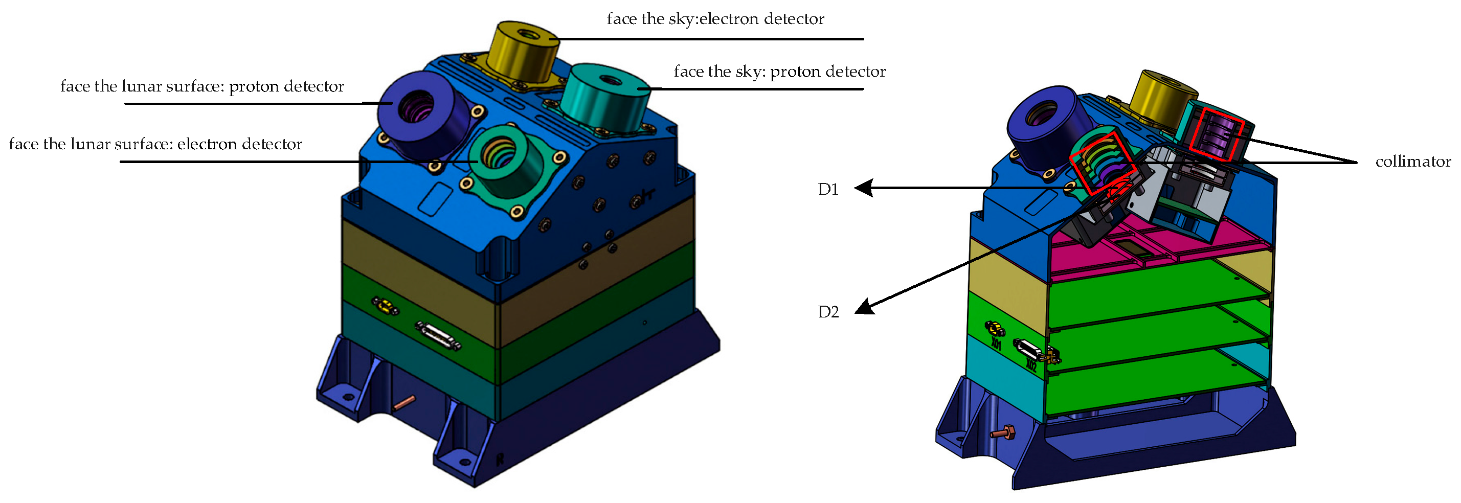
4.2.1. Sensor System

The sensor system of the high-energy particle detector includes a particle collimation system, a dE/dx detection system, a total energy measurement system, and an anti-coincidence system. The ΔE E method is employed to distinguish and measure particle energy and type. The collimation system consists of an external structure and a light-blocking layer. The external structure defines the detection field of view, shielding against interference from obliquely incident particles. The light-blocking layer, made of a 15 μm double-sided aluminum-plated Kapton film, protects against sunlight and micrometeoroids.
In Figure 4, detectors A and B, both silicon semiconductor detectors, serve as ΔE detectors to measure the dE/dx value of particles and form a fixed detection field of view. Detectors A and B operate within separate measurement fields, with Detector B also being used for dose and LET spectrum measurements. Detector C is a CsI scintillator detector designed to block high-energy charged particles and measure the total energy of the incident particles. The optical signal from the CsI scintillator is collected by a light-collecting device positioned on the bottom surface of the CsI detector. The intensity of these optical signals is correlated with the energy deposited by particles, and by analyzing the signal strength of the photodiode, the energy deposited by particles can be inferred. Detector E, a silicon semiconductor detector, serves as an anti-coincidence detector to differentiate between penetrating and stopping particles.

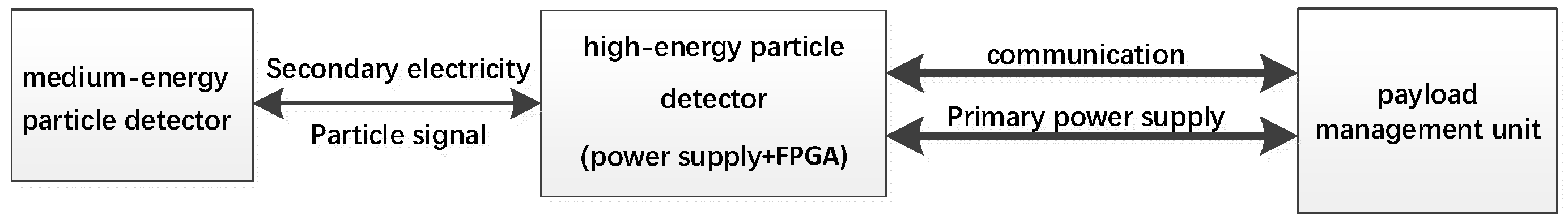
4.2.2. Electronics Design
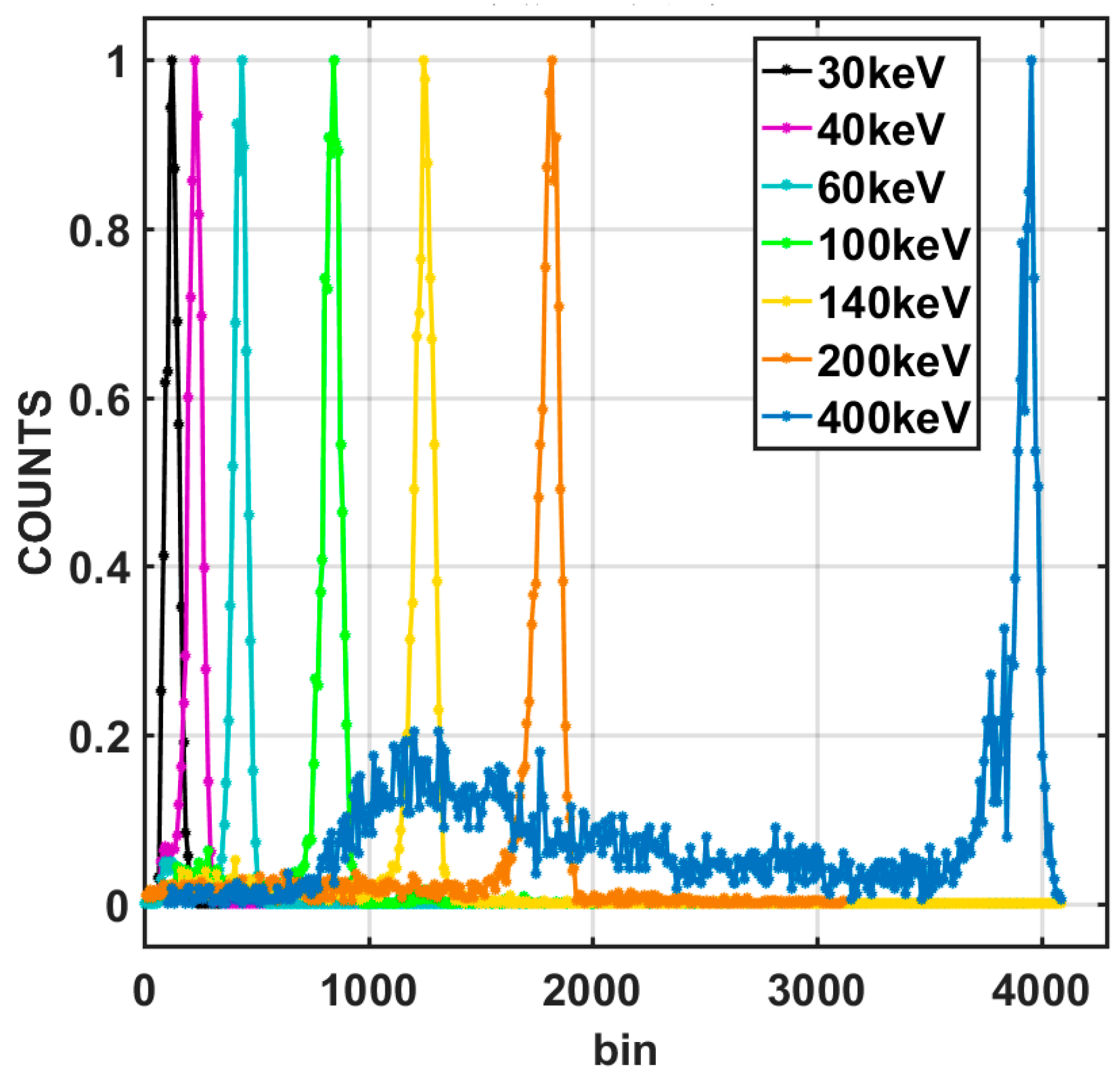
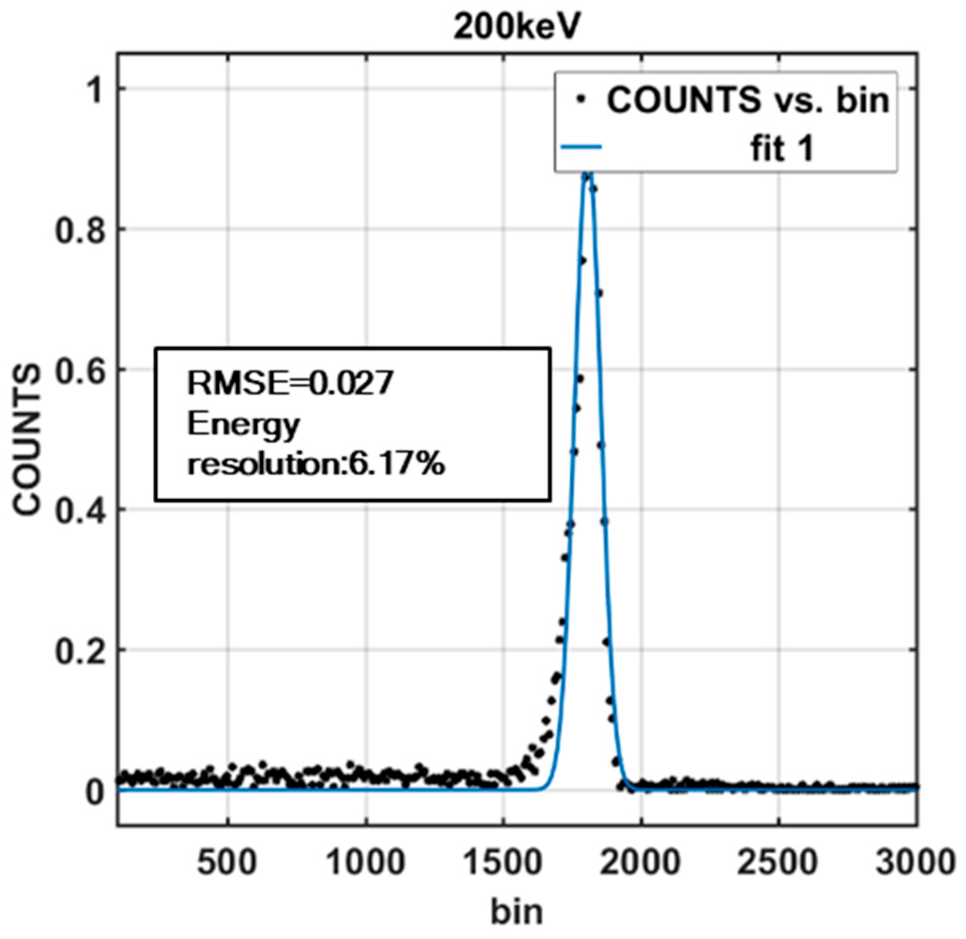
The electronics of the high-energy particle detector comprise analog and digital circuits. The analog circuit is incorporated to amplify and hold the charge signal generated by the sensor. The digital circuit collects and processes the peak level of the signal. The processing chip, an FPGA, handles data packaging, storage, and communication with the bus. Given the wide dynamic range of energies measured by the detectors, single-stage amplification is insufficient. Instead, a multi-stage amplification method is employed to amplify the signals, meeting the requirements of the dynamic range. Each sensor utilizes more than one preamplifier, and each preamplifier exhibits linearity within its respective charge measurement range. Similarly, multiple main amplifiers are employed, with each corresponding to different measurement ranges, as shown in Figure 5.
Trigger Mechanism: When a particle incident on a sensor generates a charge signal, it is converted into a voltage signal through the preamplifier and main amplifier. Simultaneously, a synchronous trigger pulse signal is generated by a comparator. The detector recognizes this trigger pulse signal, and upon its arrival, initiates the acquisition of particle signals.

5. Calibration

Calibration tests for the medium- and high-energy particle detectors were conducted using a range of particle accelerators. The calibration process utilized several accelerators, selected based on the penetration ability and energy range of the particles required for different sensors. The accelerators included the Huairou Electron Accelerator, the HI-13 Tandem Accelerator at the Institute of Atomic Energy, the Proton Accelerator at the Xinjiang Institute of Physics and Chemistry, and the Heavy Ion Accelerator at the Lanzhou Institute of Modern Physics.
The calibration process comprised six key aspects: energy spectrum calibration, energy resolution calibration, energy linearity calibration, detection efficiency calibration, particle contamination resistance calibration, and detector angle calibration [31].
During energy resolution and detection efficiency calibration, the energy of the accelerator beam particles was adjusted, and the instrument’s response was evaluated to calibrate these parameters. Calibration was conducted using multiple accelerators, with electronics-based calibration being employed for parts that could not be measured directly by the accelerators. For the energy particle detector, the measurement range was divided into multiple energy bins, with each bin representing an energy range. Calibration involved determining the boundaries of these energy bins. For particles of the same energy, the energy signals they generate in the sensors follow a Gaussian distribution. By adjusting the beam energy and performing data fitting, the beam energy at which the counts in adjacent bins are equal can be found, representing the actual measurement boundary of the energy bin. For ranges not covered by the accelerator, linear extrapolation based on the test results within the covered range was used [32].
Electron beams of different energies were generated using the electron accelerator in Huairou, Chinese Academy of Sciences, to irradiate the medium-energy detector. The deposited-energy spectra produced by different energy electrons were obtained. The energy resolution was calculated using the standard calibration energy resolution formula [33]. In Figure 6, the horizontal axis represents the numerical channel address of the pulse amplitude of the deposited energy, while the vertical axis represents the normalized count at each channel address. Different colors represent different incident energies.
By applying a Gaussian fitting formula to each particle response spectrum, a Gaussian fit can be obtained. Figure 7 reveals that the instrument’s resolution corresponding to an electron incidence of 200 keV is 6.17%. This resolution was obtained for the detector that faces the sky.
The detection efficiency is the ratio of the measured number of particles to the number of incident particles, accounting for the instrument’s inherent inability to detect 100% of incident particles [34]. The detection efficiency of the high-energy particle detector was 95.16%, while that of the medium-energy particle detector was 96.55%.
The calibration also evaluated the detector’s ability to discriminate between different particle species. For example, a proton probe was tested in an electron radiation environment to measure its response to electrons and assess the electron interference ratio [35]. Due to the limitations of accelerator particle sources, multiple accelerators could only provide beams of protons, electrons, alphas, boron, and carbon. After logarithmic calculation, the distributions of the different particle types varied, allowing for various particles to be distinguished. In Figure 8, the elliptical area circled in fluorescent blue represents the portion in which other particles were mixed into the proton data upon their incidence. This was confirmed with the accelerator facility, where the production of other particles during heavy-ion beam emission arises from carbon bombardment of the target. Among these, the worst performance was observed for carbon (C), with a probability of 8.51% for C being mixed with other particles.
The field of view, defined as a cone angle, was calibrated by rotating the instrument to scan the beam. The angle at which the instrument’s response changed from the start of counting to its end defined the field of view. The testing methodology for the dynamic range of flux involved utilizing an accelerator to irradiate a particle beam of a single, fixed energy while gradually increasing the incident flux of the accelerator. This allowed for the establishment of a relationship between the incident flux and the count rate of the sensor [24,32].
Radiation effects testing focused on measuring dose rate and Linear Energy Transfer (LET) spectra, determined by the energy loss of particles within the high-energy particle detector’s Sensor B. Calibration tests identified the minimum dose that elicits a response from the instrument, corresponding to the minimum deposited energy in Sensor B. The range of the LET spectrum could be determined using the range of deposited energy within Sensor B. There is some error, primarily due to the fact that all the energy is deposited in B without penetrating further, resulting in an uncertain path length. This portion of the error, as calculated through simulations, is less than 10%.
Multiple accelerators were used, based on the beam conditions and detector requirements, to comprehensively evaluate the performance of the medium- and high-energy particle detectors. For aspects not covered by the accelerators, equivalent signal source electronics calibration and GEANT4 Monte Carlo simulations were employed [36]. The calibration results are summarized in Table 2. Geant4 is primarily used for simulating the lunar surface radiation environment to establish the relationship between incident energy, deposited energy, and pulse signal amplitudes across various sensors. The types and energies of the particles produced by ground accelerators are limited. GEANT4 simulations were used to extrapolate based on actual measurement results in order to verify whether the particle indicators that the accelerator fails to measure meet the requirements.
The calibration results indicate that the detectors meet the mission requirements, with the proton energy range spanning from 30 keV to 300 MeV, the electron energy range extending from 30 keV to 12 MeV, and the heavy ion energy ranging from 8 MeV/n to 400 MeV/n. The field of view is measured at ≥40°, the flux measurement range spans five orders of magnitude, the energy resolution is 10.81% at 200 keV, the dose sensitivity is 7.48 µrad(Si)/h, and the LET spectrum range is from 0.001 to 37.014 MeV/(mg/cm2), with the linearity being ≤5.88%.

6. Conclusions

This study developed medium- and high-energy particle detection technology tailored to the lunar surface radiation environment for China’s Chang’e-7 lander. This will enable investigations of the radiation field at the lunar south pole as part of the foreseen successful operation of Change’7, and can be applied to resolve advanced scientific issues and ensure the safety of human activities on the Moon. During ground development, the functions and performance of the payload were rigorously tested. Calibration tests using accelerators and simulation analyses validated the performance indicators. The results confirmed that the detection payload satisfies the mission requirements, yielding actual response parameters and coefficients. These findings provide a foundation for future data processing and in-orbit adjustments.

Author Contributions

Conceptualization, L.W. and G.S.; methodology, L.W. and G.S.; software, L.W.; validation, H.Z., Z.Q., J.L., and W.J.; formal analysis, S.Z.; investigation, D.H.; resources, Y.S.; data curation, D.H.; writing—original draft preparation, L.W.; writing—review and editing, L.W. and G.S.; supervision, X.Z.; project administration, G.S. All authors have read and agreed to the published version of the manuscript.

Funding

This research received no external funding.

Data Availability Statement

The data presented in this study are available on request from the corresponding author. The data are not publicly available due to privacy.

Conflicts of Interest

The authors declare no conflicts of interest.

References

  1. Sawyer, R.P.; Halekas, J.S. Does Magnetic Reconnection Occur in the Near Lunar Surface Environment? Geophys. Res. Lett. 2023, 50, e2023GL104733. [Google Scholar] [CrossRef]
  2. Fatemi, S.; Holmström, M. Effects of protons reflected by lunar crustalmagnetic fields on the global lunar plasma environment. J. Geophys. Res. Space Phys. 2014, 119, 6095–6105. [Google Scholar] [CrossRef]
  3. Xu, Z.; Guo, J. Primary and albedo protons detected by the Lunar Lander Neutron and Dosimetry experiment on the lunar farside. Front. Astron. Space Sci. 2022, 9, 974946. [Google Scholar] [CrossRef]
  4. Wang, C.; Li, L.; Zhang, A.B. The solar wind and particle radiation environment onthe surface of the Moon-new observations from Chang’E-4. J. Deep. Space Explor. 2022, 9, 239–249. [Google Scholar]
  5. Wimmer-Schweingruber, F.R.; Yu, J.; Böttcher, I.S. The Lunar Lander Neutron and Dosimetry (LND) Experiment on Chang’E 4. Space Sci. Rev. 2020, 216, 250–644. [Google Scholar] [CrossRef]
  6. Shi, Q.; Zhang, J. Review of particle radiation environment of the Earth-Moon space and its impact on Lunar surficial material generation. Chin. J. Geophys. Chin. Ed. 2023, 66, 2685–2702. [Google Scholar]
  7. Wimmer-Schweingruber, R.F.; Zhang, S. Radiation Dose of LND on the Lunar Surface in Two Years. Chin. J. Space Sci. 2021, 41, 439–444. [Google Scholar]
  8. Zhang, S.Y.; Wimmer-Schweingruber, R.F. First measurements of the radiation dose on the lunar surface. Sci. Adv. 2020, 6, eaaz1334. [Google Scholar] [CrossRef] [PubMed]
  9. Pak, S.; Cucinotta, F.A. Organ dose equivalents of albedo protons and neutrons under exposure to large solar particle events during lunar human landing missions. Life Sci. Space Res. 2024, 42, 133–139. [Google Scholar] [CrossRef]
  10. Naito, M.; Hasebe, N. Radiation dose and its protection in the Moon from galactic cosmic rays and solar energetic particles: At the lunar surface and in a lava tube. J. Radiol. Prot. 2020, 40, 947–961. [Google Scholar] [CrossRef]
  11. Adams, J.H.; Bhattacharya, M. The ionizing radiation environment on the moon. Adv. Space Res. 2007, 40, 338–341. [Google Scholar] [CrossRef]
  12. Schwadron, A.N.; Baker, T. Lunar radiation environment and space weathering from the Cosmic Ray Telescope for the Effects of Radiation (CRaTER). J. Geophys. Res. Planets 2012, 117, E00H13. [Google Scholar] [CrossRef]
  13. Spence, E.H.; Case, W.A. CRaTER: The cosmic ray telescope for the effects of radiation experiment on the lunar reconnaissance orbiter mission. Space Sci. Rev. 2010, 150, 243–284. [Google Scholar] [CrossRef]
  14. Wang, C.; Jia, Y. Scientific objectives and payload configuration of the Chang’E-7 mission. Natl. Sci. Rev. 2024, 11, 329. [Google Scholar] [CrossRef]
  15. Yu, H.; Rao, W. Mission Analysis and Spacecraft Design of Chang’E-7. J. Deep. Space Explor. 2023, 10, 5567–5576. [Google Scholar]
  16. Zhang, Y.L.; Wang, X.L. Evaluation of particle acceptance for space particle telescope. Chin. Phys. C 2011, 35, 774–777. [Google Scholar] [CrossRef]
  17. Muminov, R.A.; Bogomolova, T.A. Semiconductor-detectors for telescope systems. Sov. At. Energy 1986, 61, 558–561. [Google Scholar] [CrossRef]
  18. Ploskonka, J.; Zastawniak, L. Simple method for charged-particle identification. Nucl. Instrum. Methods 1975, 126, 57–59. [Google Scholar] [CrossRef]
  19. Yu, G.; Li, X. Research on the performance and the experimental energy calibration of charged particle spectrometer-identificator. Chin. J. Nucl. Sci. Eng. 2004, 24, 12–19. [Google Scholar]
  20. Guo, J.; Zeitlin, C.; Wimmer-Schweingruber, R.F.; Hassler, D.M.; Ehresmann, B.; Rafkin, S.; von Forstner, J.L.F.; Khaksarighiri, S.; Liu, W. Radiation environment for future human exploration on the surface of Mars: The current understanding based on MSL/RAD dose measurements. Astron. Astrophys. Rev. 2021, 29, 8. [Google Scholar] [CrossRef]
  21. Ehresmann, B.; Zeitlin, C. Charged particle spectra obtained with the Mars Science Laboratory Radiation Assessment Detector (MSL/RAD) on the surface of Mars. J. Geophys. Res. Planets 2014, 119, 468–479. [Google Scholar] [CrossRef]
  22. Kozyrev, A.S.; Benkhoff, J.; Litvak, M.L.; Golovin, D.V.; Quarati, F.; Sanin, A.B. Localization of cosmic gamma-ray bursts in interplanetary space with MGNS/BepiColombo and HEND/Mars Odyssey experiments. Planet. Space Sci. 2022, 224, 105594. [Google Scholar] [CrossRef]
  23. Wimmer-Schweingruber, R.F.; Janitzek, N.P.; Pacheco, D.; Cernuda, I.; Lara, F.E.; Gómez-Herrero, R.; Mason, G.M.; Allen, R.C.; Xu, Z.G.; Carcaboso, F.; et al. First year of energetic particle measurements in the inner heliosphere with Solar Orbiter’s Energetic Particle Detector. Astron. Astrophys. 2021, 656, A22. [Google Scholar] [CrossRef]
  24. Ye, Z. Space Particle Radiation Detection Technology; Science Press: Beijing, China, 1986; pp. 77–78. [Google Scholar]
  25. An, H.; Wen, X. Detection technology and design analysis of LET spectrum of space radiation particles. Nucl. Tech. 2020, 43, 43–51. [Google Scholar]
  26. Patrick, J.W.; Stephens, L.D. Efficiency of lif-7 thermoluminescent dosimeters to high let-particles, relative to co-60 gamma-rays. Health Phys. 1976, 30, 295–296. [Google Scholar]
  27. Zhang, S.Y.; Wang, S.J. Design of the sweeping magnet in the space particle detector. Chin. J. Geophys. 2007, 50, 684–690. (In Chinese) [Google Scholar]
  28. Zhang, H.; Zhang, X.; Wang, J.; Huang, C.; Li, J.; Zong, W.; Shen, G.; Zhang, S.; Yang, Y.; Zhang, P. Design and Development of Medium Energy Proton Detector Onboard FY-3E Satellite. Aerospace 2023, 10, 321. [Google Scholar] [CrossRef]
  29. Cao, X.; Jiang, W. Design and Verification of Medium Energy Telescope Onboard HXMT Satellite. Spacecr. Eng. 2018, 27, 127–133. [Google Scholar]
  30. Zhang, S.; Wang, S. Design of sweep magnet in particles detection of space. Nucl. Electron. Detect. Technol. 2005, 06, 78–80. [Google Scholar]
  31. Hou, D.; Zhang, X.; Wang, J.; Huang, C.; Li, J.; Zong, W.; Zhang, S.; Shen, G.; Zhang, X.; Yuan, B. Calibration of the FY-3E particle sensors: High-Energy Particle Detector (HEPD). J. Instrum. 2023, 18, P07050. [Google Scholar] [CrossRef]
  32. Shen, G.H.; Zhang, X.X. Development and Calibration of a Three-Directional High-Energy Particle Detector for FY-3E Satellite. Aerospace 2023, 10, 173. [Google Scholar] [CrossRef]
  33. Shen, G.H.; Zhang, S.Y. Using Energy Particle Detection Technology on the Tiangong’s Space Station’s Wentian Laboratory Cabin Module. Aerospace 2023, 10, 373. [Google Scholar] [CrossRef]
  34. Cipolla, S.J.; Milone, J.A. Methodological effects in Si (Li) detector efficiency calibrations. Nucl. Instrum. Methods Phys. Res. Sect. A Accel. Spectrom. Detect. Assoc. Equip. 2003, 505, 273–276. [Google Scholar] [CrossRef]
  35. Chêne, G.; Mathis, F. Towards calibration and characterization of high-energy beams using charged particle retrodiffusion on a double thin carbon foil system. Nuclear Inst. Methods Phys. Res. B 2010, 268, 2015–2018. [Google Scholar] [CrossRef]
  36. Allison, J. Recent Developments in Geant4. Nucl. Instrum. Meth. A 2016, 835, 186–225. [Google Scholar] [CrossRef]
Figure 1. Block diagram of distributed system scheme.
Aerospace 11 00893 g001
Figure 2. Medium-energy particle detector.
Aerospace 11 00893 g002
Figure 3. High-energy particle probe.
Aerospace 11 00893 g003
Figure 4. Basic block diagram of the high-energy particle detector sensor system.
Aerospace 11 00893 g004
Figure 5. Electronic block diagram of the high-energy particle detector.
Aerospace 11 00893 g005
Figure 6. The response spectrum of electrons with different energies in the detector.
Aerospace 11 00893 g006
Figure 7. Energy resolution for 200 keV electrons (in the direction of the sky for medium energy).
Aerospace 11 00893 g007
Figure 8. A particle identification plot obtained from accelerator experiments (the axes are the energy depositions in the detectors).
Aerospace 11 00893 g008
Table 1. Main performance specifications of medium- and high-energy particle probes.
IndicatorsRequirement
Medium- and high-energy particle flux measurement range10–105 particles/cm2·s·sr
Medium- and high-energy particle energy rangeElectrons 30 keV~12 MeV
Protons 30 keV~300 MeV
Heavy ions 8 MeV/n~300 MeV/n
Medium- and high-energy particle field of view≥40° cone angle @30 keV~300 MeV/n
Medium- and high-energy particle flux dynamic range105@30 keV~300 MeV/n
Medium- and high-energy particle radiation effectDose sensitivity 20 μrad(Si)/h
LET spectrum 0.001~37 MeV/(mg/cm2)
Medium- and high-energy particle energy resolution≤15%@200 keV
Table 2. Calibration results of medium- and high-energy particle probes.
IndicatorsCalibration MethodsCalibration Results
Medium- and high-energy particle flux measurement rangeIrradiation was conducted using a fixed-energy particle beam from the Huairou Electron Accelerator (30 keV~1.5 MeV). The incident flux was adjusted by the accelerator system, and the counting rate of the instrument was recorded.
  • High energy: 3.84~1.15 × 10⁵ particles/cm2·s·sr
  • Medium-energy protons: 8.87~1.6 × 10⁵ particles/cm2·s·sr
  • Medium-energy electrons: 8.87~1.7 × 10⁵ particles/cm2·s·sr
Medium- and high-energy particle energy rangeA particle beam with fixed energy was directed at the single-instrument probe. The pulse amplitude signals in each sensor were recorded for various incident particle energies.
  • Protons (10 MeV, 12 MeV, 20 MeV, 30 MeV, 50 MeV, 60 MeV, 80 MeV, 95 MeV, and 120 MeV)
  • Alpha particles (30 MeV, 40 MeV, 50 MeV, and 100 MeV)
  • Boron particles (200 MeV and 300 MeV)
  • Carbon particles (52 MeV)
  • Electrons (30 keV~1.5 MeV)
  • Protons: 30 keV~10 MeV (medium energy); 8~300 MeV (high energy)
  • Electrons: 30 keV~400 keV (medium energy); 400 keV~12 MeV (high energy)
  • Heavy ions: 8 MeV/n ~400 MeV/n
Medium- and high-energy particle field of viewA fixed-energy particle beam was used to irradiate the single-instrument probe while rotating the instrument through the accelerator platform. The field of view was determined according to the angle at which the instrument transitions from no count to count and back to no count.≥40° cone angle @30 keV~300 MeV/n
Medium- and high-energy particle flux dynamic rangeIrradiation with a fixed-energy particle beam from the Huairou Electron Accelerator was employed. The incident flux was adjusted and the counting rate was recorded.105 particles/cm2·s·sr (@30 keV~300 MeV/n)
Medium- and high-energy particle radiation effectThe probe was irradiated with particle beams of varying energies, and the energy loss pulse amplitude signal in Sensor B was recorded.
  • Dose sensitivity: 7.48 µrad(Si)/h
  • LET spectrum: 0.001~37.014 MeV/(mg/cm2)
Medium- and high-energy particle energy resolutionA fixed-energy particle beam was directed at the single-instrument probe. Pulse amplitude signals for various particles were recorded as follows:
  • Alpha particles (30 MeV, 40 MeV, 50 MeV, and 100 MeV)
  • Boron particles (200 MeV)
  • Carbon particles (52 MeV and 200 MeV)
  • Protons (20 MeV, 30 MeV, 50 MeV, 80 MeV, and 95 MeV)
  • Electrons (30 keV~1.5 MeV)
10.81%@200 keV (energy resolution of the medium-energy particle detector’s lunar surface probe under irradiation by a 200 keV electron beam.)
Disclaimer/Publisher’s Note: The statements, opinions and data contained in all publications are solely those of the individual author(s) and contributor(s) and not of MDPI and/or the editor(s). MDPI and/or the editor(s) disclaim responsibility for any injury to people or property resulting from any ideas, methods, instructions or products referred to in the content.

