Article

Double and Triple Photoionization of CCl4

by
Antônio Carlos Fontes dos Santos
1,*,
Joselito Barbosa Maciel
2,
Alexandre Braga Rocha
2 and
Gerardo Gerson Bezerra de Souza
2
1
Instituto de Física, Universidade Federal do Rio de Janeiro, Rio de Janeiro 21941-972, RJ, Brazil
2
Instituto de Química, Universidade Federal do Rio de Janeiro, Rio de Janeiro 21949-900, RJ, Brazil
*
Author to whom correspondence should be addressed.
Atoms 2024, 12(12), 74; https://doi.org/10.3390/atoms12120074
Submission received: 9 November 2024 / Revised: 7 December 2024 / Accepted: 20 December 2024 / Published: 21 December 2024
(This article belongs to the Section Atomic, Molecular and Nuclear Spectroscopy and Collisions)

Abstract

:
(1) Background: Fragmentation after double and triple photoionization of the CCl4 molecule in the valence, Cl 2p, and C 1s regions have been reported; (2) Methods: We have used photoion-photoion (PIPICO) coincidence technique combined with synchrotron radiation. In addition, ab initio quantum mechanical calculations were done at multiconfigurational self-consistent and multireference configuration interaction to describe ground and inner-shell states; (3) Results: We have observed coincidences involving singly and doubly charged fragments coming from the doubly and triply ionized molecule. We have also found a well agreement between the quantum mechanical calculations and the total ion yield spectrum. It is shown that the Cl+ ion is the predominant product resulting from the fragmentation of the doubly and triply charged CCl4 molecule. The CCl+ + Cl+ pair is the dominant coincidence in the spectra from valence up to the C 1s edge; (4) Conclusions: The kinetic energy of the fragments is compatible with the Coulomb explosion model.

1. Introduction

Tetrachloromethane (CCl4) is a volatile liquid at room temperature with notable technological applications. For example, it is used as an etchant in microelectronics. However, the CCl4 molecule also poses environmental risks, contributing to atmospheric hazards and contributing to the depletion of the ozone layer [1]. Extensive research has been conducted on the photoabsorption properties of CCl4, focusing on both the valence and inner-shell regions [1,2,3,4,5,6,7,8,9,10,11,12].
It has been reported that not only the state of the dication CCl42+ but also its geometry affects the possible fragmentation pattern [5]. The measured triplet-state energies of CCl42+ are: 29.8, 31.4, 33.7, and 34.5 eV; and the singlet states 30.5, 32.9, 36.1, and 36.5 eV, and the results indicate that these doubly ionized states are stable or quasi-stable [7,8]. Lago, Santos, and de Souza [13,14] have conducted an in-depth investigation into the dissociative photoionization of a different compound chloro-substituted methane, the chloroform molecule (CHCl3) and its deuterated counterpart (CDCl3). In this work, we report valence and shallow core (Cl 2p and C 1s) level photoionization of the CCl4 molecule using tunable synchrotron radiation as the exciting source. The substitution of the hydrogen atom for a chlorine atom may allow for a substantial means for studying the differences in the fragmentation dynamics. In the case of small molecules, the fragmentation dynamics and pattern can be related to their geometry at the moment of dissociation. CCl4 is a highly symmetrical molecule. Its ground state possesses tetrahedral symmetry (Td point group, therefore non-polar). Then, it is compelling to compare the photofragmentation pattern for CCl4 with the previously investigated molecules, such as CHCl3 (C3v group) [13,14] and CH2Cl2 (C2v group) [15], to investigate the role of the molecular symmetry and/or polarity. In other terms, the impact of replacing an H atom with an Cl atom on the photofragmentation pattern.

2. Results

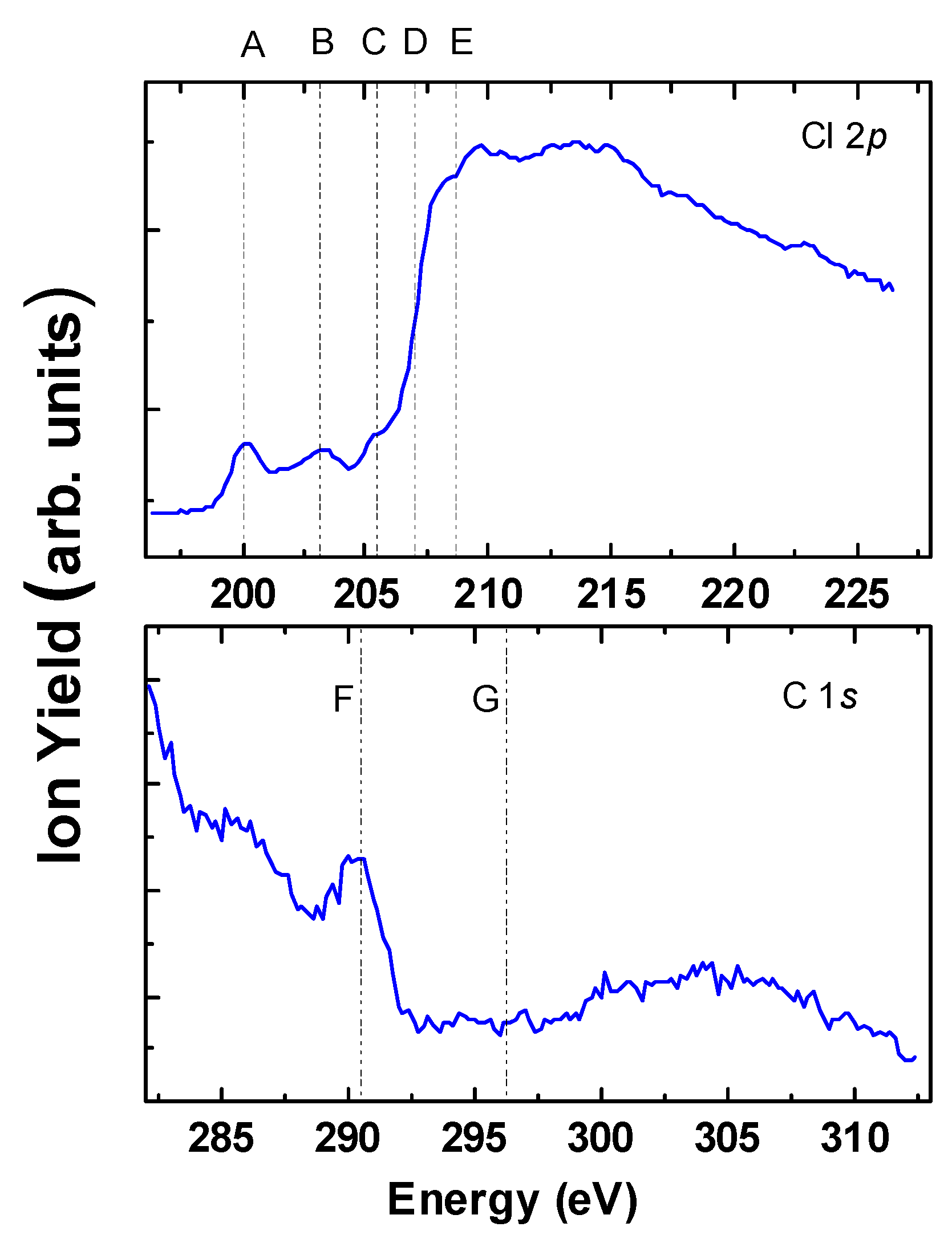
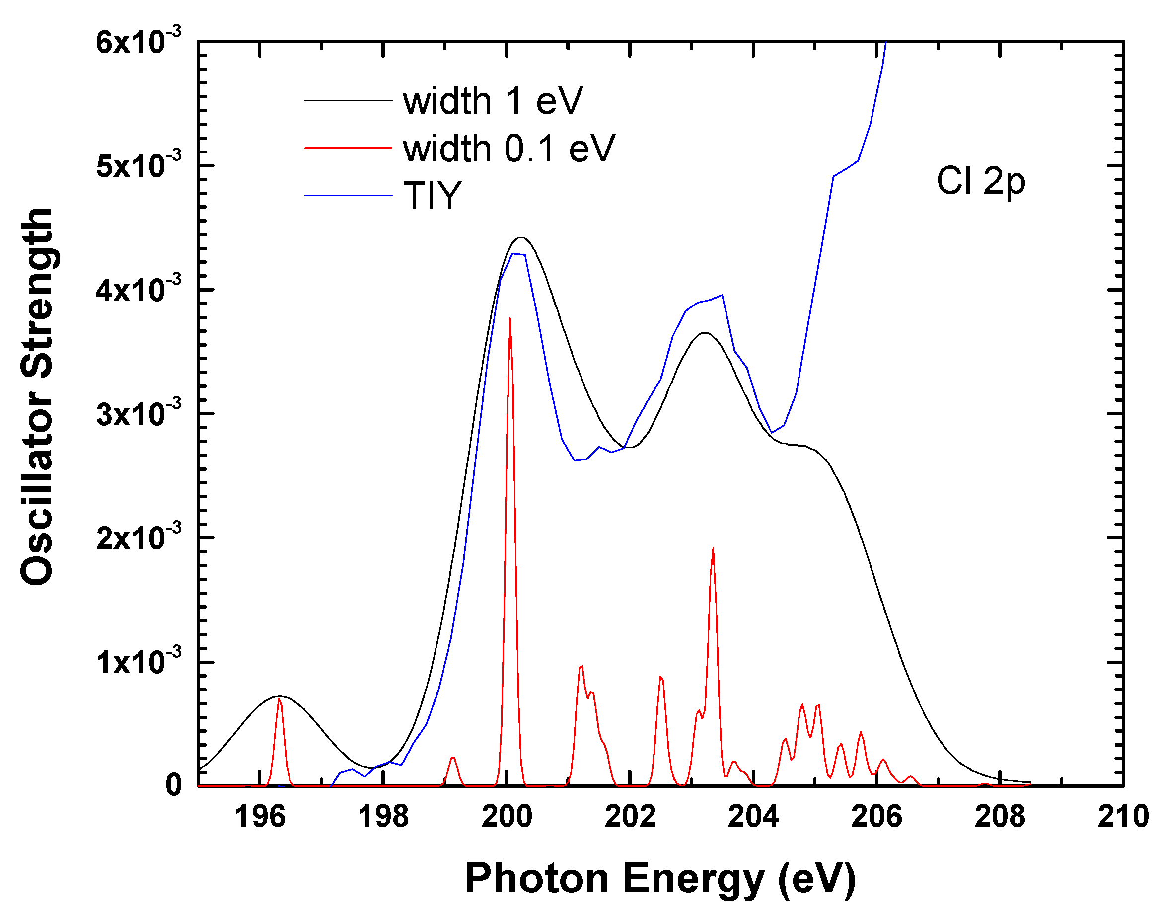
2.1. Total Ion Yield
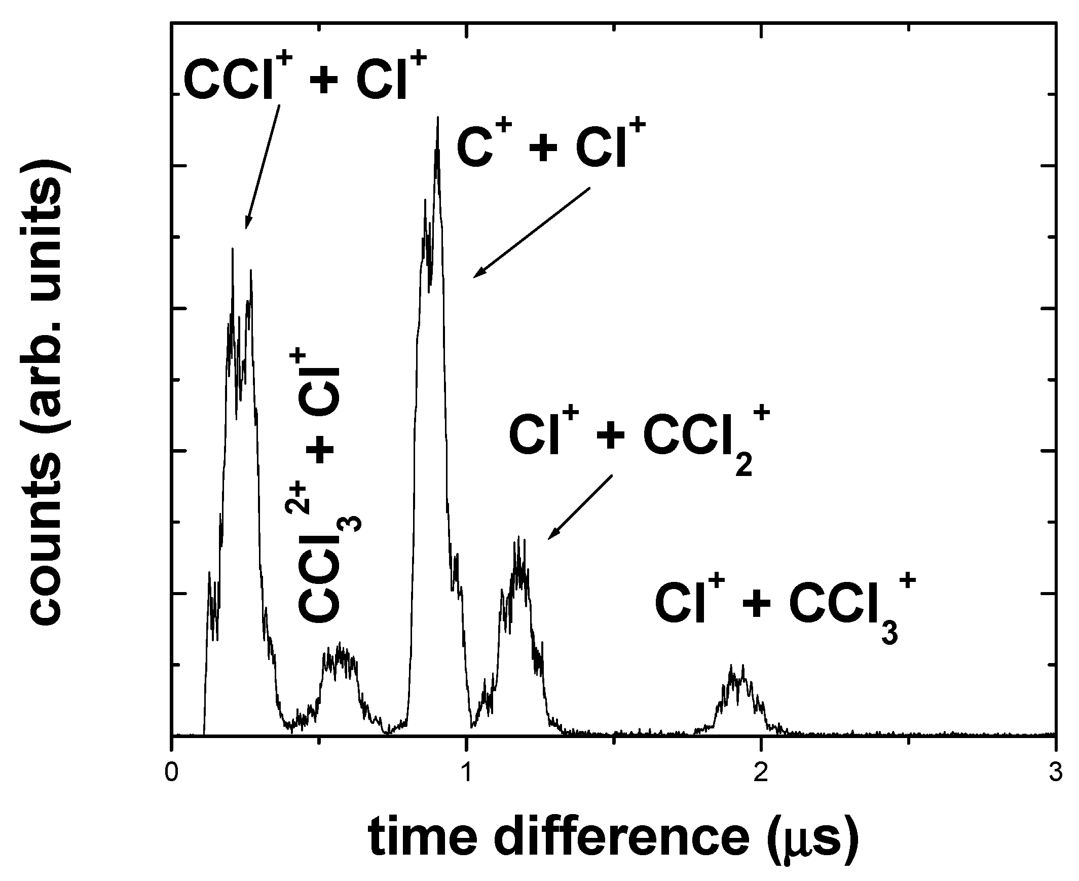
To investigate the fragmentation processes after inner-shell excitation and ionization, total ion yield (TIY) spectra were measured around the Cl 2p and C 1s edges (Figure 1). The TIY was obtained by running the photon energy around the Cl 2p and C 1s edges while recording the CCl4 charged moieties. The TIY spectrum shows a strong resemblance to the corresponding spectrum of other chlorine containing molecules [13,16]. At photon energies below the ionization potential (IP), where a core electron is excited to an unoccupied orbital, the inner vacancy is filled using resonant Auger decays, which mostly gives rise to singly ionized species, where the molecule is often unstable and breaks into smaller atomic and molecular fragments. It is well known that for photon energies above the IP, the normal Auger process prevails, giving rise to multiply charged moieties that are usually unstable in the microsecond range. The calibration of the incident photon energy was performed by scanning the monochromator across the Cl L2,3-edge, measuring the total photoion yield, and matching the observed peak structures in the yield to previously reported photoabsorption data [14]. The spectrum shows an underlying continuum owing to the direct ionization of the valence levels. The doublet structures with energies 200.0 eV (peak A) and 203.18 (peak B) in the TIY spectrum, are due to the spin-orbital splitting of the 2p3/2 (L3) and 2p1/2 (L2) levels of chlorine possessing a σ* character, in a diatomic-like picture. Feature C is allotted to a (2P3/2,1/2 → Rydberg) transition. The two ionization limits due to the spin-orbit splitting of the 2p hole, Cl 2p3/2,1/2 → ∞ (IP), are designated by the vertical lines D (207.04 eV) and E (208.73 eV) [1].
Figure 2 shows the calculated spectrum in C 1s edge. The line width was simulated by setting a Gaussian to each calculated resonance with height equating to the oscillator strength. The peaks are in good agreement with the experimental spectrum of Figure 1, but the experimental bands are wider. To simulate a spectrum as close as possible to the experimental one, we have taken a larger Gaussian width of 1 eV, also shown in Figure 1. The features indicated as F and G in Figure 1 are present in the calculated spectrum of Figure 2. It should be emphasized that this kind of calculation cannot describe the continuum part of the spectrum. So, the agreement with a pre-ionization branch of the spectrum can be considered quite good. In Figure 3, the calculated spectrum at the Cl 2p edge is presented. In this turn, given the results of C 1s, the first simulated spectrum was done with Gaussians broadening of 1 eV. The features A, B, and C of the spectrum in Figure 1 are reproduced. It should be emphasized that on one hand, the large broadening explicit the agreement with the experimental spectrum, it also hides the complex absorption manifold due to SO-coupling on the other. So, we turn to the spectrum with a Gaussian width of 0.1 eV. That is also shown in Figure 3. The difference is remarkable. The conclusion is that the low-resolution spectrum in Figure 1 makes bands merge, hiding the complex structure underlying.
It is worth emphasizing that the spectrum in Figure 3 is composed of 138 transitions. They are composed as follows. There are 12 Cl 2p orbitals. The transition from these to the first 3 empty orbitals in valence was considered. All transitions for singlet and triplet states are used as a basis to diagonalize the Breit-Pauli Hamiltonian to obtain the complete spin-orbit manifold.

2.2. PIPICO

Molecular ions with double or triple charges, such as CCl42+ and CCl43+, are predominantly unstable on a microsecond timescale. Despite the excellent signal-to-noise ratio and the high detection efficiency achieved in this study, no peaks corresponding to the doubly or triply charged parent molecule were observed. The CCl42+ dication breaks into two singly charged fragments or as one doubly charged and the other remaining neutral. To characterize the symmetric dissociative doubly ionization processes, where both fragments share the charge, photoion-photoion coincidence (PIPICO) experiments were carried out and the spectra are shown in Figure 4. The measured double-ionization energies to singlet CCl42+ are found to be 29.1 eV [5].
The PIPICO branching ratios are presented in Table 1 and shown in Figure 5, as a function of the incident photon energy. The ionic branching ratios were extracted by integrating the peaks and accepting a linear background contribution. The ion detection efficiency was assumed to be independent of the mass-to-charge ratios. Due to the limited time resolution, the Cl+ + Cl+ coincidences cannot be observed with the present experimental setup. Dramatic changes are observed in the spectra, as we move across the Cl 2p → t2 resonance. Doubly charged fragments, such as Cl2+ (1–3%), CCl2+ (~3%), Cl4+ (0.1%), CCl32+ (~0.8%), and CCl32+ (~1%), originated from the unstable doubly and/or triple charged parent molecular ions CCl42+ and CCl43+ have been reported in the photoelectron-photoion-Photoion coincidence (PEPIPICO) spectra and reported previously [6]. On the other hand, in the case of the polar CH2Cl2 molecule (C2v), only the doubly charged moieties Cl2+ (0.2–1%) and CHnCl+ (0.4–1%) were observed [12].
The peak shapes in the PIPICO spectra embody anisotropy and consequently, the symmetry, reflected in the peak shape, of the excitation as well as the kinetic energy released, reflected in the peak width, in the dissociation process and the geometry of the molecule during the fragmentation process [16,17,18]. Three different peak shapes have been reported; (i) rectangular due to decay processes with no alignment; (ii) A narrow single peak associated to decay processes where the momentum is imparted perpendicular to the spectrometer axis and (iii) a double peak structure due to dissociations where most of the momentum is imparted parallel with the spectrometer axis [17,18].

2.3. Kinetic Energy Release (KER)

Molecules have the ability to transform light energy into other forms of energy, such as kinetic energy. This can be achieved through transitions between several electronic states of the molecule. Key insights into the fragmentation process can be obtained by analyzing the average Kinetic Energy Release (KER) of the fragments. When the fragments are precisely spatially focused, and the electric field in the interaction region is uniform, the energy released during fragmentation (U0) can be calculated from the peak width (FWHM) using the expression [18].
U 12 = 1 8 m 1 + m 2 m 1 m 2 q 1 q 2 E Δ t q 1 + q 2 2
where q is the fragment charge state, E is the electric field in the interaction region, Δt is the peak width (FWHM), and m is the mass of the fragment. The indices 1, 2 are related to the ion pair. Such estimation is possible only when the fragments originate from a simple bond breaking.
A broadening of peak widths (FWHM) is observed for all fragments near the C 1s edge. This phenomenon can be explained by the presence of repulsive electronic states involved in the fragmentation of the CCl4 molecule upon C 1s core excitation. The primary factor contributing to these repulsive core-excited states is the non-ligand nature of the final σ* orbital, which causes the excess energy to be redistributed as kinetic energy shared among the fragments [17].
In the case of the fragmentation of the CCl4q+ (q = 2,3), the prevailing part of the KER is produced during the charge separation. In that case, the KER can be related to the nuclear distance between the fragments at the moment of the explosion, R, through the equation.
U ( e V ) = 14.4 q 1 q 2 R ( A o )
The case of the neutral CCl4 molecule, which possess a Td geometry, the ground-state equilibrium bond length is R = 1.751 Å [18]. Thus, we find U = 8.2 q1q2 eV (q1,2 in atomic units) for the kinetic energy release in a pure Coulombic explosion model. In Table 2 is shown the measured and calculated KER for the CCl4 moieties. Roughing speaking, there is an overall agreement between the measured and calculated KER (7–25%). The only exception is the C+ + Cl+ coincidence, which corresponds to the complete molecular fragmentation. In that case, the measured kinetic energy release is 50–90% above the values predicted by the Coulomb explosion model. Some observations should be pointed out: firstly, Equation (1) is strictly valid for a single bond breaking. Second, it was assumed that the ground-state equilibrium bond length is constant, which certainly is not the case for the C+ + Cl+ coincidence. Finally, this is an indication that the molecular state in the C+ + Cl+ + 3Cl must be strongly repulsive, meaning the two charged fragments are not fully shielded by the remaining electrons.
In the cases that the measured KER is exceeded by the calculated KER, for instance in the case of the Cl+ + CCl32+ coincidence, a naïve interpretation based on a qualitative speculation is the following: a high electron density between the two fragments shields the interaction between the ions, resulting in KER values lower than the energy predicted by the purely repulsive point-charge Coulomb potential.

3. Discussion

An analysis of the time-of-flight differences in the PIPICO spectra indicates that the peaks correspond to the decomposition of CCl42+ and in a minor proportion CCl43+ as follows:
CCl42+ → CCl3+ + Cl+
→ CCl2+ + Cl+ + Cl
→ CCl+ + C+ + 2Cl
→ C+ + Cl+ + 3Cl (d)
CCl43+ → CCl32+ + Cl+
Some general observations can be made about the mass spectra presented in Figure 4 and Figure 5 and the data tabulated in Table 1. Process (3) is a two-body decay that originates from a single bond C-Cl breaking due to the instability of the CCl42+ ions. It dominates the PIPICO spectrum at 35.0 eV and its relative contribution decreases to 8% after the C 1s edge. The CCl3+ + Cl+ coincidence presents a broad and bell-like shape (almost Gaussian), indicating that the decay process takes place with most of the momentum imparted preferentially to the spectrometer axis.
The Cl+ + CCl2+ ion pair coincidence (process 4), originated from a double bond breaking releasing an undetected neutral chlorine atom, depending on the photon energy, is the second most abundant coincidence in the PIPICO spectra. It presents several structures due to the isotopic contributions of 35Cl and 37Cl.
The Cl+ + CCl+ ion pair coincidence (process 5), originating from a multi-band breaking, dominates the PIPICO spectra above 140 eV. As seen from Figure 4, it presents a double peak structure due to decay processes where most of the momentum is imparted parallel with the spectrometer axis.
The ion par C+ + Cl+ coincidence (process 6), corresponding to the molecular atomization, increases as the photon energy increases, presenting a relative maximum in the Cl 2p resonance. This coincidence also presents a double peak structure.
The CCl32+ + Cl+ ion pair (process 7), corresponding to a triple ionization event followed by a single bond breaking, increases above Cl 2p due to the contribution of the Auger processes. It presents a rectangular peak shape indicating that the decay processes take place with no molecular alignment.
Some general observations can be made about the mass spectra presented in Figure 4 and the data tabulated in Table 1. Process (3) is a two-body decay that originates from a single bond C-Cl breaking due to the instability of the CCl42+ ions. It dominates the PIPICO spectrum at 35.0 eV and its relative contribution decreases to 8% after the C 1s edge. The CCl3+ + Cl+ coincidence presents a broad and bell-like shape (almost Gaussian), indicating that the decay process takes place with most of the momentum imparted preferentially to the spectrometer axis.
The Cl+ + CCl+ ion pair coincidence (process 5), originated from a multi-band breaking, dominates the PIPICO spectra above 140 eV. As seen from Figure 4, it presents a double peak structure due to decay processes where most of the momentum is imparted parallel with the spectrometer axis.
As in the case of the CHCl3 and CH2Cl2 molecules, it has been observed that the Cl+ ion is a major outcome of the dissociation of the doubly and triply charged CCl4 molecule. The CCl+ + Cl+ pair is the dominant coincidence in the spectra from valence up to the C 1s edge. The prominent role of the CCl+ + Cl+ pair is even more apparent in the low energy spectrum (15.3 eV). The Cl+ + CCl+ fragments pair dominates the mass spectra at lower energies while the C+ + Cl+ pair dominates the mass spectra around the C 1s region. A strong change in the equilibrium geometry is expected to occur induced by the population of the σ* orbital, and the excitation of vibrational series associated with the C–Cl bond might be expected, hence leading to a more efficient neutral Cl release. The fast increase of the C+ + Cl+ channel as the photon energy is close to the Cl 2p edge and above is an additional indication that the excitation of orbitals corresponding to the C–Cl bond is playing a role. In the case of the CHCl3 molecule, the same argument was used to explain the efficient neutral H release.

4. Materials and Methods

4.1. Experimental

The experimental apparatus used was described in detail in a previously published paper [10]. Briefly, the experiment was performed at the Center for Advanced Microstructures and Devices (CAMD), Baton Rouge, LA, USA. Photons from a toroidal grating monochromator (TGM) beamline crosses at right angles an effusive gaseous CCl4 sample inside a high vacuum chamber. The base pressure was in the 10−8 Torr range. During the experiment, the pressure was kept below 10−6 Torr. The gas needle was maintained at ground potential.
The ionized recoil moieties generated by the light beam were accelerated by a two-stage DC electric field. The fragments were detected by a pair of micro-channel plate detectors mounted in a chevron structure after being mass-to-charge analyzed by a time-of-flight spectrometer. The charged fragments produce start and stop signals to a time-to-amplitude converter (TAC). The first segment of the electric field consists of a plate-grid system with the photon beam passing in its middle and with a 500 V/cm DC electric field in the direction of the spectrometer axis. The polarization of the light beam in the same direction as the spectrometer axis. The intense extraction field (500 V/cm), was able to measure the fragments produced in all directions, minimizing any anisotropy. The ion drift tube of the spectrometer is 25.0 cm in length, while the extraction gap measures 2.0 cm. A key aspect of this time-of-flight mass spectrometer is the incorporation of a collimating electrostatic lens, enabling the efficient collection of ions with kinetic energies up to 40 eV at 100% efficiency. The CCl4 sample, purchased commercially with a high purity of 99.5%, was used as received without additional purification.

4.2. Calculations Details

Transition energies and transition dipole moments (oscillator strengths) were computed within the multiconfigurational self-consistent field for inner-shell states (IS-MCSCF) [19], followed by multireference configuration interaction (MRCI), in the ground state geometry of CCl4 molecule. All calculations were done with cc-pVTZ-DK and aug-cc-pVTZ-DK bases set for carbon and chlorine atoms respectively. Scalar relativistic effects have been considered through Douglas-Kroll-Hess Hamiltonian up to third order. The active space in the MCSCF step is composed of one configuration for each state. For the ground state, the Hartree-Fock configuration. For Cl-2p, each configuration corresponding to an excitation from 2p to an empty valence orbital was considered for all chlorine atoms, which means 12 configurations. On the other hand, for C-1s, only the Hartree-Fock configuration involves a transition from that inner-shell orbital to an empty valence. These state-averaged orbitals were used in the MRCI step to compute more states by adding two more empty orbitals. For chlorine, these were used as a basis to construct a set of singlet and triplet states at the Cl 2p excitation edge, which will be used as the basis for the full Breit-Pauli Hamiltonian diagonalization, as already done elsewhere [20], to account for spin-orbit coupling. Our results were obtained with the Molpro quantum chemistry package.

5. Conclusions

Photoionization studies of doubly and triply ionized CCl4 molecules have been conducted using synchrotron radiation as the photon source, covering both the valence and soft X-ray regions. Branching ratios were determined from PIPICO spectra, measured across the valence region and near the Cl 2p and C 1s edges. Due to their high instability on a microsecond timescale, the doubly and triply charged parent molecules were not detected in the mass spectra for this compound.

Author Contributions

Conceptualization, G.G.B.d.S.; methodology, G.G.B.d.S. and A.B.R.; formal analysis, A.C.F.d.S.; investigation, G.G.B.d.S. and J.B.M.; resources, G.G.B.d.S.; data curation, A.C.F.d.S.; writing—original draft preparation, A.C.F.d.S.; writing—review and editing, A.C.F.d.S. and A.B.R.; visualization, G.G.B.d.S.; supervision, G.G.B.d.S.; project administration, G.G.B.d.S.; funding acquisition, G.G.B.d.S. All authors have read and agreed to the published version of the manuscript.

Funding

A.C.F.S. acknowledges CNPq-Brazil (307144/2021-6).

Data Availability Statement

The raw data supporting the conclusions of this article will be made available by the authors on request.

Conflicts of Interest

The authors declare no conflicts of interest.

References

  1. Burton, G.R.; Chan, W.F.; Cooper, G.; Brion, C.E. Valence-and inner-shell (Cl 2p, 2s; C 1s) photoabsorption and photoionization of carbon tetrachloride. Absolute oscillator strength (5–400 eV) and dipole-induced breakdown pathways. Chem. Phys. 1994, 181, 147–172. [Google Scholar] [CrossRef]
  2. Hitchcock, A.P.; Brion, C.E. Inner shell excitation spectroscopy of molecules using inelastic electron scattering. J. Electr. Spectr. Rel. Phen. 1978, 14, 417. [Google Scholar] [CrossRef]
  3. Zhang, W.; Ibuki, T.; Brion, C.E. Absolute dipole differential oscillator strengths for inner shell spectra from high resolution electron energy loss studies of the freon molecules CF4, CF3Cl, CF2Cl2, CFCl3 and CCl4. Chem. Phys. 1992, 160, 435. [Google Scholar] [CrossRef]
  4. O’Sullivan, G. Chlorine L-edge absorption in CCl4 and CCl2F2. J. Phys. B. At. Mol. Opt. Phys. 1982, 15, 2385. [Google Scholar] [CrossRef]
  5. Fainelli, F.; Maracci, R.; Platania, L.; Avaldi, J. The fragmentation of CCl4 dications studied by Auger electron-ion coincidence experiments. Electron. Spectrosc. Rel. Phenom. 2001, 119, 81. [Google Scholar] [CrossRef]
  6. Stolte, W.C.; Santos, A.C.F.; de Souza, G.G.B.; Sant’anna, M.M.; Leung, K.T. Deep-core photoabsorption and photofragmentation of tetrachloromethane near the Cl K-edge. J. Chem. Phys. 2017, 146, 214306. [Google Scholar] [CrossRef] [PubMed]
  7. Wang, L.-H.; Du, X.-J.; Xu, Y.-C.; Nie, Z.-W.; Wang, D.-H.; Wang, S.-X.; Zhu, L.-F. Generalized Oscillator Strengths for the Valence Shell Excitations in Carbon Tetrachloride Studied by Fast Electron Impact. J. Phys. Chem. A 2022, 126, 453–461. [Google Scholar] [CrossRef]
  8. Kokkonen, E.; Jänkälä, K.; Patanen, M.; Cao, W.; Hrast, M.; Bučar, K.; Žitnik, M.; Huttula, M. Role of ultrafast dissociation in the fragmentation of chlorinated methanes. J. Chem. Phys. 2018, 148, 174301. [Google Scholar] [CrossRef] [PubMed]
  9. Watanabe, N.; Takahashi, M. Theoretical Study of Valence Shell Excitation by Electron Impact in CCl4. J. Phys. Chem. A 2023, 127, 1866–1873. [Google Scholar] [CrossRef] [PubMed]
  10. Santos, A.C.F.; Maciel, J.B.; de Souza, G.G.B. Valence and core level ionization of the CCl4 molecule. J. Elect. Spectr. 2007, 156, 236. [Google Scholar] [CrossRef]
  11. Fournier, P.G.; Comtet, G.; Fournier, J.; Svensson, S.; Karlsson, L.; Keane, M.P.; De Brito, A.N. Double-ionization energies of CCl 4 by double-charge-transfer and x-ray Auger-electron spectroscopies. J. Phys. Rev. A 1989, 40, 163. [Google Scholar] [CrossRef] [PubMed]
  12. Grant, R.P.; Harris, F.M.; Andrews, S.R.; Parry, D.E. Double ionization to singlet and triplet electronic states of CCl42+. Int. J. Mass Spectrom. Ion Process. 1995, 142, 117. [Google Scholar] [CrossRef]
  13. Lago, A.F.; Santos, A.C.F.; de Souza, G.G.B. Mass spectrometry study of the fragmentation of valence and core-shell (Cl2p) excited CHCl3 and CDCl3 molecules. J. Chem. Phys. 2004, 120, 9547. [Google Scholar] [CrossRef]
  14. Lago, A.F.; Santos, A.C.F.; de Souza, G.G.B. Charge separation mass spectrometry study of the chloroform molecule following valence and Cl 2p photoionization. Int. J. Mass Spectrom. 2007, 262, 187. [Google Scholar] [CrossRef]
  15. KAlcantara, K.F.; Wolff, W.; Gomes, A.H.A.; Sigaud, L.; Soriano, S.; Oliveira, V.; Rocha, A.B.; Santos, A.C.F. Fragmentation of the CH2Cl2 molecule by proton impact and VUV photons. J. Phys. B At. Mol. Opt. Phys. 2011, 44, 165205. [Google Scholar] [CrossRef]
  16. Alcantara, K.F.; Gomes, A.H.A.; Wolff, W.; Sigaud, L.; Santos, A.C.F. Strong electronic selectivity in the shallow core excitation of the CH2Cl2 molecule. J. Phys. Chem. A 2015, 119, 8822–8831. [Google Scholar] [CrossRef] [PubMed]
  17. Lindgren, A.; Gisselbrecht, M.; Burmeister, F.; Naves de Brito, A.; Kivimäki, A.; Sorensen, S.L. Molecular alignment of ammonia studied by electron-ion-ion coincidence spectroscopy. J. Chem. Phys. 2005, 122, 114306. [Google Scholar] [CrossRef] [PubMed]
  18. Burmeister, F.; Coutinho, L.H.; Marinho, R.R.T.; Homem, M.G.P.; De Morais, M.A.A.; Mocellin, A.; Björneholm, O.; Sorensen, S.L.; Fonseca, P.D.T.; Lindgren, A.; et al. Description and performance of an electron-ion coincidence TOF spectrometer used at the Brazilian synchrotron facility LNLS. J. Electron. Spectrosc. Relat. Phenom. 2010, 180, 6. [Google Scholar] [CrossRef]
  19. Rocha, A.B. Potential curves for inner-shell states of CO calculated at multiconfigurational self-consistent field level. J. Chem. Phys. 2011, 134, 024107. [Google Scholar] [CrossRef] [PubMed]
  20. Rocha, A.B. Spin-orbit splitting for inner-shell 2p states. J. Mol. Model. 2014, 20, 2355. [Google Scholar] [CrossRef] [PubMed]
Figure 1. Chlorine 2p (top) and carbon 1s (bottom) total ion yield (TIY) of the CCl4 molecule as a function of the photon energy. The vertical dashed lines represent the position of the main transitions: Line A: Cl 2p3/2 → σ*; Line B: Cl 2p1/2 → σ*; Line C: Cl 2p3/2 (IP); Line D: Cl 2p → Rydberg; Line E: Cl 2p1/2 (IP); line F: C 1s → σ*; line G: C 1s (IP).
Atoms 12 00074 g001
Figure 2. The experimental total ion yield (TIY) for C 1s (represented by magenta dots) is compared with a simulated C 1s spectrum derived from ab initio calculations of transition energies and oscillator strengths, broadened using Gaussian functions with widths of 0.1 eV (black line) and 1.0 eV (blue line). To align with the experimental data, the theoretical spectrum was shifted by 0.8 eV. For clarity in the comparison, the Cl 2p background was subtracted from the experimental TIY.
Atoms 12 00074 g002
Figure 3. Comparison between the experimental total ion yield (TIY) Cl 2p simulated spectrum, obtained by Gaussian broadening, from ab initio calculated transition energies and oscillator strength with different widths (1.0 eV and 0.1 eV). The theoretical spectrum was shifted by 10 eV to match the experimental one.
Atoms 12 00074 g003
Figure 4. PIPICO spectrum of the CCl4 molecule at 282.5 eV.
Atoms 12 00074 g004
Figure 5. PIPICO branching ratios of the CCl4 molecule as a function of the photon energy.
Atoms 12 00074 g005
Table 1. PIPICO branching ratios (%) for dissociative double and triple photoionization of CCl4.
Energy (eV)CCl+ + Cl+CCl32+ + Cl+C+ + Cl+CCl2+ + Cl+CCl3+ + Cl+
35.012.7--3.737.546.0
147.542.74.625.618.88.3
201.343.04.031.415.16.4
204.544.83.931.113.96.3
206.739.64.032.316.08.1
209.536.53.425.524.410.2
210.939.73.324.823.58.7
216.137.13.524.625.19.8
224,039.73.427.421.48.0
237.539.04.131.219.06.6
283.535.77.438.513.74.7
290.536.87.538.713.04.1
302.435.88.239.312.54.3
Table 2. Measured and calculated KER for CCl4 fragments in the PIPICO spectra.
CoincidencePhoton Energy (eV)Columb
Model
209.5 282.5302.5
Cl+ + CCl3+12.7--3.78.2
Cl + + CCl2+42.74.625.68.2
Cl+ + C+43.04.031.48.2
Cl+ + CCl32+44.83.931.116.4
Cl+ + CCl+39.64.032.38.2
Disclaimer/Publisher’s Note: The statements, opinions and data contained in all publications are solely those of the individual author(s) and contributor(s) and not of MDPI and/or the editor(s). MDPI and/or the editor(s) disclaim responsibility for any injury to people or property resulting from any ideas, methods, instructions or products referred to in the content.

