Article

Discovery of First Quinoline–Acylguanidine Hybrids as Selective Butyrylcholinesterase Inhibitors: Design, Synthesis, and Molecular Basis


by
Mayara C. dos Santos
,
Átila M. Mofati
,
Nathalia F. Nadur
,
Larissa de A. P. Ferreira
,
Lucas Caruso
,
Gleyton L. S. Sousa
,
Renata B. Lacerda
* and
Arthur E. Kümmerle
*
Laboratory of Molecular Diversity and Medicinal Chemistry (LaDMol-QM), Graduate Program in Chemistry (PPGQ), Institute of Chemistry, Federal Rural University of Rio de Janeiro, Seropédica 23897-000, RJ, Brazil
*
Authors to whom correspondence should be addressed.
Sci. Pharm. 2026, 94(4), 85; https://doi.org/10.3390/scipharm94040085
Submission received: 22 July 2026 / Revised: 11 September 2026 / Accepted: 22 September 2026 / Published: 24 September 2026
(This article belongs to the Special Issue Computer-Aided Drug Design and Molecular Synthesis)

Abstract

Alzheimer’s disease (DA) remains a major therapeutic challenge, and selective butyrylcholinesterase (BChE) inhibition has emerged as a promising strategy for symptomatic treatment, particularly in advanced stages. Herein, a series of novel quinoline–acylguanidine hybrids was designed through a bioisosteric replacement of acridine–thiosemicarbazones and synthesized via a convergent route combining the Pfitzinger reaction with acylguanidine formation. All derivatives were evaluated against BChE and AChE (acetylcholinesterase), and the 2-phenylquinoline derivative 2c (IC50 = 7.14 µM) was identified as the most potent BChE inhibitor, comparable to donepezil (IC50 = 2.39 µM), with selectivity over AChE. Structure–activity relationships revealed that the 2-phenyl substituent is essential for activity, whereas bulky groups (4-bromophenyl) or replacement with methyl abolished inhibition. Molecular docking showed that active compounds bind within the BChE catalytic anionic site with Trp110, Met465, and Trp458, while inactive analogs undergo a 180° flip of the quinoline scaffold, disrupting key interactions. In silico ADME evaluation indicated a favorable CNS drug-like profile (TPSA < 90 Å2, LogBB > −1, no Lipinski violations, and no predicted P-glycoprotein substrate liability). These findings establish the quinoline–acylguanidine scaffold as a promising new chemotype for the development of selective BChE inhibitors with potential applications in Alzheimer’s disease.

1. Introduction

Alzheimer’s disease (AD) is the leading cause of dementia worldwide and one of the greatest public health challenges of the twenty-first century. It is characterized by progressive cognitive decline, initially affecting memory and later impairing language, executive function, behavior, and the ability to perform daily activities. According to the World Health Organization, more than 55 million people currently live with dementia, with AD accounting for approximately 60–70% of all cases. As population aging accelerates, this number is expected to nearly triple by 2050, imposing a major socioeconomic burden on healthcare systems and caregivers worldwide [1].
Despite decades of research, no curative therapy is currently available for AD. Existing treatments mainly provide symptomatic relief and do not halt or reverse neurodegeneration. Approved drugs include the cholinesterase inhibitors donepezil, rivastigmine, and galantamine, the NMDA receptor antagonist memantine, and, more recently, disease-modifying monoclonal antibodies targeting Aβ aggregates. However, the modest clinical efficacy of these therapies highlights the urgent need for novel therapeutic agents capable of simultaneously modulating multiple pathological mechanisms involved in AD progression [2,3].
The cholinergic hypothesis remains one of the most validated therapeutic strategies for AD. Acetylcholinesterase (AChE) and butyrylcholinesterase (BChE) are the main enzymes responsible for acetylcholine (ACh) hydrolysis and, consequently, for regulating cholinergic neurotransmission. In the healthy brain, AChE is the predominant cholinesterase and shows high substrate specificity due to several aromatic residues lining its active-site gorge, including Tyr72, Tyr124, Tyr337, and Trp286. In contrast, BChE has a larger and more flexible catalytic gorge, in which some aromatic residues are replaced by aliphatic amino acids such as Asn68, Gln119, Ala328, Leu286, and Val288, allowing the accommodation of bulkier substrates and structurally diverse inhibitors.
As AD progresses, the expression profiles of these cholinesterases change markedly. AChE activity declines with neuronal degeneration, whereas BChE expression and activity increase by about 30–60%, partially compensating for the loss of AChE. As a result, BChE plays an increasingly important role in acetylcholine hydrolysis during advanced stages of the disease, further aggravating cholinergic dysfunction. Although AChE remains the principal cholinesterase targeted by marketed drugs, the increasing relevance of BChE during AD progression has stimulated growing interest in the development of selective BChE inhibitors, which may offer greater therapeutic benefits than conventional AChE inhibitors in patients with moderate to severe AD, while potentially reducing peripheral cholinergic adverse effects [4,5,6].
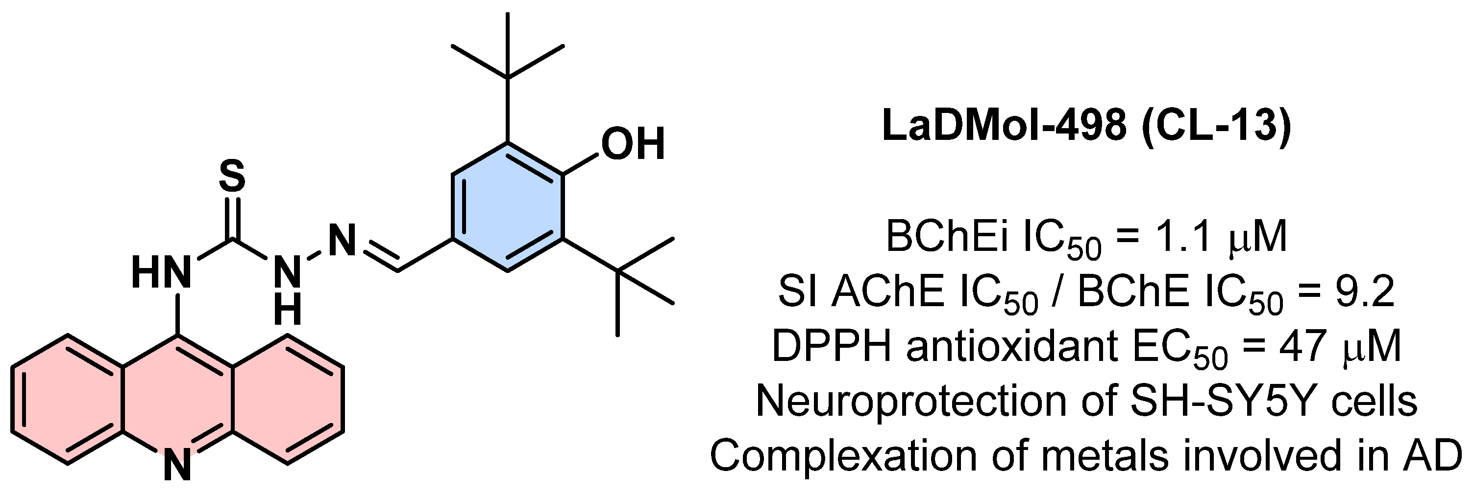
Over the past few years, our research group has been dedicated to the discovery of cholinesterase inhibitors [7,8,9]. More recently, we have demonstrated the therapeutic potential of acridine-based multitarget ligands for AD. Using an integrated strategy that combined rational molecular design, synthetic chemistry, computational studies, and biological evaluation, we developed a series of acridine–thiosemicarbazone hybrids, leading to the identification of compound LaDMol-498 (CL-13) as a promising lead candidate (Figure 1) [10]. This derivative exhibited potent BChE inhibition, along with antioxidant and neuroprotective activities, effective metal-chelating properties, and no detectable cytotoxicity toward human neuronal cells (SH-SY5Y). Furthermore, in vivo studies showed that LaDMol-498 (CL-13) significantly improved cognitive performance in experimental models of AD without affecting locomotor activity, thereby validating this medicinal chemistry strategy for the discovery of multitarget anti-Alzheimer agents [10].
Building on the knowledge gained from acridine–thiosemicarbazone derivatives, the present work reports the synthesis, molecular modeling, and enzymatic inhibition evaluation of designed quinoline–acylguanidine hybrids as a novel class of selective BChE inhibitors, as described in Section 3.1.

2. Materials and Methods

2.1. General Experimental Details

All chemicals and solvents were purchased from Sigma-Aldrich (Saint Louis, MO, USA) and used without further purification. All reactions were monitored by thin-layer chromatography (TLC) on 0.25 mm thick Merk 60 F254 silica gel plates with a fluorescent UV254 indicator. Chromatograms were viewed under ultraviolet light (254–365 nm). 1H and 13C NMR spectra were recorded on either a 400 MHz or 500 MHz Brucker Avance spectrometer (AC-400 or AC-500) in DMSO-d6 or CDCl3, using TMS as the internal standard. The multiplicities were identified as s (singlet), d (doublet), t (triplet), q (quartet), dd (double doublet), and m (multiplet). Flash chromatography was performed using an Accelerated Chromatographic Isolation system (Biotage Isolera, model ISO-4SV). Characterization data for synthetic intermediates is available in the Supplementary Material. Purity assessment of all final compounds was performed by HPLC, confirming purity levels ≥ 95%.

2.2. General Synthetic Procedures

2.2.1. General Procedure for the Synthesis of 2-Substituted-4-Carboxylic Acid Intermediates (7) [11]

In a bottom flask containing isatin (6) (3.34 mmol), KOH (13.59 mmol), ethanol (8 mL), and water (2 mL), we added acetophenone or 1-(4-bromophenyl)ethenone or 1-(4-fluorophenyl)ethenone (1 eq of each) or acetone (24 eq), and the mixture was stirred at 90 °C for 24 h or until the consumption of the reagents was evidenced by TLC. The mixture was allowed to cool, the reaction volume was reduced on a rotary evaporator, crushed ice was added, and a 20% HCl solution was added dropwise while monitoring the pH until pH 4.5. The formed precipitate was filtered and washed with distilled water. After the product was dry, it was washed with dichloromethane and dried at 40 °C. Yield: 45–60%.
  • 2-phenylquinoline-4-carboxylic acid (7a)
Compound 7a was obtained as a pale-yellow solid (45% yield): m.p. 217–218 °C.
1H RMN (500 MHz) DMSO-d6/TMS (δ-ppm): 8.67 (d, J = 8.1 Hz, 1H), 8.47 (s, 1H), 8.30 (d, J = 7.1 Hz, 2H), 8.18 (d, J = 8.3 Hz, 1H), 7.86 (t, J = 7.1 Hz, 1H), 7.71 (dd, J = 11.8, 4.6 Hz, 1H), 7.56 (m, 3H). 13C RMN (125 MHz) DMSO-d6/TMS (δ-ppm): 168.1, 156.3, 148.9, 138.4, 138.1, 130.7, 130.5, 130.3, 129.4, 128.3, 127.7, 125.9, 123.9, 119.6.
  • 2-(4-fluorophenyl)quinoline-4-carboxylic acid (7b)
Compound 7b was obtained as a pale-yellow solid (60% yield): m.p. 198–199 °C.
1H RMN (500 MHz) DMSO-d6/TMS (δ-ppm): 8.61 (d, J = 8.4 Hz, 1H), 8.40 (s, 1H), 8.33 (dd, J = 8.6, 5.6 Hz, 2H), 8.13 (d, J = 8.4 Hz, 1H), 7.83 (t, J = 7.4 Hz, 1H), 7.68 (t, J = 7.5 Hz, 1H), 7.38 (t, J = 8.8 Hz, 2H). 13C RMN (125 MHz) DMSO-d6/TMS (δ-ppm): 168.2, 164.8, 162.8, 155.2, 148.7, 138.9, 134.8, 130.7, 130.1, 130.0, 129.9, 128.2, 125.9, 123.8, 119.2, 116.4, 116.3.
  • 2-(4-bromophenyl)quinoline-4-carboxylic acid (7c)
Compound 7c was obtained as a pale-yellow solid (53% yield): m.p. 248–250 °C.
1H RMN (500 MHz) DMSO-d6/TMS (δ-ppm): 8.66 (d, J = 8.1 Hz, 1H), 8.48 (s, 1H), 8.27 (d, J = 8.6 Hz, 2H), 8.17 (d, J = 8.3 Hz, 1H), 7.87 (t, J = 8.2 Hz, 1H), 7.77 (d, J = 8.6 Hz, 2H), 7.73 (t, J = 7.1 Hz, 1H). 13C RMN (125 MHz) DMSO-d6/TMS (δ-ppm): 168.0, 155.1, 148.8, 138.4, 137.5, 132.4, 130.9, 130.2, 129.7, 128.5, 125.9, 124.3, 124.0.
  • 2-methylquinoline-4-carboxylic acid (7d)
Compound 7d was obtained as a pale-yellow solid (57% yield): m.p. 247–249 °C.
1H RMN (500 MHz) DMSO-d6/TMS (δ-ppm): 8.61 (d, J = 8.4 Hz, 1H), 8.00 (d, J = 8.4 Hz, 1H), 7.82 (s, 1H), 7.77 (t, J = 7.6 Hz, 1H), 7.62 (t, J = 7.6 Hz, 1H), 2.71 (s, 3H). 13C RMN (125 MHz) DMSO-d6/TMS (δ-ppm): 168.2, 159.2, 148.6, 136.9, 130.1, 129.3, 127.3, 125.8, 123.2, 123.1, 24.9.

2.2.2. General Procedure for the Synthesis of Guanidinylating Intermediates (8) [12]

To a solution of quinoline carboxylic acid 8a–d (2 mmol) in 10 mL of dry dichloromethane, 5 equivalents of thionyl chloride (SOCl2) were added under stirring. Then, two drops of dimethylformamide (DMF) were added, and the mixture was stirred at 40 °C for two hours, when TLC indicated complete consumption of the starting acid. The reaction mixture was completely evaporated under vacuum. tert-butyl (amino (methylthio)methylene)carbamate (5) (2.01 mmol) was solubilized in dry dichloromethane (10 mL) and triethylamine (1 mL). The previously evaporated acid chloride was diluted in dichloromethane (10 mL) and added dropwise to the previous solution. The mixture was continuously stirred for about two hours at room temperature, or until the starting reagents were completely consumed. The mixture was diluted with dichloromethane and washed with water. The organic phase was dried with anhydrous sodium sulfate and then evaporated under vacuum. The product was purified by column chromatography using a hexane/ethyl acetate mixture as the mobile phase and/or recrystallization from acetone. The product was obtained as a solid in 30–60% yields.
  • tert-butyl (Z)-((2-methylquinoline-4-carboxamido)(methylthio)methylene)carbamate (8a)
Compound 8a was obtained as a brown oil (51% yield).
1H RMN (500 MHz) CDCl3/TMS (δ-ppm): 12.51 (s, 1H), 8.78 (d, J = 7.9 Hz, 1H), 8.07 (d, J = 8.3 Hz, 1H), 7.96 (s, 1H), 7.72 (d, J = 6.6 Hz, 1H), 7.56 (t, J = 7.4 Hz, 1H), 2.80 (s, 3H), 1.50 (s, 9H). 13C RMN (125 MHz) CDCl3/TMS (δ-ppm): 158.8, 129.4, 129.2, 126.7, 125.9, 123.3, 28.0, 15.1, 13.4.
  • tert-butyl (Z)-((methylthio)(2-phenylquinoline-4-carboxamido)methylene)carbamate (8b)
Compound 8b was obtained as a brown oil (56% yield).
1H RMN (500 MHz) CDCl3/TMS (δ-ppm): δ 12.57 (s, 1H), 8.88 (d, J = 8.5 Hz, 1H), 8.63 (s, 1H), 8.24 (d, J = 8.4 Hz, 1H), 8.19 (d, J = 7.5 Hz, 2H), 7.76 (t, J = 7.5 Hz, 1H), 7.61 (t, J = 7.6 Hz, 1H), 7.56 (dd, J = 17.1, 9.8 Hz, 2H), 7.49 (t, J = 7.2 Hz, 1H), 2.58 (s, 3H), 1.57 (s, 9H). 13C RMN (125 MHz) CDCl3/TMS (δ-ppm): δ 177.2, 173.7, 156.8, 150.9, 149.4, 142.2, 139.3, 130.3, 129.6, 129.6, 129.0, 127.4, 127.4, 125.9, 124.3, 120.6, 84.1, 28.0, 15.2.
  • tert-butyl (Z)-((2-(4-fluorophenyl)quinoline-4-carboxamido)(methylthio)methylene)carbamate (8c)
Compound 8c was obtained as a brown oil (62% yield).
1H RMN (500 MHz) CDCl3/TMS (δ-ppm): δ 12.56 (s, 1H), 8.86 (d, J = 8.5 Hz, 1H), 8.57 (s, 1H), 8.19 (dd, J = 12.7, 8.0 Hz, 3H), 7.76 (t, J = 7.6 Hz, 1H), 7.60 (t, J = 7.6 Hz, 1H), 7.23 (t, J = 8.6 Hz, 2H), 2.57 (s, 3H), 1.57 (s, 9H). 13C RMN (125 MHz) CDCl3/TMS (δ-ppm): δ 177.2, 174.0, 165.1, 163.0, 155.8, 151.0, 149.5, 142.5, 135.5, 130.3, 129.9, 129.4, 129.4, 127.5, 126.0, 124.3, 120.3, 116.1, 116.0, 84.2, 77.4, 77.1, 76.9, 28.1, 15.3.
  • tert-butyl (Z)-((2-(4-bromophenyl)quinoline-4-carboxamido)(methylthio)methylene)carbamate (8d)
Compound 8d was obtained as a brown oil (33% yield).
1H RMN (500 MHz) CDCl3/TMS (δ-ppm): 12.55 (s, 1H), 8.86 (d, J = 8.5 Hz, 1H), 8.57 (s, 1H), 8.21 (d, J = 8.4 Hz, 1H), 8.07 (d, J = 8.4 Hz, 2H), 7.76 (t, J = 7.5 Hz, 1H), 7.67 (d, J = 8.4 Hz, 2H), 7.61 (t, J = 7.6 Hz, 1H), 2.56 (s, 3H), 1.57 (s, 9H). 13C RMN (125 MHz) CDCl3/TMS (δ-ppm): 177.0, 173.8, 155.5, 150.9, 149.5, 142.6, 138.1, 132.1, 130.3, 129.8, 128.9, 127.6, 125.9, 124.4, 124.2, 120.0, 84.2, 28.2, 15.9.

2.2.3. General Procedure for the Synthesis of Tert-Butyl (2-Substituted-Quinoline-4-Acylguanidine) Carbamate (9a–j) [13]

In a solution of compounds 8a–d (1 mmol) in 5 mL of dichloromethane in a round-bottom flask, 1 equivalent of Et3N, 1 equivalent of N,N-diisopropylethylamine (DIPEA) and 1,1 equivalents of the amines of interest were added. The mixture was stirred at room temperature for 24 h when the TLC indicated the complete consumption of the reactants. The mixture was then extracted in a liquid–liquid extraction containing water and dichloromethane. The organic layer was dried with anhydrous sodium sulfate and then evaporated under vacuum. The products obtained were purified by column chromatography using dichloromethane/methanol or hexane/ethyl acetate mixtures as the mobile phases. The products obtained had a yield of 50 to 97%.
  • tert-butyl (N-(N-benzylcarbamimidoyl)-2-phenylquinoline-4-carboxamide) carbamate (9a)
Compound 9a was obtained as a brownish solid (60% yield): m.p. 125–127 °C.
1H RMN (400 MHz) CDCl3/TMS (δ-ppm): 12.4 (s, 1H), 9.1 (s, 1H), 8.7 (d, J = 7.9 Hz, 1H), 8.3 (s, 1H), 8.1 (d, J = 8.1 Hz, 1H), 8.1 (d, J = 8.5 Hz, 2H), 7.5–7.4 (m, 4H), 7.3 (d, J = 4.4 Hz, 4H), 1.5 (s, 9H). 13C RMN (100 MHz) CDCl3/TMS (δ-ppm): 179.0, 156.9, 156.8, 153.4, 149.2, 144.2, 139.5, 137.2, 129.9, 129.3, 128.8, 127.7, 127.5, 126.7, 126.2, 124.4, 119.6, 83.9, 45.1, 28.0.
  • tert-butyl (N-(N-phenethylcarbamimidoyl)-2-phenylquinoline-4-carboxamide) carbamate (9b)
Compound 9b was obtained as a brownish solid (65% yield): m.p. 131–132 °C.
1H RMN (400 MHz) CDCl3/TMS (δ-ppm): 12.3 (s, 1H), 8.8 (d, J = 8.5 Hz, 2H), 8.5 (s, 1H), 8.3–8.1 (m, 3H), 7.7 (t, J = 7.0 Hz, 1H), 7.6–7.4 (m, 5H), 7.2 (dd, J = 19.5, 6.0 Hz, 3H), 3.8 (dd, J = 13.8, 7.0 Hz, 2H), 3.0 (t, J = 7.5 Hz, 2H), 1.5 (s, 9H). 13C RMN (100 MHz) CDCl3/TMS (δ-ppm): 178.9, 156.9, 153.3, 149.3, 144.4, 139.6, 138.3, 130.0, 129.3, 128.8, 128.7, 128.7, 127.5, 126.7, 126.7, 126.2, 119.7, 83.9, 42.9, 35.7, 28.1.
  • tert-butyl (N-(N-(4-fluorobenzyl)carbamimidoyl)-2-phenylquinoline-4-carboxamide) carbamate (9c)
Compound 9c was obtained as a brownish solid (76% yield): m.p. 153–154 °C.
1H RMN (400 MHz) CDCl3/TMS (δ-ppm): 9.1 (s, 1H), 8.7 (d, J = 8.4 Hz, 1H), 8.3 (s, 1H), 8.2 (d, J = 8.2 Hz, 1H), 8.1 (d, J = 7.3 Hz, 2H), 7.7 (t, J = 7.3 Hz, 1H), 7.5 (t, J = 7.1 Hz, 3H), 7.4 (s, 1H), 7.4–7.3 (m, 2H), 7.0 (t, J = 8.6 Hz, 2H), 4.7 (d, J = 5.7 Hz, 2H), 1.5 (s, 9H). 13C RMN (100 MHz) CDCl3/TMS (δ-ppm): 179.1, 163.2, 161.3, 156.9, 156.8, 153.4, 149.2, 144.2, 139.4, 133.0, 130.0, 129.3, 128.9, 127.5, 126.7, 126.1, 124.3, 119.5, 115.6, 84.0, 44.5, 28.2.
  • tert-butyl (N-(N-(4-chlorobenzyl)carbamimidoyl)-2-phenylquinoline-4-carboxamide) carbamate (9d)
Compound 9d was obtained as a brownish solid (88% yield): m.p. 162–163 °C.
1H RMN (400 MHz) CDCl3/TMS (δ-ppm): 12.2 (s, 1H), 9.4 (s, 1H), 8.6 (d, J = 8.4 Hz, 1H), 8.2 (s, 1H), 8.0 (t, J = 9.0 Hz, 3H), 7.7 (t, J = 7.2 Hz, 1H), 7.5 (d, J = 7.5 Hz, 4H), 7.4 (s, 3H), 4.6 (d, J = 6.0 Hz, 2H), 1.5 (s, 9H). 13C RMN (100 MHz) CDCl3/TMS (δ-ppm): 177.9, 156.7, 156.0, 152.3, 148.9, 144.5, 138.6, 138.1, 131.9, 130.2, 130.2, 130.0, 129.4, 129.3, 128.8, 127.4, 126.5, 124.1, 118.9, 83.8, 44.3, 28.1.
  • tert-butyl (N-(N-benzylcarbamimidoyl)-2-(4-fluorophenyl)quinoline-4-carboxamide) carbamate (9e)
Compound 9e was obtained as a whitish solid (92% yield): m.p. 143–144 °C.
1H RMN (400 MHz) CDCl3/TMS (δ-ppm): 12.4 (s, 1H), 9.1 (s, 1H), 8.7 (d, J = 8.5 Hz, 1H), 8.3 (s, 1H), 8.2–8.0 (m, 3H), 7.7 (t, J = 7.6 Hz, 1H), 7.5 (t, J = 7.7 Hz, 1H), 7.3 (m, 5H), 7.1 (t, J = 8.6 Hz, 2H), 4.7 (d, J = 5.8 Hz, 2H), 1.5 (s, 9H). 13C RMN (100 MHz) CDCl3/TMS (δ-ppm): 157.1, 155.8, 153.5, 137.4, 129.9, 129.6, 129.5, 129.4, 128.9, 127.8, 127.6, 126.9, 126.3, 124.4, 119.3, 115.9, 115.8, 84.1, 45.2, 28.2.
  • tert-butyl (N-(N-phenethylcarbamimidoyl)-2-(4-fluorophenyl)quinoline-4-carboxamide) carbamate (9f)
Compound 9f was obtained as a pale-yellow solid (97% yield): m.p. 145–146 °C.
1H RMN (400 MHz) CDCl3/TMS (δ-ppm): 12.35 (s, 1H), 8.95–8.77 (m, 2H), 8.42 (s, 1H), 8.27–8.09 (m, 3H), 7.74 (t, J = 7.5 Hz, 1H), 7.56 (t, J = 7.6 Hz, 1H), 7.37–7.02 (m, 7H), 3.81 (dd, J = 13.7, 6.8 Hz, 2H), 3.00 (t, J = 7.4 Hz, 2H), 1.55 (s, 9H). 13C RMN (100 MHz) CDCl3/TMS (δ-ppm): 157.0, 155.7, 153.4, 138.4, 129.54, 128.8, 126.9, 126.8, 126.4, 124.5, 119.3, 116.0, 115.8, 83.9, 43.0, 35.8, 28.1.
  • tert-butyl (2-(4-bromophenyl)-N-(N-(4-chlorobenzyl)carbamimidoyl)quinoline-4-carboxamide) carbamate (9g)
Compound 9g was obtained as a brownish solid (49% yield): m.p. 165–166 °C.
1H RMN (400 MHz) CDCl3/TMS (δ-ppm): 12.3 (s, 1H), 9.1 (s, 1H), 8.7 (d, J = 8.3 Hz, 1H), 8.25 (s, 1H), 8.1 (d, J = 8.5 Hz, 1H), 7.9 (d, J = 8.5 Hz, 2H), 7.7 (t, J = 7.6 Hz, 1H), 7.6 (d, J = 8.5 Hz, 2H), 7.5 (t, J = 7.2 Hz, 1H), 7.3 (d, J = 8.4 Hz, 2H), 7.2 (d, J = 8.4 Hz, 2H), 4.7 (d, J = 5.8 Hz, 2H), 1.5 (s, 9H). 13C RMN (100 MHz) CDCl3/TMS (δ-ppm): 179.1, 178.9, 157.0, 155.5, 153.4, 149.2, 144.4, 138.2, 135.8, 133.5, 131.9, 129.5, 129.0, 128.9, 128.8, 126.1, 124.4, 123.99, 118.9, 84.2, 44.5, 28.0.
  • tert-butyl (2-(4-bromophenyl)-N-(N-(4-fluorobenzyl)carbamimidoyl)quinoline-4-carboxamide) carbamate (9h)
Compound 9h was obtained as a brownish solid (55% yield): m.p. 144–145 °C.
1H RMN (400 MHz) CDCl3/TMS (δ-ppm): 12.3 (s, 1H), 9.1 (s, 1H), 8.7 (d, J = 8.4 Hz, 1H), 8.2 (s, 1H), 8.1 (d, J = 8.4 Hz, 1H), 8.0 (d, J = 8.5 Hz, 2H), 7.7 (t, J = 8.1 Hz, 1H), 7.6 (d, J = 8.5 Hz, 2H), 7.5 (t, J = 7.6 Hz, 1H), 7.3 (m, 2H), 7.0 (t, J = 8.6 Hz, 2H), 4.7 (d, J = 5.8 Hz, 2H), 1.5 (s, 9H). 13C RMN (100 MHz) CDCl3/TMS (δ-ppm): 178.9, 156.9, 155.4, 153.3, 149.2, 144.4, 138.3, 131.9, 130.0, 129.5, 129.2, 129.2, 129.0, 126.9, 126.1, 124.4, 123.9, 118.9, 115.8, 115.6, 84.2, 44.4, 28.0.
  • tert-butyl (N-(N-(4-chlorobenzyl)carbamimidoyl)-2-methylquinoline-4-carboxamide) carbamate (9i)
Compound 9i was obtained as a brownish solid (45% yield): m.p. 95–96 °C.
1H RMN (400 MHz) CDCl3/TMS (δ-ppm): 12.3 (s, 1H), 9.1 (s, 1H), 8.6 (d, J = 8.4 Hz, 1H), 8.0 (d, J = 8.4 Hz, 1H), 7.7–7.6 (m, 2H), 7.4 (t, J = 7.6 Hz, 1H), 7.3 (d, J = 8.4 Hz, 2H), 7.3–7.2 (m, 2H), 4.7 (d, J = 5.7 Hz, 2H), 2.7 (s, 3H), 1.5 (s, 9H). 13C RMN (100 MHz) CDCl3/TMS (δ-ppm): 179.1, 158.8, 157.0, 153.3, 148.9, 143.7, 136.0, 133.9, 129.0, 126.1, 123.5, 122.2, 84.2, 44.5, 28.2, 13.5.
  • tert-butyl (N-(N-(4-fluorobenzyl)carbamimidoyl)-2-methylquinoline-4-carboxamide) carbamate (9j)
Compound 9j was obtained as a brownish solid (48% yield): m.p. 97–98 °C.
1H RMN (400 MHz) CDCl3/TMS (δ-ppm): 12.3 (s, 1H), 9.0 (s, 1H), 8.7 (d, J = 8.5 Hz, 1H), 8.0 (d, J = 8.4 Hz, 1H), 7.7 (s, 1H), 7.6 (t, J = 7.2 Hz, 1H), 7.4 (t, J = 7.6 Hz, 1H), 7.3–7.3 (m, 2H), 7.0 (t, J = 8.6 Hz, 2H), 4.7 (d, J = 5.7 Hz, 2H), 2.7 (s, 3H), 1.5 (s, 9H). 13C RMN (100 MHz) CDCl3/TMS (δ-ppm): 179.1, 163.3, 158.5, 156.9, 153.3, 148.8, 143.6, 133.1, 129.4, 129.3, 129.1, 128.9, 126.1, 126.1, 122.2, 115.8, 115.6, 84.0, 44.5, 28.0, 25.3.

2.2.4. General Procedure for the Synthesis of 2-Substituted-Quinoline-4-Acylguanidine Derivatives (2a–j) [8]

To a solution of compounds 9a–j in dichloromethane (5 mL) was added 10–20 equivalents of trifluoroacetic acid (TFA). The mixture was stirred at room temperature until the complete consumption of the reactant was monitored by TLC (24–48h). The solution was then evaporated under vacuum to afford the corresponding products in quantitative yields.
  • N-(N-benzylcarbamimidoyl)-2-phenylquinoline-4-carboxamide (2a)
Compound 2a was obtained as a brownish solid m.p. 127–129 °C.
1H RMN (400 MHz) DMSO-d6/TMS (δ-ppm): δ 9.6 (s, 1H), 9.2 (s, 1H), 9.1 (s, 1H), 8.4 (s, 1H), 8.3 (d, J = 7.3 Hz, 2H), 8.2 (d, J = 8.3 Hz, 1H), 8.2 (d, J = 8.4 Hz, 1H), 7.9 (t, J = 7.6 Hz, 1H), 7.7 (t, J = 7.6 Hz, 1H), 7.5 (dq, J = 14.2, 7.1 Hz, 3H), 7.5–7.4 (m, 4H), 7.3 (dd, J = 8.4, 5.7 Hz, 1H), 4.7 (d, J = 6.1 Hz, 2H). 13C RMN (100 MHz) DMSO-d6/TMS (δ-ppm): 167.7, 155.7, 153.1, 148.0, 139.1, 137.8, 135.7, 130.8, 130.3, 129.8, 129.0, 128.7, 128.0, 127.8, 127.4, 127.3, 124.8, 122.4, 118.0, 44.4. HPLC purity: 99%. HRMS [M + H]+ calcd for C24H20N4O: 381.1710; m/z [M]+ found: 381.171.
  • N-(N-phenethylcarbamimidoyl)-2-phenylquinoline-4-carboxamide (2b)
Compound 2b was obtained as a brownish solid m.p. 153–155 °C.
1H RMN (400 MHz) DMSO-d6/TMS (δ-ppm): 9.2 (t, J = 5.3 Hz, 1H), 9.1 (s, 1H), 8.9 (s, 1H), 8.4 (s, 1H), 8.3 (d, J = 7.2 Hz, 2H), 8.2 (t, J = 8.6 Hz, 2H), 7.9 (t, J = 7.7 Hz, 1H), 7.7 (t, J = 7.7 Hz, 1H), 7.5 (dq, J = 14.2, 7.0 Hz, 3H), 7.3 (d, J = 4.3 Hz, 4H), 7.2 (dd, J = 8.0, 4.3 Hz, 1H), 3.6 (dd, J = 14.2, 6.6 Hz, 2H), 2.9 (t, J = 7.5 Hz, 2H). 13C RMN (100 MHz) DMSO-d6/TMS (δ-ppm): 167.7, 155.7, 152.8, 148.0, 139.2, 137.9, 137.8, 130.8, 130.3, 129.8, 129.0, 128.9, 128.5, 128.0, 127.3, 126.7, 124.7, 122.3, 117.9, 42.6, 39.5, 33.6. HPLC purity: 99%. HRMS [M + H]+ calcd for C25H22N4O: 395.1867; m/z [M]+ found: 395.187.
  • N-(N-(4-fluorobenzyl)carbamimidoyl)-2-phenylquinoline-4-carboxamide (2c)
Compound 2c was obtained as a brownish solid m.p. 120–121 °C.
1H RMN (400 MHz) DMSO-d6/TMS (δ-ppm): 12.4 (s, 1H), 9.7 (s, 1H), 9.1 (s, 2H), 8.4 (s, 1H), 8.3 (d, J = 7.3 Hz, 2H), 8.3 (d, J = 8.6 Hz, 1H), 8.2 (d, J = 8.4 Hz, 1H), 7.9 (t, J = 7.5 Hz, 1H), 7.7 (t, J = 7.5 Hz, 1H), 7.6–7.5 (m, 3H), 7.5 (dd, J = 7.8, 5.8 Hz, 2H), 7.2 (t, J = 8.8 Hz, 2H), 4.6 (d, J = 5.9 Hz, 2H). 13C RMN (100 MHz) DMSO-d6/TMS (δ-ppm): 168.2, 163.3, 161.2, 159.2, 156.2, 153.5, 148.4, 139.5, 138.2, 131.2, 130.7, 130.2, 130.1, 130.0, 129.4, 128.4, 127.7, 125.2, 122.8, 118.5, 116.0, 115.8, 44.1. HPLC purity: 96%. HRMS [M + H]+ calcd for C23H19FN4O: 399.1616; m/z [M]+ found: 399.161.
  • N-(N-(4-chlorobenzyl)carbamimidoyl)-2-phenylquinoline-4-carboxamide (2d)
Compound 2d was obtained as a brownish solid m.p. 113–114 °C.
1H RMN (400 MHz) DMSO-d6/TMS (δ-ppm): 9.5 (s, 1H), 9.2 (s, 1H), 8.8 (s, 1H), 8.3 (s, 1H), 8.2–8.1 (m, 4H), 7.9 (m, 1H), 7.7 (m, 1H), 7.6–7.5 (m, 3H), 7.4 (m, 4H), 4.6 (d, 2H). 13C RMN (100 MHz) DMSO-d6/TMS (δ-ppm): 174.0, 159.0, 158.8, 158.5, 158.3, 155.0, 148.5, 137.6, 135.5, 132.8, 132.4, 131.2, 130.2, 128.8, 128.8, 129.0, 128.5, 125.5, 124.4, 123.2, 118.7, 118.4, 116.3, 72.8, 44.1. HPLC purity: 96%. HRMS [M + H]+ calcd for C23H19ClN4O: 415.1320; m/z [M]+ found: 415.132.
  • N-(N-benzylcarbamimidoyl)-2-(4-fluorophenyl)quinoline-4-carboxamide (2e)
Compound 2e was obtained as a pale-yellow solid m.p. 183–185 °C.
1H RMN (400 MHz) DMSO-d6/TMS (δ-ppm): δ 9.9 (s, 1H), 9.2 (sl, 2H), 8.4 (s, 1H), 8.4–8.3 (m, 2H), 8.2 (d, J = 8.3 Hz, 1H), 8.1 (d, J = 8.4 Hz, 1H), 7.9 (t, J = 7.5 Hz, 1H), 7.7 (t, J = 7.5 Hz, 1H), 7.4–7.3 (m, 7H), 4.7 (d, J = 5.5 Hz, 2H). 13C RMN (100 MHz) DMSO-d6/TMS (δ-ppm): 168.3, 164.9, 155.1, 153.7, 148.4, 139.5, 136.2, 134.8, 131.2, 130.1, 130.1, 130.0, 129.1, 128.4, 128.2, 127.8, 125.2, 122.7, 118.5, 116.4, 116.2, 44.8. HPLC purity: 96%. HRMS [M + H]+ calcd for C23H19FN4O: 399.1616; m/z [M]+ found: 399.161.
  • N-(N-phenetylcarbamimidoyl)-2-(4-fluorophenyl)quinoline-4-carboxamide (2f)
Compound 2f was obtained as a pale-yellow solid m.p. 181–182 °C.
1H RMN (400 MHz) DMSO-d6/TMS (δ-ppm): δ 12.5 (s, 1H), 8.4 (s, 1H), 8.4–8.3 (m, 2H), 8.2 (d, J = 8.4 Hz, 1H), 8.1 (d, J = 8.4 Hz, 1H), 7.9 (t, J = 7.5 Hz, 1H), 7.7 (t, J = 7.5 Hz, 1H), 7.4 (t, J = 8.3 Hz, 2H), 7.3 (d, J = 12.8 Hz, 4H), 7.2 (s, 1H), 3.6 (d, J = 6.4 Hz, 2H), 2.9 (t, J = 7.2 Hz, 2H). 13C RMN (100 MHz) DMSO-d6/TMS (δ-ppm): δ 168.3, 164.9, 163.0, 155.1, 153.5, 148.3, 139.5, 138.3, 134.8, 131.2, 130.2, 130.1, 129.3, 128.9, 128.4, 127.0, 125.1, 122.7, 118.4, 116.4, 116.2, 43.0. HPLC purity: 95%. HRMS [M + H]+ calcd for C25H21FN4O: 413.1772; m/z [M]+ found: 413.177.
  • 2-(4-bromophenyl)-N-(N-(4-chlorobenzyl)carbamimidoyl)quinoline-4-carboxamide (2g)
Compound 2g was obtained as a brownish solid m.p. 130–132 °C.
1H RMN (500 MHz) DMSO-d6/TMS (δ-ppm): 9.7 (s, 1H), 9.2 (s, 1H), 9.0 (s, 1H), 8.4 (s, 1H), 8.2 (d, J = 8.4 Hz, 3H), 8.2 (d, J = 8.4 Hz, 1H), 7.9 (t, J = 7.6 Hz, 1H), 7.8 (d, J = 8.5 Hz, 2H), 7.7–7.7 (m, 1H), 7.5 (dd, J = 18.3, 8.5 Hz, 4H), 4.6 (d, J = 6.1 Hz, 2H). 13C NMR (126 MHz, DMSO-d6) δ 174.9, 158.8, 158.5, 158.3, 158.0, 156.2, 148.5, 132.2, 132.0, 133.1, 130.6, 130.2, 129.8, 129.4, 129.1, 129.0, 127.8, 125.3, 118.6, 115.5, 115.4. 44.2. HPLC purity: 99%. HRMS [M + H]+ calcd for C24H18BrClN4O: 493.0426; m/z [M]+ found: 493.043.
  • 2-(4-bromophenyl)-N-(N-(4-fluorobenzyl)carbamimidoyl)quinoline-4-carboxamide (2h)
Compound 2h was obtained as a yellowish solid m.p. 133–134 °C.
1H RMN (400 MHz) DMSO-d6/TMS (δ-ppm): 9.7 (s, 1H), 9.1 (d, J = 23.0 Hz, 2H), 8.4 (s, 1H), 8.2 (d, J = 8.1 Hz, 3H), 8.2 (d, J = 8.3 Hz, 1H), 7.9 (t, J = 7.4 Hz, 1H), 7.8 (s, 2H), 7.7 (t, J = 7.5 Hz, 1H), 7.5–7.4 (m, 2H), 7.2 (t, J = 8.7 Hz, 2H), 4.6 (d, J = 5.9 Hz, 2H). 13C RMN (100 MHz) DMSO-d6/TMS (δ-ppm): δ 168.0, 163.2, 161.3, 159.3, 158.8, 158.5, 155.1, 153.4, 148.4, 139.4, 139.8, 137.3, 132.4, 131.5, 130.2, 130.1, 130.0, 128.7, 125.2, 124.6, 122.9, 118.1, 116.1, 115.9, 44.3. HPLC purity: 99%. HRMS [M + H]+ calcd for C24H18BrFN4O: 477.0721; m/z [M]+ found: 477.072.
  • N-(N-(4-chlorobenzyl)carbamimidoyl)-2-methylquinoline-4-carboxamide (2i)
Compound 2i was obtained as a beige solid m.p. 106–108 °C.
1H RMN (500 MHz) DMSO-d6/TMS (δ-ppm): δ 9.7 (s, 1H), 9.1 (bs, 2H), 8.2 (d, J = 8.4 Hz, 1H), 8.1 (d, J = 8.4 Hz, 1H), 7.8 (t, J = 7.6 Hz, 1H), 7.7 (s, 1H), 7.7 (d, J = 7.3 Hz, 1H), 7.5 (d, J = 8.0 Hz, 2H), 7.5 (d, J = 8.0 Hz, 2H), 4.7 (d, J = 6.0 Hz, 2H), 2.7 (s, 3H). 13C RMN (126 MHz) DMSO-d6/TMS (δ-ppm): 168.5, 158.9, 158.8, 158.6, 135.3, 132.9, 130.8, 129.8, 129.1, 127.8, 125.3, 122.2, 121.5, 70.2, 54.0, 44.2, 24.5. HPLC purity: 96%. HRMS [M + H]+ calcd for C19H17ClN4O: 353.1164; m/z [M]+ found: 353.116.
  • N-(N-(4-fluorobenzyl)carbamimidoyl)-2-methylquinoline-4-carboxamide (2j)
Compound 2j was obtained as a brownish solid m.p. 84–86 °C.
1H RMN (400 MHz) DMSO-d6/TMS (δ-ppm): 9.6 (s, 1H), 9.1 (d, J = 45.0 Hz, 2H), 8.2 (d, J = 8.4 Hz, 1H), 8.0 (d, J = 8.4 Hz, 1H), 7.8 (t, J = 7.6 Hz, 1H), 7.8 (s, 1H), 7.7 (d, J = 7.3 Hz, 1H), 7.5–7.4 (m, 2H), 7.2 (t, J = 8.8 Hz, 3H), 4.6 (d, J = 6.0 Hz, 2H), 2.7 (s, 3H). 13C RMN (100 MHz) DMSO-d6/TMS (δ-ppm): 168.0, 158.9, 153.6, 131.2, 130.1, 128.4, 127.9, 125.3, 122.1, 121.5, 116.0, 115.8, 44.2, 24.7. HPLC purity: 95%. HRMS [M + H]+ calcd for C19H17FN4O: 337.1459; m/z [M]+ found: 337.146.

2.3. Cholinesterase Inhibition Assay

The inhibitory activity against cholinesterases was measured using an adapted Elman’s method [14]. All solutions were prepared in 0.02 molar T-HCl buffer (pH = 7.5). Stock solutions were prepared in MeOH (20 mM). The experiment has been conducted in triplicate. A flat-bottom, transparent 96-well plate was loaded with 150 μL of treatment solutions containing the compounds 2a–j and a reference internal inhibitor (donepezil. AChE IC50 = 0.007 μM; BChE IC50 = 2.625 μM) prepared at eight serially diluted concentrations. Vehicle control (MeOH—final concentration of 0.15% v/v) and untreated cells (negative control) were used in comparison. 60 μL of 5,5′-dithiobis (2-nitrobenzoic acid) (DTNB) and 30 μL of electric eel acetylcholinesterase (eeAChE) or equine serum butyrylcholinesterase (eqBChE) solution (0.20 U/mL), prepared in the presence of bovine serum albumin (BSA, 1 mg/mL), were added to each well. The absorbance was measured using an iMark microplate reader (Bio-Rad) equipped with a λ = 415 nm light filter to establish the blank reference. After 10 min of incubation at 30 °C, 24 μL of acetylcholine iodide (ACTI, 2.75 mM) or S-butyrylthiocholine iodide (BCTI) was added to initiate the enzymatic reaction. Absorbance at 415 nm was then recorded ten times at 30 s intervals.
Enzymatic activity was expressed as a percentage relative to the untreated controls after subtracting the blank values. Finally, IC50 values were calculated through non-linear regression analysis of the dose–response curves using GraphPad Prism 7.0 software.

2.4. In Silico Assays

2.4.1. Molecular Docking

To investigate the molecular basis of the observed enzymatic inhibitions, a molecular docking study was conducted using butyrylcholinesterase from Equus caballus (EqBChE).
A three-dimensional model was developed via comparative modeling. This process utilized the protein sequence from the UniProtKB/Swiss-Prot database (entry Q9N1N9) and the human BChE structure (hBChE, PDB ID: 4BDS, resolution: 2.1 Å) as a template [8,15]. The resulting model demonstrated 89.89% sequence identity and 87% coverage relative to the human enzyme. Additionally, the model was compared with the AlphaFold2-predicted structure (UniProt Q9N1N9), showing an RMSD of 0.392 Å, further supporting its structural reliability.
To validate the docking protocol, redocking experiments were performed using GOLD 5.6 (CCDC) to ensure the reproduction of reliable binding poses for the cholinesterases. The binding site was defined as a 15 Å radius sphere centered on the Trp110 residue of EqBChE, which has been established in the literature as a key residue of the butyrylcholinesterase catalytic active site [16,17]. This radius was sufficient to encompass both the catalytic active site (CAS) and the peripheral anionic site (PAS). Structural water molecules were removed prior to the simulations.
The ligand structures were constructed and optimized using the Spartan’14 software [Wavefunction, Inc., Irvine, CA, USA] with the semi-empirical PM6 method. Within the GOLD docking program, results were evaluated based on “fitness scores”. These dimensionless values serve as a metric for docking quality, where higher scores indicate superior binding poses and theoretical affinity.

2.4.2. Theoretical Pharmacokinetic Profile Evaluation

To investigate the pharmacokinetic profile of the synthesized quinoline derivatives (2a–j), an in silico ADME (Absorption, Distribution, Metabolism, and Excretion) study was conducted using the SwissADME web tool (Swiss Institute of Bioinformatics). The molecular structures were converted into Canonical SMILES format and submitted to the platform to estimate key physicochemical parameters and drug-likeness properties [18,19]. The evaluation focused on the Lipinski Rule of Five (molecular weight, lipophilicity as LogP, and the number of hydrogen bond donors and acceptors) to assess the oral bioavailability potential. The compounds were also evaluated concerning aqueous solubility and P-gp substrate. Other parameters, including topological polar surface area (TPSA), were also recorded to provide a comprehensive pharmacokinetic characterization of the compounds.
Microscopic acid dissociation constants (pKa) were predicted using the Rowan pKa workflow implemented within the Rowan Scientific platform [20]. All calculations were performed in rapid mode considering a pKa window of 2 to 12. Solvation effects were accounted for using water as the solvent. This workflow utilizes fast conformer ensemble generation coupled with machine-learned interatomic potentials to evaluate the Gibbs free energies (ΔG) of the respective conjugate acid-base pairs, mapping the computed values to pKa values through established linear free-energy relationships [21].
To characterize the potential of the synthesized quinoline derivatives to reach the central nervous system (CNS), their blood–brain barrier (BBB) permeation was predicted using the Blood–Brain Barrier Permeability (LogBB) Predictor platform http://ssbio.cau.ac.kr/software/logbb_pred/ (accessed on 10 July 2026). The molecular structures in their protonated state were submitted to the platform to calculate the LogBB values based on established quantitative structure–activity relationship (QSAR) models. Predicted logBB values ≥ −1 is considered ‘BBB Permeable’ (CNS+), while LogBB ≤ −1 are considered ‘BBB Non-Permeable’ (CNS-) [22].

3. Results and Discussion

3.1. Design of Compounds

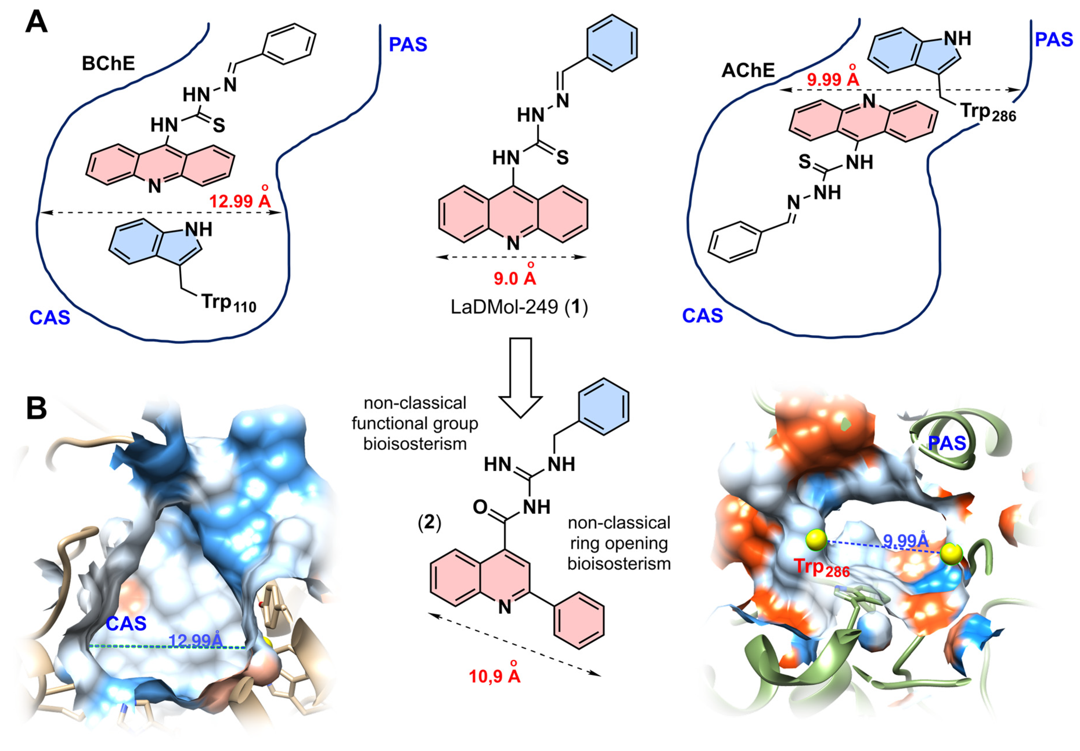
Recently, our research group reported a series of acridine-thiosemicarbazone derivatives as selective BChE inhibitors. Among them, the derivative LaDMol-249 (1), which lacks substituents on the imine-linked phenyl ring, exhibited the most potent BChE inhibitory potency within the series (AChE IC50 = 2.47 µM; BChE IC50 = 0.71 µM; SI AChE IC50/BChE IC50 = 3.5) (Figure 2A) [10]. Molecular docking simulations revealed that the acridine scaffold, owing to its planarity, which distinguishes it from the tetrahydroacridine core found in tacrine, is unable to accommodate within the narrower catalytic anionic site (CAS) of AChE. Consequently, this core is directed toward the enzyme’s peripheral site (PAS) (Figure 2A). To design compounds with enhanced BChE selectivity, we employed a non-classical ring-opening bioisosterism strategy, replacing the acridine core with a 2-phenyl-substituted quinoline scaffold (Figure 2B). The quinoline nucleus is well-documented for establishing crucial π−stacking interactions with tryptophan residues [23], a structural requirement deemed essential for the potency observed in the prototype series and others from our group. This modification aimed to slightly extend the length of the conjugated π-systems (from 9.0 Å in acridine to 10.9 Å in phenyl-quinoline), thereby maintaining the ability of the phenyl-quinoline system to interact within the BChE CAS (12.99 Å) while sterically hindering its fit into the narrower AChE PAS (9.99 Å) (Figure 2A,B). Finally, the thiosemicarbazone linker was replaced by an acylguanidine moiety. While preserving the optimal spacing between the aromatic systems, this substitution was intended to increase conformational freedom and enhance aqueous solubility by reducing the overall planarity of the compounds. Although acylguanidines remain underutilized in medicinal chemistry relative to their unsubstituted guanidine counterparts, this class exhibits remarkable pharmacological advantages. The acyl substitution confers substantially reduced basicity (approximately 4–5 orders of magnitude lower pKa values) [24], which translates into superior absorption (considerable portion remains uncharged at physiological pH), distribution, and target engagement (sufficiently basic to undergo key interactions) profiles across diverse biological systems [25,26].

3.2. Chemistry

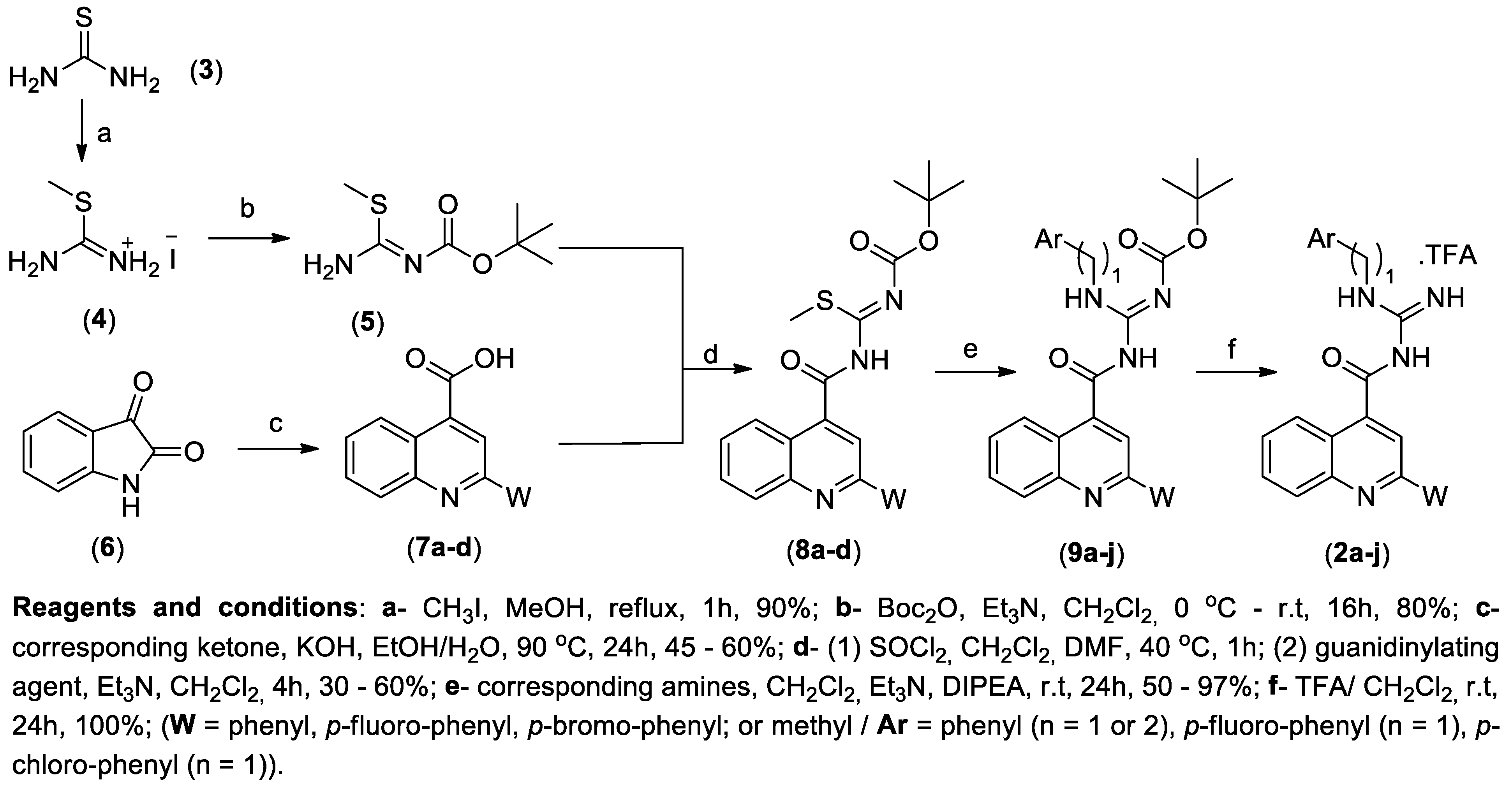
The synthesis of the 10 designed quinoline–acylguanidine hybrids (2a–j) was executed through a multi-step convergent strategy and is shown in Scheme 1. The first step was the synthesis of S-methylisothiourea (4), as previously described by Hickey [27], through the alkylation of urea (3) with methyl iodide. Subsequently, one of the nitrogens was protected with di-tert-butyl dicarbonate in dichloromethane under basic conditions, furnishing the N-Boc-S-methylisothiourea (5) (80% yield), named guanidinylating agent [12]. The quinoline core was synthesized via the Pfitzinger reaction, involving the condensation of substituted isatins (6) with various ketones under basic conditions (Scheme 1) [11]. This reaction provided the corresponding quinoline-4-carboxylic acid intermediates (7a–d) in moderate to good yields (45–60%). The versatility of the Pfitzinger approach allowed for the introduction of diverse substituents at the C-2 position of the quinoline ring, which is important for exploring the structure–activity relationship (SAR).
The resulting carboxylic acid intermediates (7a–d) were treated with thionyl chloride in dichloromethane and DMF to yield the corresponding acyl chlorides, which were subsequently reacted with the guanidinylating agent (5) to afford the key intermediates (8a–d). Thus, the protected acylguanidines (9a–j) were synthesized via the condensation of intermediates 8a–d with the appropriate benzylamines or phenethylamine, in yields ranging from 50 to 97%. In the final synthetic step, the Boc protecting groups were removed under acidic conditions to yield the corresponding deprotected acylguanidines (2a–j) (Scheme 1) [8]. The reaction proceeded in a mixture of trifluoroacetic acid and dichloromethane at room temperature over 24 to 48 h, providing the free acylguanidines (2a–j) as their respective trifluoroacetate salts in quantitative yields.
The principal 1H NMR signals characterizing the acylguanidines correspond to the guanidine NH protons, which were observed between 9.7 and 9.1 ppm. These derivatives were further characterized by the presence of aromatic proton signals ranging from 8.5 to 7.4 ppm. Additionally, a doublet corresponding to the spacer methylene group for benzylic compounds, and a double triplet for phenethyl compounds, were observed at approximately 4.7 and 3.7 ppm, respectively. Furthermore, the absence of the signal corresponding to the Boc methyl groups at 1.5 ppm confirmed the successful deprotection (Figures S21–S30).

3.3. Evaluation of In Vitro Cholinesterase Inhibition

The cholinesterase (EeAChE and EqBChE) inhibition activities of the free quinoline–acylguanidine derivatives (2a–j) were determined by an adapted Ellman’s method using donepezil as a reference compound. Initial screening of compounds 2a–j at 30 µM revealed a clear selective BChE inhibition profile, in which compounds presented up to 79% inhibition of this enzyme (for compound 2c), while the same compounds showed weak inhibitions of AChE, with less than 39% inhibition at the same concentration (Table 1). Furthermore, any of the Boc-protected compounds (9a–j) showed at least 50% inhibition of both cholinesterases, suggesting, in general, that the presence of the Boc group does not favor the inhibition of these enzymes.
Apparently, the best substitution profile in the quinoline nucleus was the introduction of a phenyl group in C-2 (2-phenylquinoline) with acceptable changes in the guanidine moiety. This pattern gave maximum BChE inhibition, at 30 µM, of 78.6% and an IC50 of up to 7.1 µM (for compound 2c), comparable to the reference donepezil (2.39 µM). As all 2-phenylquinoline compounds presented similar potencies, independent of the substitution profile, even for phenetyl compound (2b), we supposed that these substituents are turned to PAS site, at the entrance of BChE gorge, which is exposed to solvent with less steric restriction, as previewed in our compound design.
The size of the substituents at the C-2 position of the 2-phenylquinoline nucleus plays an important role in activity. The unsubstituted phenyl group led to the most active compound, 2c. Introduction of a fluorine atom at the 4-position of the phenyl ring was well tolerated, affording compound 2e (BChE IC50 = 8.6 µM), whereas a bulkier substituent such as bromine impaired activity, yielding compound 2h, which showed only 36% inhibition of BChE at 30 µM. These results support our design hypothesis that the phenylquinoline nucleus can be well accommodated within the CAS. While the 4-fluorophenyl group was essentially equipotent to the phenyl group, likely due to their similar steric demand, the introduction of the bulkier 4-bromophenyl substituent probably caused unfavorable van der Waals repulsions, leading to reduced activity. Finally, replacement of the phenyl substituent was also detrimental to activity, giving compounds 2i and 2j, which showed no significant BChE inhibition (up to 20% at 30 µM). These results indicate that complete occupation of the CAS is important for activity within this series.

3.4. Molecular Modeling Evaluation

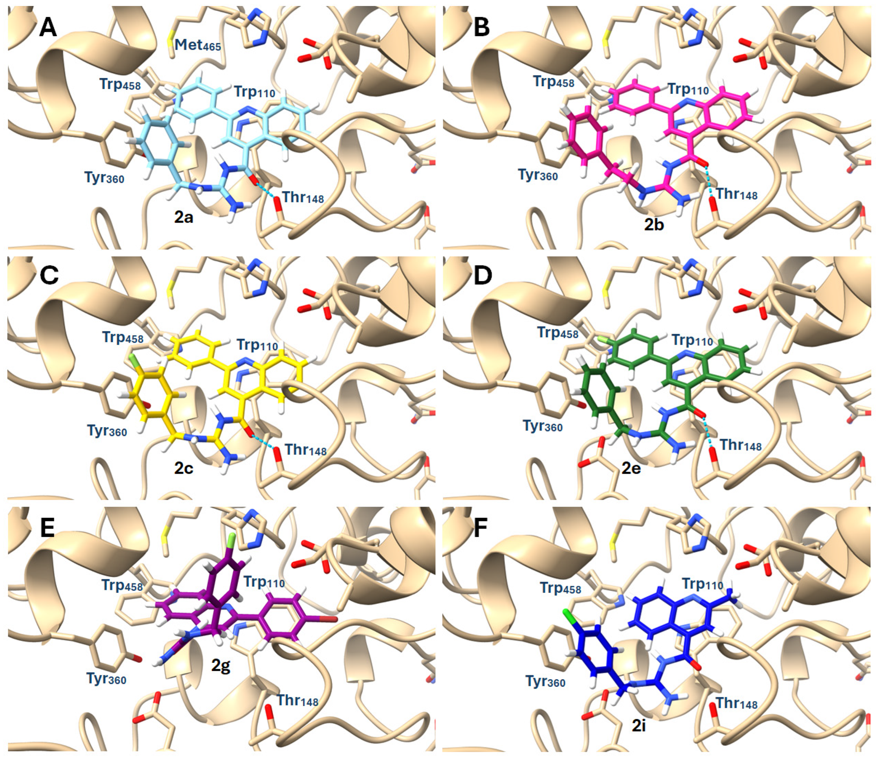
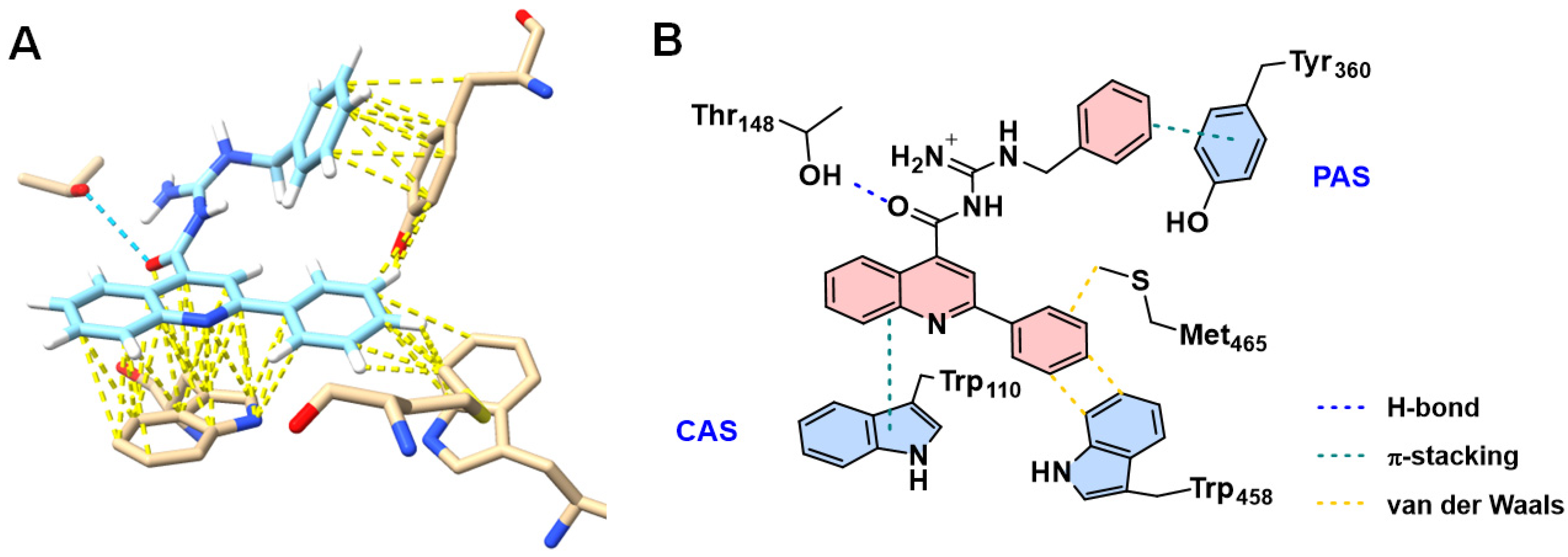
To complement the in vitro inhibition assays, molecular docking studies were performed to investigate the structure–activity relationships of the derivatives. The binding modes of the compounds were analyzed using a BChE model derived from equine serum and available from UniProt/Swiss-Model. As expected, most of the compounds were readily accommodated within the CAS of BChE, with the acylguanidine moieties oriented toward the PAS, where they established hydrogen bonds with Thr348 and hydrophobic interactions, including π-stacking, with Tyr360 (Figure 3). As anticipated, the 2-substituted-quinoline subunit formed additional hydrophobic contacts, including π-π stacking, with the key CAS residues Trp110, Met465 and Trp458, which collectively contributed to stabilization of the enzyme–ligand complexes [16,17]. Overall, the docking poses of all 2-substituted phenylquinoline derivatives (2a–c and 2e) were highly consistent (Figure 3).
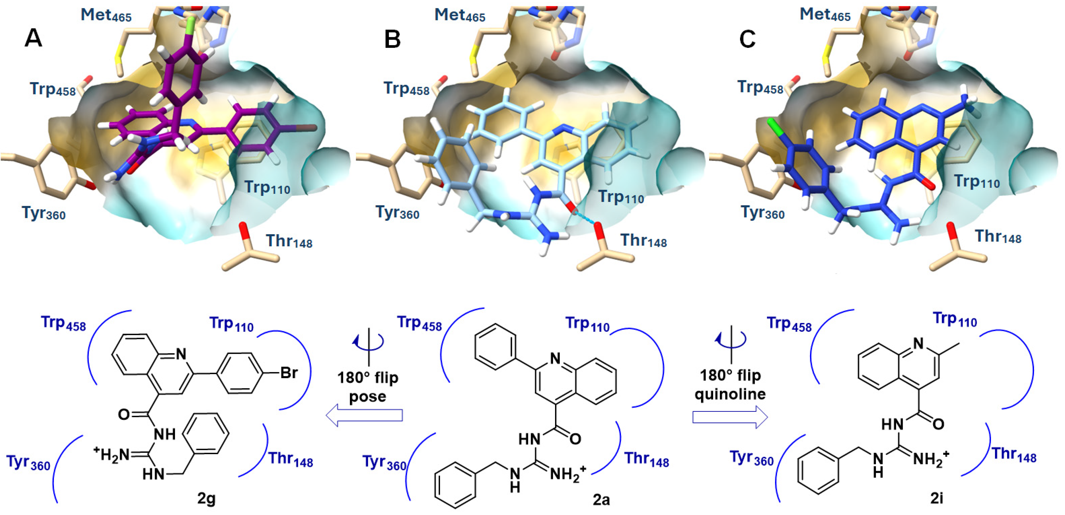
Docking studies further revealed that the inactive compounds adopted distinct binding poses, which can explain their loss of BChE inhibitory activity (Figure 4). In particular, the 2-methylphenylquinoline derivatives (2i and 2j) displayed an approximately 180° rotation of the quinoline nucleus, probably preventing the optimal interactions observed for the active analogues (2a–c and 2e) with Trp110, Met465 and Trp458 (Figure 4 and Figure 5A). Moreover, this quinoline flip rearranges the overall docking pose of compound 2i, leading to loss of the hydrogen bond between its carbonyl group and Thr148, which may contribute to the reduced stability of the complex (Figure 4). Likewise, the 4-bromophenyl-substituted quinoline derivatives (2g and 2h) exhibited a markedly different binding mode, with an approximately 180° flip of the entire structure, disrupting key interactions with Tyr380 and Thr148, consistent with the reduced activity of this subgroup. This flip can be rationalized by the docking pose of compound 2a, in which the 2-phenyl substituent occupies a sterically restricted region of the CAS, forcing the scaffold to reorient in order to relieve steric congestion and achieve a better fit within the binding site (Figure 4). All docking results led us to propose a map of required interactions based on polar and hydrophobic interactions observed for the active quinoline–acylguanidine derivatives (Figure 5B).

3.5. In Silico ADME Physico-Chemical Profile Analysis

ADME prediction is particularly important for BChE inhibitors, as their therapeutic efficacy depends not only on target potency but also on adequate brain exposure, including the ability to cross the blood–brain barrier (BBB). Early ADME assessment can help prioritize compounds with a favorable balance of absorption, distribution, metabolism, excretion, and CNS penetration. Small molecules may cross the BBB primarily through passive diffusion across the phospholipid bilayer of brain endothelial cells or through carrier-mediated processes involving transmembrane influx and efflux transporters [28]. TPSA has been identified as one of the most important physicochemical descriptors of BBB permeability, with CNS-active drugs generally exhibiting TPSA values below approximately 90 Å2 [29,30,31]. However, this threshold should be regarded as a general guideline rather than an absolute cutoff. Different physicochemical properties may be relevant to distinct transport mechanisms, including passive diffusion, carrier-mediated influx, and active efflux [28]. In a recent integrative model, TPSA, logD, molecular weight (MW), and the number of hydrogen-bond donors (HBDs) were identified as the most important overall predictors of putative BBB permeation, whereas rotatable bonds (RBs), logS, logP, and the number of hydrogen-bond acceptors (HBAs) made smaller contributions [28].
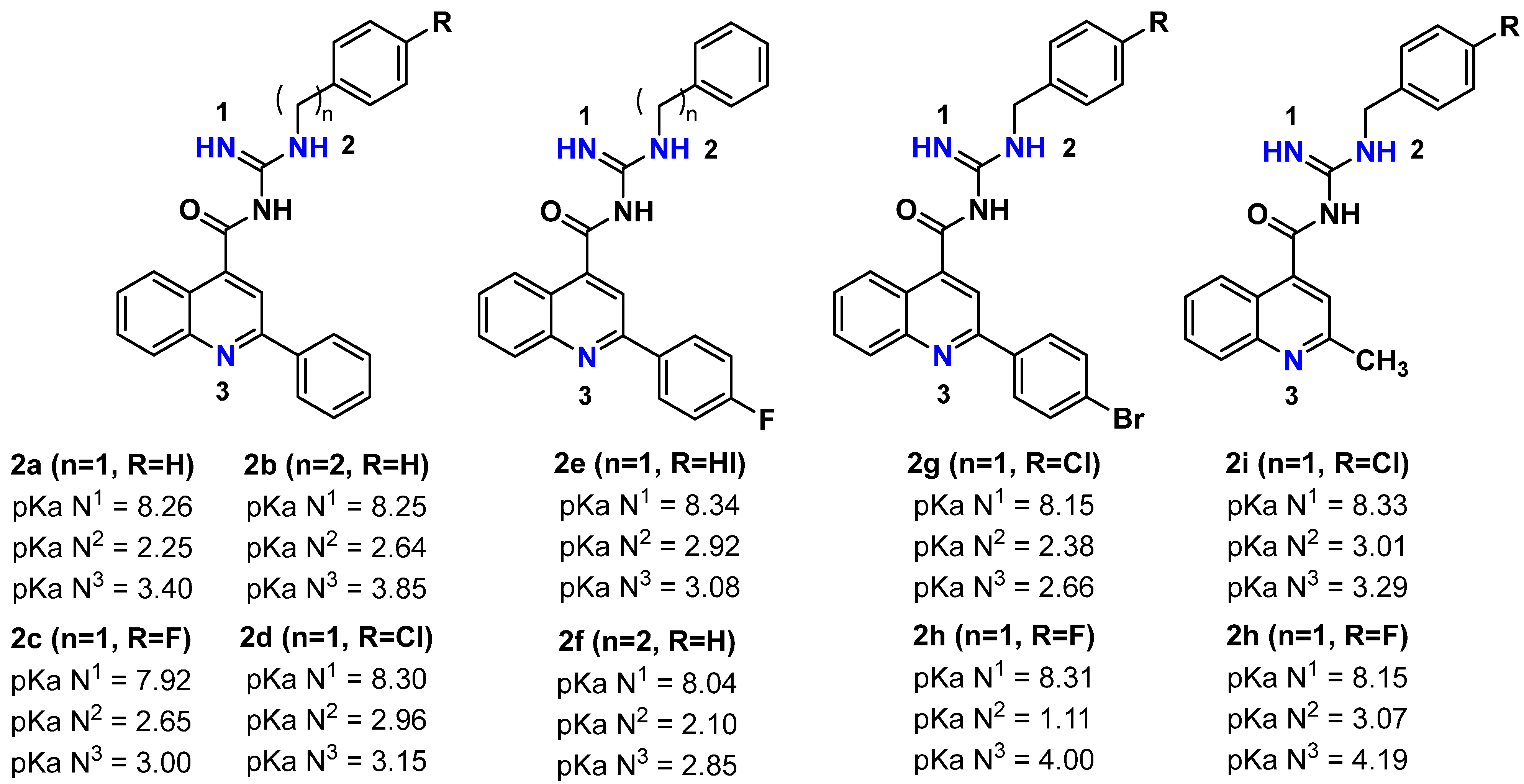
Thus, we performed an in silico evaluation of the compounds by analyzing these physicochemical parameters (Table 2, Figures S2–S12) [18,19]. The TPSA values indicated moderate polarity, with a representative value of 77.87 Å2, below the commonly recommended threshold of 90 Å2 for CNS-active compounds. Other relevant physicochemical properties were also calculated, including molecular weight (MW = 336–493 Da) and the number of hydrogen-bond donors (HBD ≤ 3). All compounds, except the inactive analogs 2g and 2h, met the established criteria for CNS-penetrant compounds (Table 2). Regarding lipophilicity, the calculated LogD values (2.49–4.54) were slightly above the optimal range of 1.5–2.7 for blood–brain barrier permeability. LogD was determined from the calculated pKa of the basic acylguanidine moiety [20,21] and the ionization state of each compound at pH 7.4 (Figure 5 and Figure S12), using the Henderson-Hasselbalch equation: LogD = LogP − log10 (1 + 10pKa-pH). To the best of our knowledge, experimental pKa determination for acylguanidines remains largely unexplored, with only a single reported measurement yielding a value near 8 [24]. Our predictions (pKa range 8.04–8.34) demonstrate excellent agreement with this limited experimental reference point (Figure 6). Moreover, we could calculate the ionized extension of compounds in a range of 81 to 90% (Figure S12).
With LogD slightly out of the ideal range, to address the potential of the synthesized quinoline derivatives (2a–j) to reach the CNS, their BBB permeation was predicted using the LogBB_Pred platform. This computational tool was employed to estimate the logarithmic ratio of the concentration of the compounds in the brain to that in the blood (LogBB). All compounds, calculated in their ionized state, presented LogBB values higher than −1 (Table S3), which means they are able to cross the BBB. Finally, the compounds were not predicted to be P-glycoprotein (P-gp) substrates and exhibited favorable drug-likeness profiles, with no violations of Lipinski’s rule of five [32], Ghose’s criteria [33], Veber’s [34] or Egan guidelines [35]. The only exception was the inactive analog 2g, which violated one criterion due to its molecular weight exceeding 480 Da.

4. Conclusions

A novel series of quinoline–acylguanidine hybrids was designed, synthesized, and evaluated as selective BChE inhibitors. Compound 2c (IC50 = 7.14 µM) was identified as the most promising derivative. SAR analysis clearly revealed that the 2-phenylquinoline core is essential for activity, whereas replacement with a methyl group (2i and 2j) or introduction of a bulky substituent as 4-bromophenyl (2g and 2h) abolishes inhibition. Molecular docking studies showed that active compounds adopt a well-defined binding pose within the BChE, occupying the CAS and PAS site, whereas inactive compounds undergo a 180° flip of the quinoline scaffold, disrupting key hydrogen bonds and hydrophobic contacts. All active derivatives exhibited a favorable in silico ADME profile, including CNS penetration (TPSA < 90 Å2, LogBB > −1) and no predicted P-gp liability. These results validate the quinoline–acylguanidine scaffold as a new chemotype for selective BChE inhibition, providing a structural and pharmacokinetic basis for further optimization toward preclinical development.

Supplementary Materials

The following supporting information can be downloaded at https://www.mdpi.com/article/10.3390/scipharm94040085/s1, Table S1. Docking Score and RMSD Values for the Redocking Validation of the eqBChE Homology Model; Table S2. Fitness scores obtained by molecular docking in BChE; Figure S1. Molecular docking best pose for 2a in eqBChE representing the main interactions proposed as responsible for biological activity. Representation of hydrophobic van der Waals surface and the orientation of the compound within the active site gorge; Figure S2. ADME, Radar graphic and druglikeness properties prediction for comp. 2a; Figure S3. ADME, Radar graphic and druglikeness properties prediction for comp. 2b; Figure S4. ADME, Radar graphic and druglikeness properties prediction for comp. 2c; Figure S5. ADME, Radar graphic and druglikeness properties prediction for comp. 2d; Figure S6. ADME, Radar graphic and druglikeness properties prediction for comp. 2e; Figure S7. ADME, Radar graphic and druglikeness properties prediction for comp. 2f; Figure S8. ADME, Radar graphic and druglikeness properties prediction for comp. 2g; Figure S9. ADME, Radar graphic and druglikeness properties prediction for comp. 2h; Figure S10. ADME, Radar graphic and druglikeness properties prediction for comp. 2i; Figure S11. ADME, Radar graphic and druglikeness properties prediction for comp. 2j; Table S3. Predicted LogBB and BBB permeability for compounds 2a–j; Figure S12. Microscopic acid dissociation constants (pKa) values. Calculated LogD from pKa of the basic acylguanidine moiety and the ionization state of compound at pH 7.4, using the Henderson-Hasselbalch equation: LogD = LogP – log10 (1 + 10pKa-pH); Figure S13. 1H NMR spectrum of compound 7a in DMSO-d6 at 500 MHz; Figure S14. 13C NMR spectrum of compound 7a in DMSO-d6 at 126 MHz; Figure S15. 1H NMR spectrum of compound 8d in CDCl3 at 500 MHz; Figure S16. 13C NMR spectrum of compound 8d in CDCl3 at 126 MHz; Figure S17. 1H NMR spectrum of compound 9a in CDCl3 at 500 MHz; Figure S18. 13C NMR spectrum of compound 9a in CDCl3 at 126 MHz; Figure S19. 1H NMR spectrum of compound 9h in CDCl3 at 400 MHz; Figure S20. 13C NMR spectrum of compound 8h in CDCl3 at 100 MHz; Figure S21. 1H NMR spectrum of compound 2a in DMSO-d6 at 400 MHz; Figure S22. 13C NMR spectrum of compound 2a in DMSO-d6 at 100 MHz; Figure S23. HPLC purity analysis of compound 2a. Isocratic elution MeOH/H2O (80/20), 254 nm; Figure S24. High resolution mass spectrometry (HRMS) spectrum of 2a in positive ionization mode; Figure S25. 1H NMR spectrum of compound 2b in DMSO-d6 at 400 MHz; Figure S26. 13C NMR spectrum of compound 2b in DMSO-d6 at 100 MHz; Figure S27. HPLC purity analysis of compound 2b. Isocratic elution MeOH/H2O (80/20), 254 nm; Figure S28. High resolution mass spectrometry (HRMS) spectrum of 2b in positive ionization mode; Figure S29. 1H NMR spectrum of compound 2e in DMSO-d6 at 400 MHz; Figure S30. 13C NMR spectrum of compound 2e in DMSO-d6 at 100 MHz; Figure S31. HPLC purity analysis of compound 2e. Isocratic elution MeOH/H2O (50/50), 254 nm; Figure S32. High resolution mass spectrometry (HRMS) spectrum of 2e in positive ionization mode; Figure S33. 1H NMR spectrum of compound 2f in DMSO-d6 at 400 MHz; Figure S34. 13C NMR spectrum of compound 2f in DMSO-d6 at 100 MHz; Figure S35. HPLC purity analysis of compound 2f. Isocratic elution MeOH/H2O (80/20), 254 nm; Figure S36. High resolution mass spectrometry (HRMS) spectrum of 2f in positive ionization mode; Figure S37. 1H NMR spectrum of compound 2h in DMSO-d6 at 400 MHz; Figure S38. 13C NMR spectrum of compound 2h in DMSO-d6 at 100 MHz; Figure S39. HPLC purity analysis of compound 2h. Isocratic elution MeOH/H2O (80/20), 254 nm; Figure S40. High resolution mass spectrometry (HRMS) spectrum of 2h in positive ionization mode.

Author Contributions

M.C.d.S., Á.M.M., G.L.S.S., experimental design, data acquisition, and compounds synthesis. R.B.L., and A.E.K. were involved in the development of research hypotheses, experimental design, founding. L.d.A.P.F., N.F.N. were involved in enzyme inhibition and biological assays. N.F.N., L.C., A.E.K. were involved in computational studies. All authors have read and agreed to the published version of the manuscript.

Funding

Fundação de Amparo a Pesquisa do Estado do Rio de Janeiro (FAPERJ E-26/210.699/2024 and E-26/200.337/2026) and National Council for Scientific and Technological Development (CNPq 305176/2025-0) for the financial support. This study was also partially supported by the Coordenação de Aperfeiçoamento de Pessoal de Nível Superior—Brasil (CAPES)—Finance Code 001.

Institutional Review Board Statement

Not applicable.

Informed Consent Statement

Not applicable.

Data Availability Statement

The original contributions presented in this study are included in the article/Supplementary Materials. Further inquiries can be directed to the corresponding authors.

Acknowledgments

The authors thank the Federal Rural University of Rio de Janeiro (UFRRJ), which was fundamental in the development of this work. We also thank Marina Amaral and Júlia Nunes from the Laboratory of Metabolomics Applied to Systems Medicine (Meta2MS)—UFRJ, for the HRMS analysis.

Conflicts of Interest

The authors declare no conflicts of interest.

Abbreviations

The following abbreviations are used in this manuscript:
AChAcetylcholine
AChEAcetylcholinesterase
ADAlzheimer’s Disease
ADMEAbsorption, Distribution, Metabolism, and Excretion
BBBBlood–Brain Barrier
BChEButyrylcholinesterase
Boctert-Butoxycarbonyl
BSABovine Serum Albumin
CASCatalytic Anionic Site
CNSCentral Nervous System
DIPEAN,N-Diisopropylethylamine
DMFDimethylformamide
DMSODimethyl Sulfoxide
DTNB5,5′-Dithiobis(2-nitrobenzoic acid)
EeAChEElectric Eel Acetylcholinesterase
EqBChEEquine Serum Butyrylcholinesterase
GOLDGenetic Optimization for Ligand Docking
HBAHydrogen Bond Acceptor
HBDHydrogen Bond Donor
HIAHuman Intestinal Absorption
IC50Half-Maximal Inhibitory Concentration
LogBBLogarithm of Brain-to-Blood Concentration Ratio
LogDLogarithm of Distribution Coefficient (at a given pH)
LogPLogarithm of Partition Coefficient (octanol–water)
LogSLogarithm of Aqueous Solubility
MWMolecular Weight
NMRNuclear Magnetic Resonance
P-gpP-glycoprotein
PAMPAParallel Artificial Membrane Permeability Assay
PASPeripheral Anionic Site
PDBProtein Data Bank
PSAPolar Surface Area
QSARQuantitative Structure–Activity Relationship
RBRotatable Bonds
Ro5Rule of Five (Lipinski)
SARStructure–Activity Relationship
SISelectivity Index or Supplementary Information
SMILESSimplified Molecular Input Line Entry System
TFATrifluoroacetic Acid
TLCThin-Layer Chromatography
TMSTetramethylsilane
TPSATopological Polar Surface Area

References

  1. Long, J.M.; Holtzman, D.M. Alzheimer Disease: An Update on Pathobiology and Treatment Strategies. Cell 2019, 179, 312–339. [Google Scholar] [CrossRef] [Scilit] [PubMed]
  2. Al-Ghraiybah, N.F.; Wang, J.; Alkhalifa, A.E.; Roberts, A.B.; Raj, R.; Yang, E.; Kaddoumi, A. Glial Cell-Mediated Neuroinflammation in Alzheimer’s Disease. Int. J. Mol. Sci. 2022, 23, 10572. [Google Scholar] [CrossRef] [Scilit] [PubMed]
  3. Zhang, J.; Zhang, Y.; Wang, J.; Xia, Y.; Zhang, J.; Chen, L. Recent Advances in Alzheimer’s Disease: Mechanisms, Clinical Trials and New Drug Development Strategies. Signal Transduct. Target. Ther. 2024, 9, 211. [Google Scholar] [CrossRef] [Scilit] [PubMed]
  4. Gambardella, M.D.; Wang, Y.; Pang, J. The Selectivity of Butyrylcholinesterase Inhibitors Revisited. Molecules 2025, 30, 4201. [Google Scholar] [CrossRef] [Scilit] [PubMed]
  5. Greig, N.H.; Utsuki, T.; Ingram, D.K.; Wang, Y.; Pepeu, G.; Scali, C.; Yu, Q.-S.; Mamczarz, J.; Holloway, H.W.; Giordano, T.; et al. Selective Butyrylcholinesterase Inhibition Elevates Brain Acetylcholine, Augments Learning and Lowers Alzheimer β-Amyloid Peptide in Rodent. Proc. Natl. Acad. Sci. USA 2005, 102, 17213–17218. [Google Scholar] [CrossRef] [Scilit] [PubMed]
  6. Goulart, P.d.N.; Caruso, L.; Nadur, N.F.; Franco, D.P.; Kümmerle, A.E.; Lacerda, R.B. Butirilcolinesterase-BuChE: Um Potencial Alvo Para O Desenvolvimento De Fármacos Para O Tratamento Da Doença De Alzheimer. Rev. Virtual Quím. 2021, 13, 90–126. [Google Scholar]
  7. de Souza, G.A.; da Silva, S.J.; Del Cistia, C.d.N.; Pitasse-Santos, P.; Pires, L.d.O.; Passos, Y.M.; Cordeiro, Y.; Cardoso, C.M.; Castro, R.N.; Sant’Anna, C.M.R.; et al. Discovery of Novel Dual-Active 3-(4-(Dimethylamino)Phenyl)-7-Aminoalcoxy-Coumarin as Potent and Selective Acetylcholinesterase Inhibitor and Antioxidant. J. Enzym. Inhib. Med. Chem. 2019, 34, 631–637. [Google Scholar] [CrossRef] [Scilit] [PubMed]
  8. Franco, D.P.; Caruso, L.; Grillo, D.C.N.; Nadur, N.F.; de Azevedo, L.L.; Pereira, T.M.; da Silva, M.C.; Lacerda, R.B.; Pinheiro, P.d.S.M.; Riger, C.J.; et al. Synthesis and Pharmacological Evaluation of Novel 1,5-Disubstituted-3-Amino-1,2,4-Triazoles Designed as Multitarget Directed Ligands for Alzheimer’s Disease Targets. ACS Omega 2026, 11, 13184–13198. [Google Scholar] [CrossRef] [Scilit] [PubMed]
  9. Nadur, N.F.; Ferreira, L.d.A.P.; Franco, D.P.; Azevedo, L.L.d.; Caruso, L.; Honório, T.d.S.; Furtado, P.d.S.; Simon, A.; Cabral, L.M.; Werner, T.; et al. Design, Synthesis, and Biological Evaluation of Novel Multitarget 7-Alcoxyamino-3-(1,2,3-Triazole)-Coumarins as Potent Acetylcholinesterase Inhibitors. Pharmaceuticals 2025, 18, 1398. [Google Scholar] [CrossRef] [Scilit] [PubMed]
  10. Leonel Silva Sousa, G.; da Silva Honório, T.; de Souza Furtado, P.; Simon, A.; Mendes Cabral, L.; Rodrigues Coutinho Pereira, G.; Emanuel Ferreira Alves, J.; Mônica Vitalino de Almeida, S.; Rodrigues Silva, V.; de Souza Santos, L.; et al. Design, Synthesis and Antiproliferative Evaluation of New Acridine-Thiosemicarbazone Derivatives as Topoisomerase IIα Inhibitors. Results Chem. 2024, 7, 101371. [Google Scholar] [CrossRef] [Scilit]
  11. Verbanac, D.; Malik, R.; Chand, M.; Kushwaha, K.; Vashist, M.; Matijašić, M.; Stepanić, V.; Perić, M.; Paljetak, H.Č.; Saso, L.; et al. Synthesis and Evaluation of Antibacterial and Antioxidant Activity of Novel 2-Phenyl-Quinoline Analogs Derivatized at Position 4 with Aromatically Substituted 4H-1,2,4-Triazoles. J. Enzym. Inhib. Med. Chem. 2016, 31, 104–110. [Google Scholar] [CrossRef] [Scilit] [PubMed]
  12. Santos, S.N.; Souza, G.A.d.; Pereira, T.M.; Franco, D.P.; Cistia, C.d.N.D.; Sant’Anna, C.M.R.; Lacerda, R.B.; Kümmerle, A.E. Regioselective Microwave Synthesis and Derivatization of 1,5-Diaryl-3-Amino-1,2,4-Triazoles and a Study of Their Cholinesterase Inhibition Properties. RSC Adv. 2019, 9, 20356–20369. [Google Scholar] [CrossRef] [Scilit] [PubMed]
  13. Liang, P.; Shen, S.; Xu, Q.; Wang, S.; Jin, S.; Lu, H.; Dong, Y.; Zhang, J. Design, Synthesis Biological Activity, and Docking of Novel Fluopyram Derivatives Containing Guanidine Group. Bioorganic Med. Chem. 2021, 29, 115846. [Google Scholar] [CrossRef] [Scilit] [PubMed]
  14. Ellman, G.L.; Courtney, K.D.; Andres, V.; Featherstone, R.M. A New and Rapid Colorimetric Determination of Acetylcholinesterase Activity. Biochem. Pharmacol. 1961, 7, 88–95. [Google Scholar] [CrossRef] [Scilit] [PubMed]
  15. Nachon, F.; Carletti, E.; Ronco, C.; Trovaslet, M.; Nicolet, Y.; Jean, L.; Renard, P.-Y. Crystal Structures of Human Cholinesterases in Complex with Huprine W and Tacrine: Elements of Specificity for Anti-Alzheimer’s Drugs Targeting Acetyl- and Butyryl-Cholinesterase. Biochem. J. 2013, 453, 393–399. [Google Scholar] [CrossRef] [Scilit] [PubMed]
  16. Cheng, Z.-Q.; Zhu, K.-K.; Zhang, J.; Song, J.-L.; Muehlmann, L.A.; Jiang, C.-S.; Liu, C.-L.; Zhang, H. Molecular-Docking-Guided Design and Synthesis of New IAA-Tacrine Hybrids as Multifunctional AChE/BChE Inhibitors. Bioorganic Chem. 2019, 83, 277–288. [Google Scholar] [CrossRef] [Scilit] [PubMed]
  17. Jiang, C.-S.; Ge, Y.-X.; Cheng, Z.-Q.; Wang, Y.-Y.; Tao, H.-R.; Zhu, K.; Zhang, H. Discovery of New Selective Butyrylcholinesterase (BChE) Inhibitors with Anti-Aβ Aggregation Activity: Structure-Based Virtual Screening, Hit Optimization and Biological Evaluation. Molecules 2019, 24, 2568. [Google Scholar] [CrossRef] [Scilit] [PubMed]
  18. Daina, A.; Michielin, O.; Zoete, V. SwissADME: A Free Web Tool to Evaluate Pharmacokinetics, Drug-Likeness and Medicinal Chemistry Friendliness of Small Molecules. Sci. Rep. 2017, 7, 42717. [Google Scholar] [CrossRef] [Scilit] [PubMed]
  19. Daina, A.; Michielin, O.; Zoete, V. iLOGP: A Simple, Robust, and Efficient Description of n-Octanol/Water Partition Coefficient for Drug Design Using the GB/SA Approach. J. Chem. Inf. Model. 2014, 54, 3284–3301. [Google Scholar] [CrossRef] [Scilit] [PubMed]
  20. Rowan Scientific Rowan Scientific. Available online: https://www.rowansci.com (accessed on 10 July 2026).
  21. Wagen, C.; Wagen, A. Efficient and Accurate pKa Prediction Enabled by Pre-Trained Machine-Learned Interatomic Potentials. ChemRxiv 2024. [Google Scholar] [CrossRef] [Scilit]
  22. Shaker, B.; Lee, J.; Lee, Y.; Yu, M.-S.; Lee, H.-M.; Lee, E.; Kang, H.-C.; Oh, K.-S.; Kim, H.W.; Na, D. A Machine Learning-Based Quantitative Model (LogBB_Pred) to Predict the Blood–Brain Barrier Permeability (logBB Value) of Drug Compounds. Bioinformatics 2023, 39, btad577. [Google Scholar] [CrossRef] [Scilit] [PubMed]
  23. Bootsma, A.N.; Doney, A.C.; Wheeler, S.E. Predicting the Strength of Stacking Interactions between Heterocycles and Aromatic Amino Acid Side Chains. J. Am. Chem. Soc. 2019, 141, 11027–11035. [Google Scholar] [CrossRef] [Scilit] [PubMed]
  24. Schmuck, C.; Lex, J. Acetate Binding within a Supramolecular Network Formed by a Guanidiniocarbonyl Pyrrole Cation in the Solid State. Org. Lett. 1999, 1, 1779–1781. [Google Scholar] [CrossRef] [Scilit]
  25. Ghorai, P.; Kraus, A.; Keller, M.; Götte, C.; Igel, P.; Schneider, E.; Schnell, D.; Bernhardt, G.; Dove, S.; Zabel, M.; et al. Acylguanidines as Bioisosteres of Guanidines: NG-Acylated Imidazolylpropylguanidines, a New Class of Histamine H2 Receptor Agonists. J. Med. Chem. 2008, 51, 7193–7204. [Google Scholar] [CrossRef] [Scilit] [PubMed]
  26. Carbone, M.; Ciavatta, M.L.; Mathieu, V.; Ingels, A.; Kiss, R.; Pascale, P.; Mollo, E.; Ungur, N.; Guo, Y.-W.; Gavagnin, M. Marine Terpenoid Diacylguanidines: Structure, Synthesis, and Biological Evaluation of Naturally Occurring Actinofide and Synthetic Analogues. J. Nat. Prod. 2017, 80, 1339–1346. [Google Scholar] [CrossRef] [Scilit] [PubMed]
  27. Hickey, S.M.; Ashton, T.D.; Khosa, S.K.; Pfeffer, F.M. An Optimised Synthesis of 2-[2,3-Bis(Tert-Butoxycarbonyl)Guanidino]Ethylamine. Synlett 2012, 23, 1779–1782. [Google Scholar] [CrossRef] [Scilit]
  28. Cornelissen, F.M.G.; Markert, G.; Deutsch, G.; Antonara, M.; Faaij, N.; Bartelink, I.; Noske, D.; Vandertop, W.P.; Bender, A.; Westerman, B.A. Explaining Blood–Brain Barrier Permeability of Small Molecules by Integrated Analysis of Different Transport Mechanisms. J. Med. Chem. 2023, 66, 7253–7267. [Google Scholar] [CrossRef] [Scilit] [PubMed]
  29. Su, Q.; Ye, Z.; Han, C.; Xiao, G.; Chen, J.; Chen, H.; Wang, Z.; Huang, S.; Wang, L.; Peng, G.; et al. QSAR Prediction of BBB Permeability Based on Machine Learning upon PETBD: A Novel Data Set of PET Tracers. J. Med. Chem. 2026, 69, 944–963. [Google Scholar] [CrossRef] [Scilit] [PubMed]
  30. Feher, M.; Sourial, E.; Schmidt, J.M. A Simple Model for the Prediction of Blood–Brain Partitioning. Int. J. Pharm. 2000, 201, 239–247. [Google Scholar] [CrossRef] [Scilit] [PubMed]
  31. Rankovic, Z. CNS Drug Design: Balancing Physicochemical Properties for Optimal Brain Exposure. J. Med. Chem. 2015, 58, 2584–2608. [Google Scholar] [CrossRef] [Scilit] [PubMed]
  32. Lipinski, C.A.; Lombardo, F.; Dominy, B.W.; Feeney, P.J. Experimental and Computational Approaches to Estimate Solubility and Permeability in Drug Discovery and Development Settings. Adv. Drug Deliv. Rev. 1997, 23, 3–25. [Google Scholar] [CrossRef] [Scilit]
  33. Ghose, A.K.; Viswanadhan, V.N.; Wendoloski, J.J. A Knowledge-Based Approach in Designing Combinatorial or Medicinal Chemistry Libraries for Drug Discovery. 1. A Qualitative and Quantitative Characterization of Known Drug Databases. J. Comb. Chem. 1999, 1, 55–68. [Google Scholar] [CrossRef] [Scilit] [PubMed]
  34. Veber, D.F.; Johnson, S.R.; Cheng, H.-Y.; Smith, B.R.; Ward, K.W.; Kopple, K.D. Molecular Properties That Influence the Oral Bioavailability of Drug Candidates. J. Med. Chem. 2002, 45, 2615–2623. [Google Scholar] [CrossRef] [Scilit] [PubMed]
  35. Egan, W.J.; Merz Kenneth, M.; Baldwin, J.J. Prediction of Drug Absorption Using Multivariate Statistics. J. Med. Chem. 2000, 43, 3867–3877. [Google Scholar] [CrossRef] [Scilit] [PubMed]
Figure 1. Acridine–thiosemicarbazone lead compound from the series used as the basis for the design of the present quinoline–acylguanidine derivatives.
Scipharm 94 00085 g001
Figure 2. Structural design of novel quinoline–acylguanidine derivatives. (A) Pictorial comparison of the accommodation of LaDMol-249 (1) within the active sites of AChE and BuChE, together with comparative measurements of the dimensions of both cholinesterase gorges and the acridine nucleus of LaDMol-249 (1). (B) Proposed quinoline–acylguanidine derivatives (2) designed through bioisosteric exchanges, along with a comparison of the dimensions of the entrance gorge of both cholinesterases. All measurements were obtained using ChimeraX 1.11.1.
Scipharm 94 00085 g002
Scheme 1. Synthesis of designed quinoline–acylguanidine compounds (2a–j).
Scipharm 94 00085 sch001
Figure 3. Molecular docking of 2a (light blue) (A) in eqBChE compared to the docking found for 2b (pink) (B), 2c (yellow) (C), 2e (green) (D), 2g (lilac) (E) and 2i (dark blue) (F). The amino acid residues highlighted constitute the main interaction sites in the CAS and PAS of the eqBChE studied.
Scipharm 94 00085 g003
Figure 4. Molecular docking poses and two-dimensional representations of 2g (lilac) (A), 2b (light blue) (B), and 2i (blue) (C) in eqBChE, highlighting the hydrophobic van der Waals surface and the orientation of the compounds within the active site gorge.
Scipharm 94 00085 g004
Figure 5. Molecular docking interactions of 2a (light blue) (A) in eqBChE, representing H-bond (blue) and hydrophobic interactions (yellow) and (B) representative two-dimensional ligand important interactions map of quinoline–acylguanidine compounds with amino acid residues of the eqBChE.
Scipharm 94 00085 g005
Figure 6. Prediction of microscopic pKa values for conjugate acids derived from basic functional groups (in blue) using the Rowan Scientific platform in rapid-mode calculation (pKa range 2–12). Aqueous solvation effects were incorporated implicitly [20].
Scipharm 94 00085 g006
Table 1. Results of inhibition and half maximum inhibitory concentration obtained for compounds 2a–j against EeAChE and EqBChE.
Scipharm 94 00085 i001
% Inhibition ± SD aIC50(µM) ± SD b
CompoundnWArAChE cBChE dBChE d
2a1PhPh9.1 ± 0.5277.6± 0.7410.08 ± 0.59
2b2PhPh9.5 ± 0.0873.4± 0.7612.66 ± 0.62
2c1Ph4-F-Ph15.7 ± 0.3978.6± 0.747.14 ± 0.38
2d1Ph4-Cl-Ph37.5 ± 0.8570.8± 0.3311.10 ± 0.51
2e14-F-PhPh21.5 ± 2.0958.4 ± 0.828.56 + 0.39
2f24-F-PhPh17.5 ± 3.7444.1 ± 1.03nd
2g14-Br-Ph4-Cl-Ph11.2 ± 0.9519.5 ± 1.25nd
2h14-Br-Ph4-F-Ph15.3 ± 0.7536.3 ± 1.32nd
2i1Me4-Cl-Ph7.5 ± 0.4920.0 ± 0.80nd
2j1Me4-F-Ph29.1 ± 2.3018.4 ± 0.69nd
Donepezil---84.4 ± 0.2490.8± 0.192.39 ± 0.11
a Maximum cholinesterase inhibition of compound at 30 µM; b Concentration required to inhibit 50% of cholinesterase activity, data obtained ± standard deviation (SD) from triplicates of three independent experiments; c Electric eel AChE; d Equine serum BChE; nd—not determined.
Table 2. In silico ADME and druglikeness properties prediction for designed compounds.
Comp.MWHBD aHBA aTPSA (Å) aLogP apKa bLogD bRB bHIA bLogBB c
2a380.443377.874.098.263.177H−0.49
2b394.443377.874.408.253.498H−0.55
2c398.433477.874.558.343.567H−0.43
2d414.893377.874.798.303.847H−0.48
2e398.433477.874.558.343.567H−0.51
2f412.463477.874.778.044.048H−0.52
2g493.783377.875.368.154.547H−0.51
2h477.333477.875.138.314.177H−0.45
2i352.823377.873.558.332.576H−0.46
2j336.363477.873.318.152.496H−0.46
Predictions made with the SwissADME web tool a Swiss Institute of Bioinformatics [18,19], the Rowan Scientific platform b [20] and the LogBB Predictor platform c http://ssbio.cau.ac.kr/software/logbb_pred/ (accessed on 10 July 2026) [22]. MW—molecular weight, HBD a—number of hydrogen bond donors, HBA a—number of hydrogen bond acceptors, TPSA a—topological polar surface area, LogP a—consensus log value of octanol-water partition coefficient, pKa b—acid dissociation constant, LogD b—logarithm of the distribution coefficient at pH 7.4, RB b—number of rotatable bonds, HIA b—human gastrointestinal absorption (H means high), LogBB c—logarithm of the brain-to-blood ratio.
Disclaimer/Publisher’s Note: The statements, opinions and data contained in all publications are solely those of the individual author(s) and contributor(s) and not of MDPI and/or the editor(s). MDPI and/or the editor(s) disclaim responsibility for any injury to people or property resulting from any ideas, methods, instructions or products referred to in the content.