Article

Biological Effects of Non-Ionizing Electromagnetic Fields at 27 GHz on Sperm Quality of Mytilus galloprovincialis

by
Roberta Pecoraro
1,*,†,
Santi Concetto Pavone
2,3,†,
Elena Maria Scalisi
1,
Carmen Sica
1,
Sara Ignoto
1,
Martina Contino
1,
Antonio Salvaggio
4,
Dimitra Marmara
5,
Gino Sorbello
2,3,
Loreto Di Donato
2,3,‡ and
Maria Violetta Brundo
1,*,‡
1
Department of Biological, Geological and Environmental Sciences, University of Catania, Via Androne 81, 95124 Catania, Italy
2
Department of Electrical, Electronics, and Computer Engineering (DIEEI), University of Catania, Viale Andrea Doria 6, 95125 Catania, Italy
3
Consorzio Nazionale Interuniversitario per le Telecomunicazioni (CNIT), Viale G.P. Usberti, 181/A Pal. 3, 43124 Parma, Italy
4
Experimental Zooprophylactic Institute of Sicily “A. Mirri”, Via Gino Marinuzzi, 3, 90129 Palermo, Italy
5
Department of Marine Sciences, University of the Aegean, University Hill, 81100 Mytilene, Greece
*
Authors to whom correspondence should be addressed.
These authors contributed equally to this work.
J. Mar. Sci. Eng. 2022, 10(4), 521; https://doi.org/10.3390/jmse10040521
Submission received: 9 January 2022 / Revised: 4 April 2022 / Accepted: 7 April 2022 / Published: 9 April 2022
(This article belongs to the Special Issue Feature Papers in Marine Biology)

Abstract

:
Recently, an increasing use of wireless internet technologies has been demonstrated. The devices which use these technologies emit in new spectral regions an electromagnetic radiation (EMF) which could interact with the male reproductive system. The aim of this study was to investigate in vitro the effect of electromagnetic fields at 27 GHz on sperm quality in Mytilus galloprovincialis. Sperm samples were collected from sexually mature males of M. galloprovincialis and placed in seawater. Once we evaluated the number and quality of spermatozoa, sperm cells were exposed to electromagnetic fields radiated by a pyramidal horn antenna. The effect of exposure was evaluated after 10, 20, 30, 40 and 60 min by a light microscope and using an Eosin test. Ten replications were performed for each time series, and statistical analysis was carried out by t-test. Sperm motility decreased after 10 min of exposure, and after 30 min most of the spermatozoa were immobile and not vital. This study provides useful data on the potential ecological impact of the high-band 5G on animal fertility, the effect of which is currently under investigation.

1. Introduction

1.1. 5G Technology

With the roll-out of 5G mobile networks and the significantly higher mobile broadband speeds, the increasingly wider use of mobile data is inevitable. This is made possible also by the use of additional higher frequency bands [1]; 5G aims to enhance the potentiality of communications, from virtual reality and autonomous vehicles to the industrial internet and smart cities. Furthermore, 5G is considered to be the core technology for the Internet of Things (IoT), where machines communicate with machines [1]. The 5G networks will work in different frequency bands and they are divided into two different groups [2]. The first group, called Frequency Range 1 (FR1), includes the frequency bands below 6 GHz, some of which have already been used by previous standards but have been extended to cover new portions of the spectrum between 410 MHz and 7125 MHz. The second group, called Frequency Range 2 (FR2), includes the frequency range between 24.25 GHz and 52.6 GHz (millimeter waves or mmWave) and has a lower range but allows a wider available bandwidth than the bands of the FR1 group. In addition, a change in exposure to the electromagnetic fields (EMFs) of humans and the environment is expected.

1.2. The Impact of 5G Technology to Health

The introduction of this new technology which operates in different frequency bands has attracted a significant amount of toxicity studies [3,4,5]. To date, however, only a few studies have been conducted on the high frequencies that will be used by 5G [2], and the data are not sufficient to conclude on the existence or not of health effects related to exposure to electromagnetic fields at frequencies around 26 GHz [6]. Tissue heating is the main mechanism of interaction between radiofrequency fields and the human body. The levels of radiofrequency exposure of current technologies cause a negligible increase in the temperature of the human body. As the frequency increases, there is less penetration into the body’s tissues and the absorption of energy becomes more limited to the surface of the body (e.g., skin and eyes). As long as the overall exposure remains below levels fixed by international guidelines, there are no consequences for public health [7]. The World Health Organization (WHO) is conducting a health risk assessment from radio frequency exposure, covering the full range of radio frequencies, including 5G, reviewing literature data on potential health risks from exposure to 5G.

1.3. The Impact of 5G Technology to Marine Coastal Marine Species

Unfortunately, little is known about the effect that this new technology could have on coastal marine species. Due to increasing pressure on the environment by humans, the biodiversity loss has become one of the greatest environmental concerns. Habitat destruction and overexploitation represent the greatest stressors to marine biodiversity, but excessive anthropization, including the installation of antennas or repeaters, can also be a threat especially for the reproduction of many species [8]. Consequently, artificial electromagnetic fields could impact on the ecological processes in sensitive species, such as spawning or feeding migrations, homing, predation and detection of sexual mates [9,10,11]. In particular, aquatic invertebrates seem to be sensitive to external factors, and their gametes may be involved at different levels of biological organization [12]. A change has been observed in the release of gametes (both spermatozoa and eggs) in seawater, a crucial aspect which decreases the reproductive success for the survival of species. Even if some parameters identified as targets of environmental stress are useful biomarkers for the evaluation of exposure to conventional pollutants (pesticides, heavy metals and chemicals substances) [13,14,15,16,17,18], the effects of electromagnetic fields have not been studied yet. Some evidence suggests that the global environment conditions are rapidly changing, and the contaminated conditions could interfere with reproductive mechanisms. In this view, some literature studies have shown biological effects on reproductivity at different frequency bands below 6 GHz [2,5]; thus, in this paper we have investigated the effect of electromagnetic fields at 27 GHz on the sperm quality of bivalve mollusk Mytilus galloprovincialis by in vitro assays. The experiments were conducted with a commercial pyramidal horn antenna with an incident density power not exceeding the international limits set by the ICNIRP for frequencies above 6 GHz; spermatozoa were exposed to electromagnetic fields for up to 1 h, and during the exposure we evaluated motility and vitality, important parameters that reflect the health status of the spermatozoa and consequently the reproductive success of species.

2. Materials and Methods

2.1. Study Design

The focus of this study was to investigate in vitro the influence of electromagnetic fields at 27 GHz on sperm quality in Mytilus galloprovincialis. First of all, the number and morphology of spermatozoa were evaluated to ensure the good quality of the samples to use; then spermatozoa were exposed to electromagnetic fields radiated by a pyramidal horn antenna. The effect of exposure was evaluated after 10, 20, 30, 40 and 60 min by optical microscope. Vitality was assessed with an Eosin test. Ten replications were performed, and statistical analysis was carried out by t-test.

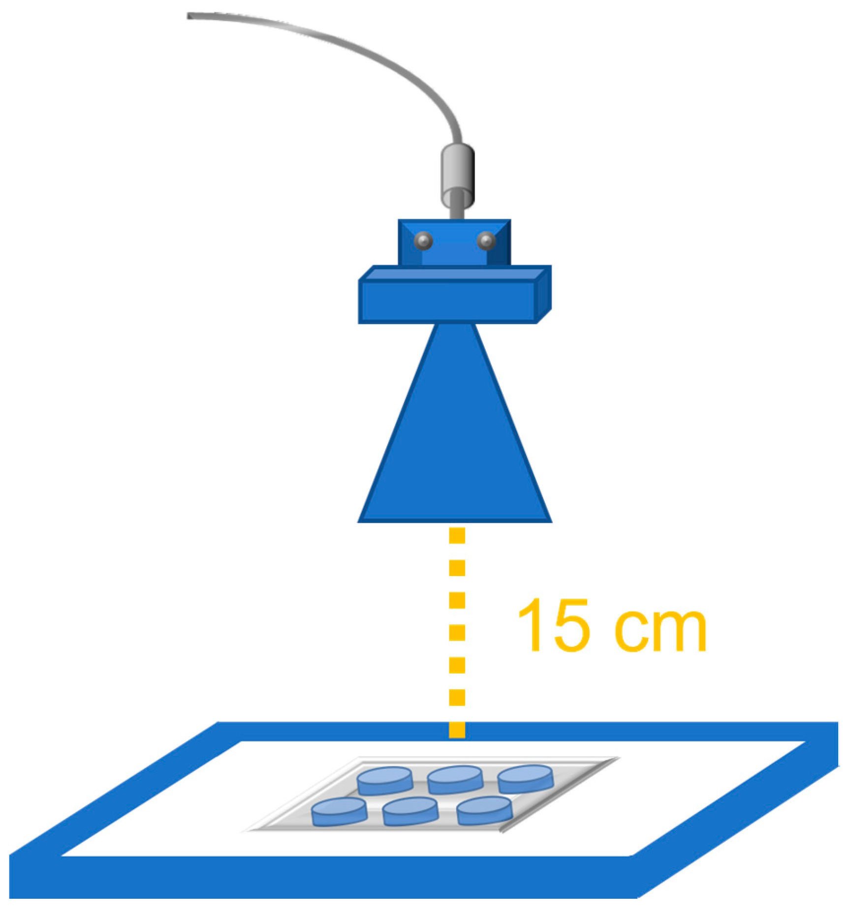
2.2. 27 GHz Antenna and Exposure Setup

The experiments were conducted using a commercial pyramidal horn antenna by XiBao Electronic Tech (model XB-HA28-20) at 27 GHz. The dimension of the antenna aperture was 4.4 × 3.51 cm, and the maximum gain reported in antenna datasheet was 19.23 dB at 26.50 GHz. The antenna was fed by a RF signal generation (R&S SMB100A) with an output power of +23 dBm that reduced to +20 dBm due to insertion loss of the coaxial cable linking the RF generator and antenna. The distance between the antenna aperture plane and the 6-well microplates was fixed to 15 cm (Figure 1) as a trade-off between the maximum available output power of the RF signal generator and the need to ensure an incident power density comparable with the ICNIRP international guideline restrictions, that is comparable to or not exceeding 10 mW/cm2 [7].

2.3. Experimental Exposure Procedure

Mussels (Mytilus galloprovincialis) were purchased from a mussel farm in Sicily, placed in plastic bags with seawater and immediately transported to the Catania University’s Laboratories. We discarded the individuals with obvious signs of breakage of the shell from the sample. Once their maturity was verified, the mussels were cleaned from debris and epibionts by manual scraping of the shell, rinsed quickly with water and stabilized for 1 h before experiments. A total of 20 individuals were selected to induce spawning eggs and sperms by applying a protocol of thermal stimulation (heat shock). Mussels were placed at a temperature of 4 °C for 3–4 h and then transferred into a tank containing water heated at 28 °C. The water used for the stimulation had a salinity of 30 ± 1‰ and pH of 8.3 and it had been filtered with a 0.22 μm filter. After thirty minutes, most of the specimens had opened the valve and resumed filtration, and they were transferred to a second tank containing seawater at a temperature of 18 °C. Once the spawning was initiated, the specimens were immediately removed and placed into an aquarium. Spermatozoa were placed in 6-well microplates in 5ml seawater/well. Control samples (negative control) were incubated only with seawater. The effect of exposure was evaluated up to 1 h (with observation intervals at 10, 20, 30, 40 and 60 min). A controlled room temperature allowed us to maintain 17 ± 1 °C in wells. Ten replications for each time series (10, 20, 30, 40 and 60) were performed.

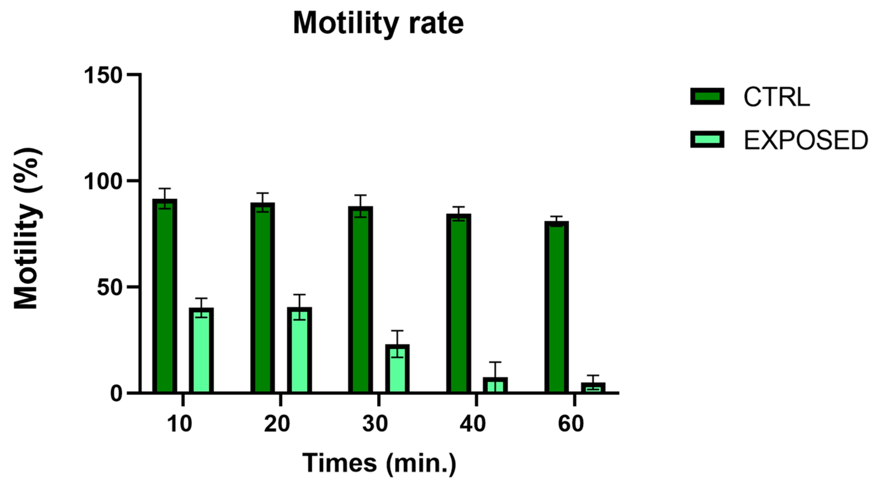
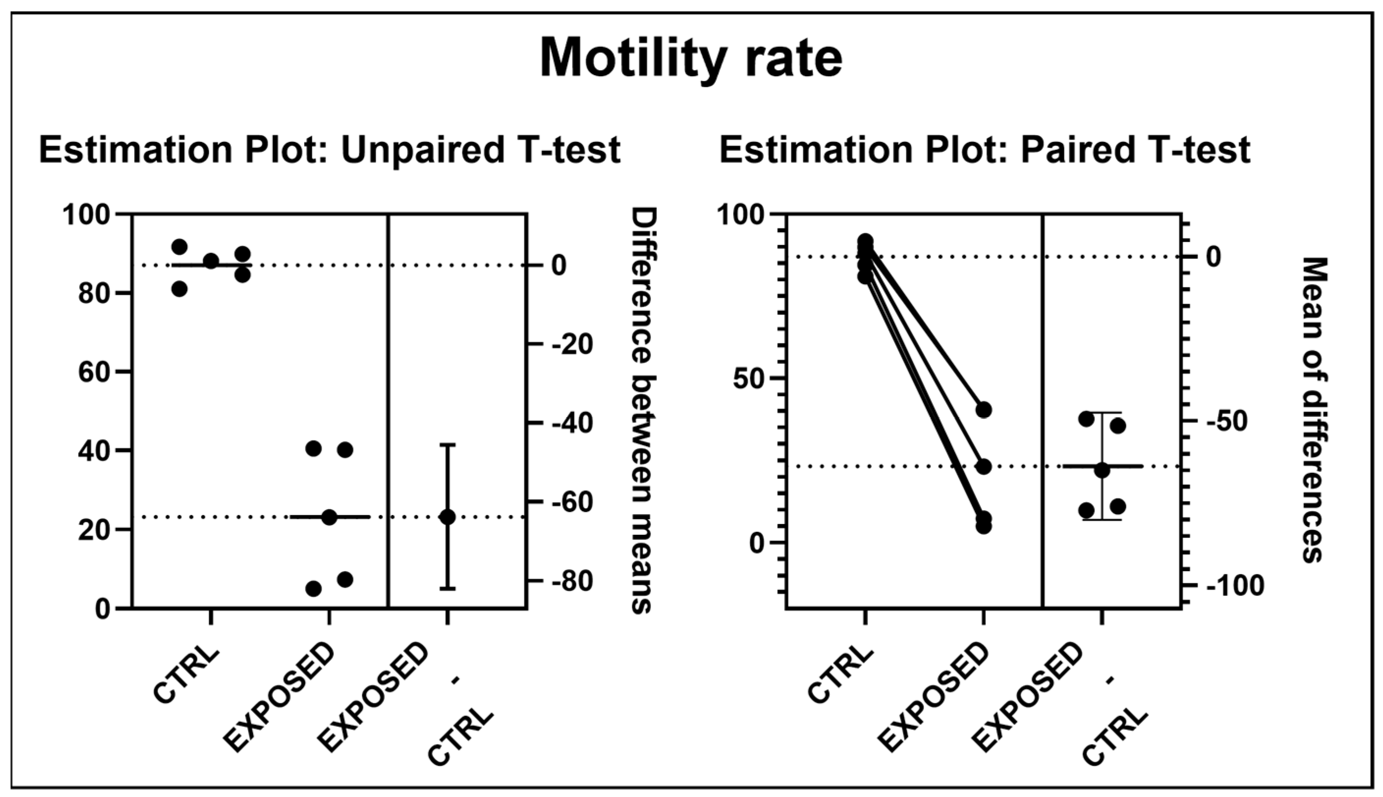
2.4. Motility Analysis

We measured the motility dividing spermatozoa into two categories: motility and no motility. For each replicate of the time series exposure, we measured the motility by placing 10 µL of sperm sample on a slide and observing it under an optical microscope (Leica DMLB) at ×400 magnification. We counted 100 spermatozoa at least.

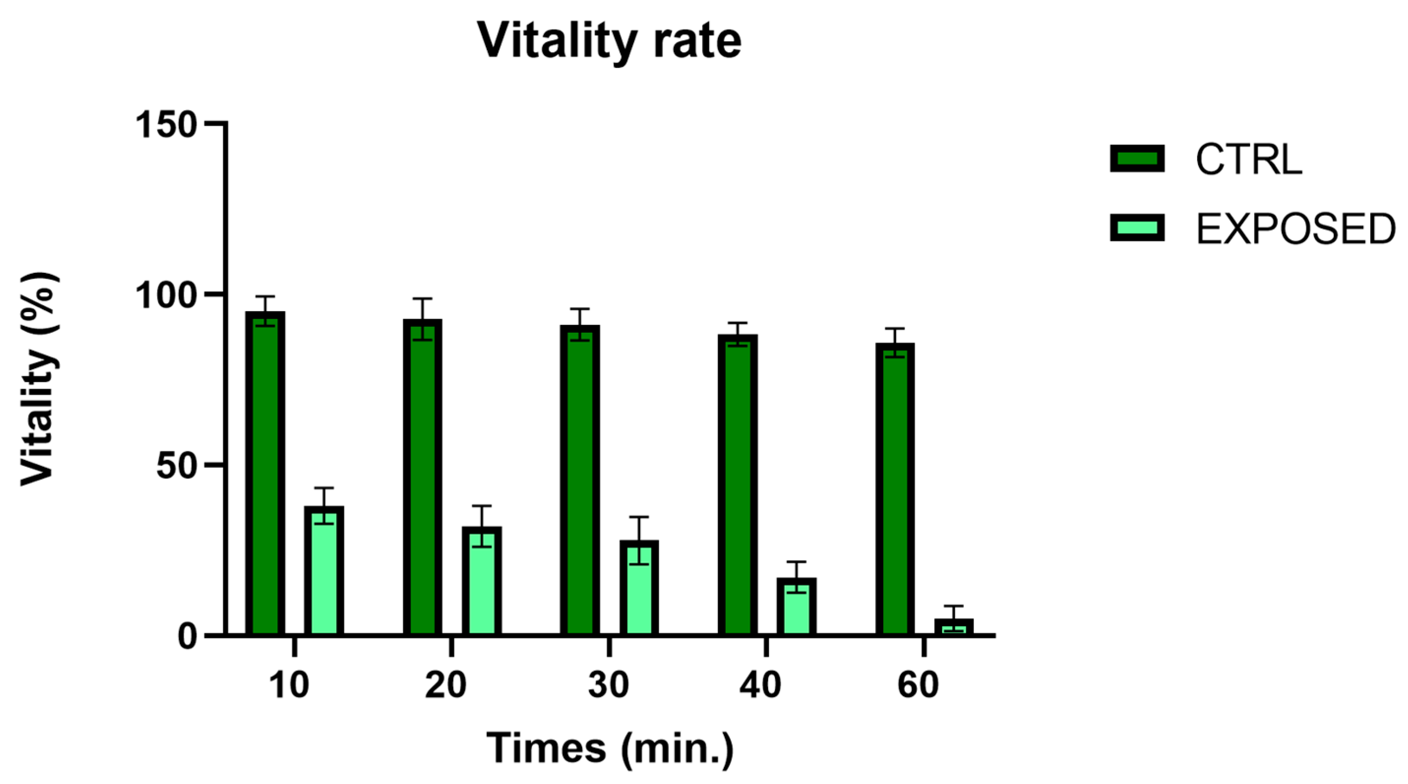
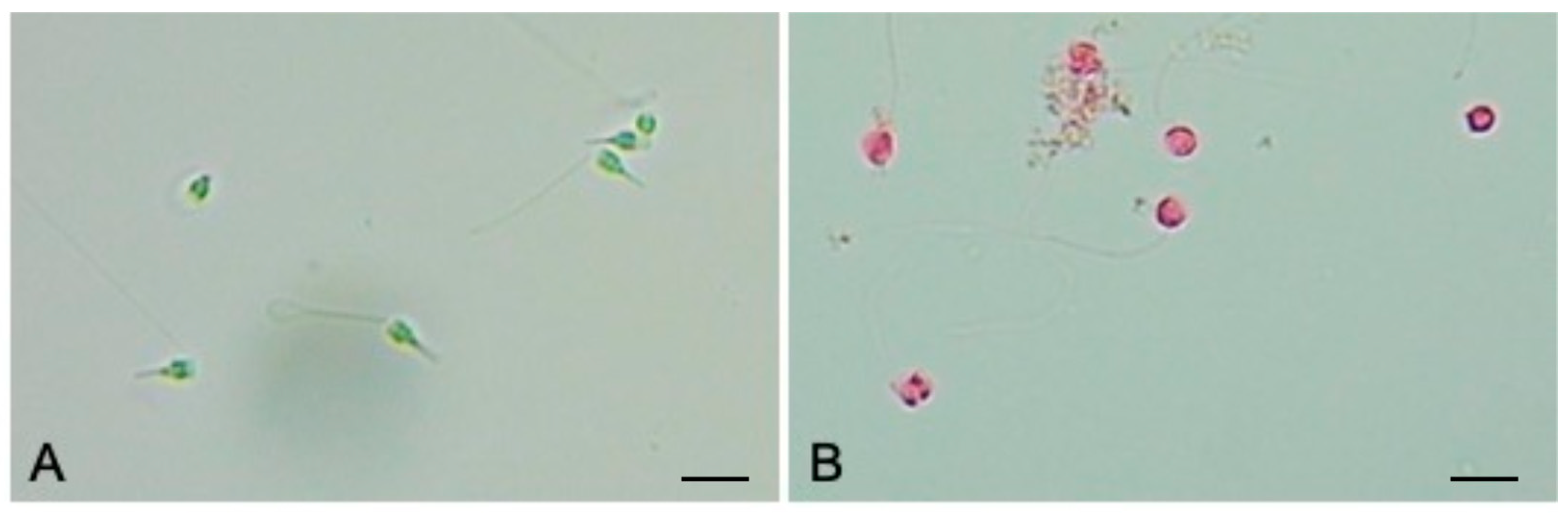
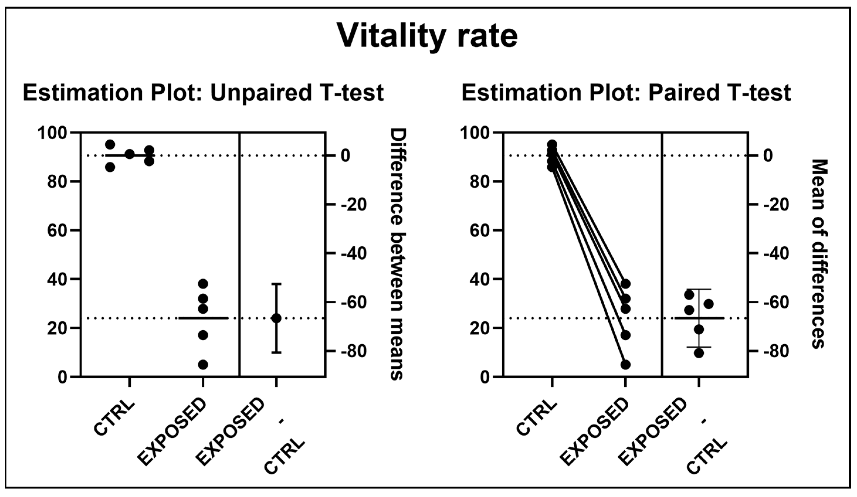
2.5. Vitality Analysis

For each replicate of the time series exposure, we evaluated the vitality of sperm samples. Using an Eosin test, we put 10 µL of sperm sample on a slide on which we added 10 µL of Eosin Y (0.5%, Bio-Optica). The observations were made under an optical microscope (Leica DMLB) at ×400 magnification. At least 100 spermatozoa were counted. Dead spermatozoa appeared in pink due to the loss of membrane integrity, compared to live spermatozoa that maintained their original coloring.

2.6. Statistical Analyses

The data obtained were processed for statistical analysis. The vitality and motility rates between spermatozoa exposed to the electromagnetic fields and the control, were compared by the t-test. Paired (Tables S1 and S2) and unpaired (Tables S3 and S4) using GraphPad Prism software (version 9.3.1) (Table S5). Bar chart graphs of motility and vitality rate were realized using GraphPad Prism software (version 9.3.1).

3. Results

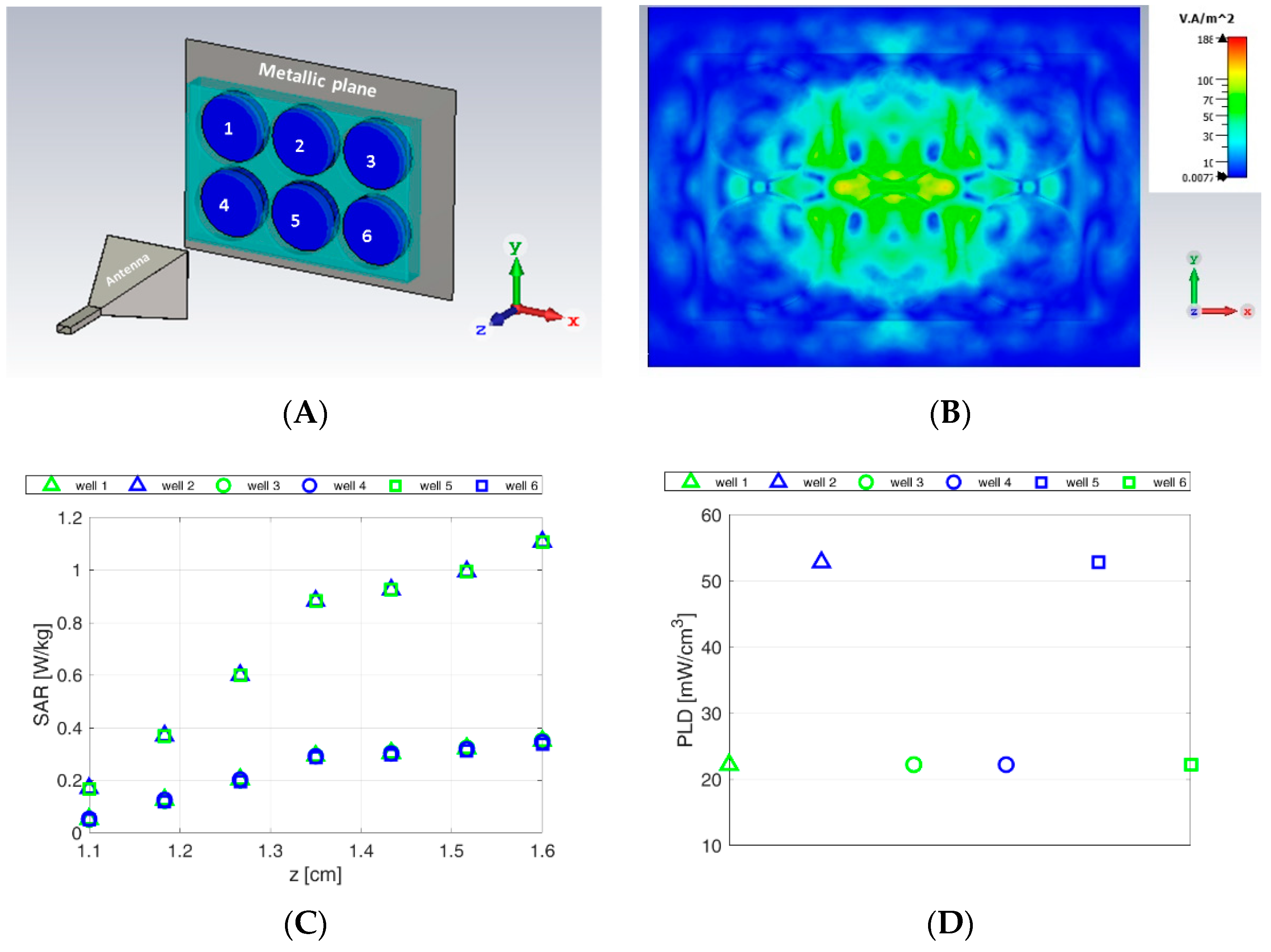
3.1. Numerical Dosimetry Analyses

To establish electromagnetic exposure conditions, a numerical simulation was performed by means of the commercial software CST Microwave Studio. In particular, in the CAD model we considered the 6-well microplates, the horn antenna and a metallic plane behind the sample holder to take into account the agitator for the oxygenation of samples placed at 1 cm below the 6-well microplates (Figure 2A). In the experimental setup, a polystyrene foam slab was inserted between the metallic plane and the sample holder to prevent the reflecting plane from being very close to the aqueous samples; however, it has not been considered in the simulation as dielectric parameters of foam can be neglected. The dielectric parameters of the materials adopted for the dosimetric analysis at a working frequency of 27 GHz are reported in Table 1.
In order to evaluate the exposure condition, three complementary metrics have been considered [7]:
-
density power of the incident field [W/m2];
-
the local specific absorption rate (point SAR) [W/Kg];
-
power loss density (PLD) deposited into the exposed aqueous samples [W/m3].
The resulting incident power density is reported in Figure 2B; as it can be seen, although the power density is not uniform above the 6-well microplates, since the far-field condition is not satisfied [7], it reaches values ranging from 30 to 100 W/m2 over the antenna footprint area. Taking into account that the metallic plate of the agitator gives rise to a total reflection, these values are comparable with 10 mW/cm2 set by the international guidelines as the exposure limit above 6 GHz, see Table 2 in [7].
As far as SAR calculation is concerned, it is worth underlining that the specific absorption rate (SAR) averaged over a 10 g cubic volume, considered by international guidelines for frequencies below 6 GHz, cannot be considered here as the total mass of the samples (5 g) is smaller than the average mass considered by ICNIRP guidelines [6]. For this reason, we report the local SAR as numerically evaluated in each cell of the grid used to discretize the samples in the wells (point SAR). The computed SAR level averaged in each layer of the aqueous samples is shown in Figure 2C. The SAR distribution is nonhomogeneous in the different aqueous samples as larger values are reached in the central wells (2 and 5). This is due to the near-field condition exposure that entails a not local plane wave in front of the field impinging on the samples. Moreover, as expected, the top layers of the aqueous samples show larger SAR values than in the deeper layers, due to strong electromagnetic discontinuities and small penetration depth in salt water at 27 GHz, (0.65 mm). Finally, the PLD also shows that power deposition into the central samples is about three times larger than that in the peripheral samples. However, as spermatozoa move randomly inside the sample, they will experience an average PLD value of about 22 mW/cm3 in the outermost samples (well number 1, 3, 4, 6) and about 52 mW/cm3 in the innermost ones (well number 2 and 5), see Figure 2D. It is worth stressing that, although nonhomogeneous exposure is not suggested as a good condition for dosimetric evaluation, in this case, as sperm vitality is observed on spermatozoa samples taken separately from each well, this may allow us to understand if there are possible effects that are associated with different power level conditions.
Finally, it is worth noticing that only numerical dosimetry has been performed because continuous thermal monitoring during exposure protocol is not viable due to the invasiveness of the most common temperature probes (thermocouple or optical fiber) at this working frequency. This notwithstanding, water temperature measurements of the aqueous sample reveal an increase of about 1 °C after the exposure time with respect to the sham samples.

3.2. Sperms Vitality and Motility Analyses

A significative decrement of sperm vitality (Figure 3) was observed already after 10 min of exposure at 27 GHz, compared to control samples. An Eosin test showed a high mortality rate of spermatozoa in all exposed samples already after 30 min (Figure 4). We observed that electromagnetic fields also with irradiation induced a significant decrease in sperm motility (Figure 5) after 10 min of exposure.
Statistically significant differences (p < 0.0001 and p < 0.0004 only for paired t-test motility) have been obtained for all times of exposure between spermatozoa exposed and controls both for vitality and motility rate (Figure 6 and Figure 7).

4. Discussion

The fifth generation of global mobile communications will also bring a new era to maritime connectivity for real-time data transmissions. The development of mobile communication technologies requires the assessment of possible risks occurring from exposures to radiofrequency electromagnetic fields. Whereas much has been written in the literature on the effect on reproduction of electromagnetic fields in terrestrial animal models [19,20,21,22,23,24], little is known about the effect on aquatic animals, except for a few studies concerning zebrafish [16,19]. Moreover, most of these effects were investigated at frequencies typical of previous cellular communications, mainly 2G and 3G, i.e., frequencies much lower than 27 GHz.
Recent evidence demonstrates that electromagnetic fields affect sperm quality, count, morphology and motility. In 2-day-old male rats exposed to EMF 1800 and 900 MHz for 2 h continuously per day for 90 days, the percentage of epididymal sperm motility was significantly higher in the 1800 MHz group (p < 0.05). The morphologically normal spermatozoa rates were higher, and the tail abnormality and total percentage abnormalities were lower in the 900 MHz group (p < 0.05) [9]. In another study conducted on rats exposed to 900 MHz for 8 weeks, the authors highlighted a statistically significant decrease in epididymal sperm count in the exposed group (p < 0.001), a significant decrease in sperm motility and a significant (p < 0.001) increase in the frequency percentage of dead spermatozoa [18]. Guo et al. [25] exposed rats for 1 month to 220 MHz and showed a decreased sperm count and survival rate of sperm (p < 0.05) and increased sperm abnormalities. No differences in body weight and development among the groups were found in rats of both sexes exposed to 2.45 GHz for 2 h/day, 5 days/week for 5 consecutive weeks, starting the day after birth [24]. On zebrafish exposed to 3.5 GHz RFR, a specific absorption rate (SAR) ≈ 8.27 W/Kg from 6 h post-fertilization (hpf) to 48 hpf did not reveal significant impacts on mortality, morphology or photomotor responses, but only a modest inhibition of startle response suggesting some levels of sensorimotor disruptions [25].
Several studies were conducted on the evaluation of exposure to submarine power cables which produce both electric and magnetic fields. Although magnetic and electric fields’ intensities decrease with distance away from the cable, marine invertebrate species are the major fauna exposed to it so they have received greater attention [26]. A high-strength magnetic field applied during sea urchin (Lytechinus pictus and Strongylocentrotus purpuratus) fertilization delayed cell division in embryos [27,28]. Moreover, Levin and Ernst [27] highlighted an increase in developmental abnormalities, but only in L. pictus. However, a 93-day exposure throughout the reproductive period of the blue mussel (Mytilus edulis) did not affect either its condition index or its gonad development index [29].
In our study, we have noted a significant decrease in the vitality of M. galloprovincialis spermatozoa after only 10 min of exposure at 27 GHz. We also observed that electromagnetic field irradiation induced a significant decrease in sperm motility after 10 min of exposure. If confirmed, a possible explanation for our observation is related to the direct action of the electromagnetic field on the phospholipid bilayer of cell membranes. This effect had been investigated, but at higher frequencies (around 60 GHz) on liposomes and on giant unilamellar vesicles [30,31,32], where the authors hypothesized a change of the polarization states of water induced by radiation, which causes a partial dehydration of the membrane and consequently a greater packing density of phospholipids.

5. Conclusions

This study indicates that electromagnetic fields at 27 GHz can affect the sperm quality in marine mussel Mytilus galloprovincialis. The significative decrease observed in sperm motility after only 10 min of exposure represents a crucial factor to be considered because it can threaten the reproductivity of the species. This study provides useful data on the potential impact of high frequency EMFs on aquatic animals and cells, which is currently poorly investigated. Future research could benefit from specific investigations into the impact of 5G to better monitor the effects on animal organisms and to fill the gap currently known about the interactions with artificial sources of electromagnetic fields.

Supplementary Materials

The following supporting information can be downloaded at: https://www.mdpi.com/article/10.3390/jmse10040521/s1, Table S1: Paired t-test Vitality; Table S2: Paired t-test Motility; Table S3: Unpaired t-test Vitality; Table S4: Unpaired t-test Motility; Table S5: excel data.

Author Contributions

Conceptualization, L.D.D. and M.V.B.; methodology, R.P., E.M.S., M.C., S.C.P. and G.S.; investigation, C.S. and S.I.; data curation, S.I., C.S., S.C.P. and G.S.; writing—original draft preparation, L.D.D. and M.V.B.; writing—review and editing, R.P., E.M.S., A.S. and M.V.B.; English language editing, D.M.; supervision, L.D.D., R.P. and M.V.B.; funding acquisition, L.D.D. and M.V.B. All authors have read and agreed to the published version of the manuscript.

Funding

This research has been funded by “Programma ricerca di Ateneo UNICT 2020-22 linea 2”.

Institutional Review Board Statement

Not applicable.

Informed Consent Statement

Not applicable.

Conflicts of Interest

The authors declare no conflict of interest.

References

  1. Attaran, M. The impact of 5G on the evolution of intelligent automation and industry digitization. J. Ambient. Intell. Hum. Comput. 2021, 1–17. [Google Scholar] [CrossRef] [PubMed]
  2. Foster, K.R.; Vijayalaxmi. Needed: More Reliable Bioeffects Studies at “High Band” 5G Frequencies. Front. Commun. Netw. 2021, 2, 721925. [Google Scholar] [CrossRef]
  3. Hardell, L.; Carlberg, M. Health risks from radiofrequency radiation, including 5G, should be assessed by experts with no conflicts of interest. Oncol. Lett. 2020, 20, 15. [Google Scholar] [CrossRef] [PubMed]
  4. Simkó, M.; Mattsson, M.O. 5G Wireless Communication and Health Effects—A Pragmatic Review Based on Available Studies Regarding 6 to 100 GHz. Int. J. Environ. Res. Public Health 2019, 16, 3406. [Google Scholar] [CrossRef] [Green Version]
  5. Mattsson, M.O.; Simkó, M.; Foster, K.R. 5G New Radio Requires the Best Possible Risk Assessment Studies: Perspective and Recommended Guidelines. Front. Commun. Netw. 2021, 2, 724772. [Google Scholar] [CrossRef]
  6. Agence nationale de sécurité sanitaire de l’alimentationde l’environnement et du travail (ANSES). Expositions aux champs électromagnétiques liées au déploiement de la technologie de communication « 5G » et effets sanitaires éventuels associés. Rapport d’expertise collective. 2021. Available online: https://www.vie-publique.fr/rapport/279567-deploiement-de-la-technologie-de-communication-5g-et-effets-sanitaires (accessed on 1 March 2022).
  7. International Commission on Non-Ionizing Radiation Protection (ICNIRP). Guidelines for limiting exposure to time-varying electric, magnetic and electromagnetic fields (up to 300 GHz). Health Phys. 1998, 74, 494–522. [Google Scholar]
  8. Dreujou, E.; Desroy, N.; Carrière, J.; Tréau de Coeli, L.; McKindsey, C.W.; Archambault, P. Determining the Ecological Status of Benthic Coastal Communities: A Case in an Anthropized Sub-Arctic Area. Front. Mar. Sci. 2021, 8, 637546. [Google Scholar] [CrossRef]
  9. Klimley, A.; Wyman, M.; Kavet, R. Assessment of Potential Impact of Electromagnetic Fields from Undersea Cable on Migratory Fish Behaviour (No. FINAL REPORT, DOE-EPRI-Ee0006382 OCS Study BOEM 2016-041); Electric Power Research Institute (EPRI): Palo Alto, CA, USA, 2016. [Google Scholar]
  10. Ohman, M.; Sigray, P.; Westerberg, H. Offshore windmills and the effects of electromagnetic fields on fish. Ambio 2007, 36, 630–633. [Google Scholar] [CrossRef]
  11. Tricas, T.; Gill, A. Effects of EMFs from Undersea Power Cables on Elasmobranchs and Other Marine Species (No. OCS Study BOEMRE 2011-09); U.S. Department of the Interior, Bureau of Ocean Energy Management (BOEM): Camarillo, CA, USA, 2011.
  12. Wildt, D.E.; Comizzoli, P.; Pukazhenthi, B.; Songsasen, N. Lessons from biodiversity—The value of nontraditional species to advance reproductive science, conservation, and human health. Mol. Reprod. Dev. 2010, 77, 397–409. [Google Scholar] [CrossRef] [Green Version]
  13. Boncel, S.; Kyzioł-Komosińska, J.; Krzyżewska, I.; Czupioł, J. Interactions of carbon nanotubes with aqueous/aquatic media containing organic/inorganic contaminants and selected organisms of aquatic ecosystems—A review. Chemosphere 2015, 136, 211–221. [Google Scholar] [CrossRef]
  14. Maresca, V.; Fusaro, L.; Sorbo, S.; Siciliano, A.; Loppi, S.; Paoli, L.; Monaci, F.; Asadi karma, E.; Piscopo, M.; Guida, M.; et al. Functional and structural biomarkers to monitor heavy metal pollution of one of the most contaminated freshwater sites in Southern Europe. Ecotoxicol. Environ. Saf. 2018, 163, 665–673. [Google Scholar] [CrossRef] [PubMed]
  15. Piscopo, M.; Notariale, R.; Rabbito, D.; Ausi, J.; Olanrewaju, O.S.; Guerriero, G. Mytilus galloprovincialis (Lamarck, 1819) spermatozoa: Hsp70 expression and protamine-like protein property studies. Environ. Sci. Pollut. Res. 2018, 25, 12957–12966. [Google Scholar] [CrossRef] [PubMed]
  16. Reitsema, R.E.; Meire, P.; Schoelynck, J. The future of freshwater macrophytes in a changing world: Dissolved organic carbon quantity and quality and its interactions with macrophytes. Front. Plant Sci. 2018, 9, 629. [Google Scholar] [CrossRef] [PubMed]
  17. Scalici, M.; Traversetti, L.; Spani, F.; Malafoglia, V.; Colamartino, M.; Persichini, T.; Cappello, S.; Mancini, G.; Guerriero, G.; Colasanti, M. Shell fluctuating asymmetry in the sea-dwelling benthic bivalve Mytilus galloprovincialis (Lamarck, 1819) as morphological markers to detect environmental chemical contamination. Ecotoxicology 2017, 26, 396–404. [Google Scholar] [CrossRef]
  18. Vassalli, Q.A.; Caccavale, F.; Avagnano, S.; Murolo, A.; Guerriero, G.; Fucci, L.; Ausió, J.; Piscopo, M. New insights into protamine-like component organization in Mytilus galloprovincialis’ sperm chromatin. DNA Cell Biol. 2015, 34, 162–169. [Google Scholar] [CrossRef]
  19. Ozlem Nisbet, H.; Nisbet, C.; Akar, A.; Cevik, M.; Karayigit, M.O. Effects of exposure to electromagnetic field (1.8/0.9 GHz) on testicular function and structure in growing rats. Res. Vet. Sci. 2012, 93, 1001–1005. [Google Scholar] [CrossRef] [Green Version]
  20. Bin-Meferij, M.M.; El-Kott, O.F. The neuroprotective effects of Moringa oleifera against mobile phone electromagnetic radiation-induced infertility in rats. Int. J. Clin. Exp. Med. 2015, 8, 12487–12497. [Google Scholar]
  21. Bilgici, B.; Gun, S.; Avci, B.; Akar, A.; Engiz, B. What is adverse effect of wireless local area network, using 2.45 GHz, on the reproductive system? Int. J. Radiat. Biol. 2018, 94, 1054–1061. [Google Scholar] [CrossRef]
  22. Saygin, M.; Asci, H.; Ozmen, O.; Cankara, F.N.; Dincoglu, D.; Ilhan, I. Impact of 2.45 GHz microwave radiation on the testicular inflammatory pathway biomarkers in young rats: The role of gallic acid. Environ. Toxicol. 2016, 31, 1771–1784. [Google Scholar] [CrossRef]
  23. Ozguner, M.; Koyu, A.; Cesur, G.; Ural, M.; Ozguner, F.; Gokcimen, A.; Delibas, N. Biological and morphological effects on the reproductive organ of rats after exposure to electromagnetic field. Saudi Med. J. 2005, 26, 405–410. [Google Scholar]
  24. Sambucci, M.; Laudisi, F.; Nasta, F.; Pinto, R.; Lodato, R.; Lopresto, V.; Altavista, P.; Marino, C.; Pioli, C. Early life exposure to 2.45 GHz WiFi-like signals: Effects on development and maturation of the immune system. Prog. Biophys. Mol. Biol. 2011, 107, 393–398. [Google Scholar] [CrossRef] [PubMed]
  25. Guo, L.; Lin, J.J.; Xue, Y.Z.; An, G.Z.; Zhang, J.P.; Zhang, K.Y.; He, W.; Wang, H.; Li, W.; Ding, G.R. Effects of 220 MHz pulsed modulated radiofrequency field on the sperm quality in rats. Int. J. Environ. Res. Public Health 2019, 16, 1286. [Google Scholar] [CrossRef] [PubMed] [Green Version]
  26. Albert, L.; Deschamps, F.; Jolivet, A.; Olivier, F.; Chauvaud, L.; Chauvaud, S. A current synthesis on the effects of electric and magnetic fields emitted by submarine power cables on invertebrates. Mar. Environ. Res. 2020, 159, 104958. [Google Scholar] [CrossRef]
  27. Levin, M.; Ernst, S. Applied DC magnetic fields cause alterations in the time of cell divisions and developmental abnormalities in early sea-urchin embryos. Bioelectromagnetics 1997, 18, 255–263. [Google Scholar] [CrossRef]
  28. Zimmerman, S.; Zimmerman, A.; Winters, W.; Cameron, I. Influence of 60-Hz magnetic fields on sea urchin development. Bioelectromagnetics 1990, 11, 37–45. [Google Scholar] [CrossRef] [PubMed]
  29. Bochert, R.; Zettler, M. Long-term exposure of several marine benthic animals to static magnetic fields. Bioelectromagnetics 2004, 25, 498–502. [Google Scholar] [CrossRef]
  30. D’Agostino, S.; Della Monica, C.; Palizzi, E.; Di Pietrantonio, F.; Benetti, M.; Cannatà, D.; Cavagnaro, M.; Sardari, D.; Stano, P.; Ramundo-Orlando, A. Extremely high frequency electromagnetic fields facilitate electrical signal propagation by increasing transmembrane potassium efflux in an artificial axon model. Sci. Rep. 2018, 8, 9299. [Google Scholar] [CrossRef] [PubMed]
  31. Cosentino, K.; Beneduci, A.; Ramundo-Orlando, A.; Chidichimo, G. The influence of millimeter waves on the physical properties of large and giant unilamellar vesicles. J. Biol. Phys. 2013, 39, 395–410. [Google Scholar] [CrossRef] [PubMed] [Green Version]
  32. Di Donato, L.; Cataldo, M.; Stano, P.; Massa, R.; Ramundo-Orlando, A. Permeability changes of cationic liposomes loaded with carbonic anhydrase induced by millimeter waves radiation. Radiat. Res. 2012, 178, 437–446. [Google Scholar] [CrossRef] [Green Version]
Figure 1. Experimental exposure setup.
Jmse 10 00521 g001
Figure 2. Experimental setup for numerical dosimetry. (A) CAD model (6-well microplates numbered for understanding following figures). (B) Incident power density at a distance of 0.5 cm from the air–water interface of the aqueous samples. (C) Average point SAR in each sample layer along depth z-direction. (D) Average PLD for each aqueous sample in the wells.
Jmse 10 00521 g002
Figure 3. Vitality percentage at different times of exposure. The data represent the mean of observation performed tenfold by the same observer to avoid subjective differences in vitality evaluation.
Jmse 10 00521 g003
Figure 4. Vitality evaluation with Eosin Test on Mytilus galloprovincialis spermatozoa. (A) Untreated sample. (B) After exposure to 27 GHz for 30 min. Scale bar: 4 μm.
Jmse 10 00521 g004
Figure 5. Motility percentage at different times of exposure. The data represent the mean of observation performed tenfold by the same observer to avoid subjective differences in motility evaluation.
Jmse 10 00521 g005
Figure 6. Line plot comparing mean vitality rate of M. galloprovincialis sperm samples exposed to 27 GHz than control.
Jmse 10 00521 g006
Figure 7. Line plot comparing mean motility rate of M. galloprovincialis sperm samples exposed to 27 GHz than control.
Jmse 10 00521 g007
Table 1. Dielectric parameters adopted for the numerical dosimetry.
ComponentMaterialDielectric ConstantLoss TangentMass Density
Horn antenna Perfect electric conductor (good metal)--
Aqueous sample Salt water (30‰ salinity)23.641.271029 [Kg/m3]
6-weel microplatesPolystyrene 2.50-
Ground planePerfect electric conductor (good metal)--
Publisher’s Note: MDPI stays neutral with regard to jurisdictional claims in published maps and institutional affiliations.

