Article

Effect of Integration of Linseed and Vitamin E in Charolaise × Podolica Bulls’ Diet on Fatty Acids Profile, Beef Color and Lipid Stability

by
Valeria Maria Morittu
1,
Anna Antonella Spina
1,*,
Piera Iommelli
2,*,
Anselmo Poerio
1,
Francesco Vincenzo Oliverio
1,
Domenico Britti
1 and
Raffaella Tudisco
2
1
Centro Interdipartimentale di Servizi Veterinari per la Salute Umana ed Animale, Dipartimento di Scienze della Salute, Università Magna Græcia di Catanzaro, 88100 Catanzaro, Italy
2
Dipartimento di Medicina Veterinaria e Produzioni Animali, Università di Napoli Federico II, 80100 Napoli, Italy
*
Authors to whom correspondence should be addressed.
Agriculture 2021, 11(11), 1032; https://doi.org/10.3390/agriculture11111032
Submission received: 14 September 2021 / Revised: 15 October 2021 / Accepted: 17 October 2021 / Published: 21 October 2021

Abstract

:
Dietary supplementation with oilseeds improves the fatty acid profiles of meat, but results are often inconsistent. This study aimed to assess the effects of dietary linseed and vitamin E supplementation on fatty acid profile, cholesterol content and color stability of beef samples. Dorsal subcutaneous fat samples were subjected to lipid stability assessment. Eighteen young bulls (385 ± 15 kg BW, age 8–9 months) were allocated into three homogeneous groups, each receiving ad libitum wheat straw and concentrate only (CON = 5.5 kg/day), concentrate with linseed (LIN = 80 g/kg, i.e., 440 g/head/day), and concentrate with linseed plus vitamin E (L + E = 80 g/kg, i.e., 440 g/head/day + 2500 IU/head/day of Vitamin E). Group L+E showed significantly lower cholesterol content, lower n-6/n-3 ratio and a higher PUFA percentage compared to the CON group. Meat color was affected by feeding LIN with a decrease in a*, b*, and C* compared to the CON group. The experimental diets increased H° values compared to the CON group. A positive effect of vitamin E in protecting lipids of dorsal subcutaneous depots from oxidation was detected in group L+E compared to group LIN. The supplementation with extruded linseeds in the diet had positive effects on the nutritional profile of the meat. When vitamin E was included, linseed did not alter the color of meat, and the lipid stability of the subcutaneous fat improved.

1. Introduction

Consolidated human dietary recommendations suggest that saturated fatty acids (SFA) and cholesterol intake should be reduced and monounsaturated fatty acids (MUFA) and polyunsaturated fatty acids (PUFA) intake should be increased, especially the n-3 series [1,2,3,4]. Indeed, MUFA and PUFA can have important beneficial health effects, such as reducing hypercholesterolemia, and also play essential metabolic roles in the prevention of chronic inflammatory diseases [3]. Moreover, the prevention of these chronic diseases as a function of a low PUFA n-6/n-3 ratio has been demonstrated [4].
In addition to grazing, other animal feeding strategies have been suggested to increase the concentration of beneficial lipids. Several studies have been conducted on diet supplementation with linseeds to produce beef with enhanced levels of components with potential health benefits [5,6,7], but with inconsistent results.
Increasing the content of healthy fatty acids in meat may have deleterious effects on its appearance and shelf life. Indeed, high PUFA content of meat can reduce its stability to oxygen and heighten its susceptibility to rancidity [8,9,10]. Oxidation of meat pigments is recognizable by the development of brown discoloration replacing the bright cherry-red color. This represents a problem, because color and fat are parameters directly related to the perception of beef quality that influence the consumer at the time of purchase [10]. One way to address these adverse effects is to enrich the diet of animals with antioxidants such as vitamin E, which has been demonstrated to alter ruminal PUFA hydrogenation in beef cattle [11].
Several studies have been carried out on the influence of dietary linseed supplementation on meat FA profiles from the beef of typical Italian breeds, Piemontese [5], Maremmana [7] and Podolica [12] but, to the best of our knowledge, no studies have been carried out to determine the effects of linseed on the meat characteristics of Podolica × Charolaise young bulls.
Podolica is an autochthonous cattle breed raised in the southern region of Italy characterized by its rusticity. In most meat farms, the Podolica is usually bred by crossing with other specialized meat breeds, especially the Charolaise.
The present study was undertaken to investigate the effects of adding extruded linseeds and vitamin E to the diet of Charolaise × Podolica young bulls on the fatty acid profile of intramuscular fat and on the quality of meat during the storage.

2. Materials and Methods

2.1. Animals and Diets

The trial was performed according to the Animal Welfare and Good Clinical Practice [13] and was approved by the Animal Ethic Committee local (protocol number: 50/03.04.2015). The experiment was carried out using 18 Charolaise × Podolica young bulls (385 ± 15 kg body weight (BW), age 8–9 months/249–279 days) raised on pasture until the beginning of the trial. Animals, coming from 18 different Podolica cows and 3 Charolaise bulls (6 young bulls per sire), were divided, according to BW and sire, into three homogenous groups: concentrate only (CON = 5.5 kg/day), concentrate with extruded linseed (LIN = 80 g/kg, i.e., 440 g/head/day), or concentrate with extruded linseed plus vitamin E (Rovimix E 100, Istituto delle Vitamine S.p.A, Milan, Italy) (L + E = 80 g/kg, i.e., 440 g/head/day + 2500 IU/head/day of Vitamin E).
The concentrate ingredients are reported in the Supplementary Materials, whereas the chemical composition and nutritive value of the feeds are shown in Table 1. Bulls were housed in three paddocks with straw bedding. All animals received their ration twice a day at a fixed time (07:00 a.m.–03:00 p.m.) and had free access to wheat straw and water. After eight days of adaptation to the experimental diet, the animals were treated for 91 ± 18 days and then slaughtered in 6 batches of three animals per week, one animal/group/batch. None of the animals had health problems during the study. BW was monthly measured and, when bulls reached the endpoint, they were transported to a licensed slaughterhouse located within 20 min from the experimental farm. Animals fasted for 12 h before slaughter, which was performed according to EU Regulations [14]. The interval between the first and the last slaughter was 35 days.

2.2. Sample Collection and Animals Perfomances

Animals were weighed before slaughter and the weight of the hot and cold carcass (after 24 h of refrigeration at 4 °C) was recorded according to ASPA methods [16]. Thirty-five minutes after the animals’ slaughter, the pH0 was measured on the right side of the carcass on the Longissimus thoracis muscle (LT) between the 12th and 13th ribs with a pH-meter (Hanna Instruments, San Benedetto del Tronto, AP, Italy). Carcasses were chilled at 4 °C for 24 h in a cold room, then pH24 was measured after 24 h post-mortem. Hot dressing percentage was calculated as the ratio of hot carcass weight (HCW) to slaughter weight, while the final carcass weights (FCW) were recorded after 24 h of chilling at 4 °C to determine shrink losses (shrink losses = (HCW − FCW)/HCW × 100).
On the right side of the carcass, the LT muscle was removed from the vertebra L1 to the vertebra T8 and sampled for subsequent analysis [16,17]. In particular, six 2.5 cm-thick steaks were taken from the caudal end towards the cranial end of the LT muscle and assigned to the various analysis in the following order: color assessment, chemical composition, cholesterol content, drip loss, cooking loss and fatty acid analysis. Finally, an aliquot of 200 g of subcutaneous fat was collected from each animal at T12 and T13 vertebrae level. All samples were vacuum-packed and stored at 4 °C except subcutaneous fat and steaks intended for chemical and fatty acid analysis that were frozen at −20 °C until analysis.
Drip and cooking loss analysis were determined about 27 h post-mortem on samples of 200 g of weight. For drip loss, meat samples were taken out of the bag and placed in an airtight container for 24 h at 4 °C in a common laboratory refrigerator. The drip loss value was calculated as the difference between the initial weight and final weight and expressed as a percentage of the initial weight.
For the determination of cooking loss, the steaks were placed in open plastic bags and cooked in a boiling distilled water bath until a temperature of 75 °C at their geometric centers was measured by a digital thermometer. The samples were then cooled in melting ice to a temperature of 20 °C and then gently dried with absorbent paper before being weighed. The cooking loss was calculated as the difference between raw and cooked weight and expressed as a percentage of the raw weight.

2.3. Meat Chemical and Cholesterol Analysis

Meat samples were analyzed for dry matter, crude protein (CP), crude fiber, ether extract (EE) and crude ash according to official AOAC methods [18]. For cholesterol analysis, the samples were subjected to method described by Almeida et al. [19]. After that, they were analyzed by high-performance liquid chromatography (HPLC Jasco AS-2057 PLUS, Thermo Scientific, Waltham, MA, USA). The HPLC apparatus consisted of a system including a column Lune 54 C18, size 250 × 4, 6mm (Altmann Analytik GmbH & Co. Munich, Germany), an injection volume of 70 µL, with an ultraviolet detector. The mobile phase (flow rate = 1 mL/min) consisted of methanol for HPLC. The resulting chromatograms were processed at 210 nm. Quantification for each sample was achieved by internal standardization (Cholesteryl linoleate, Sigma-Aldrich, St. Louis, MO, USA).

2.4. Meat Fatty Acids Analysis

The samples of LT were subjected to lipid extraction and methylation. Meat fatty acid composition was assessed as reported by Almeida et al. [19]. The fatty acid methyl esters (FAME) were separated and quantified by a gas chromatograph (Thermo GC VEGA 2000 series, Milan, Italy) with a flame ionization detector (FID) and SP2560 capillary column 100 m in length, 0.25 mm in diameter and had a film thickness of 0.25 μm (Sigma, St Louis, MO, USA). The column temperature was initially set at 40 °C for 5 min and gradually ramped up to 200 °C, where it remained to the end of the cycle (80 min). The temperature of the injector and the flame-ionization detector was 240 and 280 °C, respectively. Hydrogen was used as a carrier gas at a flow of 1 mL/min. A 1-μL aliquot of the sample was injected in the GC. Fatty acids were quantified using tridecanoic acid methyl ester (C13:0; Fluka, Sigma-Aldrich Milano, Milano, Italy), as an internal standard. Fatty acid methyl esters were identified by comparing the retention times with pure Fame standards (Supelco 37 Component FAME Mix, St Louis, MO, USA).

2.5. Meat Color Determination

After removing the steaks from the packaging, they were placed on a plastic tray with an airtight lid and kept in the dark at 4 °C.
In particular, for the observation of the day 1, the meat samples were removed from the vacuum packaging and, based on other similar studies in the literature [20,21,22], we waited 30 min before instrumental reading. This waiting time was necessary to allow the phenomenon of “blooming” [20,21,22].
For the following days (day 2–6) the meat samples were placed on a plastic tray with an airtight lid, in a refrigerator at 4 °C and in the dark, simulating a domestic conservation. Color readings were taken at three randomly selected locations on the surface of each piece.
Muscle color was measured in the CIEL*a*b* space [23], with a measured area diameter of 8 mm, “A” standard illuminant, observer angle 10° [24]. The colorimeter used was a Konica-Minolta CM-700d spectrophotometer (Konica Minolta Holdings, Inc., Osaka, Japan) calibrated before analyzing the samples against black and white standards. The instrumental reading took place every day at the same time (10:00 am) at a storage time of 24 h, 2 days, 3 days, 4 days, 5 days, and 6 days after packaging. The individual values of the Lightness (L*), redness (a*), and yellowness (b*) were recorded every day, and hue angle (H°) and chroma (C*) indexes were calculated as H° = tan−1 (b*/a*) expressed in degrees, and C* = (a*2 + b*2)0.5 [24]. The relative content of oxyhemoglobin, methemoglobin and myoglobin were calculated from measurements at wavelengths of 473, 525, 572 and 730 nm [25].

2.6. Lipid Oxidation

The determination of the hexanal content in the frozen (−80 °C) subcutaneous fat samples was performed to evaluate the state of the lipid oxidation as described by Panseri et al. [26]. Briefly, a TRACE GC Ultra gas chromatograph, complete with a mass spectrometer (Tace DSQII, Thermo-Fisher Scientific, Waltham, MA, USA) and with an Rtx.Wax column with the dimensions of 30 m by 0.25 mm by 0.25 mm (Restek, Bellefonte, PA, USA) was used. The hexanal was identified by comparing the retention time with the compounds of the standard and the internal standard.

2.7. Statistical Analysis

Statistical analysis was carried out using a commercial software (GraphPad Prism version 8.3.0 for Windows, GraphPad Software, San Diego, CA, USA). Data on animal performances, drip and cooking loss, meat chemical composition as well as lipid stability to oxidation were tested for normality by Kolmogorov–Smirnov test and then analyzed by ordinary one-way ANOVA followed by Tukey’s multiple comparisons test. Some data required log transformation (drip loss, C22:4 n-6) or exponential transformation (C18:3 n-3) to normalize distributions before performing one-way ANOVA. For C20:5 n-3, the data distributions could not be normalized and the non-parametric Kruskal–Wallis test was performed. The data relating to color were analyzed according to the mixed-effect model including the effect of the “Diet” (=CTR, LIN, L+E) as a variable between factors, of the “Day” (day 1–6) as a variable within factors and the “Diet × Day” interaction. Tukey’s multiple comparison test was used to test the difference between means. The results were expressed as Means expected magnitudes (Least-Squares Means) ± SEM and the significance level was established for p < 0.05.
Statistical processing was carried out with GraphPad PRISM version 8.3.0 software for Windows, La Jolla, CA, USA.

3. Results

3.1. Animal Perfomances, Drip and Cooking Loss Analyses

The average weight of the bulls at the start of the experiment did not differ among the groups, as well as the slaughter weight, although there was a trend towards statistical significance (p = 0.09) for the initial live weight (Table 2). In fact, the L+E group had a lighter average initial body weight (370 kg) than the LIN group (419 kg) (Table 2). Finally, there were no significant differences in hot carcass weight among the three groups, or in the other parameters evaluated (Table 2). No differences were detected in muscle pH0 and pH24 among diets (Table 2). The water holding capacity, measured by the drip losses and the cooking losses was not affected by diet (Table 2). Drip loss was, overall, very low, ranging between 1.23 and 1.78% of the initial weight of the sample, while cooking loss was between 21.68 and 24.96%.

3.2. Proximate and Cholesterol Analyses

Dietary treatment did not affect LT muscle chemical composition, which showed no significant differences in dry matter, protein, fat, and ash content (Table 3). However, the fat content was close to statistical significance (p = 0.05) with lower values in the CON and L+E groups than in the group LIN. The meat of L+E animals had lower cholesterol content compared to LIN and CON groups (p = 0.010) (Table 3).

3.3. Fatty Acids Analysis

The fatty acid profile is shown in Table 4. The saturated fatty acids content (SFA) in the LT was significantly affected by linseed supplementation. In particular, SFA decreased in the LIN and L+E groups (p = 0.025), compared to the CON group.
Monounsaturated fatty acids (MUFA) decreased in the group L+E (p = 0.006) compared to the CON group.
The PUFA content in the LT was affected for both experimental groups (Table 4).
In particular, the content of PUFA in the LIN and L+E group was higher compared to the CON group. The meat from young bulls fed linseed and linseed with Vitamin E showed an increase in n-3 (p < 0.0001), above all in the L+E group. The n-6 fatty acids increased significantly only in the L+E group (p = 0.008).
Meat from the linseed and linseed with Vitamin E groups had a lower n-6/n-3 ratio (p = 0.001). The n-6/n-3 ratio is highly influenced by linseed supplementation that significantly increased also the concentration of total CLA (Conjugated Linoleic Acid) (p = 0.009).

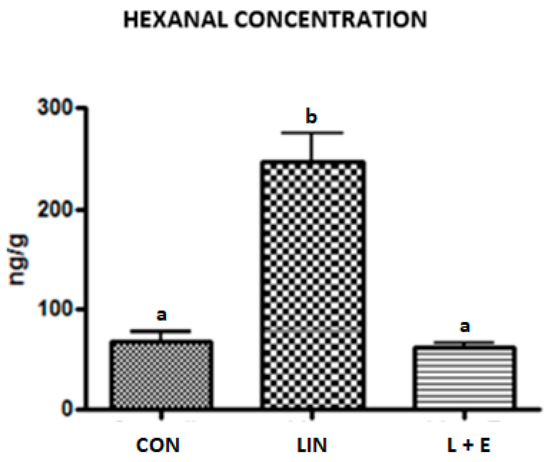
3.4. Lipid Stability to Oxidation

The results obtained (Figure 1) indicate that the LIN group had a much greater lipid oxidation index compared to CON and L+E groups. Intramuscular fat of the group L+E, although richer in PUFA, had a hexanal content approximately 4-fold lower compared to the LIN group.

3.5. Color Determinations

Results of meat color are shown in Table 5. Lightness (L*) of meat was not influenced by supplementation of linseeds with or without vitamin E (p = 0.326), while during storage was observed an increment in the L* of the samples from the second day (p < 0.0001).
In the subsequent days, L* remained stable, settling on values between 44 and 45 and indicating an increase compared to the initial average figure, equal to approximately 41.
Over time, a* had an increasing trend between the first and second day and then showed a slow and constant decrease. Similar to a*, the yellowness (b*) was also influenced by diet (p = 0.045) and the storage time (p < 0.0001). In particular, the CON diet led to a greater b* value (p < 0.05) compared to the LIN group, whereas L+E samples show intermediate yellowness values. During the storage period, a similar trend was observed for b* as for redness. Beef color hue (H°) was influenced both by diet (p < 0.0001) and time of storage (p < 0.0001). The interaction Diet × Day was also significant (p < 0.0001).
Chroma (C*) was influenced both by diet (p = 0.014) and storage time (p < 0.001). Meat from the CON group had a more saturated color than LIN while the L+E samples showed intermediate values. Over time, C* underwent an increase from day 1 to day 2 (p < 0.0001. In the subsequent period, the trend of C* was of a progressive decrease. However, the decline was very small.
The relative proportion of myoglobin forms did not vary among the groups whereas, as expected, we observed a progressive increase in MMb (%) during storage due to the oxidation of the other two forms of the pigment. In particular, a marked increase in OMb (%) occurred as well as a sharp decrease in DMb (%) between the first and second day was observed (Table 5).

4. Discussion

4.1. Animal Performances, Drip and Cooking Loss

The body weight response is in line with that reported on both Podolica breed by other authors [27] and the Charolaise breed [28]. The effects of linseed supplementation in the diet on the performance of the animals and the characteristics of the carcass were consistent with those reported by previous studies [28]. In contrast, Albertì et al. [6] reported a significant reduction in dressing percentage (60.4% vs. 62.6% for linseed and control groups, respectively), using whole linseeds at a lower level of integration (5%). Tarricone et al. [12], feeding Podolica bulls with a diet based on extruded linseeds (3%), obtained a final body weight higher than the control group. These authors, however, point out that these bulls were heavier compared to those used in other studies on the same breed [27,28] and probably attribute this to the longer duration of the feeding period in their study. According to Juárez et al. [11] linseeds (10.7 % of DM) supplemented diet led to an increase in final body weight, average daily gain and weight of the hot carcass while according to Mach et al. [29] using a higher level of linseed (15.8% on DMI), animal performances were not affected. The high level of Vitamin E in the L+E diet (2500 IU/head/day) did not affect animal growth or carcass weight (Table 3) as well as observed by Carter et al. [30] by using a high level of vitamin E (2000 IU/head/day) for 7, 14, or 28 days. Similarly, the inclusion of vitamin E at low/modest levels [11,31] did not affect animal performance or carcass quality. Cuvelier et al. [32], on samples from different breeds bulls, reported values of drip losses between 2.0 and 3.4%, and of cooking losses, between 29.3 and 33.1%.
The mean of the pH at 24 h (5.73 ± 0.09) could reflect low levels of stress before slaughter, avoiding detrimental effects on meat quality [33]. In fact, a pH of 5.73 is considered an adequate pH value that does not affect the phenomenon of dark cutting carcasses [26,27].
Dawson et al. [34] recorded a cooking loss of 25.2–27.9% in bulls of similar slaughter weight of that of the current study. Furthermore, Corazzin et al. [3] reported a cooking loss value of about 30% and that the dietary treatment did not affect this parameter. The authors explain that their results on the cooking loss could be due to the lack of effects on ultimate pH that is related to the post-mortem protein denaturation.
Tenderness and juiciness of the meat are closely related properties [35] and are directly proportional to the amount of water present in cooked meat, as this gives plasticity to the myofibrillar proteins preventing their stiffening and hardening [36]. Our results demonstrated that experimental diets did not increase drip and cooking losses.

4.2. Proximate and Cholesterol Analyses

Chemical composition was not affected by dietary treatment (Table 3), although we observed an almost significant difference in intramuscular fat content among groups, possibly related to a low homogeneity of the BW of the young bulls (Table 2). From this point of view, the higher fat content in LIN group meat is in accordance with Corazzin et al. [3] that reported an increase in intramuscular fat in animals fed linseed supplemented diets. On the other hand, in the L+E group this effect of linseed supplementing may have been hidden by the lower BW of the animals. Cholesterol content was reduced by feeding linseeds with vitamin E (Table 3), whereas feeding linseed alone did not cause changes. In particular, the meat of L+E animals had lower cholesterol content (28.63%) compared to the other groups. This result was very encouraging since the extent of the reduction was significant, reaching 39% compared to the value of the CON group. This aspect is important for the consumer because guidelines for healthy eating recommend that intake of cholesterol should not exceed 300 mg per day [37].
Other authors did not find a significant response in the reduction in cholesterol content caused by the addition of linseeds [7] or by the addition of linseeds and vitamin E in the diet [28]. However, the reason for the decrease in cholesterol level only in the L+E group and not in the LIN group remains to be investigated. This could probably be explained by the higher level of n-3 PUFA observed in the group L+E (Table 4). We hypothesize that the association of linseed with high levels of Vitamin E in the diet may be responsible for the decrease in cholesterol in meat.
The role of vitamin E in the metabolism of cholesterol remains to be determined. According to Özer et al. [38] and Bozaykut et al. [39] the effect of vitamin E on cholesterol could be partially mediated by a group of receptors connected to the metabolism of lipids and Peroxisome Proliferator-Activated receptors (PPARs). They provided evidence for the role of vitamin E supplementation on gene expression through the upregulation of peroxisome PPAR gamma (PPARG), nuclear factor-erythroid 2-related factor (NFE2L2), induction of their downstream targets ATP-binding cassette transporter A1 (ABCA1), glutathione S-transferase α (GSTA1) and through the inhibition of matrix metalloproteinase-1 (MMP1).

4.3. Fatty Acids Analysis

The level of total SFA (Table 4) was higher than that reported by other authors [5,6,28]. These differences with respect to those other studies are attributable to the various breeds, diets, production systems, or slaughter weight. The levels of MUFA were affected by diet and the linseed with the vitamin E decreased levels of MUFA. Decreases in total MUFA content in the present experiment might be related to decreases in total trans-18:1 when feeding linseed and linseed with Vitamin E. However, also C18:1 n-9 cis decreased in the L+E group compared to the CON group. This result is not in agreement with the findings of precedent studies [5,28,39] that showed an increasing of MUFA and in particular of C18:1 trans, which is a by-product of biohydrogenation in the rumen, in meat of animals supplemented with unsaturated fatty acids.
However, the significant decrease in the L+E group recorded here was mainly ascribable to the decrease in oleic acid, passing from an initial value of 29.50% to a final value of 26.58%. Vitamin E in high doses probably has a role that should be investigated on the biohydrogenation of the rumen and of the derived by-products.
Confirming these results, Juarez et al. [11] reported an interaction between flaxseed and high levels of vitamin E associated with a variation in levels in C18:1 n-9 cis. According to these authors vitamin E could influence ruminal pathways of PUFA biohydrogenation, acting either as an inhibitor of bacteria producing trans 18:1 or as an electron acceptor for Butyrivibrio fibrisolvens [40].
Total PUFA content was greater in the LIN and L+E groups compared to the CON group, especially the n-6 PUFA content that was greater in the L+E group compared to the other two groups. The L+E and LIN groups showed higher levels in the total n-3 content, partly confirming that vitamin E has a role in PUFA hydrogenation. The C18:3 n-3 increased only in the L+E group and not in the CON and LIN groups, although it reached levels no greater than 1.85%. Additionally, Conte et al. [7] reported that the higher level of C18:3 n-3 was probably associated with a significant increase in the intermediate products of ruminal biohydrogenation of C18:3n-3: C18:2t11,c15, C18:1c11 and C18:1c12.
The most abundant polyunsaturated fatty acid was linoleic acid which, in contrast to what was expected, was higher in the LIN and L+E groups than in the CON group. L+E. In this regard Conte et al. [7] found an increase, although it was not statistically significant, in the content of linoleic acid in the polar fat (PL) of the meat of animals fed with linseed compared to bulls fed with a standard diet. This aspect should be investigated more thoroughly.
Other results with Italian beef cattle breeds such as Piemontese [41] and Maremmana [7] demonstrated that inclusion of 10% of DM of ground flaxseed and 20% of DM of extruded linseeds, respectively, modified the FA profile of the meat, increasing the n-3 PUFA content and lowering the n-6/n-3 PUFA ratio. These studies suggested that the proper selection of breed and diet allows an improvement of the nutritional value of meat for human consumption. Thus, this ratio is influenced by the FA composition of the diet administered to the animals. In the present study, linseed supplementation decreased the n-6/n-3 ratio of intramuscular lipids from 7.23 to 5.4 (Table 4), approaching the maximum value recommended for the human diet.
The addition of linseeds with or without vitamin E had an effect on the total CLA level, which increased in the two experimental diets compared to in the CON group. Some authors have reported that linseed has no effect on CLA levels [42], while others have found an increase in CLA 9c-11t [6,29]. The increased levels of CLA in animals receiving the experimental diets could, in turn, be explained by the roles of C18:2 n-6 cis and C18:3 n-3 and therefore to a greater biohydrogenation in the rumen of these FA in the supplemented diets. In fact, some studies have indicated that diets containing seeds rich in linoleic acid (LA) and alpha-linoleic acid (ALA) stimulate the production of CLA 9c-11t in the rumen [42]. Meat from bulls fed linseed was enriched with n-3 fatty acid due to the amount of linseed in the diet (8%). These fatty acids compete for the same enzymes responsible for desaturation and elongation and for their incorporation into lipid tissues [41]. Linseed supplementation increased the availability of linolenic acid in the muscle, resulting in an enhanced synthesis of its elongation and desaturation products such as EPA (Eicosapentaenoic acid), DPA (Docosapentaenoic acid) and DHA (Docosahexaenoic acid). These fatty acids have various beneficial effects on human health [43]. Additionally, Barton et al. [28] observed that in Longissimus thoracis of animals fed diet supplemented with linseeds there was greater CLA concentration, which in turn may have reflected a greater synthesis of CLA in the rumen. Moreover, CLAs are also produced by the enzymatic system stearoyl-CoA desaturase (SCD) present in the adipose tissue and in the mammary gland whose expression can be positively modulated by the dietary supplementation of flax seeds [44].
In the present study, n-6/n-3 ratio was lower in the LIN and L+E groups compared to the CON group and was closer to that recommended by the literature [45]. The n-6/n-3 ratio considered as optimal is in fact < 5 [46]. The beneficial role of linseeds in modifying the n-6/n-3 ratio has been confirmed in other similar studies using various doses of linseeds and breeds [7,12,32,34,47].
The role of linseeds in improving the fatty acid profile is similar to that found in animals fed on pasture compared to concentrate-based diets [48] the latter, indeed, worsened the n-6/n-3 ratio [49].

4.4. Lipid Stability to Oxidation

The stability of fat to oxidation is an important factor in food products because it influences other factors such as odor, color, flavor, texture and overall nutritional value [50]. According to Wood et al. [47] the fatty acid profile of intramuscular fat affects stability and PUFA is more susceptible to oxidative rancidity than MUFA and SFA. In our study, oxidation stability was analyzed in the subcutaneous fat of the carcass measuring the amount of hexanal present in ng/g in the fat. This molecule is considered an indicator of the extent of lipid alteration, being one of the major oxidation products of fat during storage. It confers an odor not appreciated by the consumer, described as “grass”, and has a very low perception threshold in water [51]. In particular, meats that oxidize produce secondary oxidative compounds such as hexanal, pentanal, 2.4-decadienal, 2.3-octanedione, and 2-octenal [52].
The extent of lipid oxidation can be assessed by various methods such as the Thiobarbituric Acid Reactive Substances Method (TBARS), commonly used as indices to assess the degree of lipid oxidation by considering the by-products of the oxidation reaction [53]. TBARS values and sensory scores are well correlated in meat products [53].
In addition, there is also the hexanal analysis which has been recommended as a secondary measure of lipid oxidation in raw or precooked meats [53]. As expected, intramuscular fat of the group L+E, although richer in PUFA, had a hexanal content approximately 4-fold lower compared to the group LIN. This can be explained by the antioxidant effect of vitamin E, which interrupts the auto-oxidation process of unsaturated fatty acids and therefore the formation of the hexanal during oxidation of linoleic acid [26].
Our results are in agreement with those from Scollan et al. [54], who observe through TBARS assay that increasing n-3 PUFA content in meat several negative sensory attributes score higher while the duration of the meat color is reduced.
Another study showed a lower degree of increase in lipid oxidation of the meat of bulls fed different levels of vitamin E, analyzed through hexanal analysis [55]. These authors reported that hexanal values, which are used as indicators of flavor, were correlated also with other flavors detected by a sensory panel [55].

4.5. Color Determination

In accordance with Dawson et al. [34] and Albertí et al. [6] also in our experiment, the beef color was not influenced by supplementation of linseeds with or without vitamin E, while during storage was observed an increment in the L* of the samples from the second day. This increase in L* was unexpected based on the literature, but it can be explained as the effect of an insufficient “blooming” time between the opening of the vacuum pack and the consequent oxygenation of the sample before color measurement.
In this regard, previous studies on the instrumental analysis of the color in bovine LT have obtained very different results. As matter of fact, it is stated that a time of 12 min from the sectioning of the carcass is sufficient for color stabilization [21], but based on other experiences, a period of “blooming” is recommended to be 30 min or more [21]. Based on the experience gained with this study, it is considered useful to respect a blooming time of approximately 60 min, as recommended by Lee et al. [31].
These values agree with those reported in a similar test performed by Albertí et al. [6] on Pyrenean young bulls and by King et al. [56] on Charolaise bulls. Instead, these values are slightly higher than those measured by another study [32] in Belgian Blue, Limousine and Aberdeen Angus bulls (between 37.4 and 44.2%). Both the type of diet and time influenced redness value (a*) of the Longissimus thoracis samples. Redness value (a*) was higher in the CON group than L+E whereas the LIN group samples showed the lowest values. This led us to hypothesize a positive effect of vitamin E with supplementation of linseeds. In contrast, other research in dairy calves [29] or dual-purpose cattle [34] feeding diets enriched in linseeds during finishing, did not report any significant effect on the color (L*, a*, b*, C*, H°) of fresh meat. Over time, a* has had an increasing trend between the first and second day and then showed a slow and constant decrease. This indicates that at the time of the first measurement (day 1), myoglobin was probably still undergoing oxygenation and that a time of exposure to the air lasting 30 min may not have been sufficient to convert the deoxymyoglobin present in the samples to oxymyoglobin. This hypothesis is also confirmed by the pigment status results discussed below.
Overall, a* was satisfactory in all groups for the entire period considered, with values comparable to those published in a review on dietary vitamin E supplementation for beef quality [57], and greater than those of Cuvelier et al. [32] who, in other meat breeds, obtained an a* between 15 and 18 on days 2 and 8, respectively. Similar to a*, the yellowness (b*) was also influenced by diet and also for the storage period, a similar trend was observed for b * as for redness.
Diet and time of storage influenced the beef color hue (H°). The Diet × Day interaction was also significant. In general, except for days 1 and 6, the CON group showed lower hue values than other groups. Since the hue value decreases as the red component increases compared to the yellow one, the CON group had more red meat than other groups. Nevertheless, these values are below those reported by Albertí et al. [58] in an experiment similar to ours. A similar work [12] on the Podolica breed fed with 3% extruded linseed did not influence the colorimetric response of the Longissimus lumborum (Ll) samples. However, all color measurements (L*, a*, b*) of this study were slightly lower for the linseed-fed bulls than for those fed the control diet. Chroma (C*) is directly proportional to color saturation and was influenced both by diet and storage time. Over time, C* underwent an increase from day 1 to day 2, likely due to the “blooming” effect on day 1. In the subsequent period, the trend of C* was of a progressive decrease. However, the decline was very small.
In fact, in terms of stability, the value of C* over time varied similarly to that reported by King et al. [56] in a test with Charolaise bull meat. The decrease recorded over 6 days, equal to 2.2 points, indicates a “physiological discoloration” and is linked to the superficial accumulation of metmyoglobin. A C* value of 18 has been proposed as a cut-off point to indicate the end of the shelf life of beef [59]. Although the supplementation with linseeds produced a decline in saturation, this reduction never reached the cut-off point, contrary to the study of Dawson et al. [34].
Regarding the percentage of OMb, DMb and MMb, the marked increase in OMb (%) as well as the sharp decrease in DMb (%) observed between the first and second day it can be attributed to the oxygenation of the pigment following the opening of the vacuum packs. Overall, the proportion of OMb was found for all dietary protocols to be above 60% of total myoglobin and always above the minimum OMb threshold of 50% indicated by Van den Oord and Wesdorp [60] for the acceptability of beef color. Even the MMb values, for all groups, were much lower than the maximum threshold of acceptability for meat color reported by Chan et al. [61] equal to 50% of MMb. We considered the result obtained to be encouraging in terms of stability of the color, despite the high content of intramuscular PUFA in experimental diets (Table 4).

5. Conclusions

The dietary supplementation with extruded linseed at 8% of the concentrate increased the percentage of n-3 fatty acids in the intramuscular fat and reduced the n-6/n -3 ratio in beef from Charolaise × Podolica young bulls. Vitamin E in high doses (2500 IU) seems to act in synergy with linseeds to further improve the profile of fatty acids and reduce cholesterol content in meat. Furthermore, addition of vitamin E did not harm color stability during storage and, despite the high PUFA content, maintained good lipid stability of the subcutaneous fat. These findings should be confirmed on a larger number of animals, and blood and ruminal tests should be included to assess the systemic responses of the animals.

Supplementary Materials

The following are available online at https://www.mdpi.com/article/10.3390/agriculture11111032/s1, Table S1: Ingredients of the basal concentrate fed to young bulls.

Author Contributions

Conceptualization, V.M.M. and A.A.S.; methodology, A.A.S., F.V.O. and A.P.; software, V.M.M.; validation, A.A.S. and V.M.M.; formal analysis, A.A.S. and F.V.O.; investigation, A.A.S., A.P. and P.I.; resources, R.T. and D.B.; data curation, P.I. and A.A.S.; writing—original draft preparation, A.A.S., V.M.M. and P.I.; writing—review and editing, P.I. and R.T.; visualization, V.M.M., R.T. and D.B.; supervision, V.M.M., R.T. and D.B. All authors have read and agreed to the published version of the manuscript.

Funding

This research was financially self-funded by the Interdepartmental Services Centre of Veterinary for Human and Animal Health, Department of Health Science, Magna Græcia University, Catanzaro, 88100, Italy. This research received no external funding.

Institutional Review Board Statement

The study was conducted according to the guidelines of the Declaration of Helsinki and approved by the Ethics Committee of Magna Graecia University of Catanzaro (protocol code 50/03.04.2015).

Data Availability Statement

The data presented in this study are available from the corresponding author.

Acknowledgments

The authors are grateful for the contribution provided by the “Basile” farm and by “Oliverio Carni” in carrying out this research. In particular, the authors thank for the technical assistance G. Oliverio.

Conflicts of Interest

The authors declare that they have no conflicts of interest. The animal husbandry practices of the farms simulated were approved by Italian veterinarians.

References

  1. Amine, E.K.; Baba, N.H.; Belhadj, M.; Deurenberg-Yap, M.; Djazayery, A.; Forrestre, T.; Galuska, D.A.; Herman, S.; James, W.P.T.; M’Buyamba Kabangu, J.R.; et al. Diet, nutrition and the prevention of chronic diseases. World Health Organ. Tech. Rep. Ser. 2003, 916, 1–49. [Google Scholar] [CrossRef] [Green Version]
  2. Maki, K.C.; Eren, F.; Cassens, M.E.; Dicklin, M.R.; Davidson, M.H. ω-6 Polyunsaturated Fatty Acids and Cardiometabolic Health: Current Evidence, Controversies, and Research Gaps. Adv. Nutr. 2018, 9, 688–700. [Google Scholar] [CrossRef]
  3. Corazzin, M.; Bovolenta, S.; Sepulcri, A.; Piasentier, E. Effect of whole linseed addition on meat production and quality of Italian Simmental and Holstein young bulls. Meat Sci. 2012, 90, 99–105. [Google Scholar] [CrossRef] [PubMed]
  4. EFSA Panel on Dietetic Products and Allergies (NDA), N. Scientific Opinion on Dietary Reference Values for fats, including saturated fatty acids, polyunsaturated fatty acids, monounsaturated fatty acids, trans fatty acids, and cholesterol. EFSA J. 2010, 8, 1461. [Google Scholar] [CrossRef] [Green Version]
  5. Renna, M.; Brugiapaglia, A.; Zanardi, E.; Prandini, A.; Moschini, M.; Sigolo, S. Fatty acid profile, meat quality and flavour acceptability of beef from double-muscled Piemontese young bulls fed ground flaxseed. Ital. J. Anim. Sci. 2019, 18, 355–365. [Google Scholar] [CrossRef] [Green Version]
  6. Albertí, P.; Beriain, M.J.; Ripoll, G.; Sarriés, V.; Panea, B.; Mendizabal, J.A.; Purroy, A.; Olleta, J.L.; Sañudo, C. Effect of including linseed in a concentrate fed to young bulls on intramuscular fatty acids and beef color. Meat Sci. 2014, 96, 1258–1265. [Google Scholar] [CrossRef]
  7. Conte, G.; Serra, A.; Casarosa, L.; Ciucci, F.; Cappucci, A.; Bulleri, E.; Corrales-Retana, L.; Buccioni, A.; Mele, M. Effect of linseed supplementation on total longissimus muscle lipid composition and shelf-life of beef from young Maremmana bulls. Front. Vet. Sci. 2019, 5, 1–15. [Google Scholar] [CrossRef]
  8. Faustman, C.; Sun, Q.; Mancini, R.; Suman, S.P. Myoglobin and lipid oxidation interactions: Mechanistic bases and control. Meat Sci. 2010, 86, 86–94. [Google Scholar] [CrossRef] [PubMed]
  9. Amaral, A.B.; Da Solva, M.V.; Lannes, S.C.D.S. Lipid oxidation in meat: Mechanisms and protective factors—A review. Food Sci. Technol. 2018, 38, 1–15. [Google Scholar] [CrossRef] [Green Version]
  10. Wu, D.; Sun, D. Colour measurements by computer vision for food quality control—A review. Trends Food Sci. Technol. 2013, 29, 5–20. [Google Scholar] [CrossRef]
  11. Juárez, M.; Dugan, M.E.R.; Aalhus, J.L.; Aldai, N.; Basarab, J.A.; Baron, V.S.; McAllister, T.A. Effects of vitamin E and flaxseed on rumen-derived fatty acid intermediates in beef intramuscular fat. Meat Sci. 2011, 88, 434–440. [Google Scholar] [CrossRef]
  12. Tarricone, S.; Colonna, M.A.; Giannico, F.; Facciolongo, A.M.; Jambrenghi, A.C.; Ragni, M. Effects of dietary extruded linseed (Linum usitatissimum L.) on performance and meat quality in Podolian young bulls. S. Afr. J. Anim. Sci. 2019, 49, 781–789. [Google Scholar] [CrossRef] [Green Version]
  13. Sellick, J. Enhancing the protection of animals used for scientific purposes. Environ. Law Manag. 2011, 23, 75–82. [Google Scholar]
  14. European Council. European Council Regulation (EC) 1099/2009 of 24 September 2009 on the Protection of Animals at the Time of Killing. Official Journal of the European Union. L 303. 18 November 2009. Available online: https://eur-lex.europa.eu/legal-content/EN/TXT/?uri=OJ:L:2009:303:TOC (accessed on 8 September 2021).
  15. Noziere, P.; Sauvant, D.; Delaby, L. INRA Feeding System for Ruminants; Wageningen Academic Publishers: Wageningen, The Netherlands, 2018; ISBN 9086862926. [Google Scholar]
  16. ASPA (Animal Science and Production Association). Metodiche per la Determinazione delle Caratteristiche Qualitative della Carne (Procedures for Meat Quality Evaluation); University of Perugia: Perugia, Italy, 1996. [Google Scholar]
  17. ASPA (Animal Science and Production Association). Metodologie Relative alla Macellazione degli Animali di Interesse Zootecnico ed alla Valutazione e Dissezione della Loro Carcassa (Procedures for Carcass Quality Evaluation); ISMEA: Roma, Italy, 1991. [Google Scholar]
  18. Latimer, G.W. AOAC International Official Methods of Analysis of AOAC International, 20th ed.; AOAC International: Rockville, MD, USA, 2016; Volume 3172. [Google Scholar]
  19. De Almeida, J.C.; Perassolo, M.S.; Camargo, J.L.; Bragagnolo, N.; Gross, J.L. Fatty acid composition and cholesterol content of beef and chicken meat in Southern Brazil. Rev. Bras. Cienc. Farm. J. Pharm. Sci. 2006, 42, 109–117. [Google Scholar] [CrossRef] [Green Version]
  20. Tapp, W.N.; Yancey, J.W.S.; Apple, J.K. How is the instrumental color of meat measured? Meat Sci. 2011, 89, 1–5. [Google Scholar] [CrossRef]
  21. Rentfrow, G.; Linville, M.L.; Stahl, C.A.; Olson, K.C.; Berg, E.P. The effects of the antioxidant lipoic acid on beef longissimus bloom time. J. Anim. Sci. 2004, 82, 3034–3037. [Google Scholar] [CrossRef]
  22. Insausti, K.; Beriain, M.J.; Purroy, A.; Alberti, P.; Lizaso, L.; Hernandez, B. Colour stability of beef from different Spanish native cattle breeds stored under vacuum and modified atmosphere. Meat Sci. 1999, 53, 241–249. [Google Scholar] [CrossRef]
  23. OIV Compendium Of International Analysis Of Methods -OIV Chromatic Characteristics Method OIV-MA-AS2-11 Determination of Chromatic Characteristics According to CIELab. 2006. Available online: https://www.oiv.int/public/medias/2478/oiv-ma-as2-11.pdf (accessed on 14 October 2021).
  24. Hunt, M.C.; King, A.; Barbut, S.; Clause, J.; Cornforth, D.; Hanson, D.; Lindahl, G.; Mancini, R.; Milkowski, A.; Mohan, A. AMSA Meat Color Measurement Guidelines; American Meat Science Association: Champaign, IL, USA, 2012; Volume 61820, ISBN 8005172672. [Google Scholar]
  25. Krzywicki, K. Assessment of relative content of myoglobin, oxymyoglobin and metmyoglobin at the surface of beef. Meat Sci. 1979, 3, 1–10. [Google Scholar] [CrossRef]
  26. Panseri, S.; Soncin, S.; Chiesa, L.M.; Biondi, P.A. A headspace solid-phase microextraction gas-chromatographic mass-spectrometric method (HS-SPME-GC/MS) to quantify hexanal in butter during storage as marker of lipid oxidation. Food Chem. 2011, 127, 886–889. [Google Scholar] [CrossRef]
  27. Ragni, M.; Toteda, F.; Tufarelli, V.; Laudadio, V.; Facciolongo, A.; Dipalo, F.; Vicenti, A. Feeding of extruded flaxseed (linum usitatissimum L.) and pasture in podolica young bulls: Effects on growth traits, meat quality and fatty acid composition. Pak. J. Zool. 2014, 46, 1101–1109. [Google Scholar]
  28. Barton, L.; Marounek, M.; Kudrna, V.; Bures, D. MEAT Growth performance and fatty acid profiles of intramuscular and subcutaneous fat from Limousin and Charolais heifers fed extruded linseed. Meat Sci. 2007, 76, 517–523. [Google Scholar] [CrossRef] [PubMed]
  29. Mach, N.; Devant, M.; Díaz, I.; Font-Furnols, M.; Oliver, M.A.; García, J.A.; Bach, A. Increasing the amount of n-3 fatty acid in meat from young Holstein bulls through nutrition. J. Anim. Sci. 2006, 84, 3039–3048. [Google Scholar] [CrossRef] [PubMed] [Green Version]
  30. Carter, J.N.; Gill, D.R.; Krehbiel, C.R.; Confer, A.W.; Smith, R.A.; Lalman, D.L.; Claypool, P.L.; Mcdowell, L.R. Vitamin E supplementation of newly arrived feedlot calves 1, 2. J. Anim. Sci. 2005, 83, 1924–1932. [Google Scholar] [CrossRef] [PubMed] [Green Version]
  31. Lee, S.K.; Panjono; Kang, S.M.; Kim, T.S.; Park, Y.S. The effects of dietary sulfur and Vitamin E supplementation on the quality of beef from the longissimus muscle of Hanwoo bulls. Asian-Australas. J. Anim. Sci. 2008, 21, 1059–1066. [Google Scholar] [CrossRef]
  32. Cuvelier, C.; Clinquart, A.; Hocquette, J.F.; Cabaraux, J.F.; Dufrasne, I.; Istasse, L.; Hornick, J.L. Comparison of composition and quality traits of meat from young finishing bulls from Belgian Blue, Limousin and Aberdeen Angus breeds. Meat Sci. 2006, 74, 522–531. [Google Scholar] [CrossRef] [PubMed]
  33. Grandin, T. The Effect of Stress on Livestock and Meat Quality Prior to and During Slaughter. Int. J. Study Anim. Probl. 1980, 1, 313–337. [Google Scholar]
  34. Dawson, L.E.R.; Fearon, A.M.; Moss, B.W.; Woods, V.B. Effects of substitution of a proportion of the concentrate in grass silage/concentrate-based diets with extruded linseed on performance and meat quality of dairy bulls. Anim. Feed Sci. Technol. 2010, 156, 10–18. [Google Scholar] [CrossRef]
  35. Otremba, M.M.; Dikeman, M.E.; Milliken, G.A.; Stroda, S.L.; Chambers Iv, E.; Chambers, D. Interrelationships between descriptive texture profile sensory panel and descriptive attribute sensory panel evaluations of beef Longissimus and Semitendinosus muscles. Meat Sci. 2000, 54, 325–332. [Google Scholar] [CrossRef]
  36. Hughes, J.M.; Oiseth, S.K.; Purslow, P.P.; Warner, R.D. A structural approach to understanding the interactions between colour, water-holding capacity and tenderness. Meat Sci. 2014, 98, 520–532. [Google Scholar] [CrossRef]
  37. Fernandez, M.L.; Calle, M. Revisiting dietary cholesterol recommendations: Does the evidence support a limit of 300 mg/d? Curr. Atheroscler. Rep. 2010, 12, 377–383. [Google Scholar] [CrossRef]
  38. Özer, N.K.; Şirikçi, Ö.; Taha, S.; Şan, T.; Moser, U.; Azzi, A. Effect of Vitamin E and Probucol on Dietary Cholesterol-Induced Atherosclerosis in Rabbits. Free Radic. Biol. Med. 1998, 24, 226–233. [Google Scholar] [CrossRef]
  39. Bozaykut, P.; Karademir, B.; Yazgan, B.; Sozen, E.; Siow, R.C.M.; Mann, G.E.; Ozer, N.K. Effects of vitamin e on peroxisome proliferator-activated receptor γ and nuclear factor-erythroid 2-related factor 2 in hypercholesterolemia-induced atherosclerosis. Free Radic. Biol. Med. 2014, 70, 174–181. [Google Scholar] [CrossRef] [PubMed]
  40. Yoshimura, E.H.; Santos, N.W.; Machado, E.; Agustinho, B.C.; Pereira, L.M.; De Aguiar, S.C.; Franzolin, R.; Gasparino, E.; Santos, G.T.; Zeoula, L.M. Effects of dairy cow diets supplied with flaxseed oil and propolis extract, with or without vitamin E, on the ruminal microbiota, biohydrogenation, and digestion. Anim. Feed Sci. Technol. 2018, 241, 163–172. [Google Scholar] [CrossRef]
  41. Marino, R.; Della Malva, A.; Caroprese, M.; De Palo, P.; Santillo, A.; Sevi, A.; Albenzio, M. Effects of whole linseed supplementation and treatment duration on fatty acid profile and endogenous bioactive compounds of beef muscle. Animal 2019, 13, 444–452. [Google Scholar] [CrossRef]
  42. Raes, K.; Haak, L.; Balcaen, A.; Claeys, E.; Demeyer, D.; De Smet, S. Effect of linseed feeding at similar linoleic acid levels on the fatty acid composition of double-muscled Belgian Blue young bulls. Meat Sci. 2004, 66, 307–315. [Google Scholar] [CrossRef]
  43. Givens, D.I.; Gibbs, R.A. Very long chain n-3 polyunsaturated fatty acids in the food chain in the UK and the potential of animal-derived foods to increase intake. Nutr. Bull. 2006, 31, 104–110. [Google Scholar] [CrossRef]
  44. Tudisco, R.; Chiofalo, B.; Lo Presti, V.; Morittu, V.M.; Moniello, G.; Grossi, M.; Musco, N.; Grazioli, R.; Mastellone, V.; Lombardi, P.; et al. Influence of feeding linseed on SCD activity in grazing goat mammary glands. Animals 2019, 9, 786. [Google Scholar] [CrossRef] [PubMed] [Green Version]
  45. Simopoulos, A.P. Essential fatty acids in health and chronic disease 1,2. Am. J. Clin. Nutr. 1999, 70, 560–569. [Google Scholar] [CrossRef] [Green Version]
  46. Yu, M.; Gao, Q.; Wang, Y.; Zhang, W.; Li, L.; Wang, Y.; Dai, Y. Unbalanced omega-6/omega-3 ratio in red meat products in China. J. Biomed. Res. 2013, 27, 366–371. [Google Scholar] [CrossRef] [Green Version]
  47. Wood, J.D.; Enser, M.; Fisher, A.V.; Nute, G.R.; Sheard, P.R.; Richardson, R.I.; Hughes, S.I.; Whittington, F.M. Fat deposition, fatty acid composition and meat quality: A review. Meat Sci. 2008, 78, 343–358. [Google Scholar] [CrossRef]
  48. Tudisco, R.; Calabrò, S.; Cutrignelli, M.I.; Moniello, G.; Grossi, M.; Gonzalez, O.J.; Piccolo, V.; Infascelli, F. Influence of organic systems on Stearoyl-CoA desaturase in goat milk. Small Rumin. Res. 2012, 106, 37–42. [Google Scholar] [CrossRef]
  49. Kalač, P.; Samková, E. The effects of feeding various forages on fatty acid composition of bovine milk fat: A review. Czech J. Anim. Sci. 2010, 55, 521–537. [Google Scholar] [CrossRef] [Green Version]
  50. Fernández, J.; Pérez-Álvarez, J.A.; Fernández-López, J.A. Thiobarbituric acid test for monitoring lipid oxidation in meat. Food Chem. 1997, 59, 345–353. [Google Scholar] [CrossRef]
  51. García-Llatas, G.; Lagarda, M.J.; Romero, F.; Abellán, P.; Farré, R. A headspace solid-phase microextraction method of use in monitoring hexanal and pentane during storage: Application to liquid infant foods and powdered infant formulas. Food Chem. 2007, 101, 1078–1086. [Google Scholar] [CrossRef] [Green Version]
  52. Trout, G.R.; Dale, S. Prevention of warmed-over flavor in cooked beef: Effect of phosphate type, phosphate concentration, a lemon juice/phosphate blend, and beef extract. J. Agric. Food Chem. 1990, 38, 665–669. [Google Scholar] [CrossRef]
  53. Ahn, J.; Grün, I.U.; Fernando, L.N. Antioxidant properties of natural plant extracts containing polyphenolic compounds in cooked ground beef. J. Food Sci. 2002, 67, 1364–1369. [Google Scholar] [CrossRef]
  54. Scollan, N.; Hocquette, J.F.; Nuernberg, K.; Dannenberger, D.; Richardson, I.; Moloney, A. Innovations in beef production systems that enhance the nutritional and health value of beef lipids and their relationship with meat quality. Meat Sci. 2006, 74, 17–33. [Google Scholar] [CrossRef]
  55. Yang, A.; Brewster, M.J.; Beilken, S.L.; Lanari, M.C.; Taylor, D.G.; Tume, R.K. Warmed-Over Flavor and Lipid Stability of Beef: Effects of Prior Nutrition. J. Food Sci. 2002, 67, 3309–3313. [Google Scholar] [CrossRef]
  56. King, D.A.; Shackelford, S.D.; Kuehn, L.A.; Kemp, C.M.; Rodriguez, A.B.; Thallman, R.M.; Wheeler, T.L. Contribution of genetic influences to animal-to-animal variation in myoglobin content and beef lean color stability. J. Anim. Sci. 2010, 88, 1160–1167. [Google Scholar] [CrossRef] [PubMed] [Green Version]
  57. Liu, Q.; Lanari, M.C.; Schaefer, D.M. A review of dietary vitamin E supplementation for improvement of beef quality. J. Anim. Sci. 1995, 73, 3131–3140. [Google Scholar] [CrossRef] [PubMed]
  58. Albertí, P.; Campo, M.M.; Beriain, M.J.; Ripoll, G.; Sañudo, C. Effect of including whole linseed and vitamin E in the diet of young bulls slaughtered at two fat covers on the sensory quality of beef packaged in two different packaging systems. J. Sci. Food Agric. 2017, 97, 753–760. [Google Scholar] [CrossRef] [Green Version]
  59. MacDougall, D.B. Changes in the colour and opacity of meat. Food Chem. 1982, 9, 75–88. [Google Scholar] [CrossRef]
  60. Van den Oord, A.H.A.; Wesdorp, J.J. Analysis of pigments in intact beef samples: A simple method for the determination of oxymyoglobin and ferric myoglobin in intact beef samples using reflectance spectrophotometry. Int. J. Food Sci. Technol. 1971, 6, 1–13. [Google Scholar] [CrossRef]
  61. Chan, W.K.M.; Hakkarainen, K.; Faustman, C.; Schaefer, D.M.; Scheller, K.K.; Liu, Q. Dietary Vitamin E Effect on Color Stability and Sensory Assessment of Spoilage in Three Beef Muscles. Meat Sci. 1996, 42, 387–399. [Google Scholar] [CrossRef]
Figure 1. Effect of diet (CON, LIN, L+E) on hexanal production in frozen subcutaneous fat of young bulls of Charolaise × Podolica by chromatography/mass spectrometry (GC/MS). Different letters are statistically different for p < 0.001.
Agriculture 11 01032 g001
Table 1. Chemical composition of feeds (% DM), nutritive value (MJ/kg DM) and fatty acids profile (% total FA).
ConcentrateExtruded LinseedWheat Straw
Chemical composition (% DM)
Crude Protein17.222.03.8
Ether extract5.335.03.1
Crude fiber7.57.538.2
Ash7.14.55.9
NEF, MJ/kg DM8.410.32.5
Fatty acid profile (% total FA)
C16:017.014.0-
C18:06.44.1-
C18:1n-920.9618.5-
C18:2n-642.2211.9-
C18:3n-30.542.5-
DM, dry matter; NEF, Net energy for fattening calculated according to INRA 2018 standard for ruminants [15].
Table 2. Performance and slaughter traits of the 18 Charolaise × Podolica young bulls (mean values) after 91 ± 18 days on feed.
Diet
ItemCONLINL+ESEMp-Value
Initial live weight (kg)40041937024.980.093
Slaughter weight (kg)52253148025.670.171
Daily weight gain (kg/d)1.361.251.230.070.270
Hot carcass weight (kg)29930726220.200.151
Hot dressing (%)57.0357.5754.411.290.192
Shrink loss (%)0.981.000.980.060.929
pH06.496.616.580.090.645
pH245.685.735.770.080.665
Drip loss (%)1.781.231.460.530.708
Cooking loss (%)21.6823.5224.961.590.161
Table 3. Meat composition of the Longissimus thoracis muscle from 18 young Charolaise × Podolica bulls fed CON, LIN, or L+E diet.
Diet
ItemCONLINL+ESEMp-Value
Dry matter (%)27.3726.5326.380.860.561
Crude ash (%)1.081.011.010.070.542
Crude Protein (%) 21.8222.2421.650.390.474
Ether extract (%)2.923.732.800.330.050
Cholesterol (mg/100 g)45.93a49.81a28.63b5.470.010
Within a row, means without a common letter differ (p < 0.05).
Table 4. Effects of linseed and of high levels Vitamin E in dietary supplementation on principal intramuscular fatty acid composition (% total fatty acids) in the Longissimus thoracis from young bulls Charolaise × Podolica.
Diet
ItemCONLINL+ESEMp-Value
SFA 52.22a48.66b48.02b0.840.025
MUFA 35.81a34.15ab33.53b0,990.006
PUFA 11.97b17.19a20.69a1.220.001
n−6 09.89b14.32ab17.14a1.840.008
n−3 1.37c2.49b3.15a0.239**
n−6 to n−3 ratio7.21a5.75b5.43b0.570.001
CLA *0.31b0.40a0.40a0.030.009
C14:02.952.232.450.290.113
C16:027.9828.2227.220.470.201
C18:020.0017.6718.540.900.095
Total trans C18:1 3.18a2.084b1.99b0.320.009
C18:1 n-9 cis29.50a29.69ab26.58b0.990.036
C18:2 n-6 cis7.86b11.72a13.86a1.540.003
C18:3 n-30.94b1.26b1.85a0.16**
CLA 9–110.29b0.39a0.40a0.030.003
C20:2 n-60.040.070.060.020.204
C20:3 n-60.330.370.470.080.275
C22:4 n-6 1.66b1.90ab2.62a0.290.017
C20:5 n-3 0.12b0.38ab0.68a0.05**
C22:5 n-30.28b0.59a0.54a0.06**
C22:6 n-30.02b0.10a0.10a0.020.003
Within a row, means without a common letter differ (p < 0.05). **: p-value < 0.0001. * CLA (conjugated linoleic acid) = mixture of unseparated isomers.
Table 5. Color of the Longissimus thoracis of Charolaise × Podolica young bulls according to the diet (DIET) and the day of storage (DAY).
DietDay p-Value
ItemCONLINL+E123456SEMDietDayDiet x Day
L*44.1643.5944.6940.84b44.95a44.91a44.82a44.81a44.54a0.500.326**0.078
a*22.51a19.31c20.67b20.26cd22.63a21.55b20.94bc19.98d19.66cd1.060.007**0.905
b*17.16a15.48b16.58ab13.51d17.61a17.22ab16.99ac16.44c16.58c0.700.045**0.592
37.30b38.65a38.68a33.57e37.94d38.68c39.12b39.52a40.28a0.75****0.0004
C*28.31a24.78c26.52b24.36d28.69a27.59b26.97bc25.88cd25.73bd1.180.014**0.975
OMb%70.6363.5664.0757.94fe75.81a70.32b66.56c63.87d62.73cde4.010.054**0.203
DMb%12.2916.1714.5434.32a9.19c9.84c10.64bc11.38b10.56b4.190.067****
MMb%17.0820.2721.47.74f14.99e19.84d22.81c24.75ac26.71ab2.210.140**0.146
Within a row, means without a common letter differ (p < 0.05). C, control diet; LIN, diet integrated with linseeds; L+E, diet integrated with linseeds and vitamin E. OMb, oxymyoglobin; DMb, deoxymyoglobin; MMb, metmyoglobin. **: p-value < 0.0001.
Publisher’s Note: MDPI stays neutral with regard to jurisdictional claims in published maps and institutional affiliations.

