Article

Morphological Characterization and Determination of Aflatoxigenic and Non-Aflatoxigenic Aspergillus flavus Isolated from Sweet Corn Kernels and Soil in Malaysia

by
Rahim Khan
1,
Farinazleen Mohamad Ghazali
1,*,
Nor Ainy Mahyudin
2,3 and
Nik Iskandar Putra Samsudin
1,4
1
Department of Food Science, Faculty of Food Science and Technology, Universiti Putra Malaysia, UPM Serdang 43400, Malaysia
2
Department of Food Service and Management, Faculty of Food Science and Technology, Universiti Putra Malaysia, UPM Serdang 43400, Malaysia
3
Laboratory of Halal Science Research, Halal Products Research Institute, Universiti Putra Malaysia, UPM Serdang 43400, Malaysia
4
Laboratory of Food Safety and Food Integrity, Institute of Tropical Agriculture and Food Security, Universiti Putra Malaysia, UPM Serdang 43400, Malaysia
*
Author to whom correspondence should be addressed.
Agriculture 2020, 10(10), 450; https://doi.org/10.3390/agriculture10100450
Submission received: 28 March 2020 / Revised: 3 June 2020 / Accepted: 4 June 2020 / Published: 1 October 2020

Abstract

:
This study aimed to morphologically characterize and determine the aflatoxigenic and non-aflatoxigenic Aspergillus flavus isolates. Forty isolates of A. flavus were obtained from sweet corn kernels and soil samples collected from Kampong Raja, Rose Valley, Kea, and Klebang farms in Malaysia. They were cultured on potato dextrose agar (PDA), dichloran rose-bengal chloramphenicol (DRBC), Aspergillus flavus and Aspergillus parasiticus agar (AFPA), and coconut cream agar (CCA). Macromorphological characteristics were determined by observing the colony color and texture, while the micromorphological characteristics were determined by examining the spore color, size, structure, conidiophore structure, and vesicle shape. The production of aflatoxin was determined by direct visualization of the UV fluorescence of A. flavus colonies on CCA. Aflatoxin was qualitatively detected in 18 (45%) isolates of A. flavus using UV fluorescence screening while the remaining 22 (55%) isolates did not exhibit any aflatoxin production. The highest incidence of A. flavus (30%) and aflatoxin production (15%) was recorded in samples from Kampong Raja. On the other hand, isolates from Rose Valley (17%) and Kea (12%) were non-aflatoxigenic. Klebang recorded a 25% incidence of A. flavus in which 15% were aflatoxigenic while 10% were non-aflatoxigenic. The occurrence of aflatoxin-producing A. flavus emphasizes the need for the measure to eradicate their presence in food crops. A biological control treatment utilizing the non-aflatoxigenic strains to compete with the aflatoxigenic ones is underway. Validation of aflatoxin production through high performance liquid chromatography is also ongoing.

1. Introduction

Living in the present time is dangerous, not simply because of accidents or ecological threats, but also because of the foodstuff that we consume in our routine life. Most of the foods we consume are contaminated by microorganisms as well as their toxins. The intake of food materials affected by toxigenic fungi causes the induction of mycotoxicosis (toxic syndrome). Mycotoxicosis is a disease caused by the ingestion of mycotoxins produced by toxigenic fungi on specific foodstuffs. Mycotoxins are secondary metabolites produced by several species of fungi that significantly affect the quality of food products, thus causing serious problems for animals and humans [1]. In agricultural commodities, the most significant and economically relevant mycotoxins are aflatoxins (AF), fumonisins (FUM), deoxynivalenol (DON), ochratoxin A (OTA), zearalenone (ZEA), patulin (PAT), and citrinin (CIT) [2]. Among these mycotoxins, AF, produced by Aspergillus flavus and A. parasiticus, have been receiving substantial attention because of their sporadic incidence in agricultural commodities, and their health effects in different animals such as poultry, fish, swine, and cattle [3].
AF are extremely potent carcinogens, predominantly found in cereal crops, like corn, rice, nuts, and sorghum [4]. Currently, several AF have been discovered; however, the most significant AF are AFB1, AFB2, AFG1, and AFG2 [5]. Food contamination with aflatoxigenic Aspergillus spp. and the subsequent production of AF could take place pre-, during, and post-harvest [6,7,8,9,10]. Food contamination by AF depends on vulnerability, environmental factors, fungal community, and their capacity to penetrate the food. Humans could be exposed to AF through sporadic intake of adulterated foodstuff, causing nutritional deficiencies, immunosuppression, and hepatocellular carcinoma [11].
Although most of the symptoms are associated with chronic AF exposure, in some exceptional cases, however, a high level of AF could cause various clinical symptoms including bile duct proliferation, edema, anorexia, hepatitis, kidney malfunctioning, and fatigue [12,13]. Despite the developments in food safety, endemic aflatoxicosis still occurs in various developing countries. According to Qian et al. [14], Ross et al. [15], and Wang et al. [16], the exposure of AF could increase the incidence of hepatocellular carcinoma by 2.4 to 5.5 times, especially in those who have been infected by the hepatitis B virus. The exposure of AF can severely affect the human immune system. Currently, information on the effects of the chronic exposure of AF on human immune systems exists.
The exposure of AF is also responsible for stunting in children. Gong et al. [17] found a significant negative relationship between the new-born children growth and AF exposure. Similarly, Shuaib et al. [18] presented a close relationship between AF exposure and the increased incidence of miscarriage and jaundice. Various research have also been conducted on the evaluation of AF toxicity on animals’ health. AF exposure causes serious diseases in animals such as pulmonary and tracheal exudates in horses and pulmonary edema, mucosal accretion, capillarity flimsiness, and icterus lesions in pigs. Aspergillus flavus and A. parasiticus are ubiquitous filamentous mold, growing under different environmental conditions [19]. They can be both saprophytic and pathogenic, copiously growing on various organic nutrient resources [20]. They are also capable of living in diverse temperatures ranging between 12 °C and 48 °C, with optimum growth temperatures of 28 °C to 37 °C [21,22].
Owing to the tropical climate of Malaysia, the temperature remains around 28 °C to 31 °C [23], and humidity ranges from 60% to 80% [24] all year round, which provides a suitable environment for A. flavus to contaminate the food and food products, and produce AF. To the best of our knowledge, no work has been documented thus far on the prevalence of aflatoxigenic and non-aflatoxigenic strains of A. flavus isolated from sweet corn kernels and soil samples collected from the Cameron Highlands region. Therefore, this study was aimed to identify and determine the aflatoxin production potential of A. flavus isolated from the sweet corn kernels and soil of Cameron Highlands, Malaysia.

2. Materials and Methods

2.1. Description of the Study Site

In this study, the samples were collected from Kampong Raja, Rose Valley, Kea, and Klebang farms of the Cameron Highlands, Malaysia. Cameron Highlands is the smallest district of Pahang, sharing its borders with Kelantan in the north and Perak in the west, and it is known for its plantations of tea, vegetables, and fruits that are supplied nationwide. Like all the tropical hilly regions, the annual rainfall in the Cameron Highlands is around 2500–3000 mm per year. There are two conspicuous rainy phases in the Cameron Highlands each year, one from September to December, and the second from February to May. The average temperature of Cameron Highlands ranges from 25 °C to 28 °C with the highest being recorded from June to August (33 °C) [25].

2.2. Sample Collection

2.2.1. Sweet Corn Kernels

A total of 80 sweet corn cobs (20 cobs from each farm) were collected, kept in sterile plastic bags, and sealed properly. The cobs were kept at room temperature and examined the day after collection. The sweet corn kernels were sampled from different parts of the cob using closed spear driven through to the top and sides to obtain 300 g of incremental samples [26]. All the samples were randomly sampled.

2.2.2. Soil Sampling

The intermittent design was followed across the sweet corn farm to obtain the points where the soil samples were collected using a trowel. Firstly, 9 m was diagonally measured from one corner, which was the first point from where the topsoil up to 5 cm deep was collected. Then, 9 m horizontally and 9 m vertically were measured, from where the second sample was collected. The process was continued until 16 samples, giving a composite soil sample from 50 to 60 g collected in sterilized plastic bags. Finally, the soil samples were kept at room temperature and analyzed the day after collection.

2.3. Isolation and Identification of Aspergillus flavus

Several growth media including potato dextrose agar (PDA; general medium), dichloran rose bengal chloramphenicol (DRBC; enumeration medium) agar, and Aspergillus flavus and Aspergillus parasiticus agar (AFPA; selective medium) were used for the isolation of A. flavus isolates from sweet corn and soil samples.

2.3.1. Preparation of Growth Media

The growth media (PDA, 39 g/L dH2O, DRBC, 31.5 g/L dH2O, AFPA, 45.5 g/L dH2O) were prepared following the manufacturer’s instruction (PDA; Oxoid, UK, DRBC and AFPA; Hardy Diagnostics, USA) supplemented with chloramphenicol (Fisher, UK) and autoclaved (Hirayama, Japan) at 121 °C and 15 psi for 15 min. The sterilized media were left to cool for 20 min and transferred into 90 mm Ø Petri plates. The Petri plates were kept at 4 °C until inoculation.

2.3.2. Aspergillus flavus from Sweet Corn Samples

One gram of each sample (sweet corn) was washed with distilled water and sterilized with sodium hypochlorite solution. The samples were then inoculated on PDA plates supplemented with 0.1 mg of chloramphenicol to prevent bacterial contamination. Three replicates for each sample were made. The uninoculated agar plate served as a negative control while the plate inoculated with A. flavus American Type Culture Collection (ATCC) 200026 (synonym: NRRL 3357; Northern Regional Research Laboratory) served as a positive control. The uninoculated and inoculated plates were incubated at 28 °C for 7 d and examined daily for growth and spore formation. Following 7 d of incubation, the fungal colonies were sub-cultured on freshly prepared PDA and incubated for 7 d. Aspergillus section Flavi colonies were observed under a stereo–binocular microscope (Magnus M24). Representative isolates of Aspergillus section Flavi were then single spored as described by Ho and Ko [27], Choi, Hyde and Ho [28], and Singh, Kumar and Bandyopadhyay [29] to obtain axenic cultures. The axenic cultures of Aspergillus section Flavi were sub-cultured on PDA and incubated for 7 d to observe the morphological characteristics [30,31].

2.3.3. Aspergillus flavus from Soil Samples

In a 7 mL sterile polystyrene tube, 1 g of soil was suspended in 3 mL of distilled water, mixed for 20 min on a Roto-Shaker prior to plating onto DRBC [32]. Then, 1 mL of soil solution was plated onto the surface of the agar. The uninoculated agar plate served as negative control while the plate inoculated with ATCC 200026 (NRRL 3357) served as a positive control. The uninoculated and inoculated plates were incubated at 28 °C for 7 d. Aspergillus section Flavi colonies were observed under a stereo–binocular microscope (Magnus M24). The representative isolates of Aspergillus section Flavi were then single spored as described by Ho and Ko [27], Choi, Hyde and Ho [28], and Singh, Kumar and Bandyopadhyay [29] to get pure cultures. Isolates of A. flavus were identified by the colony morphology and morphological keys as described by Klich [33].

2.3.4. Morphological Confirmation of Aspergillus flavus

To confirm the identification of A. flavus by colony reverse color, the isolates were cultured on AFPA as described by Pitt, Hocking and Glenn [34] and incubated at 28 °C for 2–3 d.

2.4. Determination of Aflatoxigenic Potential of Aspergillus flavus

Coconut cream agar (CCA) was used to screen the ability of A. flavus to produce aflatoxins, thus distinguishing the aflatoxigenic from the non-aflatoxigenic isolates. CCA was prepared in Petri dishes as described by Davis, Iyer and Diener [35] and Fente et al. [36]. Spores from 7-d old A. flavus cultures were suspended in dH2O comprising 0.025% Tween 80. Then, 10 µL of spore suspension was aseptically inoculated at the center of the Petri plates containing CCA and 0.1 mg chloramphenicol [35]. The Petri plates were incubated at 28 °C for 7 d. Following incubation, the plates were examined under UV light (365 nm) for the fluorescence screening of aflatoxins, and the results were recorded.

3. Results

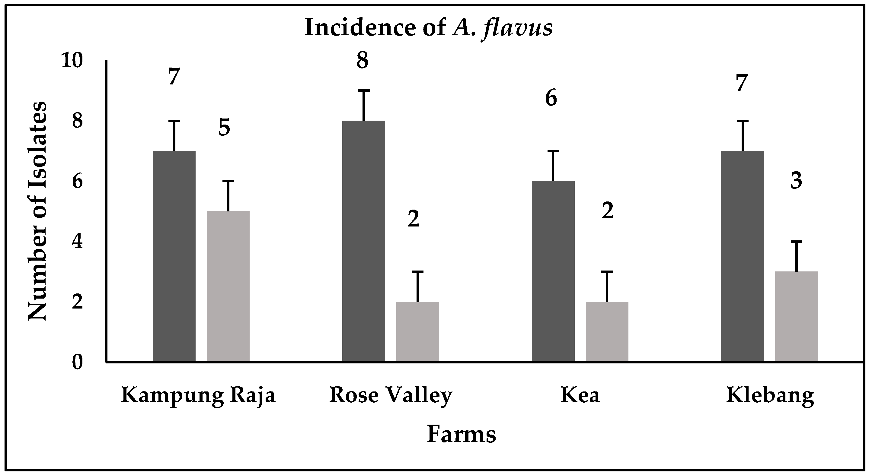
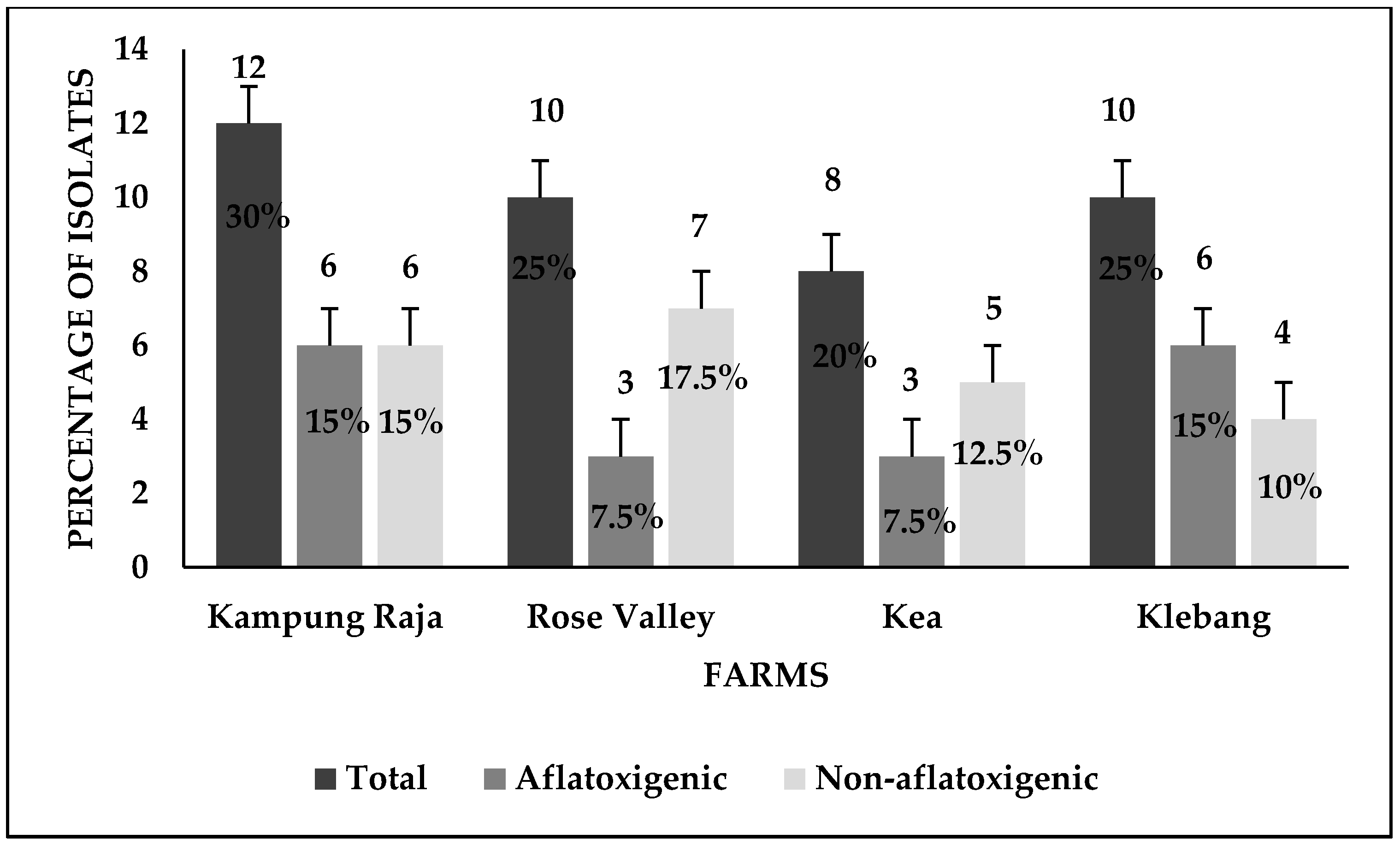
3.1. Detection of Aspergillus flavus

Forty isolates of A. flavus were detected from the sweet corn and soil samples (Figure 1). The A. flavus isolates were identified through the observation of their morphological characteristics as per the key descriptions by Klich [33] and Clayton [37]. The percentage detection of A. flavus in Kampong Raja, Rose Valley, Kea, and Klebang farms was 30% (12), 25% (10), 20% (8), and 25% (10), respectively. Moreover, the recovery of A. flavus isolates from sweet corn kernels was 30% (12) while soil samples were 70% (28).

3.2. Morphological Characterization of Aspergillus flavus

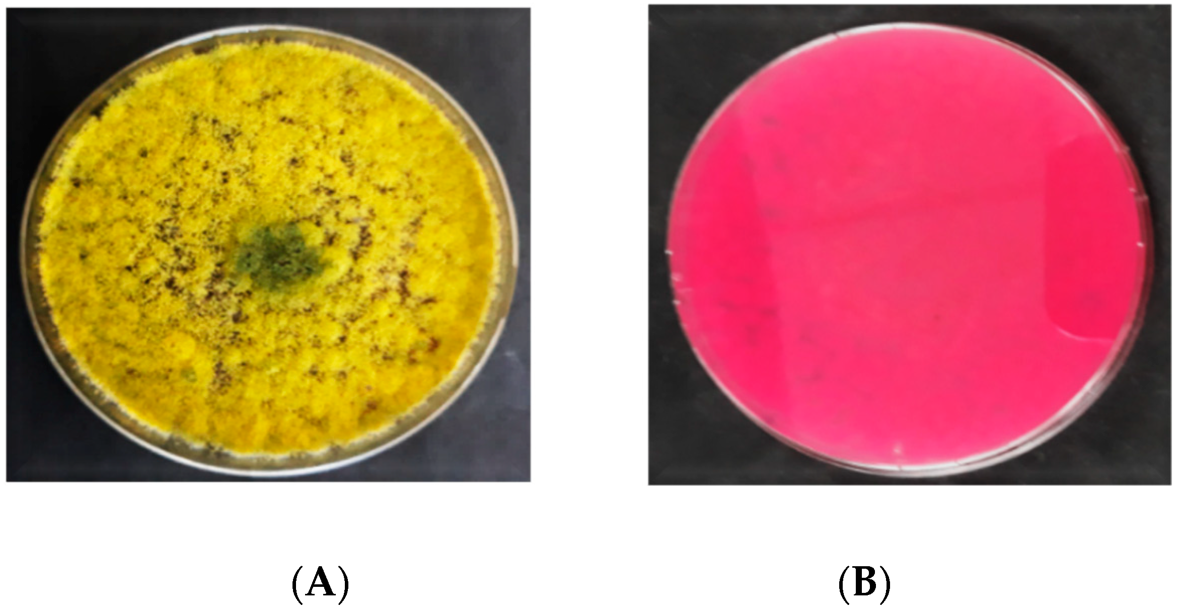
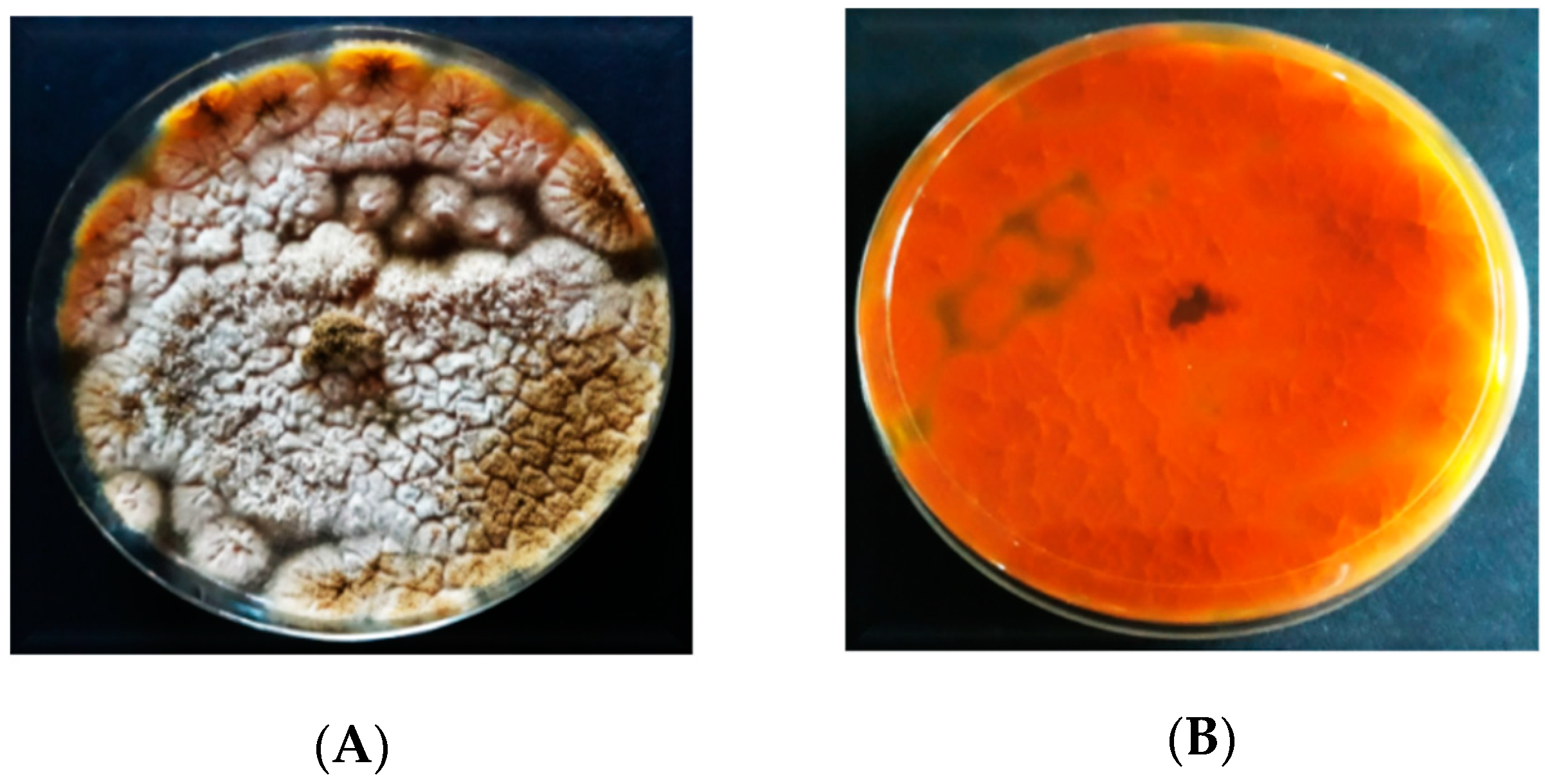
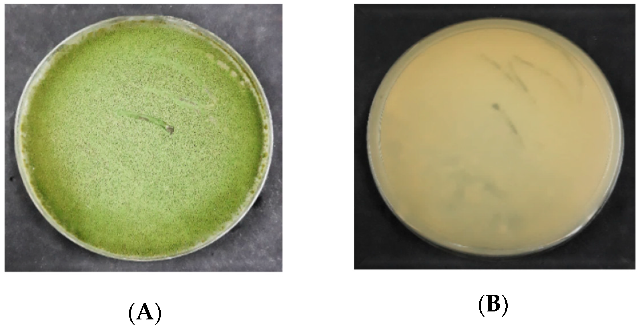
3.2.1. Macroscopic Characteristics of Aspergillus flavus on PDA

The morphology of A. flavus colonies on PDA is presented in Figure 2. In the beginning, the mycelial color of A. flavus was white. Following 3 d of incubation, the A. flavus colony formed olive-green conidia that dominated the colony’s appearance. Generally, the colonies were flat at their borders while raised in the middle. On PDA, all the A. flavus isolates produced exudates (droplets) that are brown in color or colorless. In some isolates, a compact mass of fungal mycelia (sclerotia) was produced, which were dark brown. Moreover, the colony diameter was ranged from 65 to 75 mm, surrounded by a white circle. The reverse of the colonies was pale in color.

3.2.2. Macroscopic Characteristics of Aspergillus flavus on DRBC

On DRBC (Figure 3), the A. flavus isolates initially produced white mycelia that were smooth and upraised. Following 3 d of incubation, they produced dark yellow conidia, which transformed into the olive green after 6 d. No sclerotia were produced by A. flavus isolates on DRBC. The exudates (liquid droplets) produced were colorless and embedded in the mycelia. The A. flavus isolates did not yield any soluble pigments on DRBC. The reverse sides of A. flavus colonies were colorless. The light purple is the original color of DRBC. The colonies’ diameter of A. flavus ranged from 50 to 70 mm.

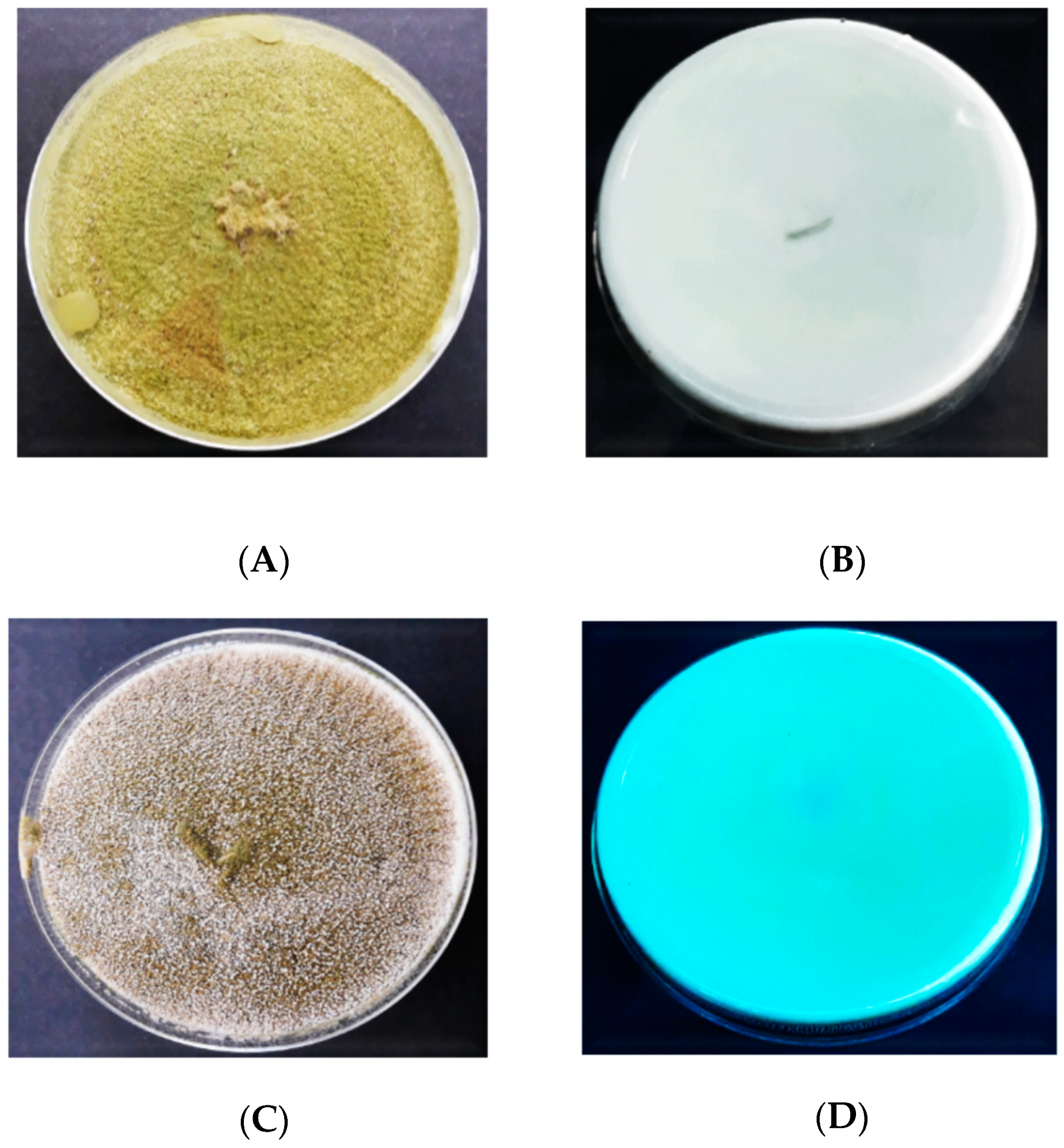
3.2.3. Macroscopic Characteristics of Aspergillus flavus on AFPA

The inoculation of A. flavus on AFPA resulted in the colonies having various profiles and dimensions (Figure 4). The colonies’ color of A. flavus on AFPA was whitish brown with dispersed conidia. On AFPA, the isolates of A. flavus did not produce exudates or soluble pigments. A. flavus produced a bright orange or golden color on the reverse due to the reaction between aspergillic acid with ferric ammonium citrate contained in the medium.

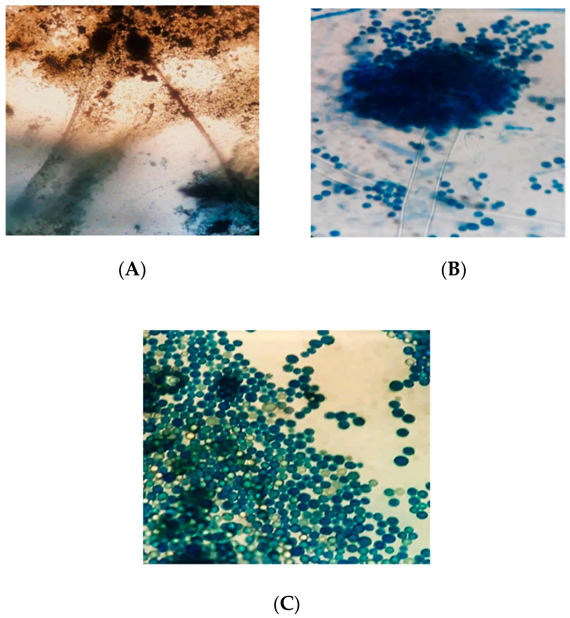
3.3. Microscopic Characteristics of Aspergillus flavus

The microscopic characteristics of A. flavus are shown in Figure 5. Under the microscope, the conidiophores of A. flavus isolates were colorless, thick walled, roughed, and bearing vesicles. The diameter of the conidiophores ranged from 800 to 1200 µm. The vesicle shape of A. flavus isolates was globose to sub-globose. The diameter of the vesicles ranged from 1800 to 2000 µm. The cells were uniseriate or biseriate. In the biseriate cells, the phialides grew on the metulae whereas, in uniseriate cells, they grew on the vesicles. The metulae enclosed the vesicles’ surface and emitted in all directions. The conidia were globose, thin walled, slightly roughed, and ranged from 250 to 450 µm in diameter.

3.4. Screening for Aflatoxin Production

Figure 6 represents the findings obtained from the UV light screening of A. flavus, displaying the fluorescent and non-fluorescent isolates of A. flavus on CCA. It was found that 18 (45%) out of 40 isolates fluoresced blue on CCA once they were exposed to UV light (365 nm), thus indicating the ability of aflatoxin production (Table 1). On the other hand, the remaining 22 (55%) isolates did not exhibit blue fluorescence in UV light, thus classified as non-aflatoxigenic isolates of A. flavus. The distribution of the aflatoxigenic and non-aflatoxigenic isolates of A. flavus in four different farms is presented in Figure 7. Of the 12 isolates from Kampong Raja, six isolates (15%) were tested positive for aflatoxin production while the remaining six (15%) were tested negative for aflatoxin production. For Rose Valley, aflatoxin was detected in three isolates (7.5%) and the remaining seven (17.5%) isolates did not produce aflatoxin. Similarly, of the eight isolates of Kea, three (7.5%) isolates were aflatoxigenic while 12% (five) of the isolates were non-aflatoxigenic. Finally, six (15%) out of 10 isolates of Klebang were aflatoxigenic whereas the remaining four (10%) isolates were found to be non-aflatoxigenic.

4. Discussion

4.1. Prevalence of Aspergillus flavus across Sweet Corn Farms in Malaysia

In Malaysia, sweet corns are grown in different environments. In unfavorable environmental conditions, sweet corn can be infected by various fungi. A. flavus has been recognized as among the most important fungal pathogens of corn. A. flavus contaminates corn both in the field as well as in storage, and the rate of infection could be different amid farms of the same region [38,39]. In this study, 40 isolates of A. flavus were identified from the fungal cultures formerly isolated from the sweet corn and soil of Kampung Raja, Rose Valley, Kea, and Klebang farms in Malaysia.
The highest occurrence (30%) of A. flavus was found in the sweet corn and soil of Kampung Raja while the lowest (20%) incidence of A. flavus was recorded for Kea farm. Rose Valley and Klebang farms comprised 25% and 25% incidence of A. flavus, respectively. The difference in the occurrence of A. flavus in the different farms could be owed to the diversity in environmental conditions between those farms. Crops grown in hot environments are more susceptible to aflatoxin-producing A. flavus and in some areas, contamination simply takes place as soon as the temperature rises with high humidity [39]. Kampung Raja is characterized by temperatures of about 25 to 33 °C with 80% humidity, which favors aflatoxin infection of sweet corn by A. flavus. While the other three farms (Rose Valley, Kea, and Klebang) are characterized by temperatures of 12–21 °C with 46–60% humidity.

4.2. Morphological Characterization of Aspergillus flavus

The morphological characterization of fungi via observing the main macro and micromorphological features cultured on diverse media is an extensively used method for fungal identifications. In this study, the morphological characterization was done to give importance to such basic identification methodologies for the fast screening of isolates in most of the developing countries, where the accessibility of unconventional tools is a great challenge. Consistent identification of A. flavus isolates was done by observing the key morphological characteristics. The physical characteristics of A. flavus including the colony color, texture and edges, as well as the exudates, soluble pigmentations, and sclerotia were studied on four different cultural media.
Overall, the A. flavus had olive-green, yellowish-green, or dark green colonies surrounded by a white circle that was eventually covered by conidia. The colonies’ textures were usually velvety or woolly with a floccose center. The A. flavus colonies produced exudates on two of the four culture media utilized in this study. Similarly, sclerotia were detected in the majority of A. flavus isolates, as it is a specific characteristic of aflatoxigenic A. flavus. In the interpretation of the taxonomic descriptions by Klich [33], the isolates’ colony morphology in this study bore a resemblance to A. flavus as shown in Figure 2 and Figure 3. On the other hand, some other Aspergillus species have the same phenotypical characteristics and which makes it difficult to differentiate them from A. flavus. It is known to have the same colony color as A. oryzae. This renders the need for comprehensive scrutiny using microscopic and macroscopic characteristics for accurate identification and classification. However, there is a consensus that, even microscopically, some characteristics are common among Aspergillus isolates [40]. The microscopic features of A. flavus such as conidiophore, vesicles, sterigmata and conidia were examined in this study.
The key microscopic features of A. flavus were rough and thick-walled conidiophores, globose vesicles with radiated sterigmata and thin walled finely rough conidia. The macroscopic and microscopic characteristics presented in Figure 2, Figure 3, Figure 4 and Figure 5 are similar to that of A. flavus characteristics described by Rodrigues et al. [41] and Diba et al. [42]. The use of PDA, DRBC, and AFPA media in this study allowed for sufficient growth and sporulation that permitted a satisfactory examination of the macroscopic and microscopic features of A. flavus. A review of the recent literature presented that these selective growth media have frequently been used for the identification of A. flavus [34]. Many A. flavus isolates isolated from corn in North-Eastern China have been identified after their morphological characterization on SZA and MEA [43].
Likewise, Diba et al. [42] characterized the Aspergillus species from medical and environmental samples through observing their macroscopic and microscopic features and stated that the utilization of PDA, DRBC, AFPA, and other growth media may stimulate the growth and sporulation process of Aspergillus species. Additionally, morphological characterization approaches have also been used on Aspergillus species in the Fars and Kerman provinces of Iran [44] and the Larkana district of the Sindh province, Pakistan [45]. Based on the size of sclerotia, A. flavus can be divided into L-strain and S-strain. The L-strain of A. flavus produces large-sized sclerotia while the S-strain A. flavus produces small sclerotia.
The relationship between the sclerotial size and aflatoxin production has been studied but the published data are unreliable. Cotty and Mellon [46] have reported a close relationship between small sclerotia and aflatoxin production. On the other hand, Abbas et al. [47] found the highest level of aflatoxins in isolates having large sclerotia. Some studies did not even find any correlation between the sclerotial size and aflatoxin production [48,49]. Thus, the sclerotial size and aflatoxin quantity have not been calculated, due to which no correlation between aflatoxin production and sclerotial type could be made.

4.3. Discrimination between Aflatoxigenic and Non-aflatoxigenic Aspergillus flavus

The screening of A. flavus isolates, based on their fluorescence characteristics under UV light at 365 nm was carried out. Once the aflatoxigenic isolates of A. flavus were exposed to UV light, they showed a blue fluorescence on the reverse of the colonies, while the non-aflatoxigenic A. flavus did not show any fluorescence (Figure 7). In this study, the use of CCA media made it possible to distinguish the aflatoxigenic A. flavus from non-aflatoxigenic ones very rapidly, albeit just qualitatively. Fluorescence was found in 18 out of 40 A. flavus (45%), thus demonstrating their aflatoxin-producing potential. In most of the developing nations, where aflatoxin determination may be delayed due to the unavailability of advanced detection instruments, culture-based techniques could be beneficial for the rapid screening of isolates.
Although the culture-based techniques are comparatively simple and economical, for aflatoxin detection however, these techniques are usually employed in combination with chromatographic methods [47]. For the precise quantification of aflatoxin, a more advanced method such as high-performance liquid chromatography equipped with fluorescence detection (HPLC-FLD) would be useful. Similarly, it has been reported that strains of A. flavus could produce B and G aflatoxins [50], a recent classification of Aspergillus section Flavi noted that such strains, though morphologically similar to A. flavus, were completely novel species [51,52]. These outcomes pointed out the fact that modern molecular approaches are mandatory to be used for A. flavus isolates’ characterization.

5. Conclusions

In conclusion, both aflatoxigenic and non-aflatoxigenic strains of A. flavus were present in the sweet corn kernels and soil of Cameron Highlands, Malaysia. Cameron Highlands being a primary source of agricultural produce could take measures to ensure that aflatoxin contamination is minimized. Non-aflatoxigenic strains indigenously present could be further harnessed into a biological control agent that can reduce the dependency on chemically synthesized pesticides. Culture media and morphology are rapid tools for the characterization of A. flavus in this study. The morphological characteristics of these isolates were similar to those explained in the published identification manuals. The screening of A. flavus isolates for aflatoxin detection under UV light (365 nm) is a fast and reliable method to distinguish the aflatoxigenic and non-aflatoxigenic isolates. Although the culture-based techniques are comparatively simple and economical, however, for the precise quantification of aflatoxin, a more advanced method such as high-performance liquid chromatography equipped with fluorescence detection (HPLC-FLD) would be useful. This further reinforces the need for measures to prevent the fungal contamination of sweet corn and emphasizes the importance of field interventions aimed at curbing agricultural soil contamination by these fungi.

Author Contributions

F.M.G., supervision, project administration, funding acquisition; R.K., methodology, writing—original draft preparation; N.I.P.S., conceptualization, and methodology assistance; N.A.M., review, resources, and data curation. All authors have read and agreed to the published version of the manuscript.

Funding

This research was funded by Geran Inisiatif Putra, Universiti Putra Malaysia, UPM/GP/2017/9568800.

Acknowledgments

The authors would like to acknowledge the financial contribution of the Ministry of Science, Technology and Innovation (MOSTI), Malaysia for funding this research under Science Fund (Grant number: 05-01-04-SF0750). The authors would like to thank Siti Nur Ezzati Yazid for her technical assistance with this study.

Conflicts of Interest

The authors declare no conflict of interest.

References

  1. IARC. Some Naturally Occurring Aromatic Amines. Mycotoxins; International Agency for Research on Cancer: Lyon, France, 1993. [Google Scholar]
  2. Richard, J.L. Some major mycotoxins and their mycotoxicoses-An overview. Int. J. Food Microbiol. 2007, 119, 3–10. [Google Scholar] [CrossRef]
  3. Afsah-Hejri, L.; Jinap, S.; Arzandeh, S.; Mirhosseini, H. Optimization of HPLC conditions for quantitative analysis of aflatoxins in contaminated peanut. Food Control 2011, 22, 381–388. [Google Scholar] [CrossRef]
  4. Filazi, A.; Sireli, U.T. Occurrence of aflatoxins in food. In Aflatoxins: Recent Advances and Future Prospects; InTech: Rijeka, Croatia, 2013; pp. 70–143. [Google Scholar]
  5. Schmidt-Heydt, M.; Rüfer, C.E.; Abdel-Hadi, A.; Magan, N.; Geisen, R. The production of aflatoxin B1 or G1 by Aspergillus parasiticus at various combinations of temperature and water activity is related to the ratio of Afls, and Aflr expression. Mycotoxin Res. 2010, 26, 46–241. [Google Scholar] [CrossRef] [Green Version]
  6. Alpsoy, L. Inhibitory effect of essential oil on aflatoxin activities. Afr. J. Biotechnol. 2010, 9, 2474–2481. [Google Scholar]
  7. Giray, B.; Girgin, G.; Engin, A.B.; Aydın, S.; Sahin, G. Aflatoxin levels in wheat samples consumed in some regions of Turkey. Food Control 2007, 18, 23–29. [Google Scholar] [CrossRef]
  8. Park, J.W.; Choi, S.Y.; Hwang, H.J.; Kim, Y.B. Fungal mycoflora and mycotoxins in Korean polished rice destined for humans. Int. J. Food Microbiol. 2005, 103, 305–314. [Google Scholar] [CrossRef]
  9. Reddy, K.R.N.; Reddy, C.S.; Muralidharan, K. Detection of Aspergillus spp. and aflatoxin B1 in rice in India. Food Microbiol. 2009, 26, 27–31. [Google Scholar] [CrossRef]
  10. Toteja, G.S.; Mukherjee, A.; Diwakar, S.; Singh, P.; Saxena, B.N.; Sinha, K.K.; Sinha, A.K.; Kumar, N.; Nagaraja, K.V.; Bai, G.; et al. Aflatoxin B1 contamination of parboiled rice samples collected from different states of India: A multi-centre study. Food Addit. Contam. 2006, 23, 411–414. [Google Scholar] [CrossRef] [PubMed]
  11. Wagacha, J.M.; Muthomi, J.W. Mycotoxin problem in Africa: Current status, implications to food safety and health and possible management strategies. Int. J. Food Microbiol. 2008, 124, 1–12. [Google Scholar] [CrossRef] [PubMed]
  12. Lizárraga-Paulín, E.G.; Moreno-Martínez, E.; Miranda-Castro, S.P. Aflatoxins and their impact on human and animal health: An emerging problem. Aflatoxins Biochem. Mol. Biol. 2011, 13, 255–282. [Google Scholar]
  13. Williams, J.H.; Phillips, T.D.; Jolly, P.E.; Stiles, J.K.; Jolly, C.M.; Aggarwal, D. Human aflatoxicosis in developing countries: A review of toxicology, exposure, potential health consequences, and interventions. Am. J. Clin. Nutr. 2004, 80, 1106–1122. [Google Scholar] [CrossRef] [PubMed]
  14. Qian, G.S.; Ross, R.K.; Yu, M.C.; Yuan, J.M.; Gao, Y.T.; Henderson, B.E.; Wogan, G.N.; Groopman, J.D. A follow-up study of urinary markers of aflatoxin exposure and liver cancer risk in Shanghai, People’s Republic of China. Cancer Epidemiol. Prev. Biomark. 1994, 3, 3–10. [Google Scholar]
  15. Ross, R.K.; Yu, M.C.; Henderson, B.E.; Yuan, J.M.; Qian, G.S.; Tu, J.T.; Gao, Y.T.; Wogan, G.N.; Groopman, J.D. Urinary aflatoxin biomarkers and risk of hepatocellular carcinoma. Lancet 1992, 339, 943–946. [Google Scholar] [CrossRef]
  16. Wang, L.Y.; Hatch, M.; Chen, C.J.; Levin, B.; You, S.L.; Lu, S.N.; Wu, M.H.; Wu, W.P.; Wang, L.W.; Wang, Q.; et al. Aflatoxin exposure and risk of hepatocellular carcinoma in Taiwan. Int. J. Cancer 1996, 67, 620–625. [Google Scholar] [CrossRef]
  17. Gong, Y.; Hounsa, A.; Egal, S.; Turner, P.C.; Sutcliffe, A.E.; Hall, A.J.; Cardwell, K.; Wild, C.P. Postweaning exposure to aflatoxin results in impaired child growth: A longitudinal study in Benin, West Africa. Environ. Health Perspect. 2004, 112, 1334–1338. [Google Scholar] [CrossRef]
  18. Shuaib, M.; Ouararhni, K.; Dimitrov, S.; Hamiche, A. HJURP binds CENP-A via a highly conserved N-terminal domain and mediates its deposition at centromeres. Proc. Natl. Acad. Sci. USA 2010, 107, 1349–1354. [Google Scholar] [CrossRef] [PubMed] [Green Version]
  19. Abbas, H.K.; Wilkinson, J.R.; Zablotowicz, R.M.; Accinelli, C.; Abel, C.A.; Bruns, H.A.; Weaver, M.A. Ecology of Aspergillus flavus, regulation of aflatoxin production, and management strategies to reduce aflatoxin contamination of corn. Toxin Rev. 2009, 28, 53–142. [Google Scholar] [CrossRef]
  20. Amaike, S.; Keller, N.P. Aspergillus flavus. Annu. Rev. Phytopathol. 2011, 49, 33–107. [Google Scholar] [CrossRef]
  21. Hell, K.; Mutegi, C. Aflatoxin control and prevention strategies in key crops of sub-Saharan Africa. Afr. J. Microbiol. Res. 2011, 5, 66–459. [Google Scholar]
  22. Yu, J. Current understanding of aflatoxin biosynthesis and future perspective in reducing aflatoxin contamination. Toxins 2012, 4, 57–1024. [Google Scholar] [CrossRef] [Green Version]
  23. Abdullah, N.; Nawawi, A.; Othman, I. Survey of fungal counts and natural occurrence of aflatoxins in Malaysian starch-based foods. Mycopathologia 1998, 143, 53–58. [Google Scholar] [CrossRef]
  24. Sulaiman, M.R.; Yee, C.F.; Hamid, A.; Yatim, A.M. The occurrence of aflatoxins in raw shelled peanut samples from three districts of Perak, Malaysia. Electron. J. Environ. Agric. Food Chem. 2007, 6, 2045–2052. [Google Scholar]
  25. Chan, N.W. Cameron Highlands: Issues and challenges in sustainable development. Malaysian J. Environ. Manage. 2009, 10, 89–114. [Google Scholar]
  26. Okoth, S.A.; Nyongesa, B.; Joutsjoki, V.; Korhonen, H.; Ayugi, V.; Kang’ethe, E.K. Sclerotia formation and toxin production in large sclerotial Aspergillus flavus isolates from Kenya. Adv. Microbiol. 2016, 6, 1–10. [Google Scholar] [CrossRef] [Green Version]
  27. Ho, W.C.; Ko, W.H. A simple method for obtaining single-spore isolates of fungi. Bot. Bull. Acad. Sin. 1997, 38, 41–44. [Google Scholar]
  28. Choi, Y.W.; Hyde, K.D.; Ho, W.H. Single spore isolation of fungi. Fungal Divers. 1999, 3, 29–88. [Google Scholar]
  29. Singh, K.P.; Kumar, D.; Bandyopadhyay, P. A new technique for single spore isolation of two predacious fungi forming constricting ring. Mycobiology 2004, 32, 197–198. [Google Scholar] [CrossRef]
  30. Raper, K.B.; Fennell, D.I. Aspergillus terreus group. In The genus Aspergillus; The Williams & Wilkins Co.: Baltimore, MD, USA, 1965; pp. 293–344. [Google Scholar]
  31. Samson, R.A.; Hoekstra, E.S.; Frisvad, J.C. Introduction to Food-and Airborne Fungi; Centraalbureau voor Schimmelcultures (CBS) Fungal Biodiversity Centre, Royal Netherlands Academy of Arts and Sciences: Utrecht, The Netherlands, 2004; Volume 7, pp. 6–389. [Google Scholar]
  32. Cotty, P.J. Comparison of Four Media for the Isolation of Aspergillus flavus Group Fungi. Mycopathologia 1994, 125, 62–157. [Google Scholar] [CrossRef] [PubMed]
  33. Klich, M.A. Identification of Common Aspergillus Species; Centraalbureau voor Schimmelcultures (CBS) Fungal Biodiversity Centre, Royal Netherlands Academy of Arts and Sciences: Utrecht, The Netherlands, 2002; pp. 426–432. [Google Scholar]
  34. Pitt, J.I.; Hocking, A.D.; Glenn, D.R. An Improved Medium for the Detection of Aspergillus flavus and A. parasiticus. J. Appl. Bacteriol. 1983, 54, 14–109. [Google Scholar] [CrossRef]
  35. Davis, N.D.; Iyer, S.K.; Diener, U.L. Improved method of screening for aflatoxin with a coconut agar medium. Appl. Environ. Microbiol. 1987, 53, 1593–1595. [Google Scholar] [CrossRef] [PubMed] [Green Version]
  36. Fente, C.A.; Ordaz, J.J.; Vazquez, B.I.; Franco, C.M.; Cepeda, A. New additive for culture media for rapid identification of aflatoxin-producing Aspergillus strains. Appl. Environ. Microbiol. 2001, 67, 62–4858. [Google Scholar] [CrossRef] [PubMed] [Green Version]
  37. Clayton, Y.M. Medically Important Fungi: A Guide to Identification. Proc. R. Soc. Med. 1977, 70, 359. [Google Scholar]
  38. Da Gloria, M.E. Aflatoxin contamination distribution among grains and nuts. In Aflatoxins–Detection, Measurement and Control; Torres-Pacheco, I., Ed.; InTech: Rijeka, Croatia, 2011; pp. 75–90. [Google Scholar]
  39. Thathana, M.G.; Murage, H.; Abia, A.L.K.; Pillay, M. Morphological characterization and determination of aflatoxin-production potentials of Aspergillus flavus isolated from maize and soil in Kenya. Agriculture 2017, 7, 80. [Google Scholar] [CrossRef] [Green Version]
  40. Cotty, P.J.; Jaime-Garcia, R. Influences of climate on aflatoxin producing fungi and aflatoxin contamination. Int. J. Food Microbiol. 2007, 119, 109–115. [Google Scholar] [CrossRef]
  41. Rodrigues, P.; Soares, C.; Kozakiewicz, Z.; Paterson, R.; Lima, N.; Venâncio, A. Identification and characterization of Aspergillus flavus and aflatoxins. In Microbiology Book Series–Communicating, Current Research and Educational Topics and Trends in Applied, Microbiology; Méndez-Vilas, A., Ed.; Formatex: Badajoz, Spain, 2007; Volume 2, pp. 527–534. ISBN 13: 978-84-611-9423-0. [Google Scholar]
  42. Diba, K.; Kordbacheh, P.; Mirhendi, S.H.; Rezaie, S.; Mahmoudi, M. Identification of Aspergillus Species Using Morphological Characteristics. Pak. J. Med. Sci. 2007, 23, 867–872. [Google Scholar]
  43. Gao, J.; Liu, Z.; Yu, J. Identification of Aspergillus Section Flavi in Maize in Northeastern China. Mycopathologia 2007, 164, 91–95. [Google Scholar] [CrossRef]
  44. Mohammadi, A.H.; Banihashemi, Z.; Haghdel, M. Identification and prevalence of aspergillus species in soils of Fars and Kerman provinces of Iran and evaluation of their aflatoxin production. Rostanilha 2009, 23, 49–50. [Google Scholar]
  45. Afzal, H.; Shazad, S.; Qamar, S.; Nisa, U. morphological identification of Aspergillus species from the soil of Larkana District (Sindh, Pakistan). Asian J. Agric. Biotechnol. 2013, 1, 17–105. [Google Scholar]
  46. Cotty, P.J.; Mellon, J.E. Ecology of aflatoxin producing fungi and biocontrol of aflatoxin contamination. Mycotoxin Res. 2006, 22, 17–110. [Google Scholar] [CrossRef]
  47. Abbas, H.K.; Zablotowicz, R.M.; Weaver, M.A.; Horn, B.W.; Xie, W.; Shier, W.T. Cultural methods for aflatoxin detection. J. Toxicol. Toxin Rev. 2004, 23, 295–315. [Google Scholar]
  48. Baquião, A.C.; De Oliveira, M.M.M.; Reis, T.A.; Zorzete, P.; Diniz, A.D.; Correa, B. Polyphasic approach to the identification of Aspergillus Section Flavi isolated from Brazil Nuts. Food Chem. 2013, 139, 32–1127. [Google Scholar] [CrossRef] [PubMed] [Green Version]
  49. Giorni, P.; Magan, N.; Pietri, A.; Bertuzzi, T.; Battilani, P. Studies on Aspergillus Section Flavi isolated from maize in northern Italy. Int. J. Food Microbiol. 2007, 113, 38–330. [Google Scholar] [CrossRef] [PubMed]
  50. Takahashi, H.; Kamimura, H.; Ichinoe, M. Distribution of aflatoxin-producing Aspergillus flavus and Aspergillus parasiticus in sugarcane fields in the southernmost islands of Japan. J. Food Prot. 2004, 67, 90–95. [Google Scholar] [CrossRef]
  51. Pildain, M.B.; Frisvad, J.C.; Vaamonde, G.; Cabral, D.; Varga, J.; Samson, R.A. Two novel aflatoxin-producing Aspergillus species from Argentinean peanuts. Int. J. Syst. Evol. Microbiol. 2008, 58, 35–725. [Google Scholar] [CrossRef] [PubMed] [Green Version]
  52. Varga, J.; Frisvad, J.C.; Samson, R.A. Two new aflatoxin producing species, and an overview of Aspergillus section Flavi. Stud. Mycol. 2011, 69, 57–80. [Google Scholar] [CrossRef]
Figure 1. Prevalence of A. flavus isolates in Kampong Raja, Rose Valley, Kea, and Klebang farms; black bars = soil samples; grey bars = maize samples.
Agriculture 10 00450 g001
Figure 2. Colony morphology of A. flavus on the potato dextrose agar (PDA); (A) = obverse, (B) = reverse.
Agriculture 10 00450 g002
Figure 3. Colony morphology of A. flavus on dichloran rose bengal chloramphenicol (DRBC); (A) = obverse, (B) = reverse.
Agriculture 10 00450 g003
Figure 4. Colony morphology of A. flavus on Aspergillus flavus and Aspergillus parasiticus agar (AFPA); (A) = obverse, (B) = reverse.
Agriculture 10 00450 g004
Figure 5. Microscopic characteristics of A. flavus; (A) = conidiophores and conidia under 10× objective; (B) = conidiophore and conidia under the 100× objective; (C) = conidia under the 100× objective of the stereo–binocular microscope (Magnus M24).
Agriculture 10 00450 g005
Figure 6. Colonies of the aflatoxigenic and non-aflatoxigenic isolates of A. flavus observed under UV light (365 nm); (A) = aflatoxigenic obverse, (B) = aflatoxigenic reverse, (C) = non-aflatoxigenic obverse, (D) = non-aflatoxigenic reverse.
Agriculture 10 00450 g006
Figure 7. The distribution of the aflatoxigenic and the non-aflatoxigenic A. flavus isolates in four sweet corn-growing farms in Malaysia. Black bars = number of A. flavus isolates and their respective percentage figure.
Agriculture 10 00450 g007
Table 1. Identification of the aflatoxin production through UV fluorescence screening.
IsolateNo.CCA
AKR11+
AKR22-
AKR33+
AKR44+
AKR55+
AKR66-
AKR77-
AKR88+
AKR99+
AKR1010-
AKR1111-
AKR1212-
ARV1313-
ARV1414+
ARV1515-
ARV1616-
ARV1717+
ARV1818+
ARV1919-
ARV2020-
ARV2121-
ARV2222-
AK2323+
AK2424+
AK2525-
AK2626-
AK2727-
AK2828+
AK2929-
AK3030-
AKL3131+
AKL3232-
AKL3333-
AKL3434+
AKL3535+
AKL3636-
AKL3737+
AKL3838+
AKL3939-
AKL4040+
AKR: A. flavus isolates from Kampung Raja. ARV: A. flavus isolates from Rose Valley. AK: A. flavus isolates from Kea. AKL: A. flavus isolates from Klebang. (-) = did not fluoresce or non-aflatoxigenic. CCA: coconut cream agar.

