Article

Characterization of Newly Discovered Phosphorite Deposits in Al-Tafeh, Jordan

1
Department of Earth and Environmental Sciences, Prince El Hassan Bin Talal Faculty of Natural Resources and Environment, The Hashemite University, P.O. Box 330127, Zarqa 13133, Jordan
2
MinXperts, P.O. Box 11121, Amman 8301, Jordan
*
Author to whom correspondence should be addressed.
Geosciences 2025, 15(11), 433; https://doi.org/10.3390/geosciences15110433
Submission received: 8 September 2025 / Revised: 5 November 2025 / Accepted: 7 November 2025 / Published: 14 November 2025
(This article belongs to the Section Sedimentology, Stratigraphy and Palaeontology)

Abstract

This study reports the discovery of a newly identified phosphorite deposit in the Al-Tafeh area of northern Jordan. Geological fieldwork investigated three outcrops and one comparison site in the Russifa area. Geochemical analyses reveal a high P2O5 content (average 24.32 wt.%), strongly correlating with CaO. There are also significant levels of trace elements, including uranium (0.045 mg/g), cadmium (0.025 mg/g), and zinc (0.099 mg/g). Mineralogical investigation reveals that francolite is the main phosphate mineral. Calcite and quartz are also present. Petrographic analysis reveals the presence of pellets, skeletal fragments, coated grains, and indicators of storm deposits, bioturbation, and fossil-rich layers. These findings indicate that the Al-Tafeh area in northern Jordan is an important yet under-explored area for phosphorite, suggesting that this discovery could have significant economic value.

1. Introduction

Phosphate rocks are important sedimentary ores and the primary source of phosphorus for fertilizers, animal feed, and industrial chemicals [1]. These deposits generally form in shallow marine environments, where elevated biological activity, organic matter accumulation, and nutrient upwelling promote the development of carbonate-fluorapatite (francolite) nodules and layers within carbonate and siliciclastic sediments [2]. The formation of phosphorites is primarily influenced by paleoceanographic conditions, sedimentation rates, and local geochemical environments [3].
Jordan is a leading global producer of phosphate rock, with vast, high-quality reserves that have played a significant role in its economy since mining began in the 1930s. The Jordan Phosphate Mines Company (JPMC) oversees exploration, extraction, and export. Phosphate deposits are essential for fertilizer production, supporting global agriculture, and Jordan’s strategic location has positioned it as a key player in the international phosphate market [4]. Major deposits are located in central and southern Jordan, including Al-Hassa, Al-Abiad, and Eshidiya [5]. The geology, mineralogy, geochemistry, and genesis of these deposits have been studied by several researchers. Ref. [6] identified marine formation conditions during the Late Cretaceous to Eocene periods. The main stratigraphic units hosting these deposits are the Al Hisa Phosphorite Formation (Campanian), Muwaqqar Chalk Marl Formation (Maastrichtian), and Wadi Shallala Formation (Eocene). Refs. [7,8] established that the Belqa Group is the key phosphatic sequence, while [9] contextualized these deposits within Tethyan phosphorogenesis. Refs. [10,11,12] identified francolite, calcite, and quartz as primary mineral phases. Ref. [13] characterized the geochemical properties and depositional environments of northwestern deposits. The genesis of phosphorites was linked to upwelling-driven marine productivity and diagenesis, with [7,9] emphasizing biogenic and chemical precipitation. Ref. [14] used carbon−14 and oxygen−18 isotope analyses to conclude that the deposits are autogenic and shaped by upwelling. Ref. [15] reconstructed paleoenvironmental conditions using sedimentological and isotopic data.
In northern Jordan, phosphorite deposits are distributed across several localities. In the northeast, ref. [16] documented middle–late Eocene to Oligocene phosphatic horizons exposed along Wadi Rewashed and Wadi Um-Qusier in the Risha region. In contrast, the Al-Kora phosphorite province in the northwestern region has been studied in detail by [17]; however, its exploitation has not been pursued due to the high population density in the area. Furthermore, more recently, ref. [18] reported the discovery of a potential phosphate resource in northeastern Jordan, near the international borders with Saudi Arabia and Iraq.
This study investigates a previously undocumented phosphate deposit in the Al-Tafeh area of northern Jordan. By characterizing its geological setting, chemical and mineral composition, and petrographic features, the research establishes the distinctiveness of this newly recognized deposit. Since phosphate is essential for fertilizer production and many industries, understanding the unique properties of this particular deposit offers valuable insights for resource evaluation and informs future development strategies, with potential economic benefits for Jordan.

2. Methodology

2.1. Geological Settings and Field Observations

Phosphate outcrops in the study area are located in Al-Tafeh, northeast of Amman. Three main sites within this region—Sites 1, 2, and 3—were investigated. For correlation purposes, Site 4, a separate outcrop in Russifa, was also included. Figure 1 illustrates the location of the study area, along with the distribution of the three investigated sites, including Site 4. The geological map of Zarqa city, which encompasses the study area, illustrates the main lithological units and the occurrence of phosphate-bearing horizons.
Site 1 begins with a hard, light crystalline limestone bed approximately 1.2 m thick, exhibiting calcite veins and affected by bioturbation at the base. Above this limestone, the contact transitions sharply to a breccia bed with dark chert fragments embedded in a phosphate matrix (Figure 2). The breccia is overlain conformably by a thick phosphorite bed, which initiates with a hard base containing dark chert extraclasts and medium-sized invertebrate bones. This phosphorite bed grades upward into a softer top, where caves have formed (Figure 3A). Above this soft top, a thin breccia bed, composed of coarse invertebrate bone and shell fragments, rests directly on the phosphorite and is capped by a bed of laminated marlstone. At the top of the marlstone, traces of bioturbation mark the transition to the cave ceiling (Figure 3B). Above the marlstone, an erosional boundary is observed below a very hard phosphorite bed containing white chert clasts (Figure 3C), which is in turn overlain by a thicker bed of soft to hard phosphorite with coarse clasts, including a complete fish skeleton (Figure 3D). Within this upper sequence, interbeds of phosphorite containing uranium are also found.
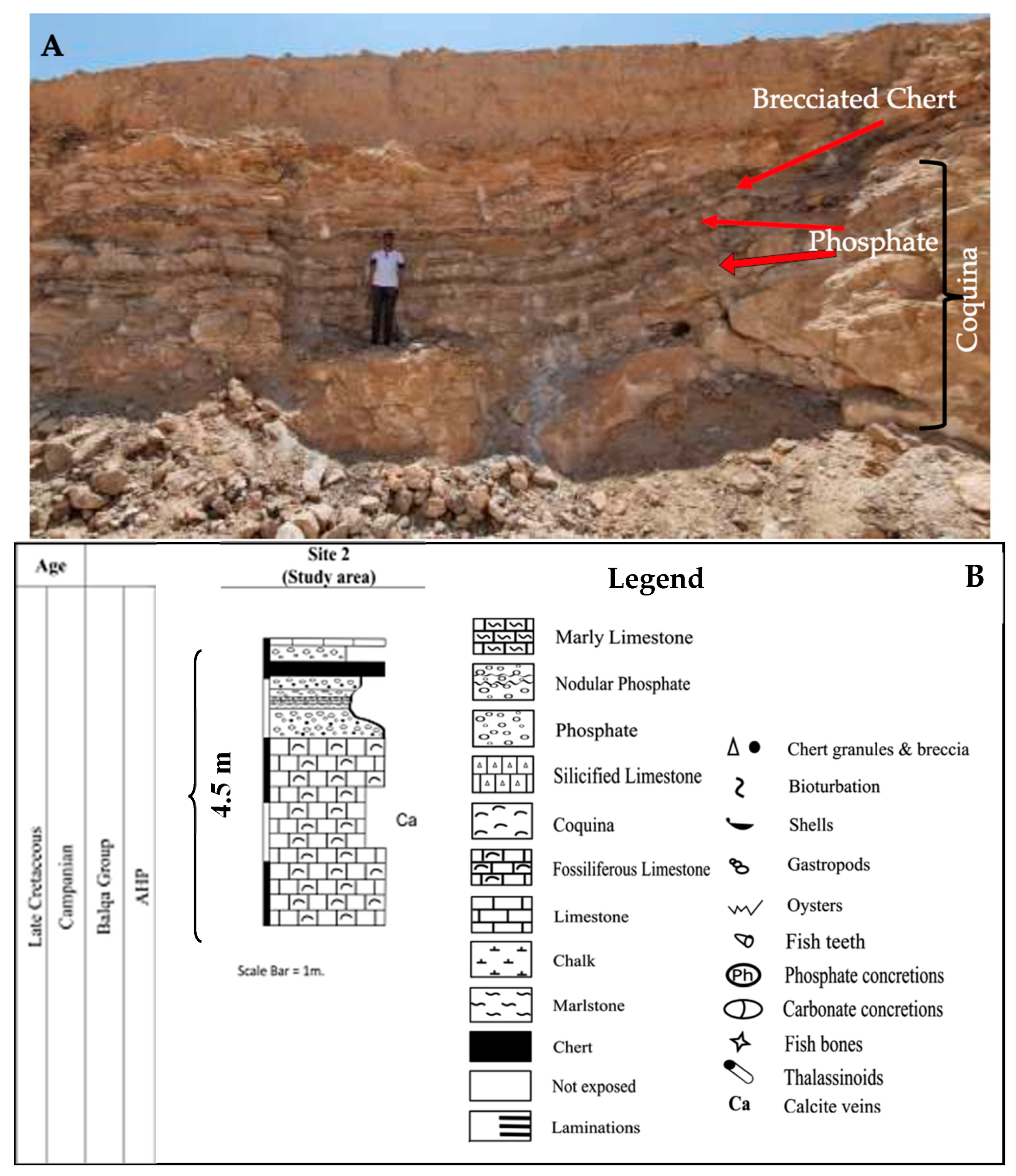
At Site 2, a clear change in rock type appears, as shown in Figure 4. This begins with two thick, hard, white, crystalline beds made of coquina. The top bed is composed of chalk and is followed by a light brown, crystalline coquina layer. Below this, the layers change and show signs of deformation, switching between coarse, dark beds of phosphate and carbonate. These beds hold fish teeth, bones, shells, and sharp pieces of dark chert. Some fragments are composed of broken chert on a phosphatic base; others are predominantly phosphate. Above these, there are light carbonate beds with chert nodules.
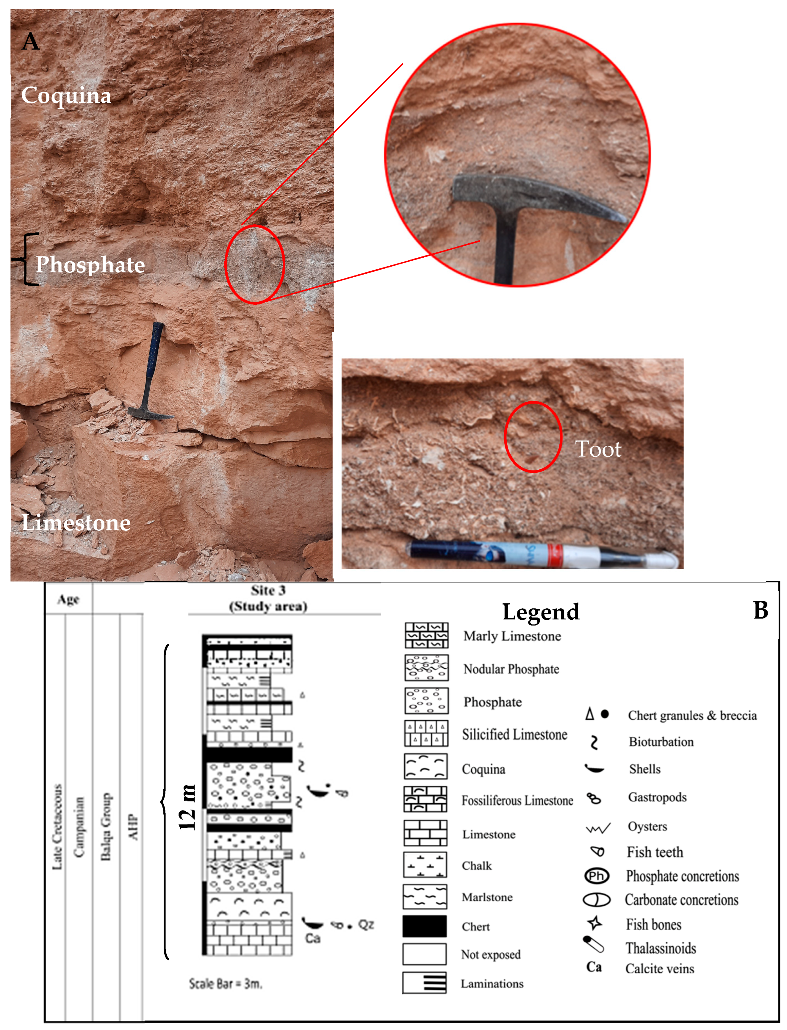
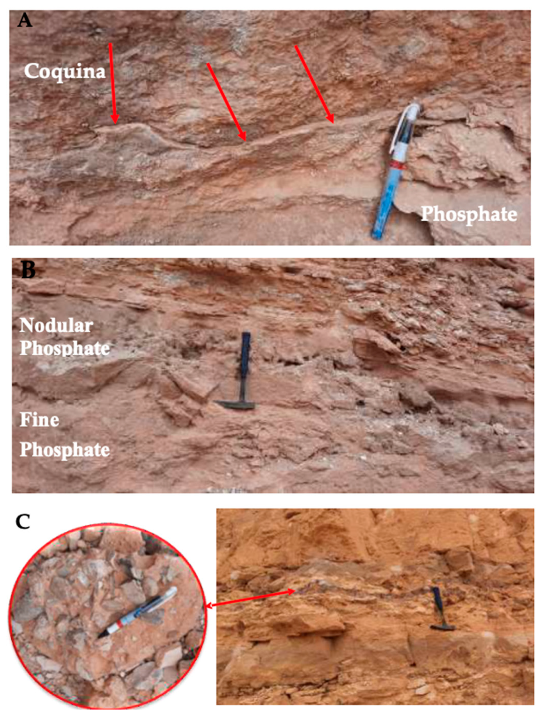
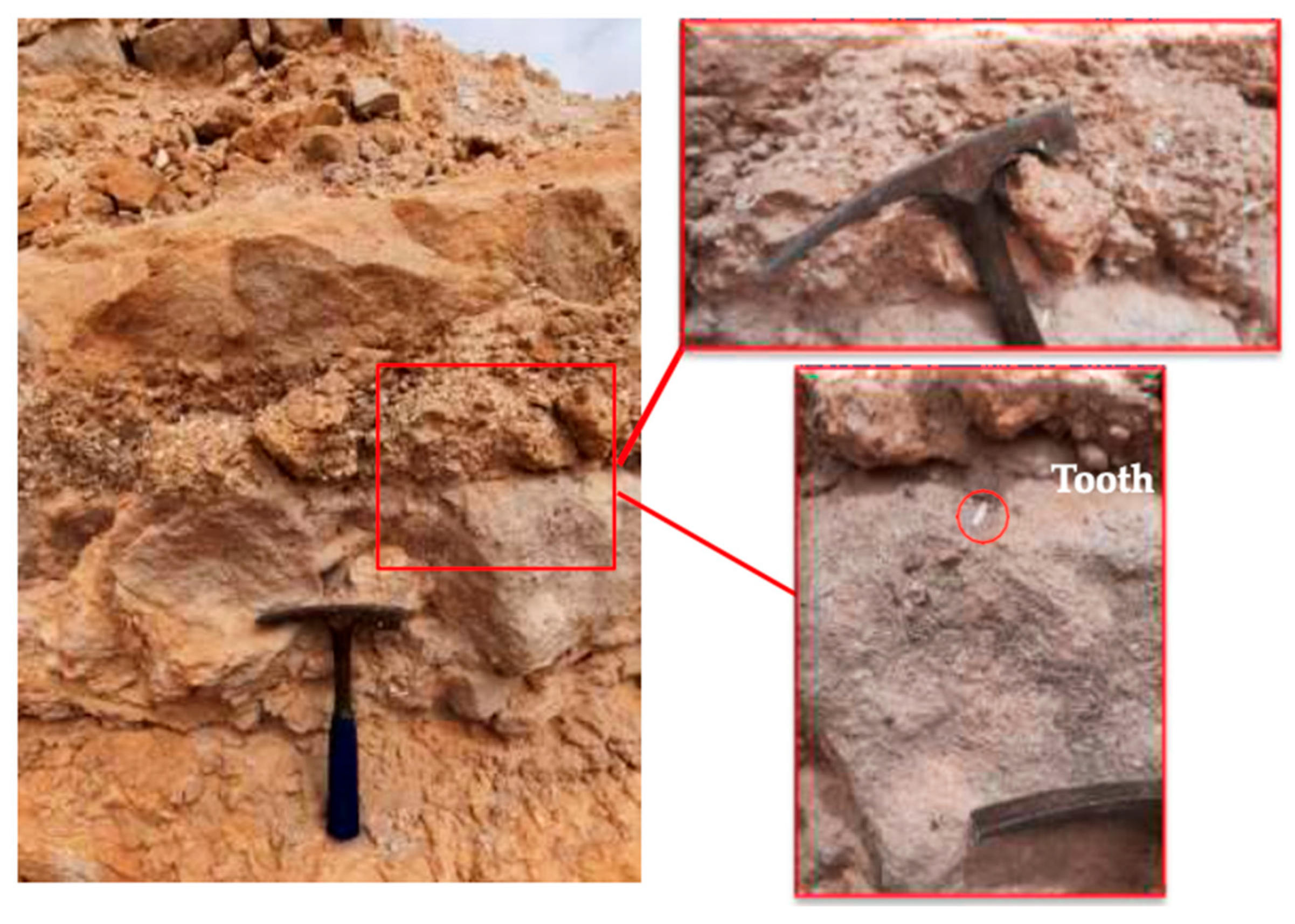
Site 3, as seen in Figure 5, consists of thick, dark, hard, massive sparitic limestone with calcite veins, followed by a thin bed of poorly sorted phosphate. This phosphate bed includes sand-sized grains with extra and intra-clasts of oyster shell fragments, carbonate, quartz, chert, shells, and shark teeth. An incrustation at the top is covered by large, fragmented oyster shells, succeeded by a medium-hard, fine-grained phosphatic bed (77 cm) and a nodular phosphatic bed (40 cm). This is followed by alternating thin white tripoli beds, chert beds, and laminated micritic limestone, showing evidence of deformation. The three layers of phosphate-containing angular, pebble-sized erosional chert fragments are at the base of the third layer (Figure 6). A bed of phosphate is followed by limestone, then a 70 cm laminated pinkish marl interbedded with three layers of dark chert. This is succeeded by 50 cm of carbonates and very thin chert, followed again by laminated pinkish marl interbedded with dark chert. Finally, a bed of marly limestone, interbedded with chert, is capped by soft, pinkish marl, culminating in a bed of hard limestone. A light-colored (65 cm) fine phosphatic bed exhibits bioturbation at both its base and top, with a magnified figure illustrating clasts of varying sizes interspersed within the phosphates (Figure 7).
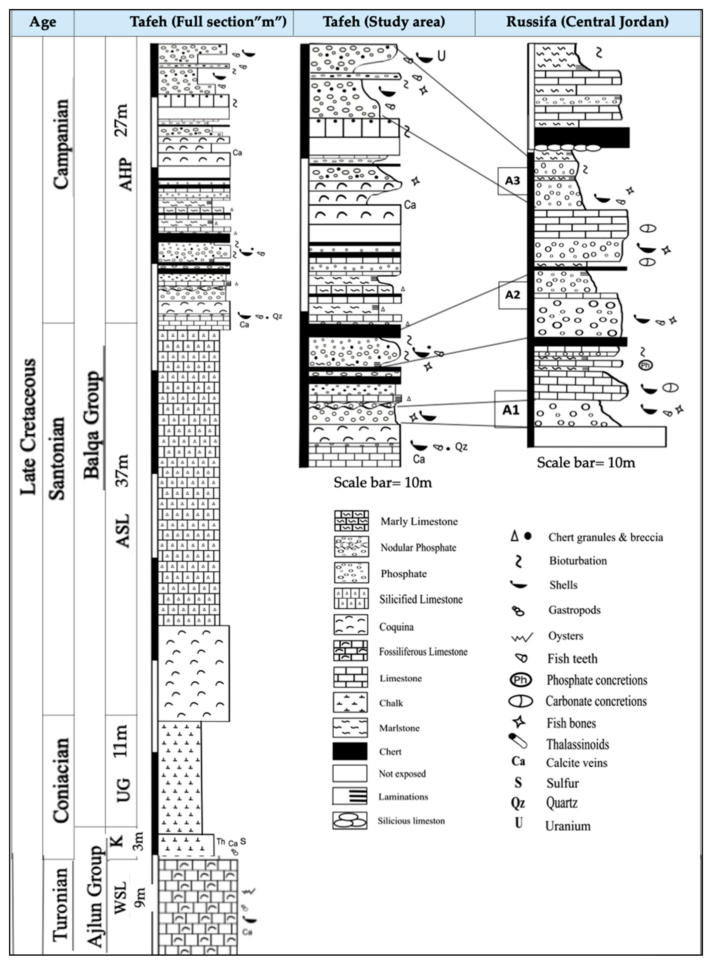
The phosphorite outcrop in the Russifa area (Site 4) was studied for lithostratigraphic comparison, to determine the extent of phosphate depositions in the area, comparing depositional environments and the arrangement of phosphate units. It was observed that the phosphate in the study area can most probably be considered an extension of the sedimentary deposits of the Russifa basin, encompassing all three of its units. The phosphate deposit in the first unit (A1) of the Russifa outcrop is characterized by its hardness and the presence of carbonate concretions. Only approximately 20 cm of this unit was exposed, and the full thickness was not revealed. The overlying layer is fine-grained, with a light gray color, and features coarse clasts that contain relatively large bone fragments, as well as vugs. Bioclasts and concretions are abundant in the carbonate layers, and the unit is concluded with successive layers of limestone and marlstone (Figure 8A). The second unit (A2) starts with a thick, hard bed of light matrix material (Rock type), with coarse clasts of medium size. Above this, laminated phosphate beds were reported, containing a high percentage of clay. This was followed by interbeds of chert and marlstone containing limestone concretions. The other layer of the same unit exhibits a high degree of crystallization, appearing hard with large, clearly visible shell fragments (Figure 8B). As for the third unit (A3), several beds of carbonates and chert are sequentially arranged. The phosphate forms a thick, soft, light gray color with coarse clasts. A clearly visible layer of silicious concretions appears, with very large sizes and a wide extent beneath a thickness of chert (Figure 8C,D). Figure 9 shows the correlation between the phosphorites of the two regions, Al-Tafeh and the Russifa, and a complete sequence of the formations in the study area. The lithostratigraphic comparison The stratigraphic comparison between the Tafeh section (north Jordan) and the Russifa section (central Jordan) reveals that both are part of the same phosphogenic system within the Jordanian Phosphorite Formation, but exhibit distinct differences in rock types. Tafeh is thicker, contains more limestone, and has chert layers, suggesting a stable, shallow sea rich in carbonates [7]. Russifa, split into units A1–A3, has more material from the land and greater variation, indicating stronger environmental changes. Even with these local differences, both were affected by changes in sea level and upwelling, which contributed to phosphate accumulation [15]. Overall, Tafeh mostly shows a carbonate setting, while Russifa has more sand and clay in the same type of area.

2.2. Laboratory Works

For lab investigations and analyses, twelve samples of phosphate were systematically selected from three outcrops based on their representativeness of varying lithologies. Major and trace elements were analyzed using Inductively Coupled Plasma (ICP-14) at the Jordan Atomic Energy CommissionMajor and trace elements were analyzed using an Inductively Coupled Plasma system (instrument ID: ICP-14) at the Jordan Atomic Energy Commission (JAEC), Amman, Jordan; the system was procured by MEDILab. The samples were first grind to a fine powder size and digested using a mixture of HF, HNO3, and HCl acids. Calibration and data accuracy were ensured using certified international phosphate rock standards (USGS GXR-1). The mineral phases were identified using X-ray diffraction (XRD) on selected representative samples at the Cementra Jordan Company Laboratory in Mafraq. The analyses were conducted with CuKα radiation at 40 kV and 30 mA, scanning from 5° to 60° 2θ at a rate of 2°/min. The obtained diffraction patterns were interpreted using JCPDS (Joint Committee on Powder Diffraction Standards) database to identify mineral phases. Thin sections were prepared from selected samples and examined under a polarizing optical microscope (Nikon type) at the Hashemite University, Jordan. using a polarizing light microscope (Nikon Eclipse LV100POL, Nikon Corporation, Tokyo, Japan) at the Department of Earth & Environmental Sciences, The Hashemite University, Zarqa, Jordan. Both plane-polarized light (PPL) and cross-polarized light (XPL) observations were used to describe the textural features, microstructures, and mineral relationships within the phosphatic rocks. Photomicrographs were taken to document the main petrographic characteristics.

3. Results

3.1. Geochemistry

Table 1 presents the average (wt.%) concentrations of major oxides for twelve samples collected from three outcrops in the study area. The results are compared to those from three Jordanian mines (Ruseifa, Al-Hisa, and Esshidyah) as documented by [20]. The table also incorporates data on phosphorites from Algeria (Djebel Onk), Syria (Khneifiss), and Morocco (Bou Craa), as reported by [21].
The results show an average CaO concentration of 52.78 wt.%. Calcium predominantly occurs in francolite and is also present in dolomite and calcite, which make up the matrix and cement of phosphorite rocks. P2O5, essential for phosphate rocks and biological processes, averages 24.32 wt.%. Elevated calcium and phosphorus concentrations together indicate robust phosphate mineralization in most samples. SiO2 averages 2.29 wt.% and is found as detrital or biogenic silica, such as chert, or within clay minerals. Al2O3 is minimal at 0.21 wt.% and is associated with clay minerals. Fe2O3 averages 0.18 wt.% and occurs as oxide-hydroxide stains on phosphate grains. MgO is present at 0.31 wt.%, occurring in francolite, clay, or dolomite. Na2O averages 0.32 wt.% and acts as a calcium substitute or impurity in apatite. K2O, at 0.02 wt.%, is a detrital contaminant external to the apatite lattice. CO2 averages 15 wt.%, reflecting a significant presence of calcite and francolite. Chlorine is present in trace amounts (0.074 wt.%). Finally, the loss on ignition (L.O.I) averages 17.45 wt.%, indicating differences in volatile content resulting from variations in mineralogical composition.
Phosphorite samples from the Al-Tafeh area are notably rich in calcium carbonate (CaO~52.8 wt.%), while their phosphate content (P2O5~24.3 wt.%) is somewhat lower than that reported for other regional deposits. Furthermore, the consistently low concentrations of silica, alumina, and iron oxides suggest that the rocks have not been significantly affected by detrital or terrigenous input. In addition, the low amounts of magnesium and sodium point to only a limited role for dolomite and evaporitic minerals in the deposit’s composition. Finally, a slight enrichment in fluorine (3.7 wt.%) reflects the strong presence of carbonate-fluorapatite, the dominant phosphate mineral phase.
Table 2 presents the average concentrations of trace elements in phosphorites from the study area and compares these values with previous research. Ref. [1] identified three primary associations for trace elements in phosphorites: substitution within apatite or adsorption onto apatite surfaces due to small crystallite size, adsorption by organic matter or association with detrital minerals, and absorption on clay or iron oxides and hydroxides. The Al-Tafeh phosphorites exhibit moderate concentrations of nickel (0.019 mg/g) and copper (0.016 mg/g), consistent with values reported by [22] for other Jordanian phosphorites and within the range observed in regional deposits such as Djebel Onk and Bou Craa. Zinc concentrations (0.099 mg/g) are lower than those in most regional deposits, whereas cadmium (0.025 mg/g) is relatively high compared to both Jordanian and international phosphorites. The elevated cadmium content suggests local geochemical enrichment during phosphate formation, potentially associated with the presence of organic matter or minor detrital input. The uranium concentration in Al-Tafeh samples (0.045 mg/g) is moderate and lower than in other Jordanian phosphorites but similar to that of Djebel Onk, which may indicate potential economic value for by-product recovery.
To examine inter-element relationships, a correlation matrix was constructed using the geochemical data of all analyzed phosphorite samples as shown in Table 3. Pearson correlation coefficients were calculated using Microsoft Excel. The results show that there is a negative correlation between P2O5 and SiO2, as P2O5 enrichment indicates the dominance of phosphate minerals, such as fluorapatite, carbonate-fluorapatite, or francolite, which are low in silica. Conversely, high SiO2 signals the presence of detrital quartz, chert, and clay minerals. In addition, a strong positive correlation typically exists between P2O5 and CaO, since apatite—the main phosphate mineral—contains calcium, a key element in phosphate deposit formation [6,7,8,20,21]. The frequent negative correlation between P2O5 and MgO further suggests that magnesium-rich minerals, such as dolomite or magnesite, are rare [19]. For trace elements, the matrix shows that as the P2O5 content increases, the levels of zircon, uranium, and copper also rise [13], indicated by R2 values of 0.1393, 0.1812, and 0.0518, which confirm these positive correlations and are consistent with the established association of rare earth elements, notably uranium’s affinity for phosphate minerals. In contrast, nickel and cadmium display a negative correlation. This geochemical pattern is well documented in Jordanian phosphorites by [13,23,24].

3.2. Mineralogical Study

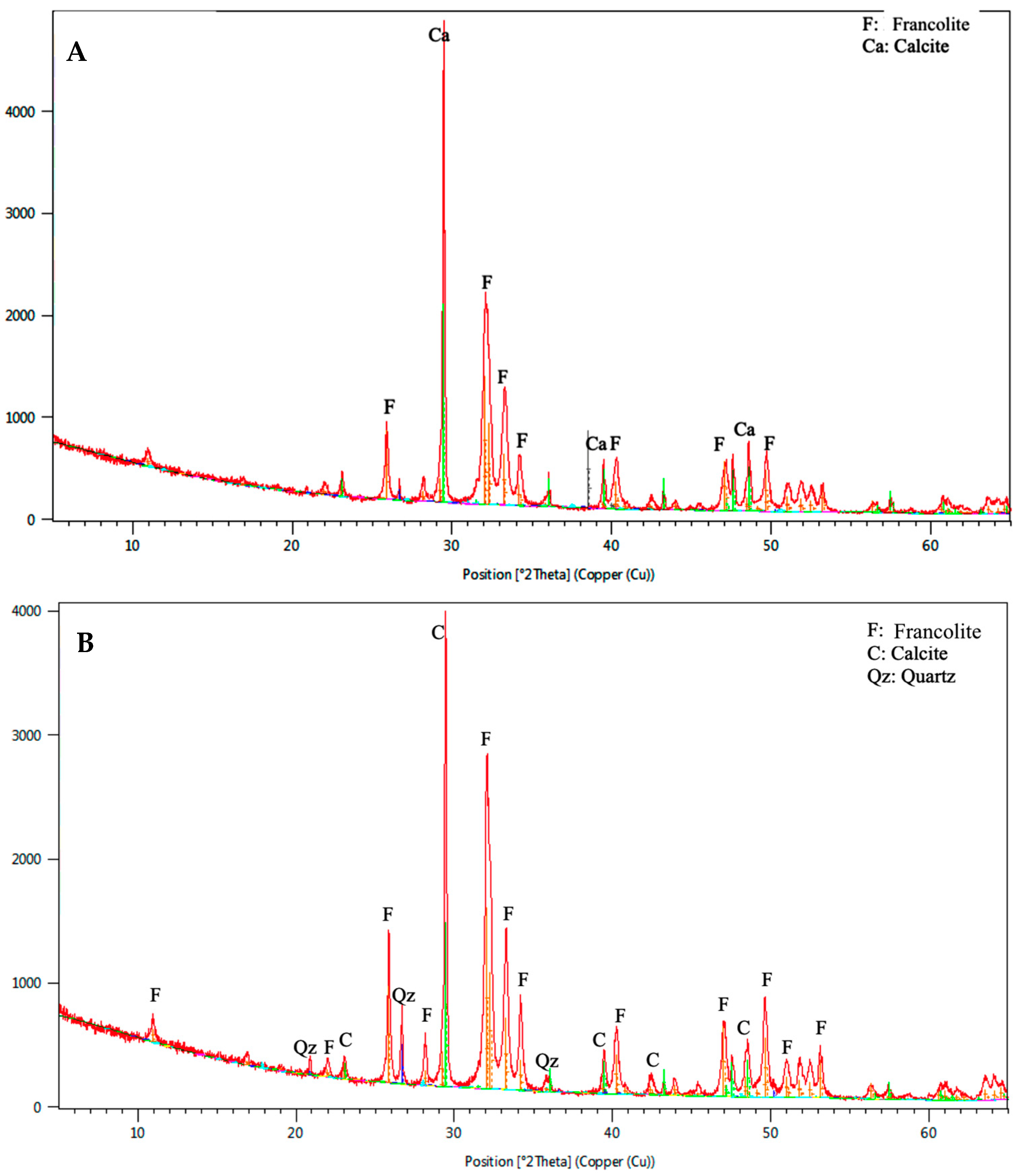
Phosphorite rocks contain various mineral phases of apatite, including fluorapatite, carbonate fluorapatite (francolite), carbonate hydroxylapatite (dahlite), and chlorapatite [1,9,22]. Figure 10A shows the XRD pattern for samples from site 1. According to the analysis, the studied samples are predominantly composed of francolite, a calcium phosphate mineral, Ca5(PO4)3F, the dominant constituent of high-grade Jordanian phosphorites [6]. Francolite typically exhibits peaks at 25.8°, 31.8°, and 32.2° (2θ). In addition to francolite, calcite (CaCO3) is also present, as indicated by peaks at 29.4° and 39.4°. Moreover, quartz (SiO2) is present in the sample from site 3, as indicated by peaks at 26.6° and 20.9° (Figure 10B). Non-phosphatic minerals in phosphate deposits can affect ore processing due to their reactivity with acids, such as calcite, and lower the ore grade.

3.3. Petrographic Description

Jordanian phosphates contain four primary particle types: pellets, intraclasts, skeletal fragments, and coated grains [19,22,25]. The main constituents of phosphatic beds in the Al-Tafeh area are consistent with those of major Jordanian phosphorites: phosphatic components, including pellets and skeletal fragments; non-phosphatic constituents, such as detrital quartz and rock fragments; a cementing material, which may be silica or calcite; and a matrix.
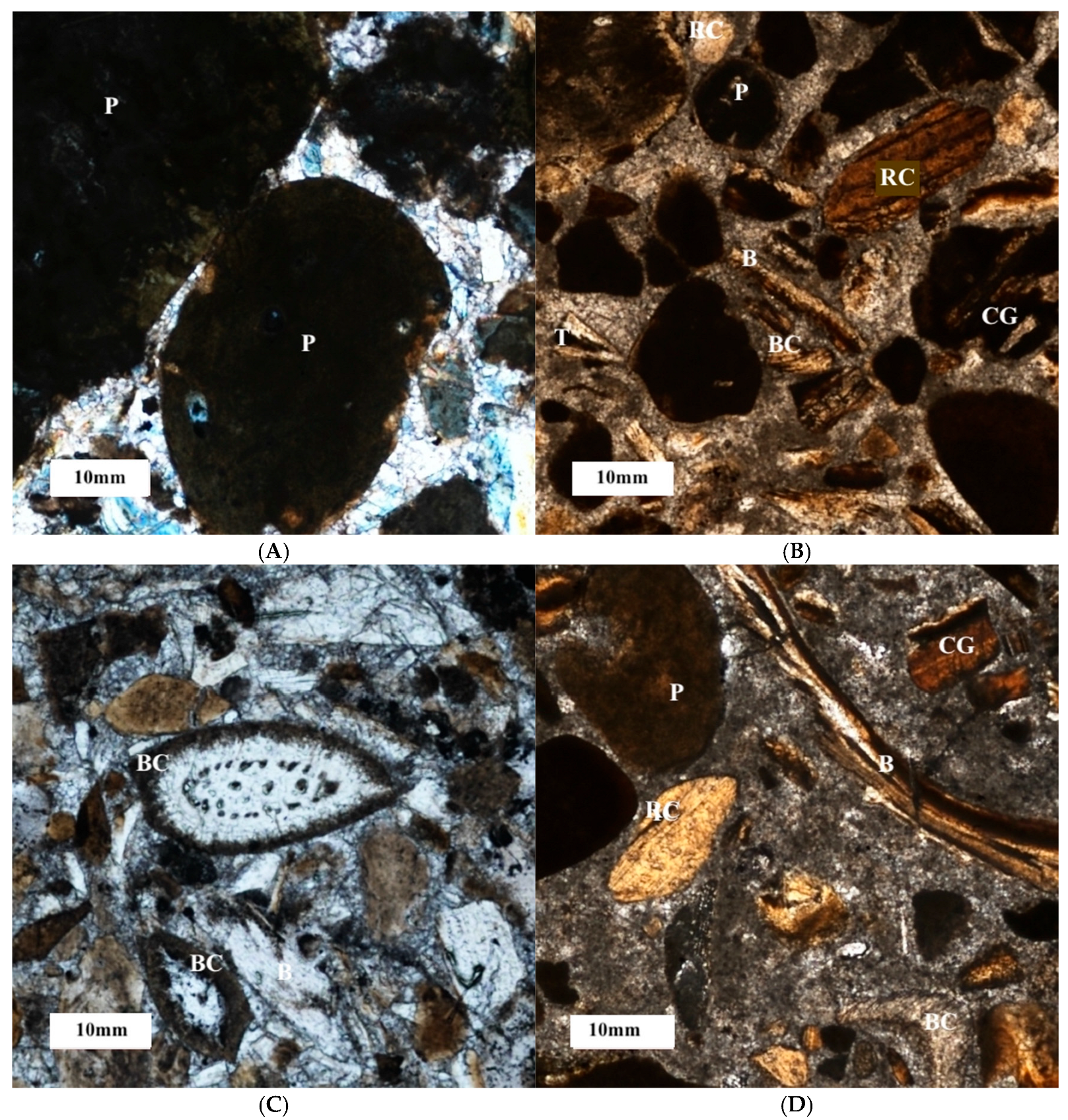
Pellets or peloids lack internal structure and are mostly rounded to subrounded with smooth boundaries. Francolite pellets, varying in size from medium to coarse and in color from light to dark brown, have a cloudy appearance and may contain organic matter, which imparts a brownish to black color. All are surrounded by calcitic cement. Under crossed Nicol, they appear isotropic (Figure 11A).
Bioclasts are composed of fossil fragments, elongated bones, and tooth fragments. These elements are typically structureless, highly angular, and anisotropic, often appearing as prismatic shreds with sharp edges in various sizes (Figure 11B). Some bioclasts have cracks filled with phosphate mud or are replaced by calcite (Figure 11C). Their occurrence suggests a depositional environment suitable for preserving vertebrate remains [24].
Rock fragments occur as grains with varied sizes and shapes, ranging from angular to subrounded (Figure 11B,D). These grains may come from the breakdown of larger phosphate rocks or other rocks, such as chert or carbonates. According to [1], chert fragments could have been transported and deposited with phosphate material, or formed in place through replacement by silica.
Coated grains comprise phosphate mud enveloping a phosphate grain or rock fragment (Figure 11B,C). Calcite also appears as a cementing material (Figure 11D).

4. Discussion

The reported phosphorite deposits in the Al-Tafeh area share many characteristics with those in the central and southern parts of Jordan. The P2O5 contents (24.32 wt.%) are within the range previously reported for Jordanian phosphorites (27.09–34.34 wt.%), such as those from Russifa, Al-Hisa, and Eshidiya [8,14,15,20]. However, the higher-grade horizons at Al-Tafeh suggest localized depositional conditions that favoured enhanced phosphate enrichment, similar to observations in the Al-Risha deposits in northeast Jordan [19]. The strong positive correlation between P2O5 and CaO, and the dominance of carbonate-fluorapatite (francolite), are consistent with mineralogical observations in other Jordanian phosphorites [7,11,20]. Additionally, uranium (up to 0.069 mg/g) and cadmium (up to 0.051 mg/g) were also reported by [20,24]. Furthermore, the enrichment of organic matter and its association with chert layers at Al-Tafeh indicate periods of upwelling and anoxic bottom-water conditions, a process widely described in Tethyan phosphorogenesis [9,13]. In terms of depositional environment, petrographic observations, including bioturbated beds, storm-induced shell accumulations, and abundant vertebrate remains, indicate a shallow-marine setting characterized by high-energy events. These findings align with interpretations from the Amman Formation phosphorites in Russifa [6,7] and are comparable to depositional models proposed for the Late Cretaceous to Eocene phosphorites of the southern Tethys [2,5]. Overall, the deposits record a dynamic paleoceanographic setting marked by high productivity, intermittent reducing conditions, and periodic high-energy events typical of an upwelling-related phosphogenic environment. The discovery supports the idea that the phosphogenic system extended farther north than previously believed, reinforcing Mikbel & Abed’s [6] conclusions that northern Jordan may have significant, underexplored phosphate resources. Altogether, these features indicate that the Al-Tafeh phosphorites represent a relatively clean and calcareous variety, enriched in francolite, and form a distinctive end-member within the broader phosphorite belt of Jordan.

5. Conclusions

The Al-Tafeh deposits add to Jordan’s known phosphorite resources and show that the Tethyan phosphogenic system reaches farther north than once thought. The deposits formed in a shallow-marine, upwelling-influenced setting characterized by high productivity and intermittent bottom-water anoxia. Geochemical data, including high P2O5 (up to 24.3 wt.%), CaO (52.8 wt.%), and enrichment in U and Cd, indicate intense phosphogenesis under suboxic to anoxic conditions with minimal detrital input. Sedimentary features such as coquina beds, brecciated chert, and bioturbated horizons reflect alternating periods of storm reworking and quiet deposition. Overall, the Al-Tafeh phosphorites help us better understand how local factors shape phosphorite formation in northern Jordan and highlight the economic promise of less-studied areas.

Author Contributions

Conceptualization, K.M.I. and M.M.; methodology, F.A.-S., K.M.I. and M.A.; investigation, F.A.-S., K.M.I., M.A. and M.M.; resources, K.M.I.; data curation, F.A.-S. and K.M.I.; writing—original draft preparation, F.A.-S., K.M.I. and M.A.; writing—review and editing, F.A.-S. and K.M.I.; visualization, K.M.I. and M.M.; supervision, K.M.I.; project administration, K.M.I. All authors have read and agreed to the published version of the manuscript.

Funding

This research received no external funding.

Data Availability Statement

Data used for the manuscript preparation have been included within the document.

Acknowledgments

The authors warmly thank the laboratories of the Jordan Atomic Energy Commission and Cementra Jordan Company for their invaluable assistance with geochemical and mineralogical analyses.

Conflicts of Interest

The authors declare no conflicts of interest.

References

  1. Cook, P.J. Volume 1: Proterozoic and Cambrian phosphorites. In Phosphate Deposits of the World; Cambridge University Press: Cambridge, UK, 2005. [Google Scholar]
  2. Föllmi, K.B. The phosphorus cycle, phosphogenesis and marine phosphate-rich deposits. Earth-Sci. Rev. 1996, 40, 55–124. [Google Scholar] [CrossRef]
  3. Glenn, C.R.; Föllmi, K.B.; Riggs, S.R.; Baturin, G.N.; Grimm, K.A.; Trappe, J.; Abed, A.M.; Galli-Oliver, C.; Garrison, R.E.; Ilyan, A.V. Phosphorus and phosphorites: Sedimentology and Environments of Formation. Eclogae Geol. Helv. 1994, 87, 747–788. [Google Scholar]
  4. US Geological Survey (USGS). Mineral Commodity Summaries: Phosphate Rock. U.S. Department of the Interior, 2022. Available online: https://www.sciencebase.gov/catalog/item/6197ccbed34eb622f692ee1c (accessed on 7 September 2025). [CrossRef]
  5. Abed, A.M. The eastern Mediterranean phosphorite giants: An interplay between tectonics and upwelling. GeoArabia 2013, 18, 67–94. [Google Scholar] [CrossRef]
  6. Mikbel, S.; Abed, A.M. Discovery of large phosphate deposits in north-west Jordan. Ann. Société Géologique Belg. 1985, 108, 169–174. [Google Scholar]
  7. Abed, A.M. On the genesis of the phosphorite–chert association of the Amman Formation in the Tel es Sur area, Ruseifa, Jordan. Sci. Géologiques Bull. Mémoires 1989, 42, 141–153. [Google Scholar] [CrossRef]
  8. Abed, A.M.; Amireh, B.S. Sedimentology, geochemistry, economic potential and palaeogeography of an Upper Cretaceous phosphorite belt in the southeastern desert of Jordan. Cretac. Res. 1999, 20, 119–133. [Google Scholar] [CrossRef]
  9. Pufahl, P.K.; Grimm, K.A.; Abed, A.M.; Sadaqah, R.M. Upper Cretaceous 11. (Campanian) phosphorites in Jordan: Implications for the formation of a south Tethyan phosphorite giant. Sediment. Geol. 2003, 161, 175–205. [Google Scholar] [CrossRef]
  10. Al-Shereideh, S.A.K.; Tarawneh, K.; El-Radaideh, N.; Saqqa, W.; Alnawafleh, H. Geology and mineralogy of Cabal Kabid phosphorite deposits, southeastern Jordan. Jordan J. Earth Environ. Sci. 2010, 3, 27–40. [Google Scholar]
  11. Al-Shereideh, S.; Saqqa, W.; El-Radaideh, N.; Tarawneh, K.; Alnawafleh, H. Calcitization and bulk chemistry of phosphate grains from the upper part of the Amman Formation, southeast Irbid, Jordan. J. Geogr. Geol. 2013, 5, 16–28. [Google Scholar] [CrossRef]
  12. Abu-Hamatteh, Z.S. Geology, geochemistry and are characteristics of the Jordanian phosphates. Cent. Eur. Geol. 2007, 50, 283–295. [Google Scholar] [CrossRef]
  13. Alnimrat, R.M.; Alqudah, M.; Al-Shereideh, S.; Alsaleh, A.R.S. Geochemical and petrological analyses of the phosphorite deposits (Amman/Al Hisa Formation) from northwest Jordan, and their insights for paleoenvironment. Arab. J. Geosci. 2022, 15, 1259. [Google Scholar] [CrossRef]
  14. Sadaqah, R.M.; Abed, A.M.; Grimm, K.A.; Pufahl, P.K. Oxygen and carbon isotopes in Jordanian phosphorites and associated fossils. J. Asian Earth Sci. 2007, 29, 803–812. [Google Scholar] [CrossRef]
  15. Alqudah, M.; Abu-Jaber, N.; Al-Rawabdeh, A.; Al-Tamimi, M. Paleoenvironmental study of the Late Cretaceous–Eocene Tethyan Sea associated with phosphorite deposits in Jordan. Appl. Sci. 2023, 13, 1568. [Google Scholar] [CrossRef]
  16. Basha, S. On the Tertiary phosphate rocks of the Risha area, NE Jordan. Dirasat 1987, 14, 211–227. [Google Scholar]
  17. Abed, A.M.; Jaber, O.; Alkuisi, M.; Sadaqah, R. Rare earth elements and uranium geochemistry in the Al-Kora phosphorite province, Late Cretaceous, northwestern Jordan. Arab. J. Geosci. 2016, 9, 187. [Google Scholar] [CrossRef]
  18. Ibrahim, K.M.; Al-Zughoul, K.; Muhtaseb, K. New occurrence of potential phosphate resource in northeast Jordan. Arab. J. Geosci. 2016, 9, 497. [Google Scholar] [CrossRef]
  19. Abu Qudaira, M. The Geology of Zarqa Area, Bulletin 58; Natural Resources Authority: Amman, Jordan, 2004; 47p. [Google Scholar]
  20. Abed, A.M.; Abu Murry, O.S. Rare earth element geochemistry of the Jordanian Upper Cretaceous Phosphorites. Arab. Gulf J. Sci. Res. 1997, 15, 41–158. [Google Scholar]
  21. Ryszko, U.; Rusek, P.; Kołodyńska, D. Quality of phosphate rocks from various deposits used in wet phosphoric acid and P-fertilizer production. Materials 2023, 16, 793. [Google Scholar] [CrossRef] [PubMed]
  22. Abed, A.M.; Al-Agha, M.R. Petrography, geochemistry and origin of the NW Jordan phosphorites. J. Geol. Soc. 1989, 146, 499–506. [Google Scholar] [CrossRef]
  23. Abed, A.M.; Sadaqah, R. Role of Upper Cretaceous oyster bioherms in the deposition and accumulation of high-grade phosphorites in central Jordan. J. Sediment. Res. 1998, 68, 1009–1020. [Google Scholar] [CrossRef]
  24. Powell, J.H.; Moh’d, B.K. Early diagenesis of Late Cretaceous chalk–chert–phosphorite hardgrounds in Jordan: Implications for sedimentation on a Coniacian–Campanian pelagic ramp. GeoArabia 2012, 17, 17–38. [Google Scholar] [CrossRef]
  25. Khaled, H.; Abed, A.M. Petrography and geochemistry of Esh-Shidiya phosphates. Dirasat 1982, 9, 81–102. [Google Scholar]
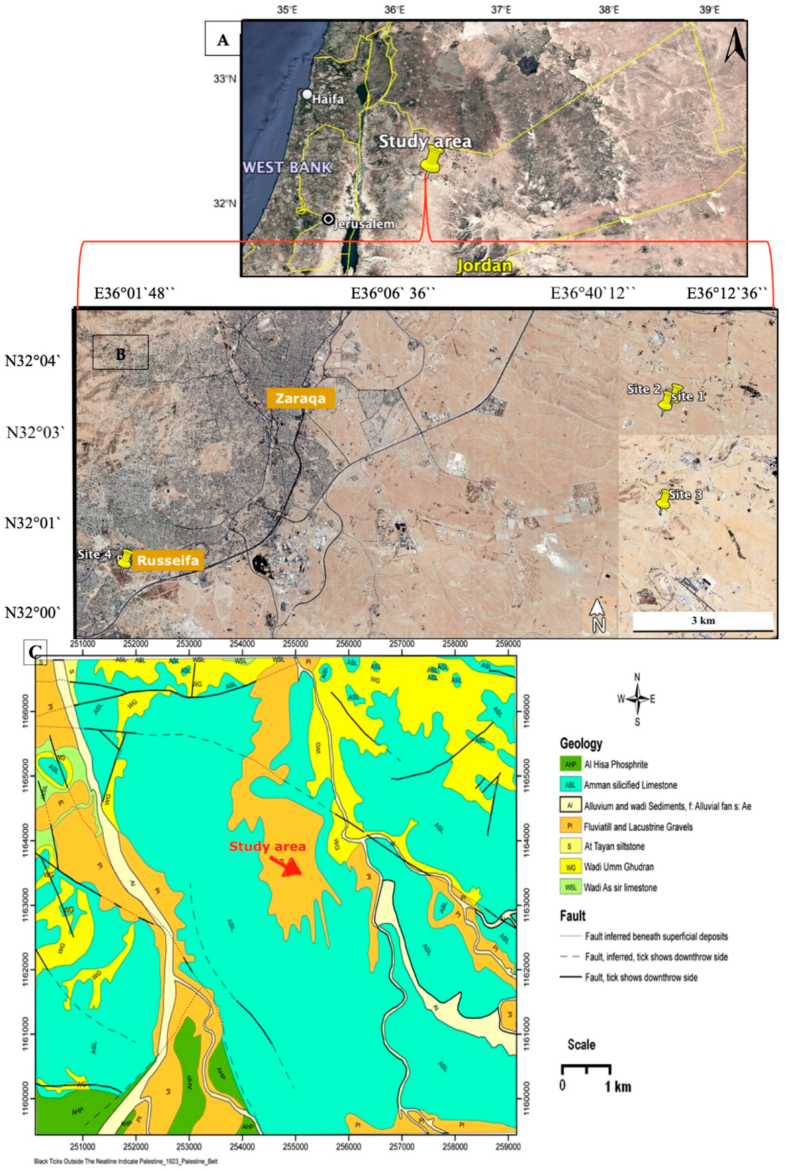
Figure 1. (A) Location map of the study area. (B) Location map of the studied outcrop sites (Google Earth 2025). (C) Geological map of Zarqa city from [19].
Geosciences 15 00433 g001
Figure 2. (A) Field photographs showing hard light crystalline limestone with a thickness of about 120 cm at the bottom, containing calcite veins (2.5 cm). A 20 cm breccia contains dark chert fragments (diameter up to 7 cm) within a phosphate groundmass. (Pen 10 cm). (B) Columnar sections of the outcrops. Legend: see Figure 1B.
Geosciences 15 00433 g002
Figure 3. Field photographs showing (A) Coarse clasts of invertebrate bones and shell fragments. (B) Phosphorite bed (275 cm), hard at the bottom with extra-clasts (dark chert) and invertebrate bones as medium-sized intra-clasts; soft at the top, forming caves. A 2 cm layer of breccia contains dark chert fragments (smaller than 5 cm) within a phosphate groundmass (near arrow). (C) Trace of bioturbation forming the cave ceiling. (D) Phosphorite bed (85 cm), soft to hard at the top, with coarse clasts; near the arrow is a complete fish skeleton. (Hummer 30 cm). Legend: see Figure 1B.
Geosciences 15 00433 g003
Figure 4. (A) Field photographs showing the phosphatic beds and dark chert beds. Some of them are brecciated chert with phosphatic matrix. The layers vary in hardness and color. (Man 184 cm). (B) Columnar sections of the outcrops. Legend: see Figure 1B.
Geosciences 15 00433 g004
Figure 5. (A) Field photographs showing A dark, hard, massive sparitic limestone with a thickness of about 133 cm appears at the bottom, which contains calcite veins. Above this is a 15 cm layer of poorly sorted phosphate (sand size) containing extra- and intra-clasts of oyster shell fragments, calcite, quartz, chert, shells, and shark teeth, as illustrated in the magnified figures. This bed has an incrustation at the top. Overlying this is a massive bed of coquina, composed of fragmented oyster shells and measuring 110 cm in thickness (Hummer 30 cm). (B) Columnar sections of the outcrops. Legend: see Figure 1B.
Geosciences 15 00433 g005
Figure 6. Field photographs showing (A) At the top of the phosphatic bed, incrustation appears near arrows and beneath an accumulation of large, fragmented oyster shells, termed “coquina.” (B) Below this is a medium-hard, fine-grained phosphatic bed (77 cm thick). This is followed by a nodular/globular phosphatic bed (40 cm), which in turn is overlain by alternating layers: 40 cm of thin white Tripoli beds, thin chert beds, and laminated micritic limestone carbonates (Hummer 30 cm). (C) At the base of the third phosphate layer, angular, pebble-sized erosional chert fragments are present, as highlighted in the magnified figure (Hummer 30 cm/Pen 10 cm). Legend: see Figure 1B.
Geosciences 15 00433 g006
Figure 7. Field photographs showing 65 cm thick, light-colored, fine-grained, bioturbated phosphate bed at the base and the top, embedded by clasts of different sizes. The magnified figures show the clasts in soft phosphate. (Hummer 30 cm). Legend: see Figure 1B.
Geosciences 15 00433 g007
Figure 8. Field photographs showing (A) Russifa outcrop showing the sequence of (A3) phosphorite unit (Hummer 30 cm); (B) Visible layer of silicious concretions appears, with very large sizes, above (A3) phosphorite unit; (C,D) Pebbles of size intra-clasts and large invertebrate bones in soft sandy phosphates in the A1 unit. (Pen 10 cm). (E) Columnar sections of the outcrops. Legend: see Figure 1B.
Geosciences 15 00433 g008
Figure 9. Vertical columnar sections of the studied outcrops at Al-Tafeh and Russifa areas.
Geosciences 15 00433 g009
Figure 10. XRD spectrum of (A) sample from site 1 and (B) sample from site 3.
Geosciences 15 00433 g010
Figure 11. Photomicrographs showing (A) pellets (P); (B) bioclasts (BC) of shell fragments, elongated bone fragments (B), sharp teeth fragments (T), rock fragments (RC) of chert, pellets (P), and coated grains (CG); (C) bioclasts (BC) of shell fragments and bone fragments (B); (D) replaced bioclasts (RC) of fossil shells with calcite, coated grains (CG), pellets (P), and elongated bone fragments (B).
Geosciences 15 00433 g011
Table 1. The average concentration of major elements (wt.%) in phosphorites from the study area compared with Jordanian phosphorites and phosphate rocks from other countries.
Elements (wt.%)Study Area
(Al-Tafeh)
Jordanian Phosphorites [20]Djebel Onk
(Algiers) [21]
Khneifiss
(Syria) [21]
Bou Craa
(Morocco) [21]
CaO 52.7833.90–53.9053.2053.4048.40
P2O524.3227.09–34.3429.0028.7029.40
SiO22.290.88–26.232.938.672.66
Al2O3 0.210.08–2.120.510.330.44
Fe2O30.180.07–0.720.640.210.55
MgO 0.310.18–0.611.310.341.01
Na2O 0.320.03–1.591.540.611.09
K2O 0.020.01–0.180.200.100.22
F 3.732.68–4.383.123.533.47
Cl0.0730.070.0120.0640.015
Table 2. The average concentration of trace elements (mg/g) in phosphorites from the study area compared with Jordanian phosphorites and phosphate rocks from other countries.
Elements (mg/g)Study Area
(Al-Tafeh)
Jordanian Phosphorites [23]Djebel Onk
(Algiers) [21]
Khneifiss
(Syria) [21]
Bou Craa
(Morocco) [21]
Ni 0.0190.0180.0150.0240.018
Cu 0.0160.0190.0110.0270.011
Zn0.0990.1490.1610.0300.152
Cd0.0250.0110.0140.0080.145
U0.0450.0900.0410.0720.035
Table 3. Correlation matrix of the elemental composition for phosphorites in the study area.
CaOP2O5SiO2Al2O3Fe2O3MgONa2OK2OCO2ClNiCuZnCdUYLa
CaO1
P2O5−0.90031
SiO2−0.51520.21931
Al2O3−0.33010.1825−0.02281
Fe2O3−0.40130.23950.12420.97491
MgO−0.49000.4038−0.23300.71910.61941
Na2O−0.88570.87050.09370.38390.36960.74261
K2O−0.28820.1636−0.08050.98340.96570.71560.35271
CO20.4826−0.3808−0.0981−0.5089−0.5023−0.5384−0.5095−0.57821
Cl0.2213−0.1942−0.3448−0.2224−0.37210.04310.0424−0.33510.35491
Ni0.6860−0.7110−0.44250.29000.17260.1025−0.54990.29190.27950.09231
Cu−0.04930.2276−0.1067−0.5189−0.5630−0.16040.1259−0.61490.70270.5260−0.12141
Zn−0.37860.37320.6182−0.5483−0.3821−0.51290.0897−0.58330.3586−0.1412−0.72140.42261
Cd0.5615−0.6588−0.1130−0.2599−0.2374−0.2556−0.5514−0.1328−0.0633−0.26680.2301−0.5202−0.15001
U−0.39810.42570.6263−0.3689−0.2004−0.49260.0643−0.42640.4202−0.2324−0.56500.46100.9254−0.38171
Y−0.83640.71150.15940.46640.45510.78110.94790.4395−0.58630.0614−0.5103−0.07070.0297−0.3555−0.06071
La−0.81900.67910.14420.63780.62700.85240.91960.6162−0.6367−0.0458−0.3775−0.1855−0.0926−0.3627−0.12430.97681
Disclaimer/Publisher’s Note: The statements, opinions and data contained in all publications are solely those of the individual author(s) and contributor(s) and not of MDPI and/or the editor(s). MDPI and/or the editor(s) disclaim responsibility for any injury to people or property resulting from any ideas, methods, instructions or products referred to in the content.