
Usefulness of 18F-FDG PET-CT in the Management of Febrile Neutropenia: A Retrospective Cohort from a Tertiary University Hospital and a Systematic Review

 ,
, 
Abstract
1. Introduction
2. Patients and Methods
2.1. Single-Center Retrospective Cohort Study
2.1.1. Design, Study Period, and Subjects
2.1.2. Data Collection
2.1.3. 18F-FDG PET-CT Technique
2.1.4. Other Imaging Techniques
2.1.5. Definitions
- -
- For fever, a single temperature equivalent to ≥38.3 °C orally or equivalent to ≥38.0 °C orally over a 1 h period;
- -
- For neutropenia, ≤500 neutrophils/mcL or ≤1000 neutrophils/mcL and a predicted decrease to ≤500/mcL in the next 48 h.
2.1.6. Usual Care
2.1.7. Data Analysis
2.2. Systematic Literature Review
3. Results
3.1. Single-Center Retrospective Cohort Study
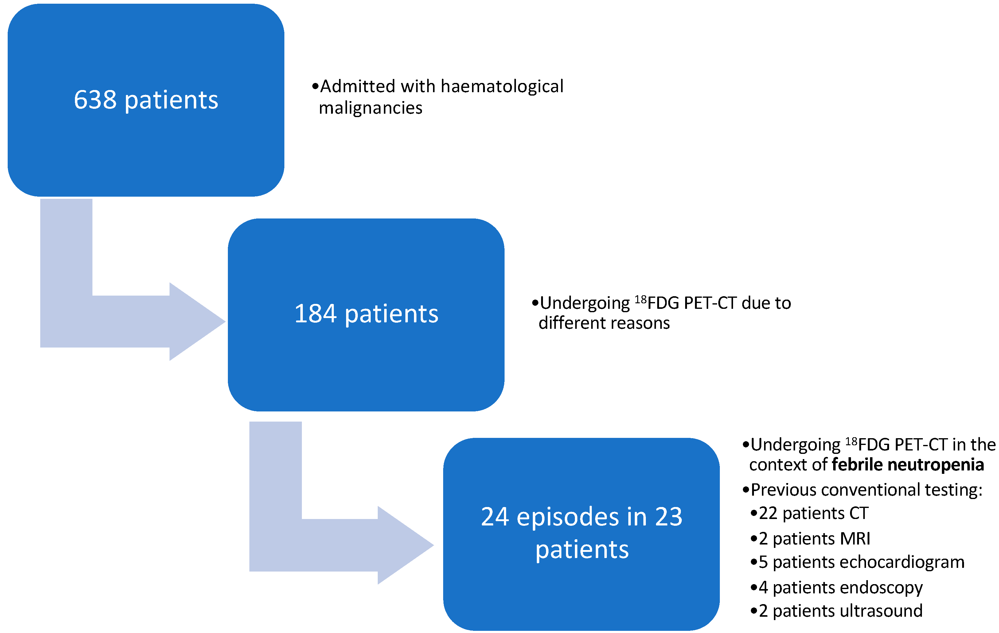
3.1.1. Characteristics of the Patients with FN That Underwent an 18F-FDG PET-CT
3.1.2. Fever Etiology (Table 2)
3.1.3. Characteristics of the 18F-FDG PET-CT as Compared with Conventional Imaging
3.2. Systematic Literature Review
Search Strategy and Inclusion
- Quality appraisal
- Results of the studies according to the methodology (Figure 2)
- Clinical trial
- 2.
- Original articles
- Studies comparing the results of conventional tests and 18F-FDG PET-CT in the same patient

| Authors | Type of Study | Study Population | FN | Compare CT + 18F-FDG-PET-CT | Relevant Results | Final Diagnosis | Limitations |
|---|---|---|---|---|---|---|---|
| Mahfouz T et al. [18] Arkansas (USA) | Retrospective study, 2005 | Multiple myeloma. (A total of 165 infectious episodes were identified in 143 patients with MM; 27 episodes of neutropenia.) | No | No | 18F-FDG-PET-CT in patients with MM identified lesions not detectable by other methods on 46 occasions, determined disseminated infection on 32, helped modify therapies in 55 episodes, and detected 20 clinically relevant silent infections. Guided diagnostic tests: No specific data. | Does not provide specific data of the neutropenic patients | Retrospective. Single center. Myeloma only. No separate data of the 27 cases of neutropenia. Does not compare CT vs. 18F-FDG-PET-CT. |
| Koh KC et al. [17] Australia | Retrosective study (case-control), 2012 | Hematologic malignancies. (100% patients with FN.) Median time from CT compared with 18F-FDG-PET-CT: 4.2 d. | Yes | Yes (in different patients) | CT (n 76) vs. 18F-FDG-PET-CT (n 37) in FN of unknown origin. An underlying cause for FN was determined in 94.6% of cases (18F-FDG-PET-CT), compared with 69.7% of controls (CT). 18F-FDG-PET-CT had a significant impact on antimicrobial utilization compared with conventional imaging (35.1% vs. 11.8%). Guided diagnostic tests: No specific data. | Infection: 67.6% in cases vs. 67.1% control group. IFI: four cases. | Single center. Does not compare CT and 18F-FDG-PET-CT scans in the same patient. |
| Vos FJ et al. [16] Netherlands | Prospective cohort study, 2012 | Hematologic malignancies (AML; MDS) and HSCT (100% with neutropenia, 26 of 28 FN). Mean time from starting chemotherapy to 18F-FDG-PET-CT: 14 d. | Yes | No | 18F-FDG-PET-CT scans were performed on patients with NF and elevated CRP > 50 mg/L. n = 28. Pathological findings were found in 26 cases (18 gastrointestinal, 9 related to CVC, and 7 related to lung). Guided diagnostic tests: yes (ultrasound in case uptake in the CVC tract). | 26/28 FDG uptake of infectious origin. IFI: seven cases. | Single center. Does not compare the performance and findings of CT and 18F-FDG-PET-CT scans in the same patient. |
| Camus V et al. [14] France | Prospective study, 2015 | Hematologic malignancies (AML; ALL) and HSCT (100% FN). Median days of neutropenia: 15. Median days of fever: 14. | Yes | Yes (in the same patient) | Usefulness of 18F-FDG-PET-CT in detecting the source of infection in FN. n = 48. In 31 cases, there was a pathological uptake. In 13, diagnosis of multiple foci/dissemination. Guided diagnostic tests: only in some patients. | Infection: 79%. IFI: three aspergillosis. | Single center. Few patients. They perform CT and 18F-FDG-PET-CT scans in the same patient, but it does not compare them with each other. |
| Guy SD et al. [19] Australia | Prospective study, 2012 | Hematological and solid malignancies. (100% FN.) Median days of neutropenia: 9. Median days of fever: 5–7. | Yes | Yes (in the same patient) | Patients with NF who undergo a 18F-FDG-PET-CT scan in addition to conventional techniques. n = 20. 18F-FDG-PET-CT identified nine infections that CT did not and had a clinical impact in 75% of patients. Compares 18F-FDG-PET-CT and conventional imaging in the same patient and provides individual patient data. Guided diagnostic tests: only in some patients. | Infection: 11/20 patients (55%) | Single center. Few patients. Does not include allogeneic HSCT. |
| Gafter-Gvili A et al. [15] Israel | Prospective study, 2013 | Hematologic malignancies (AML, ALL, lymphoma) and HSCT (100% FN). Median days of neutropenia: 11. Median days of fever: 6. | Yes | Yes (in the same patient) | Use of 18F-FDG-PET-CT in high-risk NF vs. conventional techniques, focused on IFI. n = 79. 18F-FDG-PET-CT changed diagnosis in 69% of patients and management in 55%. Guided diagnostic tests: yes. | 18F-FDG-PET-CT is useful for diagnosis in NF. Infection: 89/117 diagnoses, mainly intra-abdominal infections. IFI: 27 infections. | Single center. Focused on IFI although it also detects other sources of infection. |
| Wang SS et al. [20] | Retrospective study, 2017 | Hematologic malignancies, HSCT and solid malignancies (100% FN) | Yes | Yes (in the same patient) | To assess the impact of 18F-FDG-PET-CT on persistent or recurrent fever. n = 14. In 11 of them (79%), 18F-FDG-PET-CT had a clinical impact: in three, treatment was de-escalated, and in five, antibiotics were discontinued. 18F-FDG-PET-CT scans identified new foci in seven patients. 18F-FDG-PET-CT helped the final diagnosis in 6 out of 10 patients who had a known cause of fever. Guided diagnostic tests: No specific data. | Infection: 8/14 patients (57.1%). IFI: three cases. | Single center. Retrospective. Pediatric patients only. Few patients. Long time until the 18F-FDG-PET-CT scan is performed. |
| Douglas A et al. [3] Australia | Phase 3 randomized 1:1 multicenter clinical trial of CT vs. 18F-FDG-PET-CT/CT, 2022 | Hematologic malignancies and HSCT. (100% FN.) Median days of neutropenia: 12. Median days of fever: 8. Median time from CT compared with 18F-FDG-PET-CT: 5.5 h. | Yes | Yes (in different patients) | Total n = 65 patients in the 18F-FDG-PET-CT group and 69 in the CT group. Antibiotic adjustment occurred 82% in 18F-FDG-PET-CT and 65% in CT, most frequently reducing the spectrum of therapy, in 28 (43%) of 65 patients in the FDG-18F-FDG-PET-CT -CT group compared with 17 (25%) of 69 patients in the CT group. 18F-FDG-PET-CT is useful for adjustment of empiric therapy (cessation or reduction in antimicrobials). Guided diagnostic tests: no specific data. | Infection: microbiologically confirmed 72% in cases vs. 57% in controls (CT). IFI: 6% (vs. 4% in controls). | It does not compare the performance and findings of CT and 18F-FDG-PET-CT scans in the same patient. |
- Studies that do not compare conventional tests and 18F-FDG PET-CT in the same patient
4. Discussion
Supplementary Materials
Author Contributions
Funding
Institutional Review Board Statement
Data Availability Statement
Acknowledgments
Conflicts of Interest
References
- Douglas, A.; Thursky, K.; Slavin, M. New approaches to management of fever and neutropenia in high-risk patients. Curr. Opin. Infect. Dis. 2022, 35, 500–516. [Google Scholar] [CrossRef]
- Lewis, R.E.; Stanzani, M.; Morana, G.; Sassi, C. Radiology-based diagnosis of fungal pulmonary infections in high-risk hematology patients: Are we making progress? Curr. Opin. Infect. Dis. 2023, 36, 250–256. [Google Scholar] [CrossRef]
- Douglas, A.; Thursky, K.; Spelman, T.; Szer, J.; Bajel, A.; Harrison, S.; Tio, S.Y.; Bupha-Intr, O.; Tew, M.; Worth, L.; et al. [(18)F]FDG-PET-CT compared with CT for persistent or recurrent neutropenic fever in high-risk patients (PIPPIN): A multicentre, open-label, phase 3, randomised, controlled trial. Lancet Haematol. 2022, 9, e573–e584. [Google Scholar] [CrossRef]
- Douglas, A.; Lau, E.; Thursky, K.; Slavin, M. What, where and why: Exploring fluorodeoxyglucose-PET’s ability to localise and differentiate infection from cancer. Curr. Opin. Infect. Dis. 2017, 30, 552–564. [Google Scholar] [CrossRef]
- Gutierrez, A.; Rodriguez, B.; Velasquez, K.; Gutierrez, I.; Garcia, S.; Munez, E.; Calderon-Parra, J.; Callejas-Diaz, A.; Ramos-Martinez, A.; Fernandez-Cruz, A. Determining the usefulness of systematic 18F-FDG PET/CT for the management of invasive fungal infection (PETIFI project): A prospective national multicentre cohort study protocol. BMJ Open 2023, 13, e074240. [Google Scholar] [CrossRef]
- Hess, S. FDG-PET/CT in Fever of Unknown Origin, Bacteremia, and Febrile Neutropenia. PET Clin. 2020, 15, 175–185. [Google Scholar] [CrossRef] [PubMed]
- Bleeker-Rovers, C.P.; Vos, F.J.; van der Graaf, W.T.; Oyen, W.J. Nuclear medicine imaging of infection in cancer patients (with emphasis on FDG-PET). Oncologist 2011, 16, 980–991. [Google Scholar] [CrossRef] [PubMed]
- Vos, F.J.; Bleeker-Rovers, C.P.; Oyen, W.J.G. The use of FDG-PET/CT in patients with febrile neutropenia. Semin. Nucl. Med. 2013, 43, 340–348. [Google Scholar] [CrossRef] [PubMed]
- Contejean, A.; Maillard, A.; Canoui, E.; Kerneis, S.; Fantin, B.; Bouscary, D.; Parize, P.; Garcia-Vidal, C.; Charlier, C. Advances in antibacterial treatment of adults with high-risk febrile neutropenia. J. Antimicrob. Chemother. 2023, 78, 2109–2120. [Google Scholar] [CrossRef] [PubMed]
- Boellaard, R.; Delgado-Bolton, R.; Oyen, W.J.; Giammarile, F.; Tatsch, K.; Eschner, W.; Verzijlbergen, F.J.; Barrington, S.F.; Pike, L.C.; Weber, W.A.; et al. FDG PET/CT: EANM procedure guidelines for tumour imaging: Version 2.0. Eur. J. Nucl. Med. Mol. Imaging 2015, 42, 328–354. [Google Scholar] [CrossRef] [PubMed]
- Jamar, F.; Buscombe, J.; Chiti, A.; Christian, P.E.; Delbeke, D.; Donohoe, K.J.; Israel, O.; Martin-Comin, J.; Signore, A. EANM/SNMMI guideline for 18F-FDG use in inflammation and infection. J. Nucl. Med. 2013, 54, 647–658. [Google Scholar] [CrossRef]
- NCCN Clinical Practice Guidelines in Oncology. Prevention and Treatment of Cancer-Related Infections, Version 2. 2023. Available online: https://www.nccn.org/guidelines/guidelines-detail?category=3&id=1457 (accessed on 26 January 2024).
- Higgins, J.P.T.; Chandler, J.; Cumpston, M.; Li, T.; Page, M.J.; Welch, V.A. Cochrane Handbook for Systematic Reviews of Interventions, 2nd ed.; John Wiley & Sons: Chichester, UK, 2019. [Google Scholar]
- Camus, V.; Edet-Sanson, A.; Bubenheim, M.; Hitzel, A.; Becker, S.; David, M.; Stamatoullas, A.; Lenain, P.; Jardin, F.; Contentin, N.; et al. 18F-FDG-PET/CT imaging in patients with febrile neutropenia and haematological malignancies. Anticancer Res. 2015, 35, 2999–3006. [Google Scholar]
- Gafter-Gvili, A.; Paul, M.; Bernstine, H.; Vidal, L.; Ram, R.; Raanani, P.; Yeshurun, M.; Tadmor, B.; Leibovici, L.; Shpilberg, O.; et al. The role of 18F-FDG PET/CT for the diagnosis of infections in patients with hematological malignancies and persistent febrile neutropenia. Leuk. Res. 2013, 37, 1057–1062. [Google Scholar] [CrossRef] [PubMed]
- Vos, F.J.; Donnelly, J.P.; Oyen, W.J.G.; Kullberg, B.J.; Bleeker-Rovers, C.P.; Blijlevens, N.M.A. 18F-FDG PET/CT for diagnosing infectious complications in patients with severe neutropenia after intensive chemotherapy for haematological malignancy or stem cell transplantation. Eur. J. Nucl. Med. Mol. Imaging 2012, 39, 120–128. [Google Scholar] [CrossRef] [PubMed]
- Koh, K.C.; Slavin, M.A.; Thursky, K.A.; Lau, E.; Hicks, R.J.; Drummond, E.; Wong, P.S.; Worth, L.J. Impact of fluorine-18 fluorodeoxyglucose positron emission tomography on diagnosis and antimicrobial utilization in patients with high-risk febrile neutropenia. Leuk. Lymphoma 2012, 53, 1889–1895. [Google Scholar] [CrossRef]
- Mahfouz, T.; Miceli, M.H.; Saghafifar, F.; Stroud, S.; Jones-Jackson, L.; Walker, R.; Grazziutti, M.L.; Purnell, G.; Fassas, A.; Tricot, G.; et al. 18F-fluorodeoxyglucose positron emission tomography contributes to the diagnosis and management of infections in patients with multiple myeloma: A study of 165 infectious episodes. J. Clin. Oncol. 2005, 23, 7857–7863. [Google Scholar] [CrossRef] [PubMed]
- Guy, S.D.; Tramontana, A.R.; Worth, L.J.; Lau, E.; Hicks, R.J.; Seymour, J.F.; Thursky, K.A.; Slavin, M.A. Use of FDG PET/CT for investigation of febrile neutropenia: Evaluation in high-risk cancer patients. Eur. J. Nucl. Med. Mol. Imaging 2012, 39, 1348–1355. [Google Scholar] [CrossRef] [PubMed]
- Wang, S.S.; Mechinaud, F.; Thursky, K.; Cain, T.; Lau, E.; Haeusler, G.M. The clinical utility of fluorodeoxyglucose-positron emission tomography for investigation of fever in immunocompromised children. J. Paediatr. Child. Health 2018, 54, 487–492. [Google Scholar] [CrossRef] [PubMed]
- Madney, Y.; Shalaby, L.; Elanany, M.; Adel, N.; Nasr, E.; Alsheshtawi, K.; Younes, A.; Hafez, H. Clinical features and outcome of hepatosplenic fungal infections in children with haematological malignancies. Mycoses 2020, 63, 30–37. [Google Scholar] [CrossRef] [PubMed]
- Leroy-Freschini, B.; Treglia, G.; Argemi, X.; Bund, C.; Kessler, R.; Herbrecht, R.; Imperiale, A. 18F-FDG PET/CT for invasive fungal infection in immunocompromised patients. QJM 2018, 111, 613–622. [Google Scholar] [CrossRef]
- Gutiérrez-Martín, I.; García-Prieto, S.; Velásquez, K.; Gutiérrez-Abreu, E.V.; Diego-Yagüe, I.; Calderón-Parra, J.; Gutiérrez-Villanueva, A.; Ramos-Martínez, A.; Múñez-Rubio, E.; Callejas-Díaz, A.; et al. Usefulness of 18 F-FDG PET-CT for the management of invasive fungal infections: A retrospective cohort from a tertiary university hospital. Mycoses 2024, in press.
- Douglas, A.P.; Thursky, K.A.; Worth, L.J.; Harrison, S.J.; Hicks, R.J.; Slavin, M.A. FDG-PET/CT in managing infection in patients with hematological malignancy: Clinician knowledge and experience in Australia. Leuk. Lymphoma 2019, 60, 2471–2476. [Google Scholar] [CrossRef] [PubMed]

| P (population): Patients of all ages with febrile neutropenia in the setting of oncohematologic malignancy |
| I (intervention): 18F-FDG PET-CT in the setting of febrile neutropenia management |
| C (comparison): Conventional diagnostic tests used for febrile neutropenia workup |
| O (outcome): 18F-FDG PET-CT added value in diagnosing the cause of fever in patients with febrile neutropenia: * Final diagnosis: infection, alternative non-infectious causes of fever ruling out infection * Directed targeted diagnostic tests * Antimicrobial rationalization (discontinuation, de-escalation, or escalation of antimicrobials) * Resumption of therapy of the underlying disease |
| S (study design): RCTs; prospective or retrospective cohort studies, case control studies, case reports yield |
| Characteristic | Number (%) | |
|---|---|---|
| Number of patients | 23 | |
| Number of FN episodes | 24 | |
| Sex | ||
| Male | 13 (57%) | |
| Female | 10 (43%) | |
| Age (years) (median (IQR)/mean) | 59 (47.5–74.5)/58.6 | |
| Underlying non-oncological/haematological disease | ||
| Coronary artery disease | 0 | |
| Heart failure | 0 | |
| Peripheral arterial disease | 0 | |
| Stroke | 0 | |
| Dementia | 0 | |
| Hemiplegia | 0 | |
| COPD | 1 (4%) | |
| Diabetes | 4 (17%) | |
| Diabetes with target organ involvement | 0 | |
| Moderate–severe kidney disease | 2 (8%) | |
| Mild–moderate liver disease | 0 | |
| Severe liver disease | 0 | |
| Ulcer disease | 0 | |
| Connective tissue disease | 0 | |
| HIV-AIDS | 0 | |
| Onco-hematological disease | ||
| Acute myeloid leukemia | 12 (50%) | |
| Acute lymphoblastic leukemia | 4 (17%) | |
| Multiple myeloma | 3 (12%) | |
| Myelodysplastic syndrome | 3 (12%) | |
| Others (NK immunodeficiency) | 1 (4%) | |
| Lymphoma | 0 | |
| Stem cell transplantation | 4 (17%) | |
| Allogenic SCT | 1 (4%) | |
| HLA-haploidentical SCT | 2 (8%) | |
| Autologous SCT | 1 (4%) | |
| Umbilical cord SCT | 0 | |
| GVHD post-SCT | 2 (8%) | |
| Recent surgery (<3 months) | 3 (12%) | |
| Neutrophil counts on the day of 18F-FDG PET-CT | ||
| <100 /mcL | 12 (50%) | |
| 100–500/mcL | 10 (42%) | |
| 500–1500/mcL | 2 (8%) | |
| Fever | ||
| Persistent low-grade fever (>72 h) | 8 (33%) | |
| Persistent fever (>72 h) | 14 (58%) | |
| Relapsing fever | 1 (4%) | |
| Persistent–recurrent fever | 1 (4%) | |
| Median (IQR)/mean days of neutropenia before 18F-FDG PET-CT | 13.5 (3–74)/19.3 | |
| Median (IQR)/mean days of fever before 18F-FDG PET-CT | 13 (3–28)/13.7 | |
| Median(IQR)/mean days from conventional test to 18F-FDG PET-CT | 13 (1–29)/12.9 | |
| Median(IQR)/mean duration of antibiotic therapy until 18F-FDG PET-CT | 13.5 (3–30)/14 | |
| Sepsis prior to 18F-FDG PET-CT (3 days) | 2 (8%) | |
| Antimicrobial prophylaxis | ||
| Levofloxacin | 15 (62%) | |
| Azithromycin | 2 (8%) | |
| Cotrimoxazole | 5 (21%) | |
| Acyclovir | 11 (46%) | |
| Letermovir | 0 | |
| Entecavir | 1 (4%) | |
| Fluconazole | 1 (4%) | |
| Posaconazole | 15 (62%) | |
| Pentamidine | 1 (4%) | |
| Valganciclovir | 1 (4%) | |
| Empiric treatment pre-18F-FDG PET-CT | ||
| Cefepime | 5 (21%) | |
| Piperacillin/tazobactam | 9 (37%) | |
| Meropenem | 13 (54%) | |
| Vancomicyn | 4 (17%) | |
| Teicoplanin | 11 (46%) | |
| Echinocandin | 0 (0%) | |
| Voriconazole/isavuconazole | 1 (4%) | |
| Ambisome | 3 (12%) | |
| Others | 3 (12%) | |
| Reference test 18F-FDG PET-CT | ||
| Body CT | 16 (67%) | |
| Chest CT | 4 (17%) | |
| Abdominal CT | 3 (12%) | |
| Sinus CT | 2 (8%) | |
| MRI | 2 (8%): hepatic 1; spinal 1. | |
| Echocardiogram (TTE) | 3 (12%) | |
| Echocardiogram (TEE) | 2 (8%) | |
| Endoscopy | 2 (8%) | |
| Bronchoscopy | 2 (8%) | |
| Abdominal ultrasound | 2 (8%) | |
| Location of the 18F-FDG PET-CT uptake | ||
| Lung | 5 (21%) | |
| Brain | 0 | |
| Skin | 2 (8%) | |
| Muscle | 0 | |
| Visceral intra-abdominal | 9 (37%) | |
| Non-visceral intra-abdominal | 1 (4%) | |
| Ocular | 0 | |
| Kidney | 0 | |
| Endocarditis | 0 | |
| Endovascular/cardiac devices | 1 (4%) | |
| Bone marrow | 5 (21%) | |
| Others | 5 (21%) | |
| Type of uptake | ||
| Focal | 3 (12%) | |
| Multifocal | 17 (71%) | |
| No uptake | 4 (17%) | |
| Studies induced by 18F-FDG PET-CT result | 14 (58%) | |
| CT | 2 (8%) | |
| MRI | 0 | |
| Echocardiogram | 0 | |
| Ultrasound | 3 (12%) | |
| Endoscopy | 3 (12%) | |
| Bronchoscopy | 1 (4%) | |
| Biopsy/fine needle aspiration | 4 (17%) | |
| Other | 1 (4%) | |
| Source control of infection | ||
| Catheter removal | 1 (4%) | |
| Drain | 0 | |
| Surgery | 1 (4%) | |
| Others | 0 | |
| Antimicrobial treatment modifications | ||
| Spectrum change (total) | 16 (67%) | |
| Reduced spectrum | 2 (8%) | |
| Extended spectrum | 1 (4%) | |
| Started new treatment | 3 (12%) | |
| Antibiotic discontinuation | 10 (42%) | |
| Final diagnosis of infection | ||
| Bacterial | 2 (8%) | |
| Mycobacterial | 0 | |
| Fungal | 2 (8%) | |
| Viral | 0 | |
| Parasitic | 1 (4%) | |
| Clinical diagnosis without microbiological isolation | 1 (4%) | |
| Site of infection | ||
| Catheter | 1 (4%) | |
| Cardiac | 0 | |
| Pulmonary | 1 (4%) | |
| Urinary/genital | 0 | |
| Hepatic/biliary/splenic | 2 (8%) | |
| Intestinal/perianal/oral | 0 | |
| Sinusitis | 1 (4%) | |
| Skin and soft tissues | 0 | |
| Surgical site infection | 0 | |
| Non-infectious ethiology of fever | ||
| Engraftment syndrome | 3 (12%) | |
| Underlying hematological disease | 11 (46%) | |
| Inflammatory | 1 (4%) | |
| Gastric graft-versus-recipient disease | 2 (8%) | |
| Febrile neutropenia of unknown origin | 3 (12%) | |
| Added value of 18F-FDG PET-CT | ||
| Diagnosis of new site of infection | 4 (17%) | |
| Infection exclusion | 16 (67%) | |
| Antibiotic modification | 16 (67%) | |
| Induce other tests | 10 (42%) | |
| Allows starting chemotherapy or immunomodulators | 4 (17%) | |
| Discharge status (23 patients) | ||
| Alive | 14 (61%) | |
| Death | 9 (39%) | |
| Reference Conventional Imaging | Conventional Imaging Result | 18F-FDG PET-CT Result | 18F-FDG PET-CT vs. Conventional | Antimicrobial Therapy Modification | Induces New Diagnostic Tests | Final Diagnosis | Added Value | |
|---|---|---|---|---|---|---|---|---|
| Occult Lesions | Dissemination | |||||||
| Full-body CT scan (11 October 2019) | CT: small intestine graft-versus-recipient disease | PET-CT (29 October 2019): Pathological uptake at gastroesophageal junction with gastric extension. Gallbladder uptake. | PET > CT PET scans show gastric uptake localized gastroesophageal junction + gallbladder | No | No | Ultrasound + gastroscopy with biopsies | Gastric graft-versus-recipient disease grade 4 and cholecystitis | Yes Occult lesion (colecistitis) |
| Chest-abdomen CT scan (17 April 2020) Liver MRI (18 April 2020) | CT: Decreased bilateral pulmonary nodules. MRI: Hepatic iron overload. | PET-CT (6 May 2020): Bone uptake secondary to MM (underlying disease) + uptake in the right thyroid lobe | PET > CT: Thyroid lobe was not visible on CT | No | Withdrawal of antibiotic treatment | Thyroid ultrasound | Tumor-related fever (multiple myeloma progression). Subclinical hyperthyroidism, BMN with right dominant thyroid nodule, TIRADS 3. | Yes Occult lesion Rule out infection Unveils tumoral etiology |
| Facial-sinus CT (30 April 2019) Abdominal CT scan (16 May 2019) | Sinus CT: Periodontal disease. No osteomyelitis. Abdominal CT: No urgent pathology. | PET-CT (17 May 2019): Retroperitoneal soft tissue uptake. Left pulmonary nodule without pathological uptake. | PET > CT Retroperitoneal uptake | No | No | Endoscopic ultrasound with biopsy of retroperitoneal lesions | Tumor-related fever (refractory AML). Disease progression with hepatic infiltration. | Yes Occult lesion Rule out infection Unveils tumoral etiology |
| Full-body CT scan (30 November 2020) | CT scan: Three splenic SOLs smaller than 10 mm, suggestive of infection | PET CT (15 December 2020): Splenic lesions without increased uptake | PET > CT Rules out infectious origin | No | Withdrawal of antibiotic treatment | Control abdominal ultrasound of splenic lesions | Tumor fever (acute biphenotypic leukemia, second relapse 3 m after second allogeneic transplant) | Yes Rule out infection Antibiotic discontinuation Induces initiation of hematological treatment |
| Full-body CT scan (13 April 2022) | Body CT: Decrease in bilateral pulmonary nodules consistent with IFI. Appearance of peribronchovascular GGO in LSD, suggestive of infection. Splenomegaly of 16 cm. | PET-CT (3 May 2022): Bilateral pulmonary nodules consistent with IFI. Splenomegaly without pathological uptake. Thyroid uptake. | PET = CT Confirm infection | No | Initiates amphotericin B | Bronchoscopy | Possible pulmonary IFI | Yes Antifungal started based on 18FDG PET-CT results Diagnosis of infectious source |
| Transesophageal echocardiogram (14 October 2019) Full-body CT scan (1 October 2019) | TEE: No signs of endocarditis. CT: Bilateral pleural effusion. Mixed patchy lung consolidations, pulmonary edema vs. infectious/inflammatory. | PET-CT (30 October 2019): Bilateral pulmonary consolidations with pathological uptake, especially those located in LSI, without clear superinfection, possible pulmonary edema. No cardiac uptake. | PET = CT Rules out lung infection and endocarditis | No | No | No | Bacteremia caused by S. haemolyticus in relation to central catheter, without local complications or endocarditis | No |
| Full-body CT scan (17 October 2022) | CT: Nonspecific pulmonary consolidations. Left hydropneumothorax. | PET-CT (8 November 2022): Uncomplicated left pleural effusion. Progression of hematologic disease. | PET > TC Rules out infection | No | No | No | Tumor-related fever. Graft failure. Relapse of hematologic disease (AML). | Yes Rules out infection Unveils tumoral etiology Initiates chemotherapy for relapse after HCST |
| Colonoscopy and panendoscopy (19 July 2018) No recent CT scan | Colonoscopy and panendoscopy with colitis and ileitis. Biopsies with GVHD. | PET-TC (7 August 2018): Diffuse intestinal uptake | Not applicable | No | No | No | Cutaneous and intestinal GVHD | Yes Rules out infection Initiates immunosuppression treatment for GVHD |
| Full-body CT scan (14 October 2019) | CT: No pathologic findings | PET-CT (19 November 2019): Diffuse bone marrow uptake. No pathological uptake. | PET > CT Rules out infection | No | De-escalation of antibiotic therapy | No | Tumor-related fever origin (newly diagnosed AML, refractory disease) | Yes Rule out infection De-escalate antibiotics |
| Chest CT (15 December 2018) Bronchoscopy (21 December 2018) Full-body CT scan (29 December 2018) | Chest CT: Patchy lesions of probable infectious etiology. BAL: No microbiological isolates. CT body: Bilateral peribronchovascular pulmonary micronodular involvement of the bronchiolitis type. | PET-CT (2 January 2019): Mandibular uptake and bilateral laterocervical lymphadenopathy in keeping with underlying process. Infectious pulmonary findings in resolution. | PET-CT > CT. Rule out hidden infection. Confirms improving previous lung infection. | No | Withdrawal of antibiotic therapy | No | Tumor-related fever (refractory AML) | Yes Confirms good response De-escalate antibiotics Unveils tumoral etiology |
| Full-body CT scan (20 December 2015) | CT body: radiological improvement of pulmonary infiltrates and hepatic SOLs | PET-CT (21 December 2015): Uptake of multiple hepatic SOL and abdominal lymphadenopathy | PET-CT > CT Rule out infectious complications | No | Withdrawal of antibiotic therapy | No | Previous diagnosis of systemic infection by atypical mycobacteria 10/2015. He was readmitted due to fever without a source. Diagnosis: fever of inflammatory origin (in relation to corticosteroid decrease). | Yes Discontinue antibiotics assuming inflammatory cause Rule out infection Increased corticosteroid doses |
| Chest CT (23 June 2022) Abdomen CT (29 June 2022) | Chest CT: Isolated centrolobular opacities in LII, probably inflammatory in nature. Abdomen CT: No findings. | PET-CT (30 June 2022): No pathological uptakes | PET-CT > CT Rule out infection | No | Withdrawal of antibiotic therapy | No | Tumor-related fever (AML progression) | Yes Rule out infection Antibiotic discontinuation |
| Full-body CT scan (29 June 2020) | CT: Normal chest. Renal angiomyolipomas. Left adrenal adenoma. | PET-CT (13 July 2020): Uptake in the left upper mola, inflammatory. No other pathological uptakes. | PET-CT > CT Rule out infection | No | De-escalation of antibiotic therapy | No | Fever in relation to neutrophil recovery | Yes Rule out infection Antibiotic de-escalation |
| Transesophageal echocardiogram (14 May 2021) Full-body CT scan (19 May 2021) Left upper limb Doppler ultrasound (21 May 2023) | TEE: No endocarditis. CT: Typhlitis. No pulmonary involvement. Doppler ultrasound: Postphlebitic axillary vein changes in relation to previous catheter. | PET-CT (28 May 2021): Active process in the nostril, of probable infectious origin. Cecum uptake in relation to typhlitis. Focal colon-sigma uptake of inflammatory vs. tumor etiology. | PET-CT > TC. Unveils more occult lesions (nasal, larger colonic disease) than CT | No | Increase in dose of amphotericin B and voriconazole is associated. Surgical debridement. | Colonoscopy: Colitis. Sinus CT: No complications. Periodontal disease. Evaluation by otolaryngologist: Biopsy. Positive culture for Fusarium. | Invasive fusariosis. Persistent bacteremia due to E. faecium, in relation to CVC without distant complications on PET. AML refractory to chemotherapy. | Yes Diagnosis of infectious source |
| Full-body CT scan (13 July 2021) Spinal MRI (15 July 2021) | CT body: Indeterminate scattered bone lesions. MR: Extensive changes in intraosseous marrow in relation to rapid bone loss or remodeling from treatments. Two areas of focal hypointensity, sclerose on CT, indeterminate, not possible to specify aggressiveness. | PET (20 July 2021): Pathological and diffuse uptake in bone marrow and lymph nodes at multiple levels in relation to underlying disease | PET-CT > CT Rule out infection | No | Withdrawal of antibiotic therapy | No | Tumor-related fever (ALL relapse) | Yes Rule out infection Antibiotic discontinuation |
| Full-body CT scan (12 April 2017) | CT: Pulmonary edema. Mild ascites. Mild pleural effusion. | PET-CT (19 April 2017): No pathological uptakes | PET-CT > CT Rule out infection | No | Withdrawal of antibiotic therapy | No | Tumor-related fever (progressive myelodysplastic syndrome) | Yes Rule out infection Antibiotic discontinuation |
| Transthoracic echocardiogram (19 May 2017) Abdominal ultrasound (20 May 2017) | TTE: No relevant findings. US: Hepatomegaly. No other findings. | PET-CT (1 June 2017): Uptake in front of both psoas, more on the right side, which translates into an active process (tumor vs. infectious/inflammatory) | PET > ultrasound | No | No | Abdominal CT scan (21 June 2017): No evidence of biopsy-eligible lesions | Febrile neutropenia of unknown origin | No |
| Full-body CT scan (8 February 2022) Bronchoscopy (9 February 2022) | CT: Small peripheral pulmonary infiltrates in RUL and RI | PET-CT (18 February 2022): Multiple diffuse pathological uptakes in subcutaneous tissue. Intense uptake in the subcutaneous tract of the right supraclavicular CVC without reaching vascular territory. Pulmonary infiltrates in RUL and right base, without pathological uptake. | PET-CT > CT Detects subcutaneous pathological uptake not observed on CT scan | No | Removal of CVC. Withdrawal of antibiotic therapy. | Skin biopsy: neutrophilic lobular panniculitis | Febrile neutropenia of unknown origin (probable inflammatory vs. tumor origin due to refractory AML) | Yes Rule out infection Antibiotic discontinuation Catheter removal |
| Chest CT (16 December 2021) Transthoracic echocardiogram (17 December 2021) | CT: Mild bilateral acinar opacities (edema vs. infectious-inflammatory). TTE: No findings. | PET-CT (18 December 2021): Very mild diffuse pulmonary uptake of dubious significance | PET-CT > CT Rule out infection | No | Withdrawal of antibiotic therapy | No | Fever in relation to engraftment syndrome | Yes Rule out infection Antibiotic discontinuation |
| Full-body CT scan (18 October 2021) | CT: Splenomegaly. Hepatomegaly with simple cysts. Bilateral adrenal thickening. Chronic bronchopulmonary disease. | PET-CT (24 October 2021): Splenomegaly with two foci with pathological uptake suggestive of splenic infarctions | PET-CT > CT Rule out infection | No | Withdrawal of antibiotic therapy | No | Tumor-related fever (AML) | Yes Rule out infection Antibiotic discontinuation |
| Full-body CT scan (10 February 2022) | CT: Mild pericardial effusion. Mild bilateral pleural effusion. Mild hepatosplenomegaly. | PET-CT (15 February 2022): Bone uptake suggestive of malignancy. Pericardial effusion without pathological uptake. Focal uptake in the left colon showing mild inflammatory origin associated with diverticulum. | PET-CT > CT Rule out infection | No | Withdrawal of antibiotic therapy | No | Febrile neutropenia of unknown origin (probable inflammatory vs. tumor origin due to refractory acute myeloid leukemia) | Yes Rule out infection Antibiotic discontinuation |
| Sinus CT (25 May 2022) Full-body CT scan (26 May 2022) Transthoracic echocardiogram (10 June 2022) | Sinus CT: Periorbital and soft tissue edema of the bilateral supratemporal fossa. CT body: Discrete continuous concentric parietal thickening of the colon suggestive of nonspecific colitis. ETT: No findings. | PET-CT (7 June 2022): Diffuse pancreatic uptake suggestive of inflammation. Pulmonary edema. | PET-CT > CT Rule out infection. Mild pancreatitis possible. | No | No | No | Tumor-related fever (newly diagnosed acute myeloid leukemia) | No |
| Chest CT (26 July 2022) Abdomen CT (27 July 2022) | CT thorax: Bibasal subsegmental atelectasis. CT scan of the abdomen: Slight parietal thickening of the colon suggestive of nonspecific colitis. | PET-CT (8 August 2022): No pathological uptake | PET-CT > CT Rule out infection | No | No | No | Febrile neutropenia with probable source mucositis vs. engraftment syndrome | No |
| Full-body CT scan (27 December 2022) | CT body: Homogeneous splenomegaly. Rest without significant findings. | PET-CT (1 January 2023): Splenomegaly with high-intensity diffuse uptake | PET-CT > CT New suspected source of infection | No | Initiation of amphotericin B | PCR leishmania in blood and bone marrow biopsy review | Visceral leishmaniasis | Yes Diagnosis of infectious source |
| Full-body CT scan (11 October 2019) | CT: small intestine graft-versus-recipient disease | PET-CT (29 October 2019): Pathological uptake at gastroesophageal junction with gastric extension. Gallbladder uptake. | PET > CT PET scans show gastric uptake localized gastroesophageal junction + gallbladder | No | No | Ultrasound + gastroscopy with biopsies | Gastric graft-versus-recipient disease grade 4 and cholecystitis | Yes Occult lesion (colecistitis) |
| Chest-abdomen CT scan (17 April 2020) Liver MRI (18 April 2020) | CT: Decreased bilateral pulmonary nodules. MRI: Hepatic iron overload. | PET-CT (6 May 2020): Bone uptake secondary to MM (underlying disease) + uptake in the right thyroid lobe | PET > CT Thyroid lobe was not visible on CT | No | Withdrawal of antibiotic treatment | Thyroid ultrasound | Tumor-related fever (multiple myeloma progression). Subclinical hyperthyroidism, BMN with right dominant thyroid nodule, TIRADS 3. | Yes Occult lesion Rule out infection Unveils tumoral etiology |
| Facial-sinus CT (30 April 2019) Abdominal CT scan (16 May 2019) | Sinus CT: Periodontal disease. No osteomyelitis. Abdominal CT: No urgent pathology. | PET-CT (17 May 2019): Retroperitoneal soft tissue uptake. Left pulmonary nodule without pathological uptake. | PET > CT Retroperitoneal uptake | No | No | Endoscopic ultrasound with biopsy of retroperitoneal lesions | Tumor-related fever (refractory AML). Disease progression with hepatic infiltration. | Yes Occult lesion Rule out infection Unveils tumoral etiology |
| Full-body CT scan (30 November 2020) | CT scan: Three splenic SOLs smaller than 10 mm, suggestive of infection. | PET CT (15 December 2020): Splenic lesions without increased uptake. | PET > CT Rules out infectious origin | No | Withdrawal of antibiotic treatment | Control abdominal ultrasound of splenic lesions | Tumor fever (acute biphenotypic leukemia, second relapse 3 m after second allogeneic transplant) | Yes Rule out infection Antibiotic discontinuation Induces initiation of hematological treatment |
| Full-body CT scan (13 April 2022) | Body CT: Decrease in bilateral pulmonary nodules consistent with IFI. Appearance of peribronchovascular GGO in LSD, suggestive of infection. Splenomegaly of 16 cm. | PET-CT (3 May 2022): Bilateral pulmonary nodules consistent with IFI. Splenomegaly without pathological uptake. Thyroid uptake. | PET = CT Confirm infection | No | Initiates amphotericin B | Bronchoscopy | Possible pulmonary IFI | Yes Antifungal started based on 18FDG PET-CT results Diagnosis of infectious source |
| Transesophageal echocardiogram (14 October 2019) Full-body CT scan (1 October 2019) | TEE: No signs of endocarditis.CT: Bilateral pleural effusion. Mixed patchy lung consolidations, pulmonary edema vs. infectious/inflammatory. | PET-CT (30 October 2019): Bilateral pulmonary consolidations with pathological uptake, especially those located in LSI, without clear superinfection, possible pulmonary edema. No cardiac uptake. | PET = CT Rules out lung infection and endocarditis | No | No | No | Bacteremia caused by S. haemolyticus in relation to central catheter, without local complications or endocarditis | No |
| Full-body CT scan (17 October 2022) | CT: Nonspecific pulmonary consolidations. Left hydropneumothorax. | PET-CT (8 November 2022): Uncomplicated left pleural effusion. Progression of hematologic disease. | PET > TC Rules out infection | No | No | No | Tumor-related fever. Graft failure. Relapse of hematologic disease (AML). | Yes Rules out infection Unveils tumoral etiology Initiates chemotherapy for relapse after HCST |
| Colonoscopy and panendoscopy (19 July 2018) No recent CT scan | Colonoscopy and panendoscopy with colitis and ileitis. Biopsies with GVHD. | PET-TC (7 August 2018): Diffuse intestinal uptake | Not applicable | No | No | No | Cutaneous and intestinal GVHD | Yes Rules out infection Initiates immunosuppression treatment for GVHD |
| Full-body CT scan (14 October 2019) | CT: No pathologic findings | PET-CT (19 November 2019): Diffuse bone marrow uptake. No pathological uptake. | PET > CT Rules out infection | No | De-escalation of antibiotic therapy | No | Tumor-related fever origin (newly diagnosed AML; refractory disease) | Yes Rule out infection De-escalate antibiotics |
| Chest CT (15 December 2018) Bronchoscopy (21 December 2018) Full-body CT scan (29 December 2018) | Chest CT: Patchy lesions of probable infectious etiology. BAL: No microbiological isolates. CT body: Bilateral peribronchovascular pulmonary micronodular involvement of the bronchiolitis type. | PET-CT (2 January 2019): Mandibular uptake and bilateral laterocervical lymphadenopathy in keeping with underlying process. Infectious pulmonary findings in resolution. | PET-CT > CT. Rule out hidden infection. Confirms improving previous lung infection. | No | Withdrawal of antibiotic therapy | No | Tumor-related fever (refractory AML) | Yes Confirms good response De-escalate antibiotics Unveils tumoral etiology |
| Full-body CT scan (20 December 2015) | CT body: Radiological improvement of pulmonary infiltrates and hepatic SOLs | PET-CT (21 December 2015): Uptake of multiple hepatic SOL and abdominal lymphadenopathy | PET-CT > CT Rule out infectious complications | No | Withdrawal of antibiotic therapy | No | Previous diagnosis of systemic infection by atypical mycobacteria 10/2015. He was readmitted due to fever without a source. Diagnosis: Fever of inflammatory origin (in relation to corticosteroid decrease). | Yes Discontinue antibiotics assuming inflammatory cause Rule out infection Increased corticosteroid doses |
| Chest CT (23 June 2022) Abdomen CT (29 June 2022) | Chest CT: Isolated centrolobular opacities in LII, probably inflammatory in nature. Abdomen CT: No findings. | PET-CT (30 June 2022): No pathological uptakes | PET-CT > CT Rule out infection | No | Withdrawal of antibiotic therapy | No | Tumor-related fever (AML progression) | Yes Rule out infection Antibiotic discontinuation |
| Full-body CT scan (29 June 2020) | CT: Normal chest. Renal angiomyolipomas. Left adrenal adenoma. | PET-CT (13 July 2020): Uptake in the left upper mola, inflammatory. No other pathological uptakes. | PET-CT > CT Rule out infection | No | De-escalation of antibiotic therapy | No | Fever in relation to neutrophil recovery | Yes Rule out infection Antibiotic de-escalation |
| Transesophageal echocardiogram (14 May 2021) Full-body CT scan (19 May 2021) Left upper limb Doppler ultrasound (21 May 2023) | TEE: No endocarditis. CT: Typhlitis. No pulmonary involvement. Doppler ultrasound: Postphlebitic axillary vein changes in relation to previous catheter. | PET-CT (28 May 2021): Active process in the nostril, of probable infectious origin. Cecum uptake in relation to typhlitis. Focal colon-sigma uptake of inflammatory vs. tumor etiology. | PET-CT > TC Unveils occult lesions (nasal, larger colonic disease than CT) | No | Increase in dose of amphotericin B and voriconazole is associated. Surgical debridement. | Colonoscopy: Colitis. Sinus CT: No complications. Periodontal disease. Evaluation by otolaryngologist: Biopsy. Positive culture for Fusarium. | Invasive Fusariosis. Persistent bacteremia due to E. faecium, in relation to CVC without distant complications on PET. AML refractory to chemotherapy. | Yes Diagnosis of infectious source |
| Full-body CT scan (13 July 2021) Spinal MRI (15 July 2021) | CT body: Indeterminate scattered bone lesions. MR: Extensive changes in intraosseous marrow in relation to rapid bone loss or remodeling from treatments. Two areas of focal hypointensity, sclerose on CT, indeterminate, not possible to specify aggressiveness. | PET (20 July 2021): Pathological and diffuse uptake in bone marrow and lymph nodes at multiple levels in relation to underlying disease | PET-CT > CT Rule out infection | No | Withdrawal of antibiotic therapy | No | Tumor-related fever (ALL relapse) | Yes Rule out infection Antibiotic discontinuation |
| Full-body CT scan (12 April 2017) | CT: Pulmonary edema. Mild ascites. Mild pleural effusion. | PET-CT (19 April 2017): No pathological uptakes | PET-CT > CT Rule out infection | No | Withdrawal of antibiotic therapy | No | Tumor-related fever (progressive myelodysplastic syndrome) | Yes Rule out infection Antibiotic discontinuation |
| Transthoracic echocardiogram (19 May 2017) Abdominal ultrasound (20 May 2017) | TTE: No relevant findings. US: Hepatomegaly. No other findings. | PET-CT (1 June 2017): Uptake in front of both psoas, more on the right side, which translates into an active process (tumor vs. infectious/inflammatory) | PET > ultrasound | No | No | Abdominal CT scan (21 June 2017): No evidence of biopsy-eligible lesions | Febrile neutropenia of unknown origin | No |
| Full-body CT scan (8 February 2022) Bronchoscopy (9 February 2022) | CT: Small peripheral pulmonary infiltrates in RUL and RI | PET-CT (18 February 2022): Multiple diffuse pathological uptakes in subcutaneous tissue. Intense uptake in the subcutaneous tract of the right supraclavicular CVC without reaching vascular territory. Pulmonary infiltrates in RUL and right base, without pathological uptake. | PET-CT > CT Detects subcutaneous pathological uptake not observed on CT scan | No | Removal of CVC. Withdrawal of antibiotic therapy. | Skin biopsy: Neutrophilic lobular panniculitis | Febrile neutropenia of unknown origin (probable inflammatory vs. tumor origin due to refractory AML) | Yes Rule out infection Antibiotic discontinuation Catheter removal |
| Chest CT (16 December 2021) Transthoracic echocardiogram (17 December 2021) | CT: Mild bilateral acinar opacities (edema vs. infectious-inflammatory). TTE: No findings. | PET-CT (18 December 2021): Very mild diffuse pulmonary uptake of dubious significance | PET-CT > CT Rule out infection | No | Withdrawal of antibiotic therapy | No | Fever in relation to engraftment syndrome | Yes Rul -out infection Antibiotic discontinuation |
| Full-body CT scan (18 October 2021) | CT: Splenomegaly. Hepatomegaly with simple cysts. Bilateral adrenal thickening. Chronic bronchopulmonary disease. | PET-CT (24 October 2021): Splenomegaly with two foci with pathological uptake suggestive of splenic infarctions | PET-CT > CT Rule out infection | No | Withdrawal of antibiotic therapy | No | Tumor-related fever (AML) | Yes Rule out infection Antibiotic discontinuation |
| Full-body CT scan (10 February 2022) | CT: Mild pericardial effusion. Mild bilateral pleural effusion. Mild hepatosplenomegaly. | PET-CT (15 February 2022): Bone uptake suggestive of malignancy. Pericardial effusion without pathological uptake. Focal uptake in the left colon showing mild inflammatory origin associated with diverticulum. | PET-CT > CT Rule out infection | No | Withdrawal of antibiotic therapy | No | Febrile neutropenia of unknown origin (probable inflammatory vs. tumor origin due to refractory acute myeloid leukemia) | Yes Rule out infection Antibiotic discontinuation |
| Sinus CT (25 May 2022) Full-body CT scan (26 May 2022) Transthoracic echocardiogram (10 June 2022) | Sinus CT: Periorbital and soft tissue edema of the bilateral supratemporal fossa. CT body: Discrete continuous concentric parietal thickening of the colon suggestive of nonspecific colitis. ETT: No findings. | PET-CT (7 June 2022): Diffuse pancreatic uptake suggestive of inflammation. Pulmonary edema. | PET-CT > CT Rule out infection. Mild pancreatitis possible. | No | No | No | Tumor-related fever (newly diagnosed acute myeloid leukemia) | No |
| Chest CT (26 July 2022) Abdomen CT (27 July 2022) | CT thorax: Bibasal subsegmental atelectasis. CT scan of the abdomen: Slight parietal thickening of the colon suggestive of nonspecific colitis. | PET-CT (8 August 2022): No pathological uptake | PET-CT > CT Rule out infection | No | No | No | Febrile neutropenia with probable source mucositis vs. engraftment syndrome | No |
| Full-body CT scan (27 December 2022) | CT body: Homogeneous splenomegaly. Rest without significant findings. | PET-CT (1 January 2023): Splenomegaly with high-intensity diffuse uptake | PET-CT > CT New suspected source of infection | No | Initiation of amphotericin B | PCR leishmania in blood and bone marrow biopsy review | Visceral leishmaniasis | Yes Diagnosis of infectious source |
Disclaimer/Publisher’s Note: The statements, opinions and data contained in all publications are solely those of the individual author(s) and contributor(s) and not of MDPI and/or the editor(s). MDPI and/or the editor(s) disclaim responsibility for any injury to people or property resulting from any ideas, methods, instructions or products referred to in the content. |
© 2024 by the authors. Licensee MDPI, Basel, Switzerland. This article is an open access article distributed under the terms and conditions of the Creative Commons Attribution (CC BY) license (https://creativecommons.org/licenses/by/4.0/).
Share and Cite
Gutiérrez-Villanueva, A.; Quintana-Reyes, C.; Martínez de Antonio, E.; Rodríguez-Alfonso, B.; Velásquez, K.; de la Iglesia, A.; Bautista, G.; Escudero-Gómez, C.; Duarte, R.; Fernández-Cruz, A. Usefulness of 18F-FDG PET-CT in the Management of Febrile Neutropenia: A Retrospective Cohort from a Tertiary University Hospital and a Systematic Review. Microorganisms 2024, 12, 307. https://doi.org/10.3390/microorganisms12020307
Gutiérrez-Villanueva A, Quintana-Reyes C, Martínez de Antonio E, Rodríguez-Alfonso B, Velásquez K, de la Iglesia A, Bautista G, Escudero-Gómez C, Duarte R, Fernández-Cruz A. Usefulness of 18F-FDG PET-CT in the Management of Febrile Neutropenia: A Retrospective Cohort from a Tertiary University Hospital and a Systematic Review. Microorganisms. 2024; 12(2):307. https://doi.org/10.3390/microorganisms12020307
Chicago/Turabian StyleGutiérrez-Villanueva, Andrea, Claudia Quintana-Reyes, Elena Martínez de Antonio, Begoña Rodríguez-Alfonso, Karina Velásquez, Almudena de la Iglesia, Guiomar Bautista, Cristina Escudero-Gómez, Rafael Duarte, and Ana Fernández-Cruz. 2024. "Usefulness of 18F-FDG PET-CT in the Management of Febrile Neutropenia: A Retrospective Cohort from a Tertiary University Hospital and a Systematic Review" Microorganisms 12, no. 2: 307. https://doi.org/10.3390/microorganisms12020307
APA StyleGutiérrez-Villanueva, A., Quintana-Reyes, C., Martínez de Antonio, E., Rodríguez-Alfonso, B., Velásquez, K., de la Iglesia, A., Bautista, G., Escudero-Gómez, C., Duarte, R., & Fernández-Cruz, A. (2024). Usefulness of 18F-FDG PET-CT in the Management of Febrile Neutropenia: A Retrospective Cohort from a Tertiary University Hospital and a Systematic Review. Microorganisms, 12(2), 307. https://doi.org/10.3390/microorganisms12020307