
Biomolecular Investigation of Bartonella spp. in Wild Rodents of Two Swiss Regions
by
 , , , and
Sara Divari
1,* ,
,
Marta Danelli
1,
Paola Pregel
1,
Giovanni Ghielmetti
2,
Nicole Borel
3 and
Enrico Bollo
1 1
Department of Veterinary Science, University of Turin, Largo Braccini 2, 10095 Turin, Italy
2
Institute for Food Safety and Hygiene, Section of Veterinary Bacteriology, Vetsuisse Faculty, University of Zurich, 8057 Zurich, Switzerland
3
Institute of Veterinary Pathology, Vetsuisse Faculty, University of Zurich, 8057 Zurich, Switzerland
*
Author to whom correspondence should be addressed.
Pathogens 2021, 10(10), 1331; https://doi.org/10.3390/pathogens10101331
Submission received: 6 August 2021
/
Revised: 7 October 2021
/
Accepted: 11 October 2021
/
Published: 15 October 2021
(This article belongs to the Special Issue The Evolving Biomedical Importance of Bartonella Species Infections)
Abstract
:Rodents represent a natural reservoir of several Bartonella species, including zoonotic ones. In this study, small wild rodents, collected from two sites in rural areas of Switzerland, were screened for Bartonella spp. using molecular detection methods. In brief, 346 rodents were trapped in two rural sites in the Gantrisch Nature Park of Switzerland (Plasselb, canton of Fribourg, and Riggisberg, canton of Bern). Pools of DNA originating from three animals were tested through a qPCR screening and an end-point PCR, amplifying the 16S-23S rRNA gene intergenic transcribed spacer region and citrate synthase (gltA) loci, respectively. Subsequently, DNA was extracted from spleen samples belonging to single animals of gltA positive pools, and gltA and RNA polymerase subunit beta (rpoB) were detected by end-point PCR. Based on PCR results and sequencing, the prevalence of infection with Bartonella spp. in captured rodents, was 21.10% (73/346): 31.78% in Apodemus sp. (41/129), 10.47% in Arvicola scherman (9/86), 17.05% in Myodes glareolus (22/129), and 50% in Microtus agrestis (1/2). A significant association was observed between Bartonella spp. infection and rodent species (p < 0.01) and between trapping regions and positivity to Bartonella spp. infection (p < 0.001). Similarly, prevalence of Bartonella DNA was higher (p < 0.001) in rodents trapped in woodland areas (66/257, 25.68%) compared to those captured in open fields (9/89, 10.11%). Sequencing and phylogenetic analysis demonstrated that the extracted Bartonella DNA belonged mainly to B. taylorii and also to Candidatus “Bartonella rudakovii”, B. grahamii, B. doshiae, and B. birtlesii. In conclusion, the present study could rise public health issues regarding Bartonella infection in rodents in Switzerland.
1. Introduction
Bartonella species are Gram-negative, facultative intracellular and emerging zoonotic bacteria infecting both domestic and wild mammals [1]. Rodents are probably the most common wildlife host of Bartonella, and some rodent-associated Bartonella spp. may induce infections in humans [2].
Bartonella spp. are widespread worldwide, but the prevalence is higher in areas where the climatic conditions favor spreading of the arthropod vectors [3]. Although ectoparasites (ticks, fleas, and mites) are the principal vectors allowing Bartonella spp. transmission among animal hosts, the ecology of these bacteria is more complex and still not well understood [4].
Human Bartonellosis can manifest with various clinical signs that are often correlated with the immune status of the subject and, obviously, with the species and bacterial load of Bartonella that infect the host [5].
Some Bartonella species lead well-known human diseases, such as B. henselae, responsible for cat scratch disease, and B. quintana, causative agent of trench fever. Others are associated with different clinical conditions such as weight loss, muscle fatigue, and neurological manifestations [4] as well as emerging diseases, including endocarditis [6], chronic lymphadenopathy, bacillary angiomatosis and peliosis, uveitis, and vasculitis [7]. Bartonella infection often leads to febrile illnesses and the clinical condition may be similar to those triggered by other pathogens (e.g., Borrelia spp.) [8]. This suggests that the diseases associated to Bartonella could be under-estimated. Recently, the number of newly detected Bartonella species increased significantly and, to date, 45 different species have been isolated [9]. These were identified in humans, domestic [10,11,12] and wild animals, including bats [13], deer [14], marine mammals [15], rodents [16], and sheep [17]. Molecular evidence of Bartonella spp. was reported also in some migratory bird species and sea turtles [18,19].
Rodents represent a natural reservoir of several Bartonella species, and different Bartonella spp. could infect numerous rodent species with various prevalence worldwide [20]. B. tribocorum and B. elizabethae often associated to human bartonellosis [21,22] were identified in rats. Also B. henselae has been identified in wild rodents, such as Rattus rattus from New Zealand [23], Apodemus spp. in Denmark [24], and in the Pianosa Island, Italy [25].
Bartonella spp. are slow-growing microorganisms. They need complex media as Bartonella–Alphaproteobacteria growth medium based on an insect growth medium and culture conditions such as 5% CO2, water-saturated atmosphere [26,27,28]. Moreover they are often weak reactors to many biochemical tests [29]. These characteristics hinder their isolation and identification at species level, therefore several molecular detection methods based on specific loci have been designed for the identification of Bartonella [28,30].
In this study, small wild rodents collected from two sites in rural areas of Switzerland were screened for Bartonella spp. using molecular detection methods.
2. Results
A total of 84/116 DNA pools (72.4%) yielded Cq values lower than 35 in the qPCR analysis for Bartonella spp. 16S-23S rRNA intergenic transcribed spacer (ITS). R2, slope, primer efficiency, and Cq mean values of qPCR were 0.998, −3.386, 97.4%, and 25.16, respectively. The subsequent conventional PCR (cPCR) identified 43 out of these 84 pools (51.2%) as also positive for the Bartonella spp. citrate synthase (gltA) locus. DNA from spleen of single animals belonging to gltA positive pools were then extracted, for a total of 129/346 animals. Seventy-three (56.6%) and 64 (49.6%) out of these 129 samples showed amplicons consistent with gltA and RNA polymerase subunit beta (rpoB) loci, respectively (Table S1). Based on cPCR results (Figure S1) and following sequencing, the prevalence of infection with Bartonella spp. in captured rodents, was 73/346 (21.10%) and a significant association (p < 0.01) was observed between Bartonella spp. infection and rodent species. Prevalence recorded for Bartonella spp. in Apodemus sp. was 31.78% (41/129): 39/129 animals tested were positive for both gltA and rpoB loci by cPCR and 2/129 positive only for rpoB locus. Sequencing confirmed the results. Bartonella DNA was detected in 9/86 (10.47%) samples of Arvicola scherman, and in two specimens only gltA was amplified. gltA and rpoB loci were detected by cPCR in 24 out of 129 Myodes glareolus, but two of them were not confirmed by sequencing: therefore, the prevalence of Bartonella spp. in this rodent species was 17.05% (22/129); in details, 3/22 animals were positive only to gltA. Moreover, gltA locus amplification was observed in 1/2 (50%) samples of Microtus agrestis.
Prevalence of Bartonella DNA identified in the rodents captured in the two municipalities was 30.71% (43/140) in Riggisberg (BE) and 15.05% (31/206) in Plasselb (FR), and a significant association (p < 0.001) between trapping region and positivity to Bartonella spp. was observed. Similarly, prevalence of Bartonella DNA was significantly higher (p < 0.001) in rodents trapped in woodland areas (66/257, 25.68%) compared to those captured in open fields (9/89, 10.11%). No statistically significant association between Bartonella DNA presence and gender or age of captured rodents was observed.
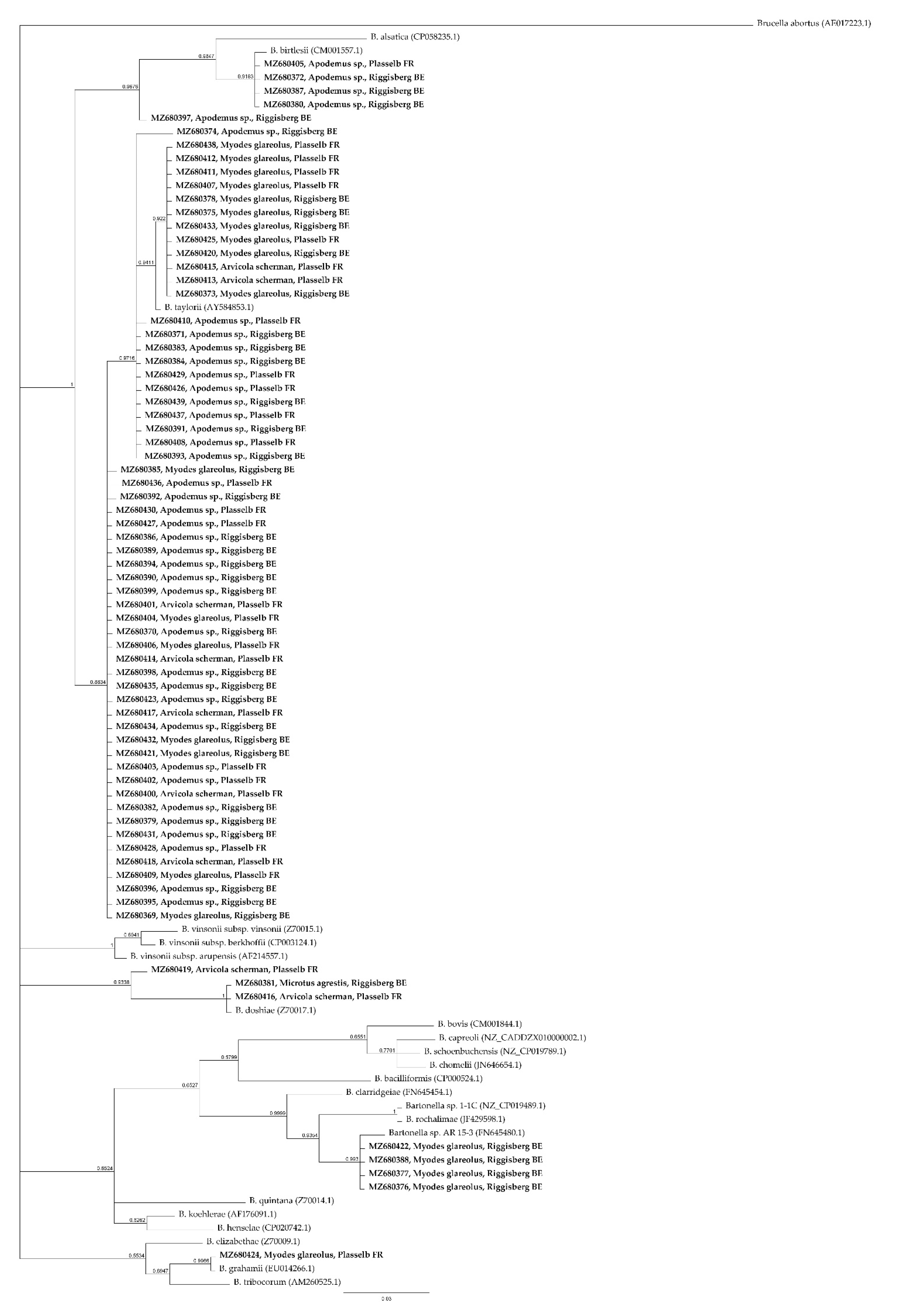
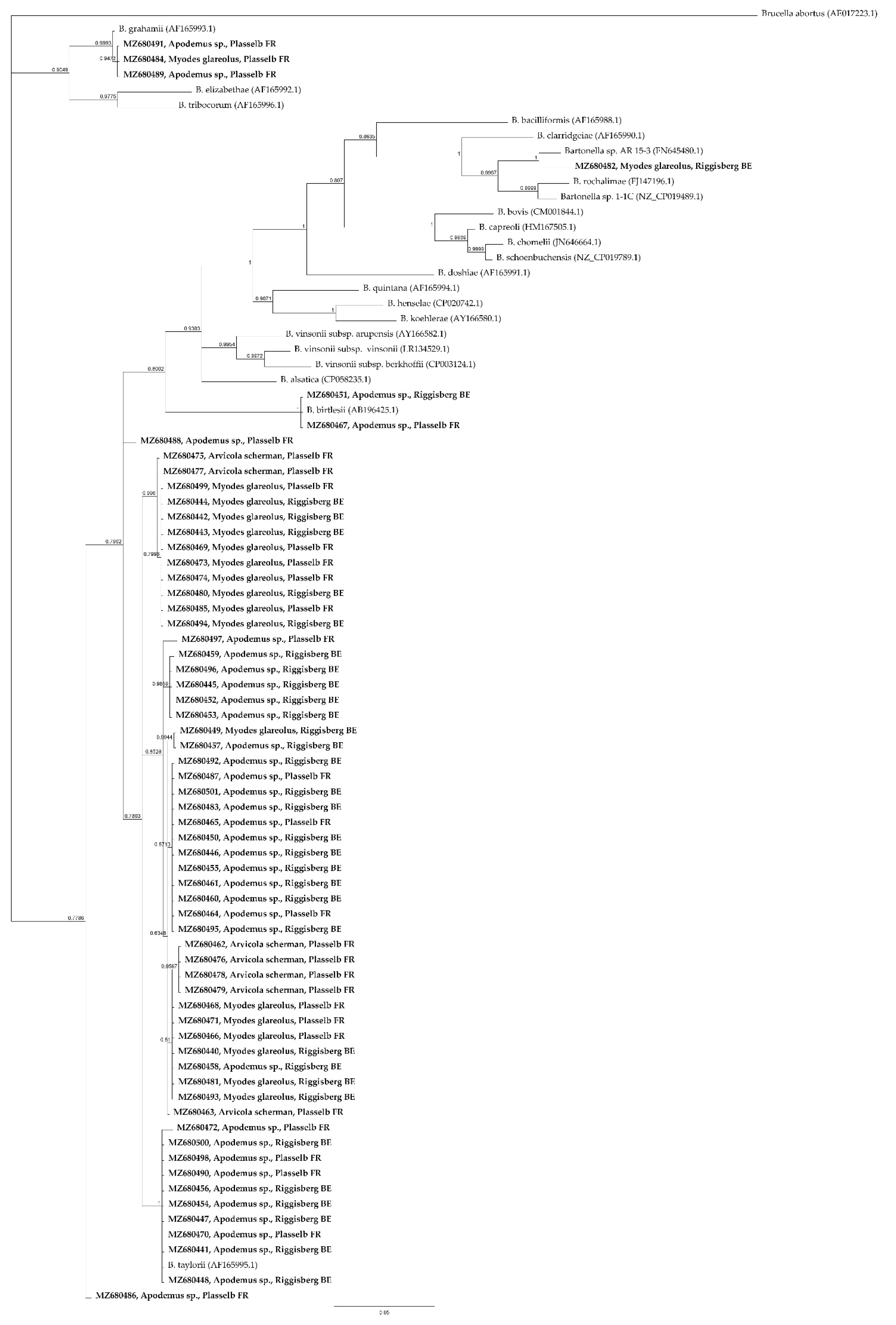
GltA sequences, amplified by cPCR, showed 100% identity to Candidatus “Bartonella rudakovii” (EF682090.1) in four Myodes glareolus out of 129 animals (3.10%). The gltA amplicon sequence in one Myodes glareolus out of 129 (0.78%) was 100% similar to B. grahamii (CP001562.1). Two Bartonella gltA sequence detected in Arvicola scherman (1/86, 1.16%) and Microtus agrestis (1/2, 50%) were 100% identical to B. doshiae (Z70017.1). B. taylorii (AF165995.1) was identified with 100% of identity sequencing rpoB amplicons (95–98% of query cover) in eight Apodemus sp. samples out of 129 (6.2%) and in two Apodemus sp out of 129 (1.6%) was identified the rpoB sequence of B. birtlesii (AB196425.1) (100% of identity). In one Apodemus sp. B. taylorii (AF165995.1, 98.15% of identity) and B. grahamii (CP001562.1, 99.65% of identity) DNA were identified by gltA and rpoB amplicons sequencing respectively. The remaining Bartonella positive animals showed a gltA and rpoB sequence identity > 96.0% and > 95.4%, respectively to closest relatives present in GenBank. In particular, considering the criteria previously established by La Scola et al. [30], B. taylorii was identified in 59/346 animals. Detailed results are shown in Supplementary Materials (Table S1). A BLASTn and phylogenetic analysis of gltA (Figure 1) and rpoB (Figure 2) loci identified in this study revealed that most of Bartonella DNA isolated are closely related to B. taylorii, followed by B. grahamii, B. birtlesii, and B. doshiae.
3. Discussion
In this study the prevalence and molecular diversity of Bartonella in small rodent populations from Switzerland were firstly described.
Wild rodents could be potential reservoirs causing Bartonella infections and more than 20 Bartonella species are associated with these small mammals [31]. They include some zoonotic species, such as B. elizabethae, B. grahamii, and B. vinsonii subsp. arupensis [20,32].
In central Europe, prevalences of Bartonella spp. ranging from 3.3 to 65.8% [33] have been observed in wild rodents. The prevalence of about 21% reported in our study was similar to the ones observed in Lithuania (24%) in 2013–2014 period [34] and Poland (11–48%) [20]. In particular, B. taylori-like DNA was the most common detected species in this study, followed by B. grahamii and B. birtlesii, three of the four most widespread species in European rodents [33].
Phylogenetic analysis based on gltA and rpoB loci demonstrated that in wild rodents of Switzerland multiple Bartonella species were identified and four genogroups were recognized, in particular B. grahamii, B. taylorii, B. doshiae and B. birtlesii. [20]. According to the lineages previously specified by Engel et al. [35], Bartonella species detected in the present study belong to lineage three and four.
B. taylorii was isolated for the first time by Birtles et al. [36]. DNA of B. taylorii strain Far East II was previously identified in rodents including Apodemus agrarius from Russian Far East in 2005 [37] and also in the present study. In Europe, B. taylorii was principally detected in Myodes sp. and Microtus sp. [38].
In this study, Bartonella DNA relative to B. grahamii was identified in a Myodes glareolus, and it is known that this bacteria may cause neuroretinitis in humans [39]. In one Apodemus sp. a probable co-infection of B. grahamii with B. taylorii was observed, confirming the wide range of hosts and the worldwide distribution of B. grahamii-like organisms, as described in Szewczyk et al., [33]. Buffet et al. [38] reported the presence of B. grahamii in Microtus spp. and Apodemus spp. in France.
Bartonella sequences identified in four Myodes glareolus trapped in the municipality of Riggisberg were 100% identical to Candidatus “Bartonella rudakovii” identified in 2007 in small wild mammals in Western Siberia (unpublished, GenBank: EF682090.1).
RpoB sequences of three Apodemus sp. captured in the present project were very close to B. birtlesii, isolated for the first time in small rodents in Germany and France [40].
From three animals, DNA similar to gltA of B. doshiae was identified (94.82–100% of identity). This species was reported in mice and voles in Europe [41] and in Sigmodon hispidus in the United States [42]. Vayssier-Taussat et al. [43] highlighted their novel potential zoonotic properties.
In this study, Apodemus sp. was the rodent genus more frequently affected by Bartonella sp. Paziewska et al. [44] obtained similar results, showing that A. flavicollis was the species in which Bartonella sp. was more present in Poland. Moreover, as in Poland, the present findings showed that B. taylorii was the most common Bartonella species, with a higher prevalence in Apodemus than in Myodes. Similarly, in 2019, a new study on the presence of Bartonella spp. in rodents was conducted in the Baltic region. The prevalence of Bartonella spp. was 54.8% and, in particular, A. flavicollis and M. agrestis were the most infected rodent species [45].
In this study, rodents trapped in woodland areas were more often infected with Bartonella spp. compared to those captured in open fields. A possible explanation for this phenomenon is the presence of vectors, such as fleas, which prefer wet conditions of woodland, allowing a better survival of larval stages [46]. In fact, ectoparasites are influenced by host characteristics, host environment, and season [46,47,48]. However, temperature and humidity conditions can affect the various vector species differently, therefore further investigations on parasites present in the considered geographical area would be useful. The distribution of reservoirs or arthropod vectors are likely the reasons why the prevalence of Bartonella infection varied among different rodent species or locations.
4. Materials and Methods
Wild rodents studied were trapped between April and November 2017 in the Gantrisch Nature Park (Switzerland) as described in Peterhans et al. [49], in the municipalities of Plasselb (canton of Fribourg) and Riggisberg (canton of Bern).
Topcat traps (Andermatt Biocontrol, Switzerland) were used in open fields, whereas live traps (Longworth, Penlon Ltd., Abingdon, UK) were placed in the woodland.
Trapped mice were visually examined and then euthanized by exposure to carbon dioxide on site. Mice were then transported refrigerated to the laboratory, where postmortem examination and sampling were performed as previous described in Peterhans et al. [49] (Table 1). The age of Arvicola was determined by measuring the weight of dry crystalline lenses [50,51]. For the remaining species, the development of the sexual organs was used as in Beerli et al. [52].
This project was performed in accordance with the Swiss Animal Welfare Act (SR 455) and the regulations of the Cantons of Bern and Fribourg (permit number BE145/16).
Lungs, spleen, liver, mandibular, and mesenteric lymph nodes were collected from 346 animals, and tissue samples of three animals were pooled, resulting in a total of 116 pools. These were homogenized and genomic DNA was obtained using DNeasy Blood and Tissue Kit (Qiagen, Hilden, Germany). A real-time PCR was performed on the pools to detect Bartonella spp. DNA as described in Divari et al. [25]. ITS region of Bartonella spp. was amplified (about 200 bp) using published primers 321s and H493as [53], [26] and amplification was performed using the CFX ConnectTM Real-Time PCR Detection System (BioRad, Hercules, CA, USA). In brief, iTaq Universal SYBER® Green Supermix (BioRad, Hercules, CA, USA) was used in the reaction mix and the protocol consisted of a 4-min step at 94 °C, followed by 45 cycles at 95 °C for 5 s and 60 °C for 20 s. A melting curve (from 65 °C to 95 °C) was obtained at the end of each run, to detect the PCR products dissociation. Examples of Tm values were described in Supplementary Materials (Table S2). The efficiency of qPCR was calculated on the slope of the standard curve constructed for ITS region amplification using scalar dilution of DNA from the positive control (Bartonella sp. FG4-1).
Pools showing a quantification cycle (Cq) less than 35 were further analyzed by cPCR, detecting a 340 bp segment of the gltA specific for Bartonella spp. cPCR was performed using previously described primers 443f and 781r [25,54] and a master mix (HotStarTaq; Qiagen, Hilden, Germany). The gltA gene fragment was amplified by a protocol consisting of a first step at 95 °C for 15 min, followed by 45 cycles at 95 °C for 5 s and 60 °C for 45 s.
PCR products were electrophoresed in 1.5% and 2% agarose gels, stained by GelRed Nucleic Acid Gel Stain (Biotium Inc., Fremont, CA, USA) and visualized under UV light. Bartonella spp. FG4-1 DNA (NCBI: txid545598) was used as a positive control and nuclease-free water included as a negative control in each PCR run. Pools showing amplicons consistent in size with the amplified locus were considered positive to Bartonella spp. infection. Therefore, DNA from spleen of single animals belonging to positive pools was extracted using DNeasy Blood and Tissue Kit (Qiagen, Hilden, Germany) and retested for gltA (as described above) and rpoB loci. For this latest gene, an 800 bp segment was amplified using previously described primers 1400f and 2300r [23,55] and amplification was conducted under the following conditions: 95 °C for 15 min, followed by 35 cycles of denaturation at 95 °C for 30 s, annealing at 53 °C, and extension at 72 °C for 1 min. At the end of the reaction, an additional extension step at 72 °C for 2 min was applied.
PCR products were visualized by electrophoresis, purified through MinElute PCR purification kit (Qiagen) and sequenced in both directions using Sanger method by a commercial sequencing provider (BMR Genomics, Padova, Italy).
The raw sequences were edited using Geneious Prime version 2021.2.2 [56] and compared to sequences deposited in NCBI using BLAST (https://blast.ncbi.nlm.nih.gov/Blast.cgi (accessed on 2 August 2021)). All sequences were deposited in the GenBank under accession numbers described in Supplementary Materials (Table S3).
GltA and rpoB sequences from this study and from GenBank were aligned (MUSCLE alignment algorithm) and phylogenetic relations were estimated through a Bayesian inference method using a GTR substitution model with the MrBayes [57] plugin in Geneious Prime version 2021.2.2 [56], with 1,100,000 chain length, 100,000 burn-in length.
Differences of Bartonella spp. prevalence among small rodent species, sampling locations, gender and age were assessed by Fisher’s exact and Chi-square test and 95% confidence intervals were set. Statistical analysis was conducted using GraphPad Prism 6 version 6.07 and a p value < 0.05 was considered significant.
5. Conclusions
Wild rodents infected with zoonotic Bartonella species were detected in two rural areas of Switzerland. However, these regions are not distant from urban areas and, thus, contacts between humans and infected rodents are possible. Therefore, the Bartonella-infected wild rodents might represent a potential pathogen reservoir in Switzerland and should be considered of public health importance.
Supplementary Materials
The following tables are available online at https://www.mdpi.com/article/10.3390/pathogens10101331/s1, Table S1: Sequencing results of Bartonella spp. positive rodents. Table S2: examples of Tm values calculated for DNA of samples analysed by qPCR. Table S3: Accession number of sequences deposited in GenBank. Figure S1: examples of cPCR products, relative to gltA and rpoB loci amplification.
Author Contributions
Conceptualization: P.P., E.B.; Methodology: S.D., P.P.; Investigation: M.D., G.G., and S.D.; Data curation: S.D., M.D.; Project administration: E.B.; Resources: N.B., G.G.; Supervision: G.G., N.B., and E.B.; Writing—original draft preparation: S.D.; Writing—review and editing: P.P., N.B., G.G., and E.B. All authors have read and agreed to the published version of the manuscript.
Funding
This research received no external funding.
Institutional Review Board Statement
This project was performed in accordance with the Swiss Animal Welfare Act (SR 455) and the regulations of the Cantons of Bern and Fribourg (permit number BE145/16).
Informed Consent Statement
Not applicable.
Data Availability Statement
The sequences obtained in the current study were submitted to GenBank under the accession numbers MZ680369 to MZ680501.
Acknowledgments
The authors are grateful to Shimon Harrus, Hebrew University of Jerusalem, Koret School of Veterinary Medicine, for providing positive control DNA.
Conflicts of Interest
The authors declare no conflict of interest.
References
- Deng, H.; Le Rhun, D.; Buffet, J.P.R.; Cotté, V.; Read, A.; Birtles, R.J.; Vayssier-Taussat, M. Strategies of exploitation of mammalian reservoirs by Bartonella species. Vet. Res. 2012, 43, 1–14. [Google Scholar] [CrossRef]
- Regier, Y.; Órourke, F.; Kempf, V.A.J. Bartonella spp.—A chance to establish One Health concepts in veterinary and human medicine. Parasites Vectors 2016, 9, 1–12. [Google Scholar] [CrossRef] [PubMed] [Green Version]
- Guptill, L. Bartonellosis. Vet. Microbiol. 2010, 140, 347–359. [Google Scholar] [CrossRef] [PubMed]
- Cheslock, M.A.; Embers, M.E. Human bartonellosis: An underappreciated public health problem? Trop. Med. Infect. Dis. 2019, 4, 69. [Google Scholar] [CrossRef] [Green Version]
- Angelakis, E.; Raoult, D. Pathogenicity and treatment of Bartonella infections. Int. J. Antimicrob. Agents 2014, 44, 16–25. [Google Scholar] [CrossRef] [PubMed]
- Breitschwerdt, E.B. Bartonellosis: One health perspectives for an emerging infectious disease. ILAR J. 2014, 55, 46–58. [Google Scholar] [CrossRef] [PubMed]
- Breitschwerdt, E.B. Bartonellosis, One Health and all creatures great and small. Vet. Dermatol. 2017, 28, 96-e21. [Google Scholar] [CrossRef] [PubMed]
- Maggi, R.G.; Mozayeni, B.R.; Pultorak, E.L.; Hegarty, B.C.; Bradley, J.M.; Correa, M.; Breitschwerdt, E.B. Bartonella spp. Bacteremia and rheumatic symptoms in patients from Lyme disease-endemic region. Emerg. Infect. Dis. 2012, 18, 783–791. [Google Scholar] [CrossRef]
- Okaro, U.; Addisu, A.; Casanas, B.; Anderson, B. Bartonella Species, an Emerging Cause of Blood-Culture-Negative Endocarditis. Clin. Microbiol. Rev. 2017, 30, 709–746. [Google Scholar] [CrossRef] [PubMed] [Green Version]
- Álvarez-Fernández, A.; Breitschwerdt, E.B.; Solano-Gallego, L. Bartonella infections in cats and dogs including zoonotic aspects. Parasites Vectors 2018, 11, 1–21. [Google Scholar] [CrossRef]
- Boularias, G.; Azzag, N.; Gandoin, C.; Bouillin, C.; Chomel, B.; Haddad, N.; Boulouis, H.J. Bartonella bovis and Bartonella chomelii infection in dairy cattle and their ectoparasites in Algeria. Comp. Immunol. Microbiol. Infect. Dis. 2020, 70, 101450. [Google Scholar] [CrossRef] [PubMed]
- Magni, E.; Bertelloni, F.; Sgorbini, M.; Ebani, V.V. Bartonella infection in asymptomatic horses and donkeys from Tuscany, Central Italy. Asian Pac. J. Trop. Med. 2017, 10, 1077–1079. [Google Scholar] [CrossRef]
- Stuckey, M.J.; Chomel, B.B.; de Fleurieu, E.C.; Aguilar-Setién, A.; Boulouis, H.-J.; Chang, C. Bartonella, bats and bugs: A review. Comp. Immunol. Microbiol. Infect. Dis. 2017, 55, 20–29. [Google Scholar] [CrossRef] [PubMed]
- Greco, G.; Zarea, A.A.K.; Sgroi, G.; Tempesta, M.; D’Alessio, N.; Lanave, G.; Bezerra-Santos, M.A.; Iatta, R.; Veneziano, V.; Otranto, D.; et al. Zoonotic Bartonella species in Eurasian wolves and other free-ranging wild mammals from Italy. Zoonoses Public Health 2021, 68, 316–326. [Google Scholar] [CrossRef]
- Harms, C.A.; Maggi, R.G.; Breitschwerdt, E.B.; Clemons-Chevis, C.L.; Solangi, M.; Rotstein, D.S.; Fair, P.A.; Hansen, L.J.; Hohn, A.A.; Lovewell, G.N.; et al. Bartonella species detection in captive, stranded and free-ranging cetaceans. Vet. Res. 2008, 39, 1–8. [Google Scholar] [CrossRef] [Green Version]
- Dahmana, H.; Granjon, L.; Diagne, C.; Davoust, B.; Fenollar, F.; Mediannikov, O. Rodents as hosts of pathogens and related zoonotic disease risk. Pathogens 2020, 9, 202. [Google Scholar] [CrossRef] [Green Version]
- Kosoy, M.; Bai, Y.; Enscore, R.; Rizzo, M.R.; Bender, S.; Popov, V.; Albayrak, L.; Fofanov, Y.; Chomel, B. Bartonella melophagi in blood of domestic sheep (Ovis aries) and sheep keds (Melophagus ovinus) from the southwestern US: Cultures, genetic characterization, and ecological connections. Vet. Microbiol. 2016, 190, 43–49. [Google Scholar] [CrossRef] [PubMed]
- Williams, H.M.; Dittmar, K. Expanding our view of Bartonella and its hosts: Bartonella in nest ectoparasites and their migratory avian hosts. Parasites and Vectors 2020, 13, 1–9. [Google Scholar] [CrossRef] [PubMed] [Green Version]
- Valentine, K.H.; Harms, C.A.; Cadenas, M.B.; Birkenheuer, A.J.; Marr, H.S.; Braun-McNeill, J.; Maggi, R.G.; Breitschwerdt, E.B. Bartonella DNA in Loggerhead Sea Turtles. Emerg. Infect. Dis. 2007, 13, 949–950. [Google Scholar] [CrossRef] [PubMed]
- Gutiérrez, R.; Krasnov, B.; Morick, D.; Gottlieb, Y.; Khokhlova, I.S.; Harrus, S. Bartonella Infection in Rodents and Their Flea Ectoparasites: An Overview. Vector Borne Zoonotic Dis. 2015, 15, 27–39. [Google Scholar] [CrossRef] [Green Version]
- Gundi, V.A.K.B.; Billeter, S.A.; Rood, M.P.; Kosoy, M.Y. Bartonella spp. in Rats and Zoonoses, Los Angeles, California, USA. Emerg. Infect. Dis. 2012, 18, 631–633. [Google Scholar] [CrossRef] [PubMed]
- Kandelaki, G.; Malania, L.; Bai, Y.; Chakvetadze, N.; Katsitadze, G.; Imnadze, P.; Nelson, C.; Harrus, S.; Kosoy, M. Human Lymphadenopathy Caused by Ratborne Bartonella, Tbilisi, Georgia. Emerg. Infect. Dis. 2016, 22, 544–546. [Google Scholar] [CrossRef]
- Helan, J.V.G.; Grinberg, A.; Gedye, K.; Potter, M.A.; Harrus, S. Molecular detection of Bartonella coopersplainsensis and B. henselae in rats from New Zealand. N. Z. Vet. J. 2018, 66, 257–260. [Google Scholar] [CrossRef]
- Engbæk, K.; Lawson, P.A. Identification of Bartonella species in rodents, shrews and cats, in Denmark: Detection of two B. henselae variants, one in cafe and the other to the long-tailed field mouse. Apmis 2004, 112, 336–341. [Google Scholar] [CrossRef] [PubMed]
- Divari, S.; Pregel, P.; Zanet, S.; Ferroglio, E.; Giannini, F.; Scaglione, F.E.; Grinberg, A.; Biolatti, B.; Bollo, E. Molecular Evidence of Bartonella spp. in Rodents: A Study in Pianosa Island, Italy. Animals 2020, 10, 2070. [Google Scholar] [CrossRef] [PubMed]
- Maggi, R.G.; Duncan, A.W.; Breitschwerdt, E.B. Novel chemically modified liquid medium that will support the growth of seven Bartonella species. J. Clin. Microbiol. 2005, 43, 2651–2655. [Google Scholar] [CrossRef] [Green Version]
- Diddi, K.; Chaudhry, R.; Sharma, N.; Dhawan, B. Strategy for identification & characterization of Bartonella henselae with conventional & molecular methods. Indian J. Med. Res. 2013, 137, 380–387. [Google Scholar]
- Gutiérrez, R.; Vayssier-Taussat, M.; Buffet, J.-P.; Harrus, S. Guidelines for the Isolation, Molecular Detection, and Characterization of Bartonella Species. Vector Borne Zoonotic Dis. 2017, 17, 42–50. [Google Scholar] [CrossRef] [Green Version]
- Breitschwerdt, E.B.; Kordick, D.L. Bartonella Infection in Animals: Carriership, Reservoir Potential, Pathogenicity, and Zoonotic Potential for Human Infection. Clin. Microbiol. Rev. 2000, 13, 428–438. [Google Scholar] [CrossRef]
- La Scola, B.; Zeaiter, Z.; Khamis, A.; Raoult, D. Gene-sequence-based criteria for species definition in bacteriology: The Bartonella paradigm. Trends Microbiol. 2003, 11, 318–321. [Google Scholar] [CrossRef]
- Gonçalves, L.R.; de Mendonça Favacho, A.R.; Roque, A.L.R.; Mendes, N.S.; Fidelis, O.L.; Benevenute, J.L.; Herrera, H.M.; D’Andrea, P.S.; de Lemos, E.R.S.; Machado, R.Z.; et al. Association of Bartonella species with wild and synanthropic rodents in different Brazilian biomes. Appl. Environ. Microbiol. 2016, 82, 7154–7164. [Google Scholar] [CrossRef] [Green Version]
- Buffet, J.P.; Kosoy, M.; Vayssier-Taussat, M. Natural history of Bartonella-infecting rodents in light of new knowledge on genomics, diversity and evolution. Future Microbiol. 2013, 8, 1117–1128. [Google Scholar] [CrossRef]
- Szewczyk, T.; Werszko, J.; Slivinska, K.; Laskowski, Z.; Karbowiak, G. Molecular Detection of Bartonella spp. in Rodents in Chernobyl Exclusion Zone, Ukraine. Acta Parasitol. 2020, 66, 222–227. [Google Scholar] [CrossRef]
- Lipatova, I.; Paulauskas, A.; Puraite, I.; Radzijevskaja, J.; Balciauskas, L.; Gedminas, V. Bartonella infection in small mammals and their ectoparasites in Lithuania. Microbes Infect. 2015, 17, 884–888. [Google Scholar] [CrossRef]
- Engel, P.; Salzburger, W.; Liesch, M.; Chang, C.-C.; Maruyama, S.; Lanz, C.; Calteau, A.; Lajus, A.; Médigue, C.; Schuster, S.C.; et al. Parallel Evolution of a Type IV Secretion System in Radiating Lineages of the Host-Restricted Bacterial Pathogen Bartonella. PLoS Genet. 2011, 7, e1001296. [Google Scholar] [CrossRef] [Green Version]
- Birtles, R.J.; Harrison, T.G.; Saunders, N.A.; Molyneux, D.H. Proposals To Unify the Genera Grahamella and Bartonella, with Descriptions of Bartonella talpae comb, nov., Bartonella peromysci comb. nov., and Three New Species, Bartonella grahamii sp. nov., Bartonella taylorii sp. nov., and Bartonella doshiae sp. nov. Int. J. Syst. Bacteriol. 1995, 45, 1–8. [Google Scholar] [CrossRef] [Green Version]
- Mediannikov, O.; Ivanov, L.; Zdanovskaya, N.; Vysochina, N.; Fournier, P.E.; Tarasevich, I.; Raoult, D. Molecular screening of Bartonella species in rodents from the Russian Far East. Ann. N. Y. Acad. Sci. 2005, 1063, 308–311. [Google Scholar] [CrossRef] [PubMed]
- Buffet, J.-P.; Pisanu, B.; Brisse, S.; Roussel, S.; Félix, B.; Halos, L.; Chapuis, J.-L.; Vayssier-Taussat, M. Deciphering Bartonella Diversity, Recombination, and Host Specificity in a Rodent Community. PLoS ONE 2013, 8, e68956. [Google Scholar] [CrossRef] [PubMed] [Green Version]
- Kerkhoff, F.T.; Bergmans, A.M.C.; Van Der Zee, A.; Rothova, A. Demonstration of Bartonella grahamii DNA in ocular fluids of a patient with neuroretinitis. J. Clin. Microbiol. 1999, 37, 4034–4038. [Google Scholar] [CrossRef] [PubMed] [Green Version]
- Bermond, D.; Heller, R.; Barrat, F.; Delacour, G.; Dehio, C.; Alliot, A.; Monteil, H.; Chomel, B.; Boulouis, H.J.; Piémont, Y. Bartonella birtlesii sp. nov., isolated from small mammals (Apodemus spp.). Int. J. Syst. Evol. Microbiol. 2000, 50, 1973–1979. [Google Scholar] [CrossRef] [PubMed] [Green Version]
- Paziewska, A.; Harris, P.D.; Zwolińska, L.; Bajer, A.; Siński, E. Recombination Within and Between Species of the Alpha Proteobacterium Bartonella Infecting Rodents. Microb. Ecol. 2011, 61, 134–145. [Google Scholar] [CrossRef] [PubMed] [Green Version]
- Abbot, P.; Aviles, A.E.; Eller, L.; Durden, L.A. Mixed infections, cryptic diversity, and vector-borne pathogens: Evidence from Polygenis fleas and Bartonella species. Appl. Environ. Microbiol. 2007, 73, 6045–6052. [Google Scholar] [CrossRef] [PubMed] [Green Version]
- Vayssier-Taussat, M.; Moutailler, S.; Féménia, F.; Raymond, P.; Croce, O.; La Scola, B.; Fournier, P.-E.; Raoult, D. Identification of Novel Zoonotic Activity of Bartonella spp., France. Emerg. Infect. Dis. 2016, 22, 457–462. [Google Scholar] [CrossRef] [PubMed] [Green Version]
- Paziewska, A.; Harris, P.D.; Zwolińska, L.; Bajer, A.; Siński, E. Differences in the ecology of Bartonella infections of Apodemus flavicollis and Myodes glareolus in a boreal forest. Parasitology 2012, 139, 881–893. [Google Scholar] [CrossRef]
- Mardosaitė-Busaitienė, D.; Radzijevskaja, J.; Balčiauskas, L.; Bratchikov, M.; Jurgelevičius, V.; Paulauskas, A. Prevalence and diversity of Bartonella species in small rodents from coastal and continental areas. Sci. Rep. 2019, 9, 12349. [Google Scholar] [CrossRef]
- Krasnov, B.R.; Khokhlova, I.S.; Fielden, L.J.; Burdelova, N.V. Effect of Air Temperature and Humidity on the Survival of Pre-Imaginal Stages of Two Flea Species (Siphonaptera: Pulicidae). J. Med. Entomol. 2001, 38, 629–637. [Google Scholar] [CrossRef]
- Krasnov, B.R.; Poulin, R.; Shenbrot, G.I.; Mouillot, D.; Khokhlova, I.S. Ectoparasitic “jacks-of-all-trades”: Relationship between abundance and host specificity in fleas (Siphonaptera) parasitic on small mammals. Am. Nat. 2004, 164, 506–516. [Google Scholar] [CrossRef]
- Kim, B.J.; Kim, S.J.; Kang, J.G.; Ko, S.; Won, S.; Kim, H.; Kim, H.C.; Kim, M.S.; Chong, S.T.; Klein, T.A.; et al. First report for the seasonal and annual prevalence of Flea-Borne Bartonella from Rodents and soricomorphs in the Republic of Korea. Vector Borne Zoonotic Dis. 2013, 13, 457–467. [Google Scholar] [CrossRef] [Green Version]
- Peterhans, S.; Landolt, P.; Friedel, U.; Oberhänsli, F.; Dennler, M.; Willi, B.; Senn, M.; Hinden, S.; Kull, K.; Kipar, A.; et al. Mycobacterium microti: Not Just a Coincidental Pathogen for Cats. Front. Vet. Sci. 2020, 7, 1–15. [Google Scholar] [CrossRef]
- Burlet, P.; Deplazes, P.; Hegglin, D. Efficient age determination: How freezing affects eye lens weight of the small rodent species arvicola terrestris. Eur. J. Wildl. Res. 2010, 56, 685–688. [Google Scholar] [CrossRef] [Green Version]
- Burlet, P.; Deplazes, P.; Hegglin, D. Age, season and spatio-temporal factors affecting the prevalence of Echinococcus multilocularis and Taenia taeniaeformis in Arvicola terrestris. Parasites Vectors 2011, 4, 6. [Google Scholar] [CrossRef] [PubMed] [Green Version]
- Beerli, O.; Guerra, D.; Baltrunaite, L.; Deplazes, P.; Hegglin, D. Microtus arvalis and Arvicola scherman: Key players in the Echinococcus multilocularis life cycle. Front. Vet. Sci. 2017, 4, 1–10. [Google Scholar] [CrossRef] [PubMed] [Green Version]
- Gutiérrez, R.; Morick, D.; Gross, I.; Winkler, R.; Abdeen, Z.; Harrus, S. Bartonellae in Domestic and Stray Cats from Israel: Comparison of Bacterial Cultures and High-Resolution Melt Real-Time PCR As Diagnostic Methods. Vector Borne Zoonotic Dis. 2013, 13, 857–864. [Google Scholar] [CrossRef]
- Gutiérrez, R.; Nachum-Biala, Y.; Harrus, S. Relationship between the presence of Bartonella species and bacterial loads in cats and cat fleas (Ctenocephalides felis) under natural conditions. Appl. Environ. Microbiol. 2015, 81, 5613–5621. [Google Scholar] [CrossRef] [Green Version]
- Renesto, P.; Gouvernet, J.; Drancourt, M.; Roux, V.; Raoult, D. Use of rpoB gene analysis for detection and identification of Bartonella species. J. Clin. Microbiol. 2001, 39, 430–437. [Google Scholar] [CrossRef] [Green Version]
- Kearse, M.; Moir, R.; Wilson, A.; Stones-Havas, S.; Cheung, M.; Sturrock, S.; Buxton, S.; Cooper, A.; Markowitz, S.; Duran, C.; et al. Geneious Basic: An integrated and extendable desktop software platform for the organization and analysis of sequence data. Bioinformatics 2012, 28, 1647–1649. [Google Scholar] [CrossRef]
- Huelsenbeck, J.P.; Ronquist, F. MRBAYES: Bayesian inference of phylogenetic trees. Bioinformatics 2001, 17, 754–755. [Google Scholar] [CrossRef] [PubMed] [Green Version]
Figure 1.
Phylogenetic tree based on the gltA (293 bp) partial sequences of Bartonella spp. Sequences identified in the present study are indicated in bold (GenBank accession number, host, and site of trapping) and sequences from GenBank are indicated as common name and GenBank accession number in bracket. The phylogenetic tree was constructed using Bayesian inference method, using a GTR substitution model with the MrBayes plugin in Geneious Prime version 2021.2.2, with 1,100,000 chain length, 100,000 burn-in length. Brucella abortus was used as outgroup.

Figure 2.
Phylogenetic tree based on the rpoB (682 bp) partial sequences of Bartonella spp. Sequences identified in the present study are indicated in bold (GenBank accession number, host, and site of trapping) and sequences from GenBank are indicated as common name and GenBank accession number in bracket. The phylogenetic tree was constructed using Bayesian inference method with the MrBayes plugin in Geneious Prime version 2021.2.2, with 1,100,000 chain length, 100,000 burn-in length. Brucella abortus was used as outgroup.


Table 1.
Origin of trapping (BE: Riggisberg; FR: Plasselb; WL: woodland; OF: open field), species, gender, age, and number of wild rodents analyzed.
Table 1.
Origin of trapping (BE: Riggisberg; FR: Plasselb; WL: woodland; OF: open field), species, gender, age, and number of wild rodents analyzed.
| Rodent Species | Site | Area | Gender | Age | Total | ||||
|---|---|---|---|---|---|---|---|---|---|
| BE | FR | WL | OF | Females | Males | Adult | Juvenile | ||
| Myodes glareolus | 55 | 74 | 128 | 1 | 66 | 63 | 84 | 45 | 129 |
| Microtus agrestis | 2 | 0 | 2 | 0 | 2 | 0 | 2 | 0 | 2 |
| Arvicola scherman | 0 | 86 | 0 | 86 | 47 | 39 | 69 | 17 | 86 |
| Apodemus sp. | 83 | 46 | 127 | 2 | 63 | 66 | 90 | 39 | 129 |
Publisher’s Note: MDPI stays neutral with regard to jurisdictional claims in published maps and institutional affiliations. |
© 2021 by the authors. Licensee MDPI, Basel, Switzerland. This article is an open access article distributed under the terms and conditions of the Creative Commons Attribution (CC BY) license (https://creativecommons.org/licenses/by/4.0/).
Share and Cite
MDPI and ACS Style
Divari, S.; Danelli, M.; Pregel, P.; Ghielmetti, G.; Borel, N.; Bollo, E. Biomolecular Investigation of Bartonella spp. in Wild Rodents of Two Swiss Regions. Pathogens 2021, 10, 1331. https://doi.org/10.3390/pathogens10101331
AMA Style
Divari S, Danelli M, Pregel P, Ghielmetti G, Borel N, Bollo E. Biomolecular Investigation of Bartonella spp. in Wild Rodents of Two Swiss Regions. Pathogens. 2021; 10(10):1331. https://doi.org/10.3390/pathogens10101331
Chicago/Turabian StyleDivari, Sara, Marta Danelli, Paola Pregel, Giovanni Ghielmetti, Nicole Borel, and Enrico Bollo. 2021. "Biomolecular Investigation of Bartonella spp. in Wild Rodents of Two Swiss Regions" Pathogens 10, no. 10: 1331. https://doi.org/10.3390/pathogens10101331
Note that from the first issue of 2016, this journal uses article numbers instead of page numbers. See further details here.