Abstract
The effects of nickel content in nickel-bearing pyrite on photocatalytic properties, light absorption properties, and oxidative decomposition of thiosulfate were studied. The leaching experiments show that the consumption of thiosulfate in the Cu2+-ethylenediamine (en)-S2O32− system increases with an increase in nickel content in nickel-bearing pyrite. The consumption of Cu(en)22+ initially increases and then decreases with an increase in leaching time. There is a clear correlation between the change trend in its consumption and the doping amount of nickel in pyrite. The XPS results show that in the Cu2+-ethylenediamine (en)-S2O32− leaching gold system (temperature 25 °C, time 35 h, solution: 0.1 mol/L S2O32−, 5 mmol/L Cu(en)22+, 200 mL solution), the nickel of pyrite-containing nickel can be transferred to the leaching solution and becomes nickel ion. In this leaching system, Cu(II), which was originally complexed with en, is reduced to Cu(I) in a short time. The consumption of Cu(en)22+ increased rapidly in the 5 h period and then decreased gradually after 5 h. The results showed that the presence of free Ni2+ in the solution facilitated the conversion of bivalent copper ions to monovalent copper ions. Free Ni2+ ions can compete with Cu2+ ions for en ligands. When ethylenediamine complexes with Ni2+, the decomposition of Cu(en)22+ into Cu(en)+ and en occurs more rapidly. And the en, which was originally to be oxidized with Cu(en)+ to form Cu(en)22+, forms Ni(en)22+. As a result, the concentration of Cu(en)22+ continues to decrease in a short period of time.
1. Introduction
As the reserves of easily processed gold deposits continue to decrease and environmental protection requirements become increasingly stringent, industrial research has turned towards finding nontoxic leaching agents that can replace cyanide [1]. Among the various gold leaching reagents, such as thiourea, polysulfides, halides, and thiosulfate, thiosulfate is considered the most promising green leaching agent with significant industrial application prospects because of its nontoxicity, high selectivity, and low cost [2,3,4,5]. However, problems such as high reagent consumption and difficulties in recovering gold complex ions from leach pulp have hindered the widespread industrialization of thiosulfate gold extraction technology [6,7].
To realize the industrial application of thiosulfate gold leaching, the high consumption of thiosulfate is an urgent technical bottleneck that must be solved. Sulfide minerals such as arsenopyrite, chalcopyrite, and galena have been reported to promote the composition of thiosulfate [8,9] because of their strong affinity for sulfur-containing substances and their semiconductor properties [10,11]. Moreover, most metal sulfides exhibit semiconducting properties which significantly affect the leaching of gold-bearing sulfides. The presence of impurities in the mineral lattice affects its composition, electronic structure, semiconductor properties, and the interaction between the leaching agent and mineral surface [9]. Additionally, different vacancy defects, impurity doping, and defect locations in the lattice affect both the sulfide ore surface and leaching characteristics [12,13,14].
Pyrite, the most common semiconductor-associated mineral in gold mines, often contains isomorphic impurities such as nickel, cobalt, and arsenic [15,16]. These impurities not only alter the semiconductor properties of pyrite but also impact the lattice defects that can affect its oxidation decomposition and surface adsorption. Although natural pyrite is widely distributed with large reserves, it typically possesses various impurities (such as nickel, silicon, arsenic.) [17,18]. Consequently, these factors introduce numerous uncertainties during experimental exploration processes.
To investigate the influence of nickel impurities in pyrite on thiosulfate gold leaching, leaching experiments were conducted using synthetic pure-phase pyrite and pyrite containing specific nickel elements. This was performed to eliminate interference from other metal impurities on the semiconductor properties of pyrite and the catalytic degradation of thiosulfate ions. Therefore, this study first synthesized pure-phase pyrite through a hydrothermal method and then synthesized pyrite containing only nickel impurities by adding a specific amount of nickel; pure-phase pyrite and nickel-containing pyrite were compared in terms of their semiconductor properties, including phase, photocatalysis, and light absorption. Additionally, the effect of nickel impurities in pyrite on the catalytic degradation of thiosulfate in a Cu2+-en-S2O32− leaching system was investigated.
2. Materials and Methods
2.1. Materials and Reagents
The reagents used in this study, including sodium thiosulfate, copper sulfate, sodium hydroxide, ferrous chloride, sodium sulfide, nickel chloride, and en, were analytically pure. All experimental solutions were prepared using ultrapure water.
2.2. Test Methods
2.2.1. Synthesis of Pure-Phase Pyrite
The basic raw materials used were 17.90 g of ferrous chloride and 43.23 g of sodium sulfide. The pH of the mixed solution was adjusted to approximately 6.6 using dilute NaOH and HCl solutions. After stirring the solution with a magnetic stirrer (MYP11-2A) for 30 min, the mixture was transferred to a high-pressure reactor. The reactor was placed in a constant-temperature vacuum drying oven (DZF-6020) at 200 °C for 24 h and then cooled to room temperature. The products in the reactor were washed, filtered, separated, and dried in a constant-temperature drying oven at 60 °C for 12 h, and the final product was synthetic pure-phase pyrite. The product was then stored in a sealed reagent bag filled with nitrogen.
2.2.2. Synthesis of Nickel-Bearing Pyrite
The synthetic procedure was the same as that for pure-phase pyrite, but the amounts of reagents used were different. The basic raw materials used were 17.90 g of ferrous chloride and 43.23 g of sodium sulfide. By adding 0.43, 1.13, 1.61, and 2.38 g of nickel chloride to the above raw materials, nickel-bearing pyrite samples with 2%, 5%, 7%, and 10% nickel contents were obtained, respectively.
2.2.3. Photocatalytic Degradation Experiment
The photocatalytic reactions were performed in a BL-GHX-V (Shanghai, China) photocatalytic reactor. The central light source of the photoreactor was a 300 W deuterium lamp. In the photocatalytic reaction, the initial concentration of methylene blue solution was 25 mg/L, and 50 mg of solid powder was added. The specific photocatalytic degradation steps have been described in the literature [19]. The methylene blue degradation rate was calculated as follows:
where C0 represents the initial concentration of methylene blue in the solution and C represents the concentration after the photocatalytic reaction.
2.2.4. Leaching Experiment and Detection Method
The leaching system was a mixed solution composed of 0.1 mol/L Na2S2O3, 0.01 mol/L en, and 0.005 mol/L CuSO4. The specific leaching experimental method has been described in detail in [20]. The analytical method for determining the gold concentration in this solution system has been extensively described in previous studies [20]. The thiosulfate concentration detection method for the leaching solution was based on previous literature [21]. The operation steps of the experiment, the consumption of thiosulfate, and the detection method of the gold concentration in the leaching solution are described in detail in the Supplementary Materials.
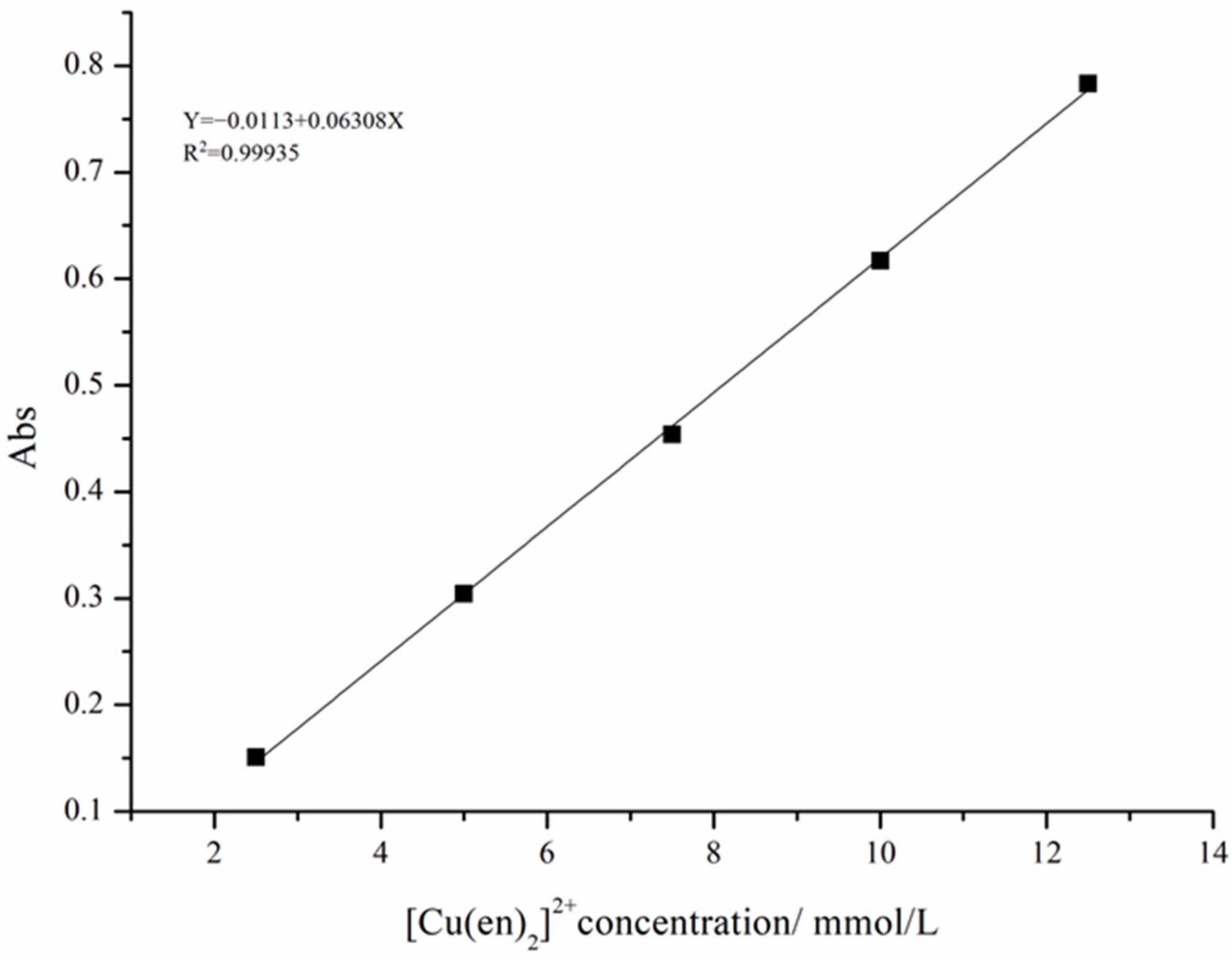
2.2.5. Determination of the Cu(en)22+ Complex Concentration
First, Cu(en)22+ standard solutions were prepared at concentrations of 2.5, 5, 7.5, 10, and 12.5 mmol/L. The absorbance of the standard solution was measured at 550 nm (Cu(en)22+ has an absorption peak at 550 nm) using deionized water as a blank. The concentration–absorbance relationship was determined by plotting the concentration of the Cu(en)22+ complex on the horizontal axis and the absorbance on the vertical axis to obtain a standard curve for the Cu(en)22+ solution (Figure 1) [22]. An aliquot of the sample liquid was periodically removed from the leaching solution using a pipette and transferred to a glass bottle. After some time elapsed, deionized water was used as the reference sample, and the absorbance of the Cu(en)22+ complex at 550 nm was measured three times in parallel using a UV-VIS spectrophotometer to determine its average value. The concentration of the Cu(en)22+ complex was calculated using the following equation:
Figure 1.
Relationship between the absorbance and Cu(en)22+ concentration (The specific parameters are shown in Table 1).
Table 1.
Data parameters obtained by linear fitting in Figure 1.
Table 1.
Data parameters obtained by linear fitting in Figure 1.
| Intercept | Slope | Statistics | |||
|---|---|---|---|---|---|
| Value | Standard Error | Value | Standard Error | Adj. R-Square | |
| C | −0.0113 | 0.00669 | 0.06308 | 8.06308 × 10⁻⁴ | 0.99935 |
2.3. Sample Characterization
The full name corresponding to the abbreviation in the text: Copper–ethylenediamine–thiosulfate (Cu2+-en-S2O32−), X-ray fluorescence spectroscopy (XRF), X-ray diffraction (XRD), ultraviolet/visible/near-infrared (UV-VIS-NIR), scanning electron microscopy (SEM), ultraviolet–visible diffuse reflection spectroscopy (UV-VIS DRS), X-ray photoelectron spectroscopy (XPS), energy-dispersive spectroscopy (EDS).
The phases of the synthetic pure-phase pyrite and nickel-bearing pyrite were detected using an XRD, and the data were analyzed using JADE software. The micromorphology and elemental composition of the samples were measured by SEM. Quantitative and qualitative analysis of leached gold foil and pyrite were performed using an XPS (X-ray photoelectron spectrometer, PHI5000 Versa ProbeII, ULVAC-PHI, Japan) [23,24]. In addition, the absorption spectra of synthetic pure-phase pyrite and nickel-bearing pyrite in the wavelength range 200–2000 nm were measured using a UV-VIS-NIR (UV-3600, Shimadzu, Japan).
3. Results and Discussion
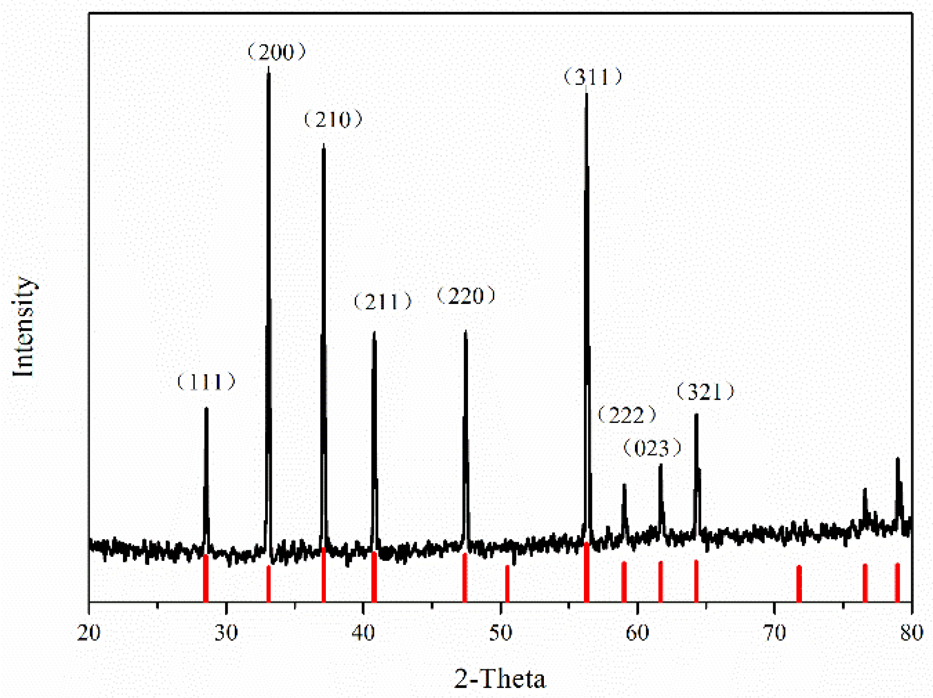
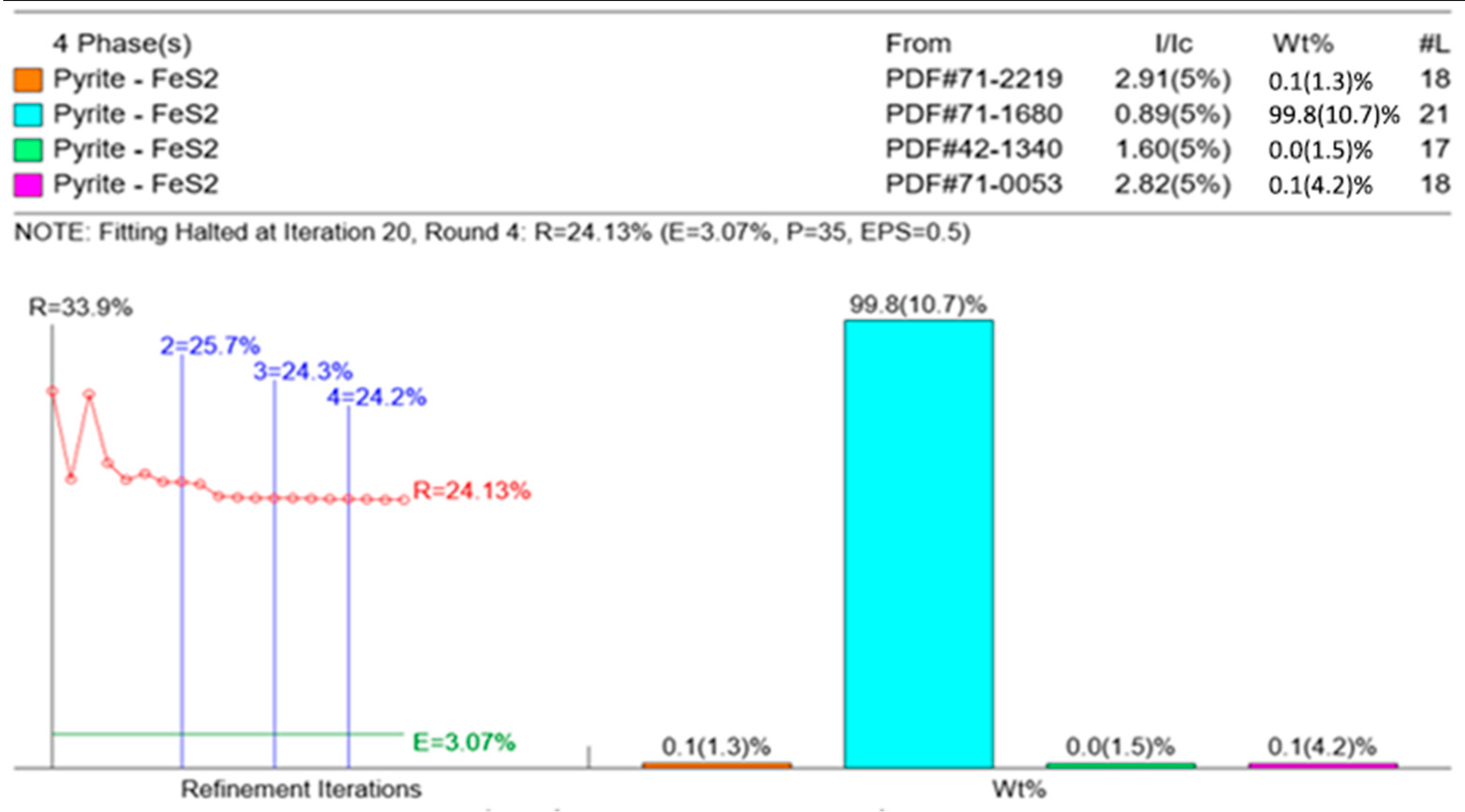
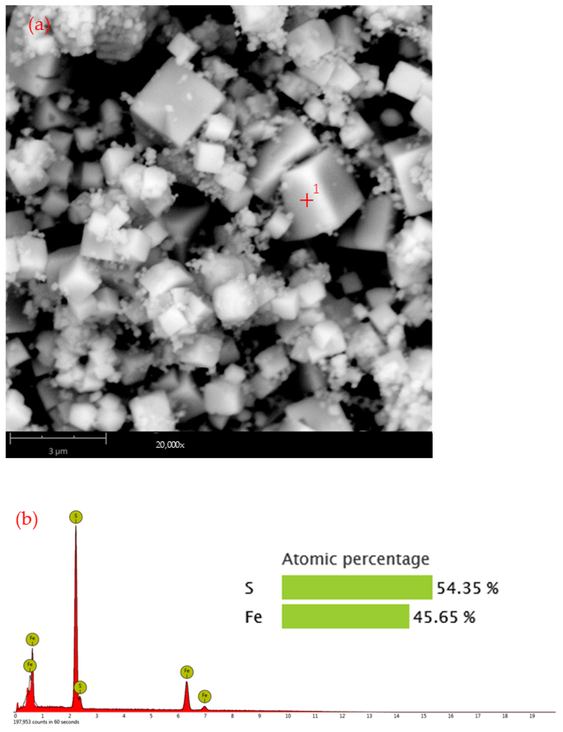
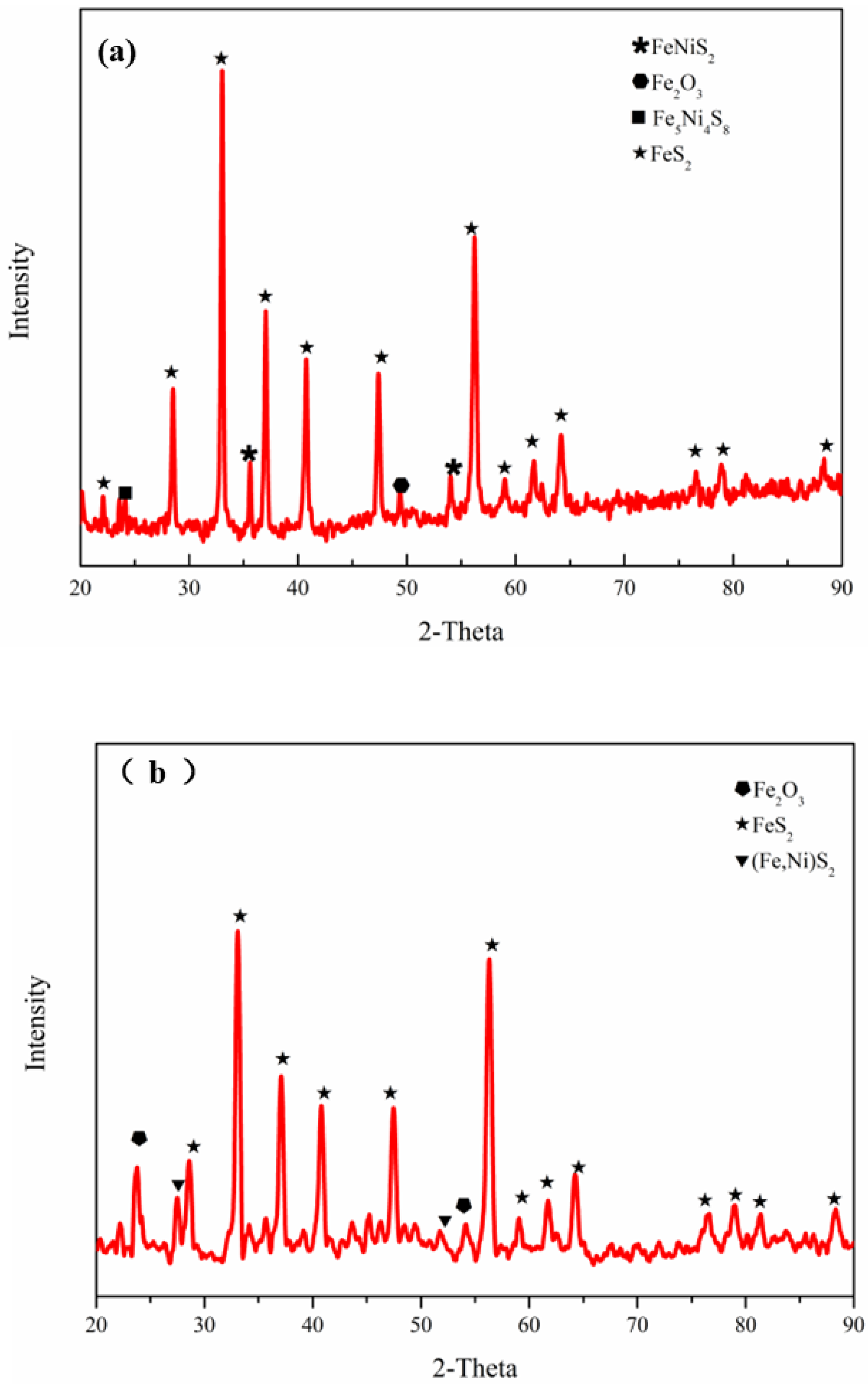
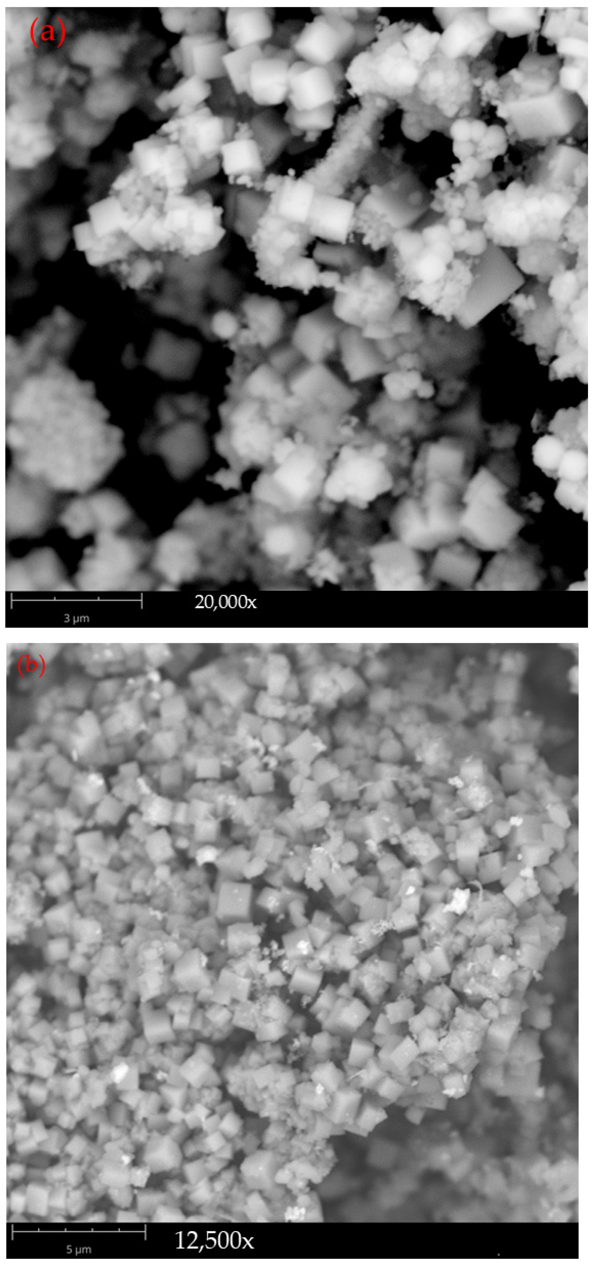
3.1. Phase and Morphology of Synthesized Pure-Phase Pyrite
Figure 2 shows the XRD pattern of the synthetic pure-phase pyrite. The diffraction peaks of the sample in the figure correspond to (111), (200), (210), (211), (220), and (311) in the pyrite-type FeS2 standard card (No.42-1340). The crystal half-peak widths of the sample were small, the diffraction peaks were sharp, and the diffraction intensities were high, indicating that the synthetic pyrite had good crystallinity, a well-developed crystal face, a complete crystal shape, and few impurities. The XRD pattern of the synthesized pyrite had almost no miscellaneous peaks, indicating that the obtained product was of high purity.
Figure 2.
XRD pattern of synthetic pyrite.
After complete quantitative phase analysis of the synthesized pyrite sample in JADE, a column chart of the content of each substance was obtained. As shown in Figure 3, the purity of pyrite in the synthetic sample was higher than that of the natural pyrite used in the experiment.
Figure 3.
Phase and purity of synthetic pyrite.
Figure 4a shows SEM images of synthetic pure-phase pyrite, demonstrating smooth and complete cubic particles with well-developed boundaries and high integrity. The particle size of large particles was approximately 3 μm, and the particle size of small particles was in the range of tens to hundreds of nanometers. Figure 4b shows the spectral analysis results and element content corresponding to point 1 in Figure 4a. It can be seen from the figure that the surface of the cube particle contains only two elements, iron and sulfur, and the specific gravity of iron and sulfur is 45.65% and 54.35%, respectively, which is very close to the theoretical ideal iron and sulfur content of pyrite.
Figure 4.
SEM of synthetic pure-phase pyrite; (a) morphology and (b) spectral diagram and weight percentage at corresponding point 1.
3.2. Influence of the Nickel Doping Content on the Properties of Pyrite
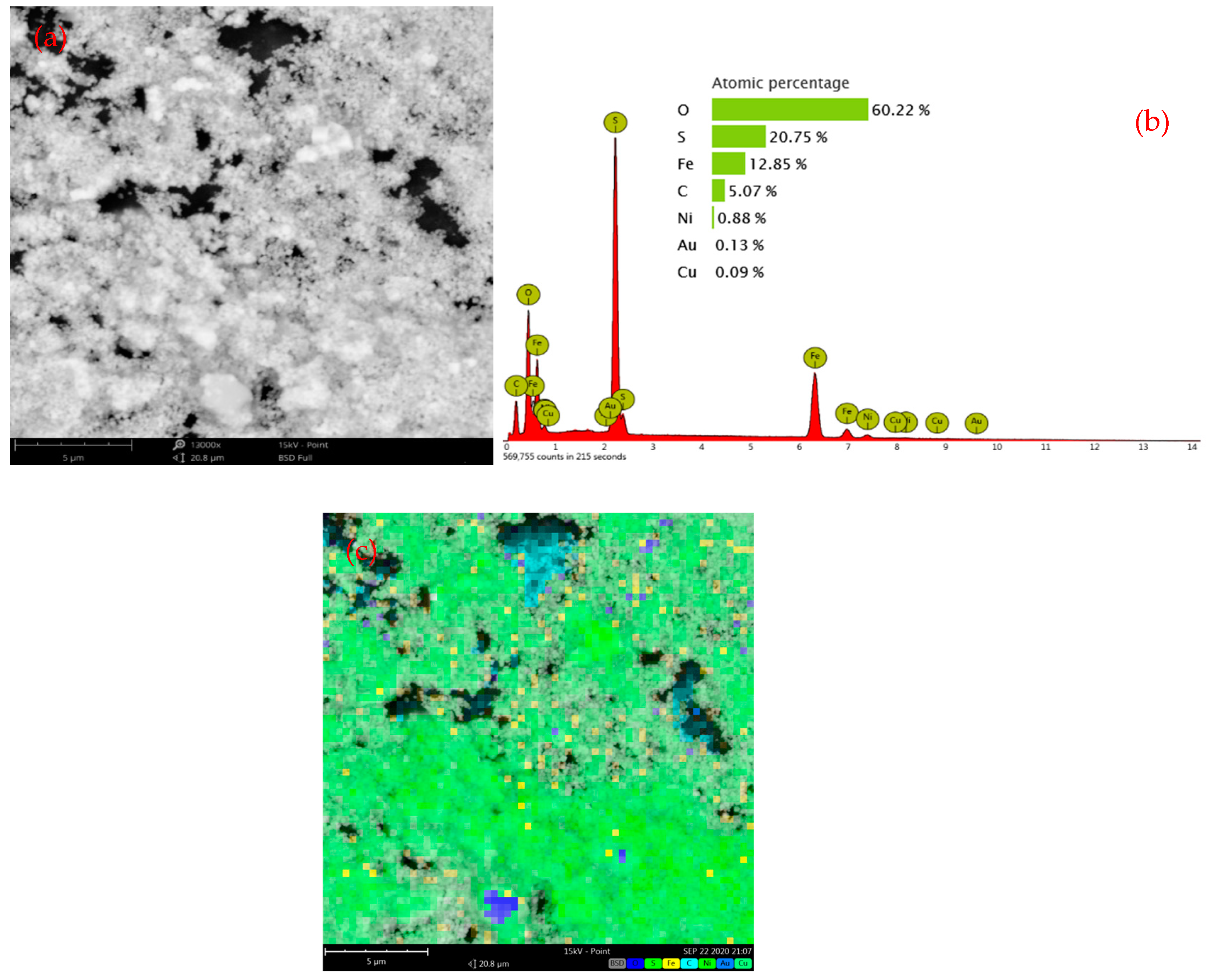
3.2.1. Phase and Morphology of Nickel-Bearing Pyrite
The XRD patterns of the synthesized nickel-bearing pyrite are shown in Figure 5, which indicate that Ni-doped pyrite contains more pyrite-type FeS2 and forms (Fe,Ni)S2 (pyrite, Ni-rich) and pentlandite (such as FeNiS2 and Fe5Ni4S8) phases. This indicates that in the synthesis of nickel-bearing pyrite, a significant portion of nickel exists in a homogeneous form within the nickel-rich pyrite, or as part of pentlandite formed from nickel sulfide and iron sulfide. In this study, pyrite containing a small amount of pentlandite and the lattice substitution of nickel are collectively referred to as nickel-bearing pyrite. By referencing the literature [25], it becomes evident that (Fe,Ni)S2 exhibits remarkable catalytic performance and high activity.
Figure 5.
XRD patterns of nickel-doped pyrite: (a) 2% nickel-doped pyrite and (b) 10% nickel-doped pyrite.
The morphological changes in the synthetic nickel-bearing pyrite are illustrated in Figure 6. Compared with the synthesized pure phase pyrite samples, the morphology and particle size of nickel-bearing pyrite changed significantly. Specifically, a large number of small particles are present in nickel-bearing pyrite mixed with 2% and 5% nickel. When 7% and 10% nickel are added, these small particles aggregate to form larger diameter clumps. Therefore, the morphology and particle size of nickel-containing pyrite samples were significantly changed by increasing the amount of nickel doping. This suggests that the introduction of Ni²+ inhibits FeS2 grain growth and thus reduces the particle size of the FeS2 sample. Due to the larger specific surface area and superior catalytic activity of small particle pyrite, the addition of nickel ions will help improve the performance of FeS2 in the catalytic degradation process.

Figure 6.
Synthetic nickel-doped pyrite surface topography: (a) 2% nickel, (b) 5% nickel, (c) 7% nickel, and (d) 10% nickel.
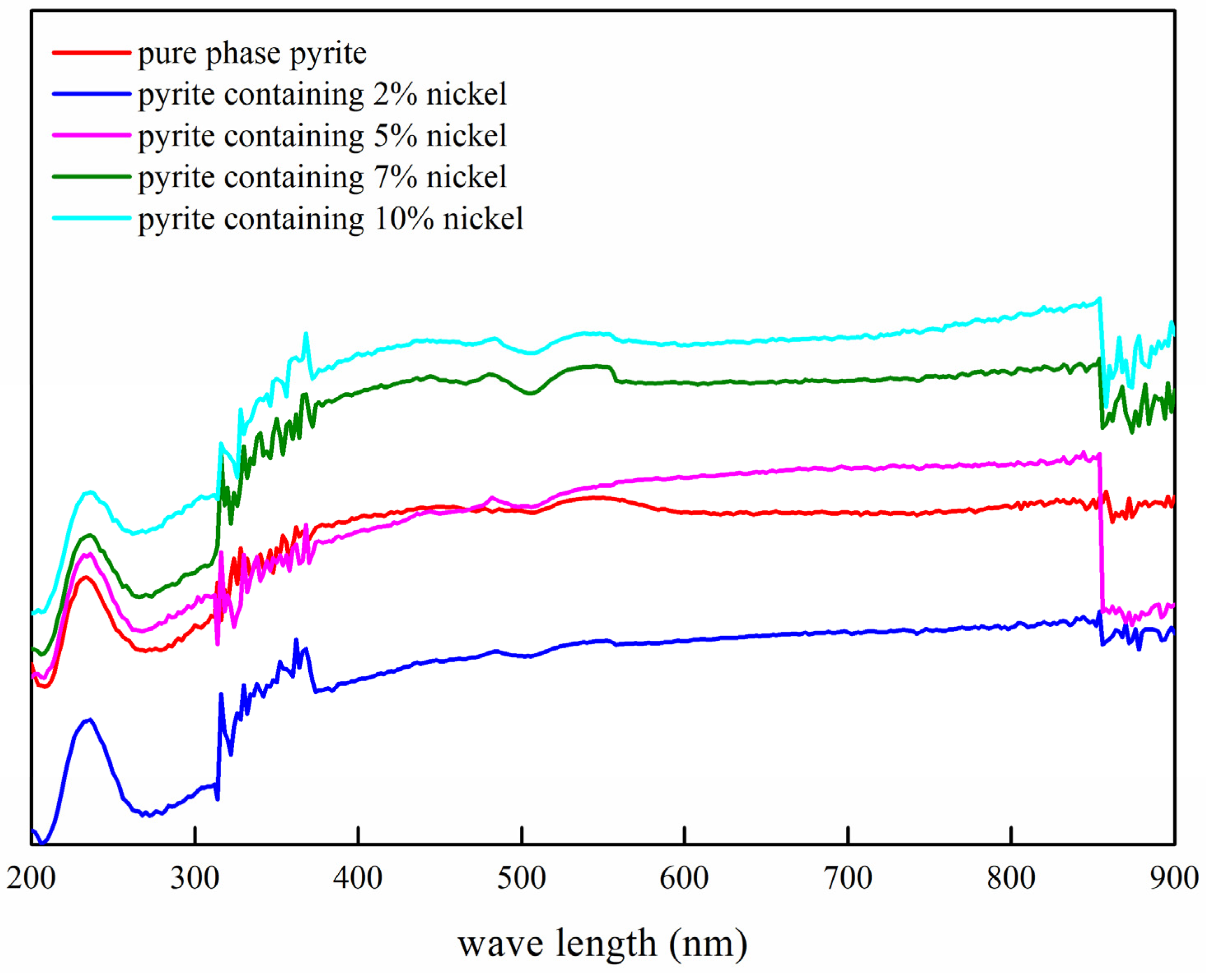
3.2.2. Influence of the Nickel Doping Amount on the Semiconductor Properties of Synthetic Products
Figure 7 illustrates the UV-VIS DRS spectrum of nickel-doped pyrite, which revealed that the light absorption coefficients of natural pyrite and synthetic pure-phase pyrite were similar, whereas those of nickel-bearing pyrite exhibited distinct trends depending on the doping amount. The light absorption coefficient gradually increased with the doping amount. This phenomenon can be attributed to the formation of a small quantity of pentlandite owing to the increased doping, resulting in an increase in the absorption coefficient and ultimately leading to enhanced light absorption.
Figure 7.
UV-VIS DRS spectra of undoped pyrite, synthesized nickel-doped pyrite, and natural pyrite.
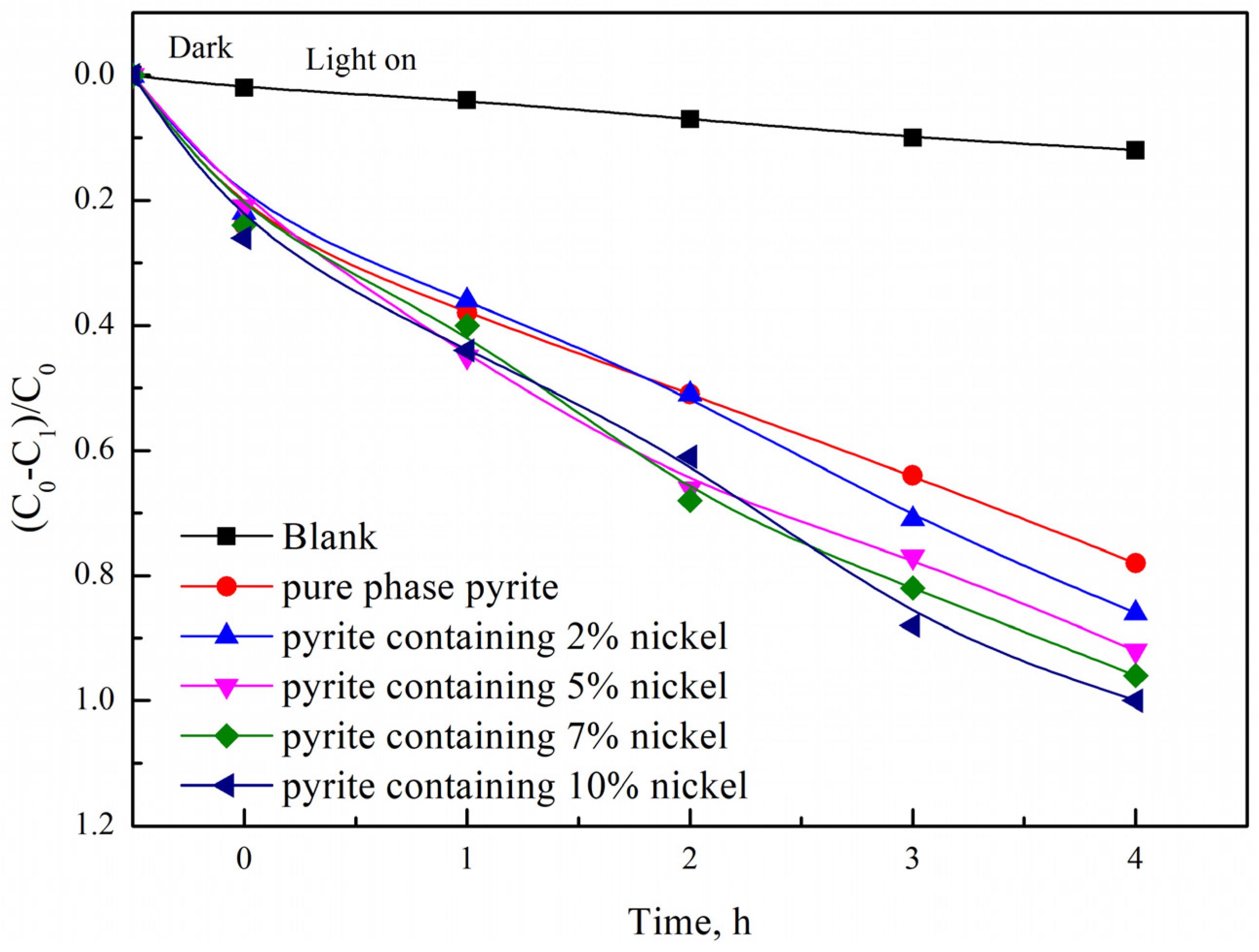
3.2.3. Influence of the Nickel Doping Amount on the Photocatalytic Degradation Properties of Synthetic Products
The degradation effects of natural pyrite, synthetic pure-phase pyrite, and nickel-bearing pyrites (with Ni-doping contents of 2%, 5%, 7%, and 10%, respectively) on methylene blue samples are illustrated in Figure 8. The degradation rate of methylene blue in the solution with nickel-bearing pyrites added increased with increasing Ni-doping content. At a Ni content of 2%, the catalytic degradation rate of nickel-bearing pyrites for methylene blue was 86%, indicating superior visible-light catalytic activity compared to that of pure-phase pyrite. The photocatalytic activity of the nickel-bearing pyrite was significantly enhanced at a nickel content of 7%. After an illumination period of 4 h, the methylene blue degradation rate reached 96%, demonstrating a noticeably better catalytic effect than that achieved using natural pyrite alone. When the nickel doping amount reached 10%, the photocatalytic degradation rate of the nickel-bearing pyrite achieved the complete removal (100%) of methylene blue. Therefore, the photocatalytic effect of nickel-bearing pyrite also increased with increasing nickel doping. The results showed that nickel doping can effectively regulate the photocatalytic properties of pyrite at different doping amounts.
Figure 8.
Changes in the photocatalytic performance of natural pyrite, undoped pyrite, and nickel-doped pyrite.
The reasons for the improvement in the visible-light catalytic activity of nickel-doped pyrite are as follows. After the inclusion of metal ions in the semiconductor photocatalyst, the metal can be used as an effective acceptor of electrons, trapping photoelectrons from the valence band in the conduction band and preventing the combination of semiconductor photoelectrons and holes, thus improving the visible-light catalytic activity of the semiconductor [11].
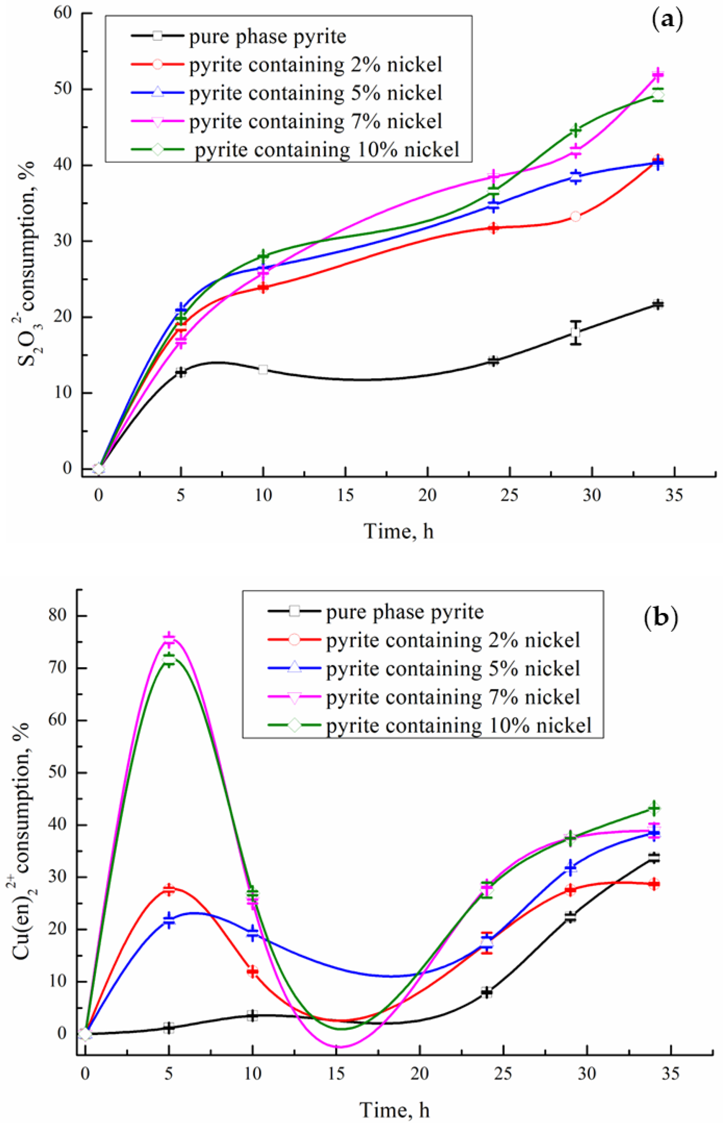
3.3. Influence of Nickel-Containing Pyrite on the Consumption of Thiosulfate and Cu(en)22+ in the System
The catalytic degradation of thiosulfate and the consumption of the copper complex by the nickel content in pyrite were investigated by introducing different nickel-doped pyrites into a Cu2+-en-S2O32− gold leaching system.
The impact of nickel-containing pyrite on thiosulfate consumption in the Cu2+-en-S2O32− gold leaching system was also investigated (Figure 9a). The nickel contents of the pyrite samples were 2%, 5%, 7%, and 10%, with the synthesized pure-phase pyrite used as the control group. When pure-phase pyrite was introduced into the system, thiosulfate consumption increased slowly within 24 h but accelerated rapidly beyond this duration. Upon the addition of nickel-containing pyrite, a significant increase in thiosulfate consumption occurred, which correlated with higher levels of nickel doping (as shown Figure 9a). This is mainly due to the catalytic action of pyrite itself on the decomposition of thiosulfate, and the introduction of nickel inhibits the growth of FeS2 grains, thus reducing the particle size of FeS2 samples. Compared with large-particle pyrite, small-particle pyrite has a larger specific surface area and superior catalytic activity. Therefore, the incorporation of nickel is helpful to improve the catalytic degradation performance of pyrite for thiosulfate. In addition, some scholars have found that the doping of nickel inside pyrite will transform it into N-type semiconductor [11,12]. With the increase in doping amount, the band gap width decreases gradually, making it easier for electrons bound to the surface to transition to the conduction band and transform into free electrons or holes. Therefore, there are more impurities in the nickel-containing pyrite that can accept electrons. When electrons in the valence band are excited into the conduction band, more holes are created [11,26], thus promoting the electron-giving process of thiosulfate. Therefore, nickel-containing pyrite significantly accelerates the oxidative decomposition of thiosulfate [27].
Figure 9.
(a) Effects of nickel doping in pyrite on thiosulfate consumption; (b) Effects of nickel dop-ing in pyrite on Cu(en)22+ consumption (in the Cu2+-en-S2O32− system, pH 9.8–10, temperature 25 °C, time 35 h, solution: 0.1 mol/L S2O32−, 5 mmol/L Cu(en)22+, 200 mL solution).
As shown in Figure 9b, in the Cu2+-en-S2O32− gold leaching system, the addition of pure-phase pyrite had little effect on Cu(en)22+ consumption compared to nickel-bearing pyrite with 2%, 5%, 7%, and 10% nickel contents. The consumption of Cu(en)22+ gradually increased with leaching time when pure-phase pyrite was added to the leaching system. However, when nickeliferous pyrite was added to the gold leaching system, the consumption of Cu(en)22+ initially sharply increased. This was particularly evident in the pyrite system with a large amount of nickel doping, where the consumption of the complex reached approximately 75% within the first 5 h. Subsequently, a slow increase in the content of Cu(en)22+ occurred during the following 5–10 h. When the leaching time exceeded 10 h, a gradual increase in the consumption of Cu(en)22+ occurred as the leaching time increased. In particular, the consumption of Cu(en)22+ also increases with an increase in nickel doping, and there is a clear correlation between the two.
3.4. Mechanistic Analysis of Cu2+-en-S2O32− Gold Leaching with Nickel-Bearing Pyrite
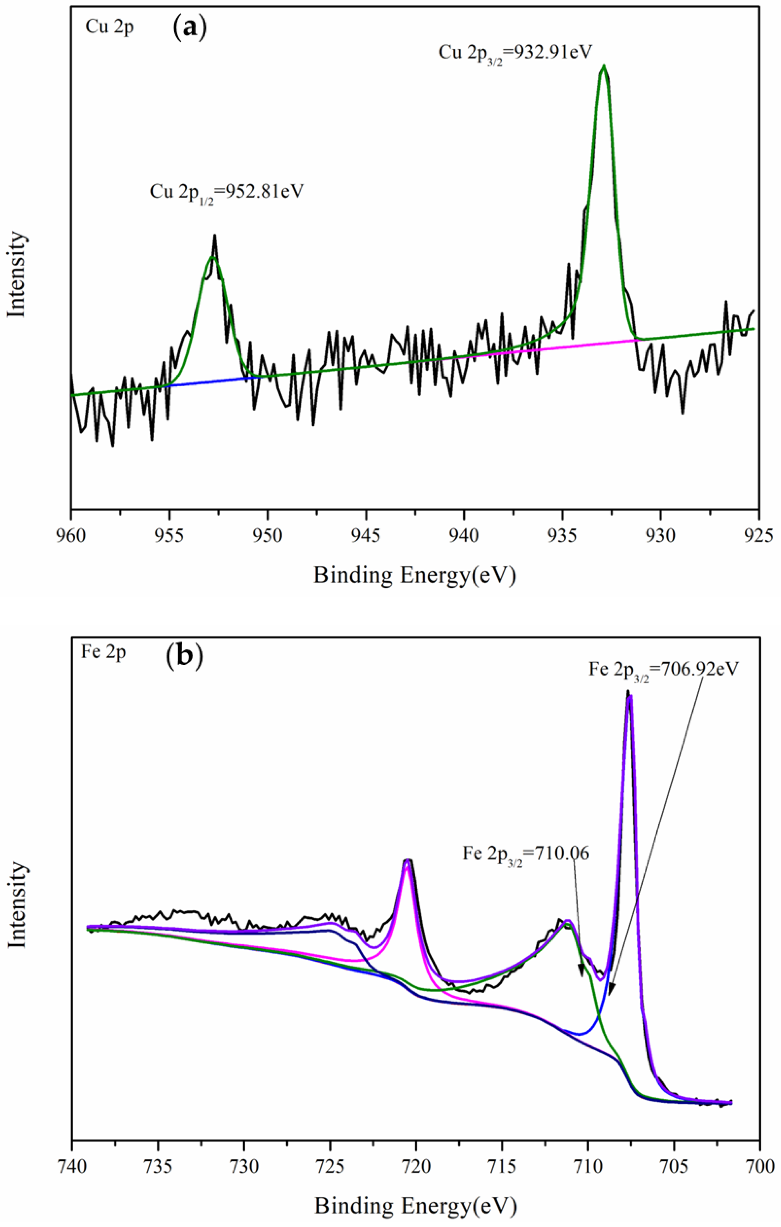
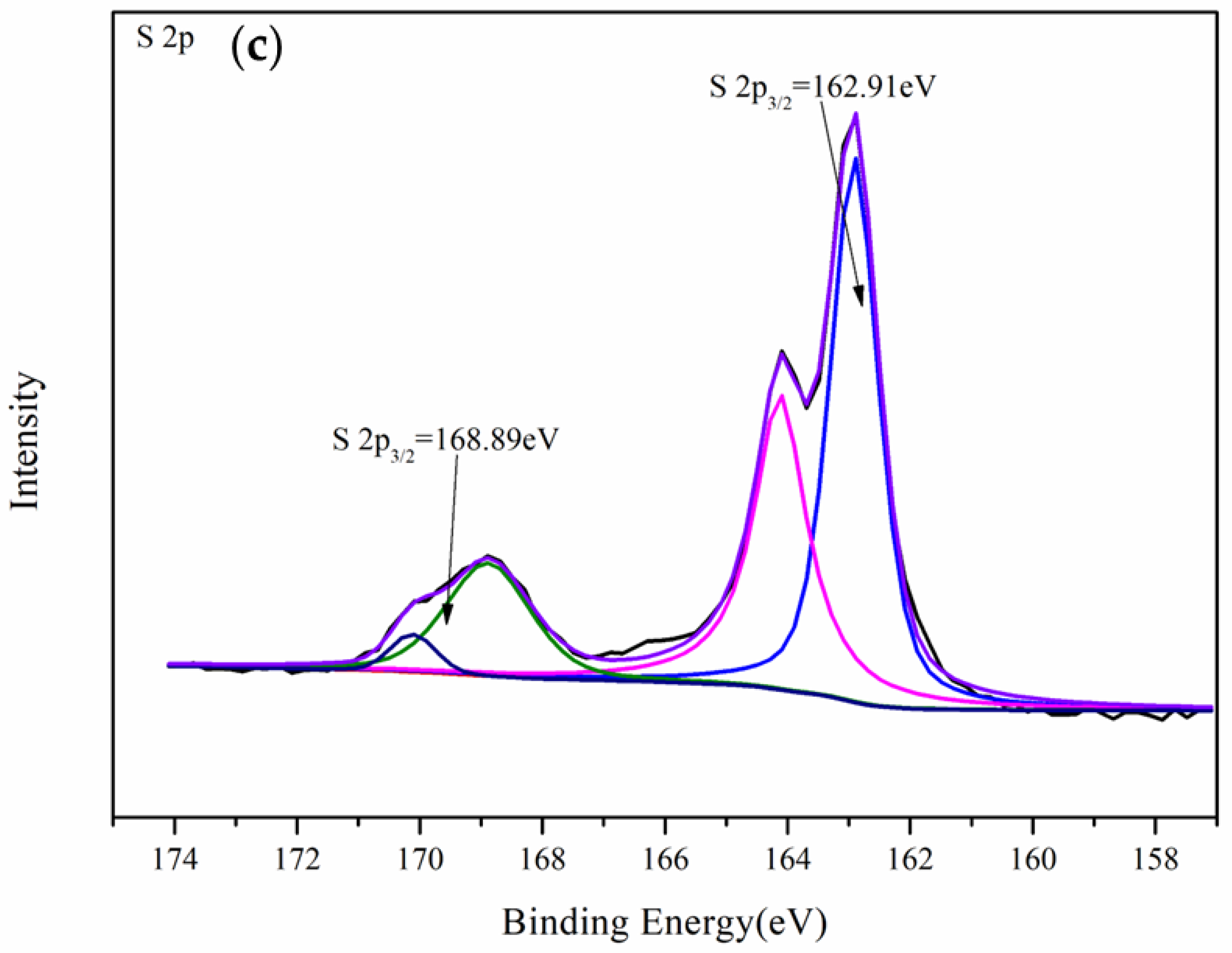
3.4.1. XPS Analysis of Gold Foil After Leaching
As shown in Figure 10a, when 7% Ni-doped pyrite was added to the Cu2+-en-S2O32− system and after fitting the Ni 2p peak of the leached gold foil surface, the peaks at 854.88, 855.43, and 856.36 eV corresponded to NiO, NiS, and NiSO4, respectively [28]. The nickel in the pentlandite was present as free NiS, and the presence of NiSO4 indicated that the nickel in the pentlandite was converted to Ni (II) during the degradation of thiosulfate. Based on the magnitude of the peak values, the primary nickel-containing compounds present on the surface of the leached gold foil were inferred to be NiO and NiS. Zhou’s [29] study revealed the chemical equation of Ni release during the dissolution and reprecipitation of pyrite, as shown in Equation (3) below:
(Fe, Ni)S2 + 3Cl− → FeS2 + NiCl3− + 2H2S + O2

Figure 10.
XPS spectrum of the gold foil surface after leaching with nickel-doped pyrite in the Cu2+-en-S2O32− gold leaching system: (a) Ni 2p, (b) Fe 2p, (c) Cu 2p, and (d) S 2p spectra (pH 9.8–10, temperature 25 °C, time 35 h, solution: 0.1 mol/L S2O3 2−, 5 mmol/L Cu(en)22+, 200 mL solution).
Due to the presence of ferrous chloride in the synthetic raw material, it is possible that the nickel in the nickel pyrite is released during the leaching process and thus dissolved in the gold leaching system.
Figure 10b displays the Fe 2p spectrum observed on the surface of the leached gold foil subsequent to the introduction of nickel-bearing pyrite into the leaching system, wherein Fe 2p3/2 exhibits three primary peaks at 706.87 eV, 710.19 eV, and 712.40 eV. The peak at 706.87 eV predominantly corresponds to FeS2, while the peak at 710.19 eV is attributed to the formation of FeS, and the peak at 712.40 eV arises from adsorption of FeSO4 onto the gold foil surface [30].
In Figure 10c, the Cu 2p3/2 peak at 932.63 eV corresponds to the binding energy of Cu2S [30,31]. This suggests that when nickel-bearing pyrite was introduced into the Cu2+-en-S2O32− gold leaching system, the only copper-containing substance present on the surface of the leached gold foil was Cu2S. This is completely different from the situation in which CuS forms the main copper passivation layer on the surface of the gold foil after the addition of natural pyrite. Therefore, the above results indicate that Cu(II), which was originally complexed with en in the system, was reduced to cuprous ions as the leaching time increased.
In Figure 10d, the S 2p3/2 peak at 162.71 eV corresponds to the binding energy of FeS2, while the peaks at 165.99 eV and 167.12 eV correspond to the binding energies of Na2SO3. The S 2p3/2 peaks at 167.86 eV and 168.97 eV correspond to the binding energies of Na2S2O3 and FeSO4, respectively [32]. The presence of SO32− and SO42− indicates significant decomposition and consumption of thiosulfate in the system.
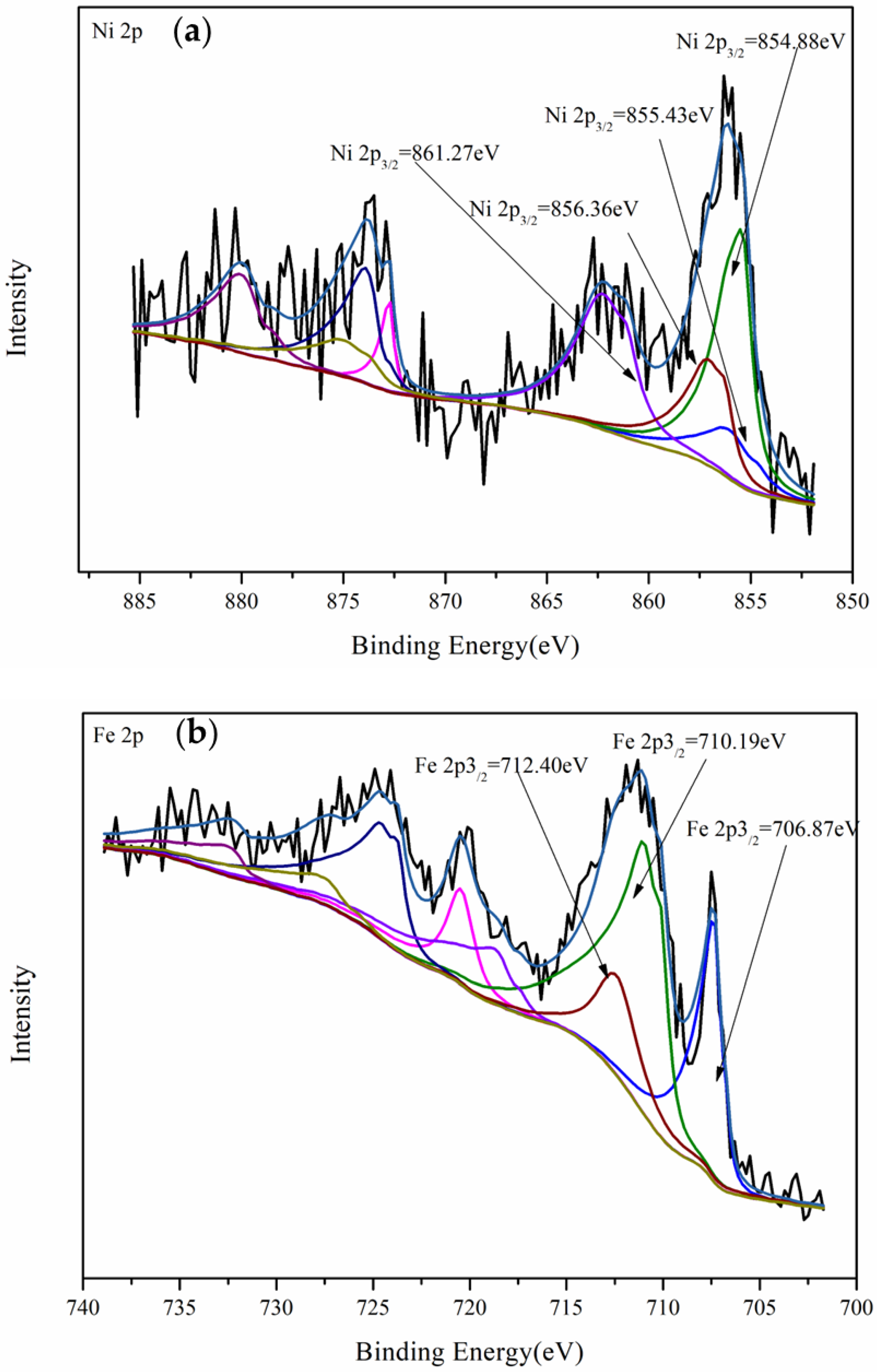
3.4.2. XPS Analysis of Nickel-Bearing Pyrite After Leaching
Figure 11a–c shows the Fe 2p, Cu 2p, and S 2p spectra, respectively, on the surface of the nickel-bearing pyrite after its addition to the Cu2+-en-S2O32− gold leaching system. In Figure 11a, the peak of Cu 2p3/2 (932.91 eV) was determined to be Cu2S [33], and no Cu2+ was present, indicating that the copper ions in the original system were more easily reduced to Cu+ under the promotion of nickel ions after the addition of nickel-containing pyrite. In Figure 11b, the peak at 706.92 eV represents the binding energy of Fe 2p3/2 in the disulfide found in pyrite, while the binding energy peak of Fe 2p3/2 in FeS was observed at 710.06 eV [30].

Figure 11.
XPS spectra of the leached pyrite with nickel-doped pyrite in the Cu2+-en-S2O32− leaching system: (a) Cu 2p, (b) Fe 2p, and (c) S 2p spectra (pH 9.8–10, temperature 25 °C, time 35 h, solution: 0.1 mol/L S2O3 2−, 5 mmol/L Cu(en)22+, 200 mL solution).
In Figure 11c, the S 2p3/2 peak at 162.91 eV corresponds to FeS2, and the S 2p3/2 peak at 168.89 eV corresponds to FeSO4 [10]. Compared with the spectral diagram of S 2p3/2 on the surface of the leached gold foil and nickel-containing pyrite, thiosulfate adsorption was not observed on the surface of the leached nickel-containing pyrite. Owing to the addition of nickel-bearing pyrite, a significant amount of thiosulfate in the system was catalytically decomposed, resulting in only a small quantity remaining. Consequently, a substantial amount of sulfate, which is the final decomposition product of thiosulfate, was adsorbed onto the surface of the leached nickel-bearing pyrite.
3.4.3. SEM Analysis of Nickel-Bearing Pyrite After Leaching
After introducing nickel-bearing pyrite into the Cu2+-en-S2O32− gold leaching system for the gold leaching test, the nickel-bearing pyrite was analyzed using SEM-EDS(as shown in Figure 12). A comparison of the surface topography of the nickel-bearing pyrite before and after the test revealed that a large number of fine particles or powder was adsorbed onto the surface of the leached nickel-bearing pyrite (Figure 12a). Moreover, the cubic particles of the leached nickel-bearing pyrite became smaller and agglomerated, forming more spherical particles or clumps. EDS analysis showed that the adsorbed materials on the surface of the leached nickel-bearing pyrite mainly contained Cu, Fe, Ni, Au, S, and other elements, indicating that nickel-bearing pyrite adsorbed some gold complexes (Figure 12b,c).
Figure 12.
(a) SEM image of the leached nickel-doped pyrite surface in the Cu2+-en-S2O32− leaching system, (b) corresponding EDS spectrum, and (c) element map (pH 9.8–10, temperature 25 °C, time 35 h, solution: 0.1 mol/L S2O3 2−, 5 mmol/L Cu(en)22+, 200 mL solution).
Owing to the high consumption of thiosulfate in the leaching system with nickel-bearing pyrite added, when the concentration of thiosulfate in the solution was very low or almost nonexistent, the stability of Au+ and the number of adsorption sites for thiosulfate and gold thiosulfate complex ions decreased. Consequently, the adsorption of gold on the mineral surfaces increased, which led to a phenomenon known as gold robbing [20].
3.4.4. Mechanism of the Catalytic Decomposition of Thiosulfate by Nickel-Bearing Pyrite in the Cu2+-en-S2O32− Gold Leaching System
During gold leaching in the Cu2+-NH3-S2O32− system, pyrite undergoes oxidation by dissolved oxygen and copper (II), which can be represented by the following simplified equation (based on the HSC 6.0 database):
Based on the effects of nickel-bearing pyrite on gold dissolution and thiosulfate and Cu(en)22+ consumption in the Cu2+-en-S2O32− leaching system as well as the surface morphology and XPS analysis of the gold foil and nickel-bearing pyrite after leaching, the main adsorbed substances on the surface of the gold foil were determined to be NiS, NiSO4, FeS2, FeSO4, and Cu2S. This indicates that nickel dissolved in the system and the free nickel ions combined with SO42− and S2−, resulting in the partial adsorption of NiS and NiSO4 onto the gold foil, which adversely affected gold dissolution. Based on the changes in Cu(en)22+ consumption in the system and the formation of Cu2S on the surface of the leached gold foil, a portion of the nickel in the nickel-bearing pyrite dissolved in the system to form numerous free Ni2+. These Ni ions accelerated the conversion of Cu2+ to Cu+.
As the complexation of en with Ni2+ made chemical Equation (5) faster and Equation (6) more difficult, the concentration of Cu(en)22+ continued to decrease over a short period of time. Cu(I) was reoxidized to Cu(II) by the dissolved oxygen in the leaching solution; however, the oxidation of the complex formed between Cu(I) and en by dissolved oxygen occurred at a slower rate than the complex reaction between nickel ions and free en (5); as shown in Table 2, the Ni(en)22+ was not as stable as Cu(en)22+. Therefore, after a certain period, the concentration of Cu(en)22+ gradually increased.
Table 2.
The equilibrium constants (logKa) for the complexation of Ni2+, Cu+, Cu2+ with en: ionic strength (I = 0), 25 °C.
The consumption of thiosulfate in the gold leaching system with the addition of nickel-containing pyrite increased sharply with increasing nickel content, indicating that the consumption of thiosulfate was related to nickel doping. First, the complexation of dissolved nickel ions with en accelerated the progress of Equation (8), thereby expediting the consumption of thiosulfate. Second, the nickel doping of pyrite increased the donor energy level of the pyrite semiconductor. The rate-controlling step of thiosulfate decomposition is the process in which oxygen accepts electrons provided by pyrite; therefore, a higher nickel doping content led to more donor impurities in the pyrite semiconductor and a narrower bandgap width, which was conducive to the rapid progress of the electron supply process in pyrite. Therefore, the process of electron acceptance by oxygen was promoted, and thiosulfate consumption increased with increasing Ni-doping content [22].
4. Conclusions
The effects of the impurity content in nickel-containing pyrite on the light absorption and photocatalytic degradation properties as well as the catalytic degradation of thiosulfate and consumption of Cu(en)22+ in the Cu2+-en-S2O32− system were studied. The main conclusions are as follows:
(1) In nickel-doped pyrite, the amount of nickel doping significantly affected the light absorption performance of pyrite. As the level of nickel doping increased, the photocatalytic performance of nickel-doped pyrite was enhanced between 2 and 4 hours.
(2) In the Cu2+-en-S2O32− gold leaching system, the passivation effect of nickel-bearing pyrite on gold dissolution was enhanced by increasing the nickel content.
(3) With an increase in the nickel content (from 2 to 10%) in pyrite, the catalytic degradation of thiosulfate was also enhanced. This is mainly because the nickel in nickeliferous pyrite forms complexes with en solution, resulting in the accelerated oxidation decomposition of thiosulfate to produce en and SO42−. Additionally, a higher nickel content leads to an increased presence of donor impurities in pyrite semiconductors. The faster the electron supply process of pyrite, the more it promotes the electron acceptance of oxygen, thereby accelerating the rate of oxidation and decomposition of thiosulfate.
Supplementary Materials
The following supporting information can be downloaded at: https://www.mdpi.com/article/10.3390/met14111256/s1.
Author Contributions
Conceptualization, X.Q. and W.Q.; Methodology, T.Z.; Investigation, T.Z.; Resources, T.Z. and W.Q.; Data curation, T.Z.; Writing—original draft, X.Q.; Writing—review & editing, X.Q.; Supervision, H.Z.; Project administration, H.Z.; Funding acquisition, T.Z. All authors have read and agreed to the published version of the manuscript.
Funding
This research was funded by Inner Mongolia University of Technology grant number [ZZ202115]. Basic Scientific Research Expenses Program of Universities directly under Inner Mongolia grant number [JY20220155]. Funded by Open Foundation of State Key Laboratory of Mineral Processing grant number [BGRIMM-KJSKL-2024-29].
Data Availability Statement
All relevant data are within the paper.
Conflicts of Interest
We declare that we have no known competing financial interests or personal relationships that could have appeared to influence the work reported in this paper.
References
- Lampinen, M.; Laari, A.; Turunen, I. Ammoniacal thiosulfate leaching of pressure oxidized sulfide gold concentrate with low reagent consumption. Hydrometallurgy 2015, 151, 1–9. [Google Scholar] [CrossRef]
- Dai, X.; Jeffrey, M.I. The effect of sulfide minerals on the leaching of gold in aerated cyanide solutions. Hydrometallurgy 2006, 82, 118–125. [Google Scholar] [CrossRef]
- Hasab, M.G.; Rashchi, F.; Raygan, S. Chloride–hypochlorite leaching and hydrochloric acid washing in multi-stages for extraction of gold from a refractory concentrate. Hydrometallurgy 2014, 142, 56–59. [Google Scholar] [CrossRef]
- Hashemzadehfini, M.; Ficeriová, J.; Abkhoshk, E.; Shahraki, B.K. Effect of mechanical activation on thiosulfate leaching of gold from complex sulfide concentrate. Trans. Nonferrous Met. Soc. China 2011, 21, 2744–2751. [Google Scholar] [CrossRef]
- Hasab, M.G.; Raygan, S.; Rashchi, F. Chloride–hypochlorite leaching of gold from a mechanically activated refractory sulfide concentrate. Hydrometallurgy 2013, 138, 59–64. [Google Scholar] [CrossRef]
- Mahandra, F.G.A. Novel Extraction Process for Gold Recovery from Thiosulfate Solution Using Phosphonium Ionic Liquids. ACS Sustain. Chem. Eng. 2021, 9, 8179–8185. [Google Scholar] [CrossRef]
- Fleming, C.A.; McMullen, J.; Thomas, K.G.; Wells, J.A. Recent advances in the development of an alternative to the cyanidation process: Thiosulfate leaching and resin in pulp. Min. Metall. Explor. 2003, 20, 1–9. [Google Scholar] [CrossRef]
- Feng, D.; Van Deventer, J.S. Ammoniacal thiosulphate leaching of gold in the presence of pyrite. Hydrometallurgy 2006, 82, 126–132. [Google Scholar] [CrossRef]
- Yang, Y.B.; Zhang, X.; Bin, X.U.; Qian, L.I.; Jiang, T.; Wang, Y.X. Effect of arsenopyrite on thiosulfate leaching of gold. Trans. Nonferrous Met. Soc. China 2015, 25, 3454–3460. [Google Scholar] [CrossRef]
- Xu, Y.; Schoonen, M.A. The stability of thiosulfate in the presence of pyrite in low-temperature aqueous solutions. Geochim. Cosmochim. Acta 1995, 59, 4605–4622. [Google Scholar] [CrossRef]
- Nie, Y.; Wang, Q.; Zi, F.; Hu, X.; Chen, Y.; Guo, L.; Yu, H. The catalytic decomposition of thiosulfate by pyrite. Appl. Surf. Sci. 2018, 436, 217–223. [Google Scholar] [CrossRef]
- Ma, W.; Chen, J.; Zhang, G.; Li, Y.; Feng, Q. The effect of As doping concentration on the electronic structure of FeS2. Chin. J. Nonferrous Met. 2017, 27, 414–422. (In Chinese) [Google Scholar]
- Chen, J.H.; Ye, C.; Li, Y.Q. Effect of vacancy defects on electronic properties and activation of sphalerite (110) surface by first-principles. Trans. Nonferrous Met. Soc. China 2010, 20, 502–506. [Google Scholar] [CrossRef]
- Caban-Acevedo, M.; Faber, M.S.; Tan, Y.; Hamers, R.J.; Jin, S. Synthesis and properties of semiconducting iron pyrite (FeS2) nanowires. Nano Lett. 2012, 12, 1977–1982. [Google Scholar] [CrossRef] [PubMed]
- Chu, F.; Sun, G.; Hu, R.; Hu, D.; Wei, C. Electron Hole-Core Characteristics of Pyrites from the Main Types of Gold Deposits in China and Affecting Factors. Acta Mineral. Sin. 2004, 24, 211–217. (In Chinese) [Google Scholar]
- Jefferson, M.; Yenial-Arslan, U.; Evans, C.; Curtis-Morar, C.; O’Donnell, R.; Parbhakar-Fox, A.; Forbes, E. Effect of pyrite textures and composition on flotation performance: A review. Miner. Eng. 2023, 201, 108234. [Google Scholar] [CrossRef]
- Li, Y.Q.; Chen, J.H.; Guo, J. DFT study of influences of As, Co and Ni impurities on pyrite (100) surface oxidation by O2 molecule. Chem. Phys. Lett. 2004, 24, 211–217. [Google Scholar] [CrossRef]
- Wu, J.; Ma, W.; Zhang, G.-F.; Feng, Q.-M. First-Principle Study of Electronic Structure and Optical Property of Cu/Co Doped FeS2. Acta Opt. Sin. 2016, 36, 1016001. (In Chinese) [Google Scholar]
- Mingyue, Z. Ni-Doped FeS: Solvothermal Synthesis and the Visible-Light Photocatalytic Properties. Chin. J. Inorg. Chem. 2015, 31, 1119–1124. (In Chinese) [Google Scholar]
- Wang, Q.; Hu, X.; Zi, F.; Yang, P.; Chen, Y.; Chen, S. Environmentally friendly extraction of gold from refractory concentrate using a copper–ethylenediamine–thiosulfate solution. J. Clean. Prod. 2019, 214, 860–872. [Google Scholar] [CrossRef]
- Nie, Y.; Yu, Q.; Hu, X.; Zi, F.; Yu, H. The effect of ammonia on the anodic process of gold in copper-free thiosulfate solution. J. Electrochem. Soc. 2016, 163, E123–E129. [Google Scholar] [CrossRef]
- Nie, Y.; Chi, H.; Zi, F.; Hu, X.; Yu, H.; He, S. The effect of cobalt and nickel ions on gold dissolution in a thiosulfate-ethylenediamine (en)-Cu2+ system. Miner. Eng. 2015, 83, 205–210. [Google Scholar] [CrossRef]
- Cao, Y.; Sun, Y.; Gao, P.; Han, Y.; Li, Y. Mechanism for suspension magnetization roasting of iron ore using straw-type biomass reductant. Int. J. Min. Sci. Technol. 2021, 31, 9–14. [Google Scholar] [CrossRef]
- Zhang, X.; Kou, J.; Sun, C. A comparative study of the thermal decomposition of pyrite under microwave and conventional heating with different temperatures. J. Anal. Appl. Pyrolysis 2019, 138, 41–53. [Google Scholar] [CrossRef]
- Lin, X.; Xia, Y.; Wei, G.; Zhou, J.; Liang, X.; Xian, H.; Zhu, J.; He, H. Distinct effects of transition metal (cobalt, manganese and nickel) ion substitutions on the abiotic oxidation of pyrite: In view of hydroxyl radical production. Geochim. Cosmochim. Acta 2022, 321, 170–183. [Google Scholar] [CrossRef]
- Xu, Y.; Schoonen, M.A.; Strongin, D.R. Thiosulfate oxidation: Catalysis of synthetic sphalerite doped with transition metals. Geochim. Cosmochim. Acta 1996, 60, 4701–4710. [Google Scholar] [CrossRef]
- Ou, Y.; Yang, Y.; Li, K.; Gao, W.; Wang, L.; Li, Q.; Jiang, T. Eco-friendly and low-energy innovative scheme of self-generated thiosulfate by atmospheric oxidation for green gold extraction. J. Clean. Prod. 2023, 387, 135818. [Google Scholar] [CrossRef]
- Feng, D.; Van Deventer, J.S. Thiosulphate leaching of gold in the presence of orthophosphate and polyphosphate. Hydrometallurgy 2011, 106, 38–45. [Google Scholar] [CrossRef]
- Zhou, Z.; Chen, Z.; Weyer, S.; Horn, I.; Huo, H.; Zhang, W.; Li, N.; Zhang, Q.; Han, F.; Feng, H. Metal source and ore precipitation mechanism of the Ashawayi orogenic gold deposit, southwestern Tianshan Orogen, western China: Constraints from textures and trace elements in pyrite. Ore Geol. Rev. 2023, 157, 105452. [Google Scholar] [CrossRef]
- Velásquez, P.; Leinen, D.; Pascual, J.; Ramos-Barrado, J.R.; Grez, P.; Gomez, H.; Schrebler, R.; Del Río, R.; Cordova, R. A chemical, morphological, and electrochemical (XPS, SEM/EDX, CV, and EIS) analysis of electrochemically modified electrode surfaces of natural chalcopyrite (CuFeS2) and pyrite (FeS2) in alkaline solutions. J. Phys. Chem. B 2005, 109, 4977–4988. [Google Scholar] [CrossRef]
- Acres, R.G.; Harmer, S.L.; Beattie, D.A. Synchrotron XPS studies of solution exposed chalcopyrite, bornite, and heterogeneous chalcopyrite with bornite. Int. J. Miner. Process. 2010, 94, 43–51. [Google Scholar] [CrossRef]
- Laajalehto, K.; Kartio, I.; Nowak, P. XPS study of clean metal sulfide surfaces. Appl. Surf. Sci. 1994, 81, 11–15. [Google Scholar] [CrossRef]
- Deroubaix, G.; Marcus, P. X-ray photoelectron spectroscopy analysis of copper and zinc oxides and sulphides. Surf. Interface Anal. 1992, 18, 39–46. [Google Scholar] [CrossRef]
Disclaimer/Publisher’s Note: The statements, opinions and data contained in all publications are solely those of the individual author(s) and contributor(s) and not of MDPI and/or the editor(s). MDPI and/or the editor(s) disclaim responsibility for any injury to people or property resulting from any ideas, methods, instructions or products referred to in the content. |
© 2024 by the authors. Licensee MDPI, Basel, Switzerland. This article is an open access article distributed under the terms and conditions of the Creative Commons Attribution (CC BY) license (https://creativecommons.org/licenses/by/4.0/).