


Abstract
Background/Objectives: Skene’s gland malignancies are extremely rare tumors of the female paraurethral region, known almost exclusively from case reports and small case series. Their diagnosis is often delayed because symptoms are nonspecific and may mimic benign periurethral disorders, urethral diverticula, primary urethral carcinoma or anterior vaginal wall tumors. This narrative review focuses on the role of imaging, particularly pelvic magnetic resonance imaging (MRI), in the diagnosis, differential diagnosis, staging and treatment planning of Skene’s gland malignancies, distinguishing evidence from confirmed Skene’s gland cases from evidence extrapolated from female urethral carcinoma. Methods: PubMed/MEDLINE, Scopus and Web of Science were searched on 27 September 2026 (publication years 2015–2026) and supplemented by reference-list screening; an explicit case definition was applied. Results: Of 191 records (93 after de-duplication), 17 reports of Skene’s gland malignancy were included (15 case reports or series and 2 systematic reviews); fewer than thirty cases have been published overall. Patients were mostly postmenopausal (median age about 70 years, range 45–91), presenting with an asymptomatic periurethral nodule, hematuria or vaginal bleeding, or lower urinary tract symptoms; no uniform staging system has been applied. Most tumors were prostatic-type adenocarcinomas treated by local excision alone; radiotherapy, chemotherapy or androgen deprivation were used mainly for nodal, metastatic or recurrent disease. After examination, ultrasonography can detect and characterize a paraurethral mass, whereas MRI best defines tumor epicenter, urethral, periurethral and anterior vaginal wall involvement and regional spread, and should precede cystourethroscopy and biopsy; CT and PET/CT are complementary for systemic staging. No single immunohistochemical marker establishes Skene’s gland origin. Conclusions: MRI is central to localizing and staging suspected Skene’s gland malignancy and thus to guiding biopsy and the choice between local excision and radical surgery, but most imaging recommendations are extrapolated from female urethral carcinoma and require validation in pooled multicenter data.
1. Introduction
The Skene’s glands, also known as paraurethral glands, are homologous to the male prostate and are located adjacent to the distal female urethra. Although benign inflammatory and cystic disorders of the Skene’s glands are relatively uncommon, malignant transformation is exceedingly rare. Skene’s gland malignancies are generally classified among female urethral adenocarcinomas, representing a minute fraction of genitourinary cancers in women.
The rarity of these tumors has resulted in a limited understanding of their natural history, optimal imaging work-up, prognostic factors, and therapeutic management. Most available data derive from isolated case reports and small retrospective series. Consequently, diagnosis is frequently delayed because symptoms such as dysuria, dyspareunia, hematuria, urinary obstruction, or recurrent urinary tract infections are nonspecific and may mimic benign periurethral lesions.
Reliable population-based epidemiological data specific to Skene’s gland malignancy are lacking, given the extreme rarity of this tumor. Population-based cancer registry data indicate that primary urethral carcinoma itself is an uncommon malignancy in women, with an estimated age-adjusted incidence below two cases per million population, increasing markedly among women older than 75 years [1]. Urethral carcinoma accounts for approximately 0.02% of all female cancers and 0.003% of female urogenital malignancies, adenocarcinoma represents only 8–10% of primary female urethral carcinomas, and female urethral adenocarcinoma as a whole is associated with a poor prognosis and low survival [2,3]. Within this already rare group of tumors, neoplasms arising from the Skene’s glands are considered exceptionally uncommon: depending on the diagnostic criteria applied, between 15 and 22 cases had been collected by successive reviews up to 2023, and fewer than thirty cases have been published worldwide to date, with a median patient age around the seventh decade of life (Section 4.1) [2,4,5]. Current knowledge of this entity therefore continues to rely almost entirely on isolated case reports and small case series rather than on population-level data.
Advances in pelvic imaging and immunohistochemical characterization have improved the recognition of periurethral neoplasms. MRI, diffusion-weighted imaging and contrast-enhanced sequences are increasingly used for the local staging and surgical planning of periurethral tumors, although their use in Skene’s gland malignancy is documented in only a few reports [2,6,7]. Concurrently, immunohistochemical studies have reinforced the concept that Skene’s gland derivatives and some of their tumors share an immunophenotype with prostatic tissue [8,9].
This narrative review aims to summarize the current evidence on Skene’s gland malignancies with a particular focus on the role of imaging in their diagnosis, differential diagnosis, local staging and treatment planning, explicitly distinguishing evidence obtained from confirmed Skene’s gland cases from evidence extrapolated from female urethral carcinoma.
2. Materials and Methods
2.1. Search Strategy and Study Selection
This manuscript was prepared as a narrative review based on a structured and reproducible literature search, addressing the diagnosis, imaging assessment, differential diagnosis and therapeutic approach of Skene’s gland malignancies. PubMed/MEDLINE, Scopus and the Web of Science Core Collection were searched; all searches were last run on 27 September 2026 and were limited to publication years 2015–2026.
In each database, two search strings were used: (i) Skene’s gland AND malignancy and (ii) Skene’s gland AND imaging, adapted to the syntax of each database (PubMed: (skene’ gland) AND (malignancy) and (skene’s glands) AND (imaging); Scopus: TITLE-ABS-KEY(“skene* gland*” AND malignan*) and TITLE-ABS-KEY(“skene* gland*” AND imaging); Web of Science: TS = (“skene* gland*” AND malignan*) and TS = (“skene* gland*” AND imaging)). To identify case reports published before 2015 or not retrieved by these strings, and to collect supporting literature, the reference lists of the included articles and of previous reviews [2,4,5] were screened, and additional targeted searches were performed using the terms “female paraurethral adenocarcinoma”, “female urethral adenocarcinoma”, “female urethral carcinoma”, “periurethral tumor”, “paraurethral mass”, “female urethral MRI”, “periurethral tumor MRI”, “ultrasonography”, “computed tomography”, “PET/CT”, “histopathology” and “immunohistochemistry”.
Records were de-duplicated and screened by title and abstract, followed by full-text assessment. For the case-level synthesis (Table 1), reports were eligible if they described one or more patients with a primary malignancy attributed to the Skene’s (paraurethral) glands, or systematically reviewed such cases. Female urethral adenocarcinomas for which a Skene’s gland origin was neither claimed nor supported were excluded from the case count and retained as comparators. Articles published in English, or in other languages with an informative English abstract, were considered. Duplicate reports of the same patient were sought by comparing age, presentation, institution and treatment across individual reports and previous reviews [2,4,5]; no duplicate patients were identified among the individually tabulated cases. Supporting literature on anatomy, imaging, pathology, differential diagnosis and treatment of female urethral and periurethral lesions was selected narratively from the same searches and from reference lists; studies dealing only with male urethral or prostatic malignancies were considered only when relevant to the homology between the Skene’s glands and the prostate or to the interpretation of prostate-associated immunohistochemical markers.
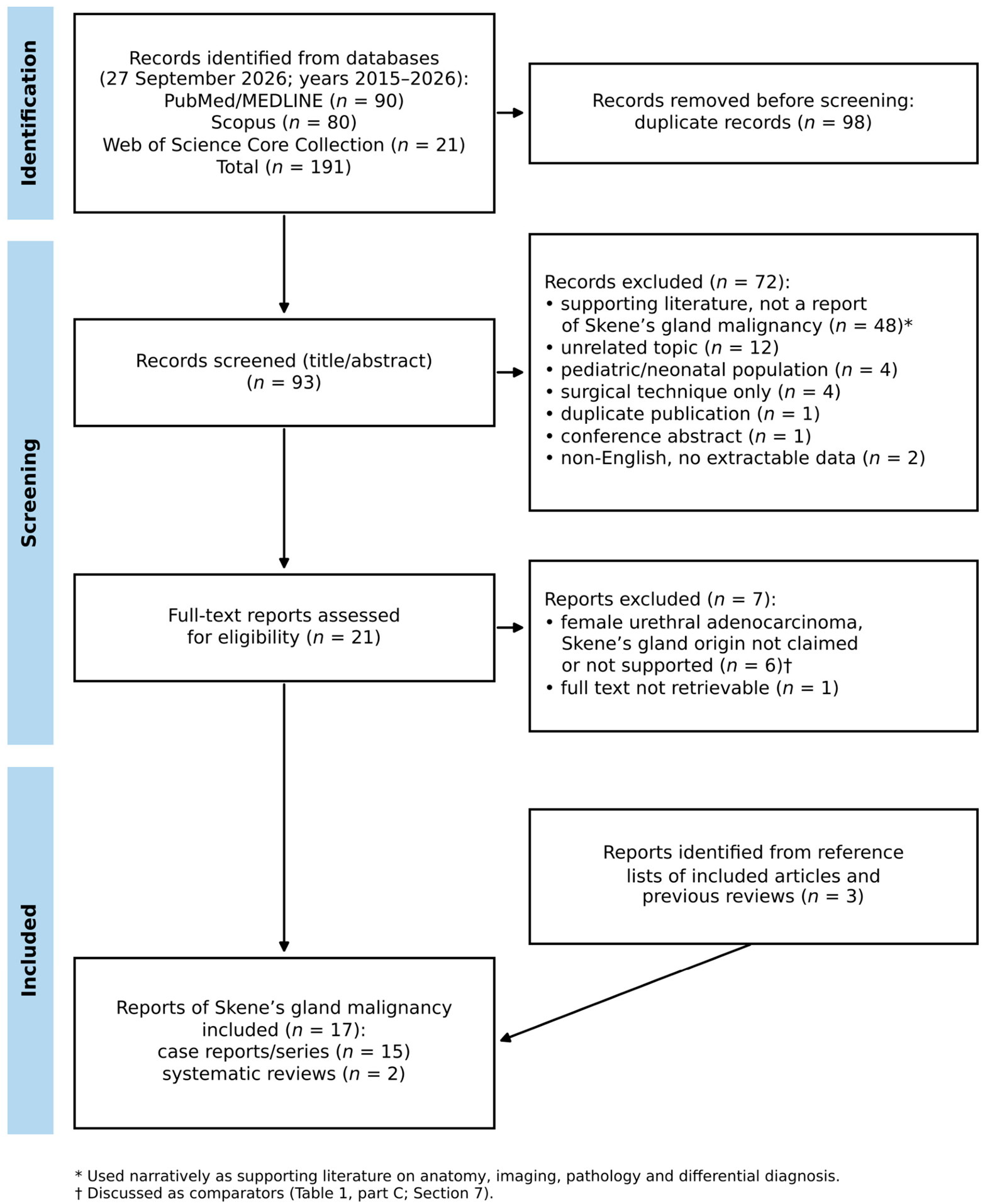
The database searches yielded 191 records (PubMed 90, Scopus 80, Web of Science 21). After removal of 98 duplicates, 93 unique records were screened, of which 72 were excluded at the title/abstract stage: 48 addressed benign periurethral disease or the imaging, pathology or management of female urethral lesions without reporting a Skene’s gland malignancy and were used as supporting literature, and 24 were unrelated to the topic, concerned pediatric patients or surgical technique only, or were duplicate publications, conference abstracts or non-English reports without extractable data. Of 21 full-text reports assessed, six female urethral adenocarcinomas without a claimed or demonstrable Skene’s gland origin and one report whose full text could not be retrieved were excluded, leaving 14 reports; three further case reports were identified from reference lists, for a total of 17 included reports (Figure 1).
2.2. Case Definition
In the absence of accepted diagnostic criteria, a tumor was classified as a Skene’s gland primary when all of the following were met: (i) a lesion centered in the periurethral/paraurethral tissues (around the distal or mid urethra, the external urethral meatus or the bladder neck and proximal urethra), as documented clinically, radiologically, endoscopically or pathologically; (ii) no evidence of another primary tumor that could account for the lesion by direct extension or metastasis (in particular colorectal, endometrial or cervical, vaginal, bladder or renal primaries); and (iii) either predominant prostatic-type differentiation (morphology resembling prostatic acinar or ductal adenocarcinoma with expression of prostate-specific antigen (PSA), prostate-specific acid phosphatase (PSAP), NKX3.1 or P501S/prostein) or morphological or immunohistochemical evidence of origin from native paraurethral glands, such as an in situ or prostatic intraepithelial neoplasia (PIN)-like precursor, a transition from benign Skene’s glands, or preserved basal cells demonstrated with a PIN4/p63 cocktail [8,10,11,12]. Tumors attributed to the Skene’s glands by the original authors that fulfilled criteria (i) and (ii) but showed a non-prostatic (intestinal, mucinous, adenosquamous or mixed) phenotype, or for which immunophenotypic data were insufficient, without demonstrated origin from native glands, were classified as probable Skene’s gland primaries and are tabulated separately (Table 1, part B).
Figure 1.
Flow diagram of the literature search and selection of reports of Skene’s gland malignancy (adapted from the PRISMA 2020 flow diagram). Records excluded as “supporting literature” were used narratively for the sections on anatomy, imaging, pathology and differential diagnosis.
2.3. Evidence Classification and Synthesis
Because direct evidence is scarce, statements and recommendations were classified according to their source as: (D) direct evidence from reported Skene’s gland malignancies; (E) evidence extrapolated from female urethral carcinoma, female urethral adenocarcinoma or benign periurethral lesions; or (X) expert inference based on anatomy and tumor extent. This classification is indicated in the text where relevant and summarized in Section 8. Because the available evidence is limited, heterogeneous and largely based on isolated cases or small series, no quantitative analysis or formal risk-of-bias assessment was performed, and the data were synthesized descriptively, with emphasis on clinical, imaging-based and pathological diagnostic evaluation and multidisciplinary decision-making.
During the preparation of this manuscript, the authors used Claude Opus 5.5 (Anthropic, San Francisco, CA, USA) to assist with executing and documenting the literature searches, preliminary screening, literature synthesis, drafting and reference formatting. All search results, screening decisions, source material, and clinical and imaging data were verified by the authors against the original publications, and the authors take full responsibility for the accuracy and content of this manuscript.
3. Anatomy and Embryological Background
The Skene’s glands are paired paraurethral glands situated near the distal urethra and opening close to the external urethral meatus. These secrete mucus through ducts opening near the external urethral meatus [13].
They originate embryologically from the urogenital sinus and are considered homologous to the male prostate gland. This embryological similarity explains the immunophenotypic overlap observed in Skene’s gland adenocarcinomas.
The periurethral location of these glands has significant implications for both diagnosis and treatment. Tumors may infiltrate the urethra, anterior vaginal wall, bladder neck, or periurethral soft tissues, complicating surgical excision and local staging.
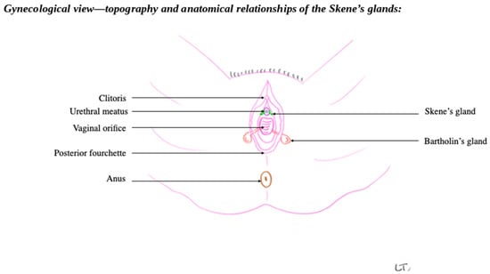
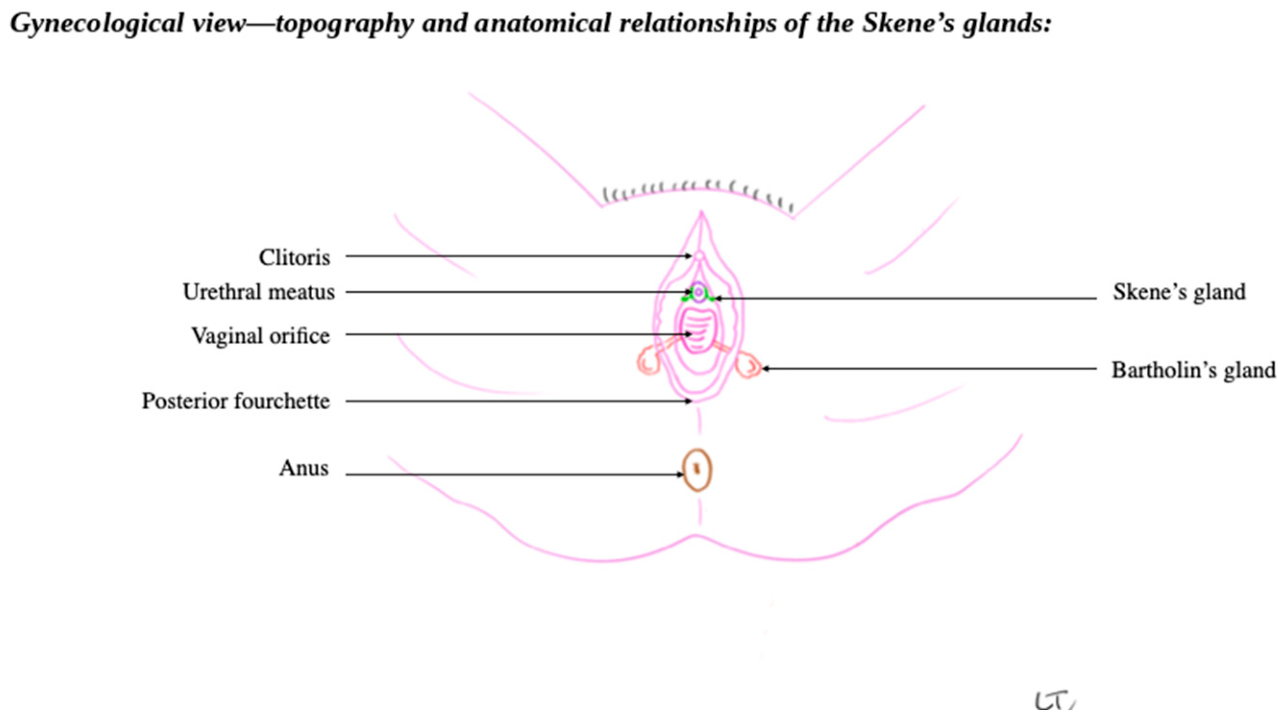
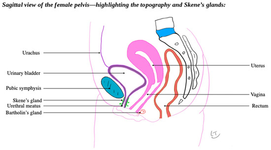
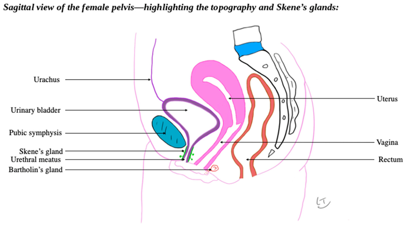
Understanding the regional anatomy is critical for interpreting imaging findings and planning radical or organ-preserving surgery. The topographic relationships of the Skene’s glands with the urethral meatus, the vaginal orifice and the neighboring Bartholin’s glands, as well as their position relative to the urinary bladder, urethra, vagina and rectum in the sagittal plane, are illustrated in Figure 2 and Figure 3.
Figure 2.
Gynecological (perineal) view showing the topography and anatomical relationships of the Skene’s glands with the clitoris, urethral meatus, vaginal orifice, Bartholin’s glands, posterior fourchette and anus. Original drawing by L.T.; “LT” in the lower right corner is the illustrator’s signature.
Figure 3.
Sagittal view of the female pelvis highlighting the topography of the Skene’s glands in relation to the urethral meatus, urinary bladder, urachus, pubic symphysis, vagina, uterus, Bartholin’s gland and rectum. Original drawing by L.T.; “LT” in the lower right corner is the illustrator’s signature.
4. Clinical Presentation and Paraclinical Assessment
4.1. Summary of Published Cases
Because Skene’s gland malignancy is known almost exclusively from isolated case reports and small series, the number of published cases depends on the diagnostic criteria applied. Slopnick et al. identified 15 cases published between 1974 and 2022 (including their own case), restricted to adenocarcinomas resembling prostatic carcinoma, with a median patient age of 71 years (range 46–88); the most frequent presentation was an asymptomatic periurethral or urethral lesion (33.3% of cases), followed by hematuria or vaginal bleeding (20.0%), and serum PSA was elevated in all eight patients in whom it was measured [4]. Applying broader criteria that also accepted non-prostatic phenotypes, Gao et al. counted 20 previously reported cases [2], and Li and Zhang identified 22 cases (median age 70 years, range 46–91; tumor diameter 0.3–9.0 cm, mean 2.6 cm) [5]. Since the latter review, at least three further cases have been published [14,15,16], and two additional reports [7,17] could not be confirmed as included in previous reviews; allowing for overlap between reviews, approximately 25 cases have therefore been reported to date. Table 1 summarizes the individually identifiable cases published since 2015, together with those identified from reference lists, grouped according to the case definition in Section 2.2; earlier cases (1974–2014) are summarized in previous reviews [2,4,5]. As shown in Table 1, reported management has ranged from limited local excision to radical cystectomy, urethrectomy or anterior pelvic exenteration with lymphadenectomy, and outcomes have varied widely, from durable disease-free survival to early death from metastatic disease, reflecting the heterogeneity of tumor stage and phenotype at presentation and the absence of a standardized treatment algorithm.
Table 1.
Individually reported cases of Skene’s gland malignancy published since 2015 or identified from reference lists, grouped according to the case definition (Section 2.2): (A) cases meeting the case definition; (B) probable cases, attributed to the Skene’s glands by the original authors, with a non-prostatic phenotype or insufficient immunophenotypic data and without demonstrated origin from native glands; (C) female urethral adenocarcinomas discussed in relation to a possible Skene’s gland origin that do not meet the case definition, shown for comparison. Earlier cases (1974–2014) are summarized in [2,4,5]. NR, not reported; NA, not available; NED, no evidence of disease; ADT, androgen deprivation therapy; LND, lymph node dissection; UTI, urinary tract infection; PSA, prostate-specific antigen; PSAP, prostate-specific acid phosphatase; AR, androgen receptor; PIN, prostatic intraepithelial neoplasia; LOH, loss of heterozygosity; FDG, fluorodeoxyglucose. See Section 5.2 and Section 7 for the imaging protocol and differential diagnosis, respectively.
4.2. Clinical Presentation and Physical Examination
The clinical presentation of Skene’s gland malignancy is nonspecific and overlaps considerably with that of benign periurethral and anterior vaginal wall conditions, which contributes to frequent diagnostic delay [24]. In a large retrospective series of periurethral and anterior vaginal wall masses, the most common presenting complaints were a palpable anterior vaginal wall or periurethral mass, dyspareunia, and lower urinary tract symptoms such as dysuria, urgency, frequency and recurrent urinary tract infection; vaginal discharge, spotting or overt bleeding, and postvoid dribbling were also reported, while malignancy accounted for only a small minority of cases in that cohort [25]. Obstructive symptoms, such as a weak or interrupted urinary stream, straining or incomplete bladder emptying, may occur when a periurethral tumor compresses or infiltrates the urethra [3,22,23]. In the specific context of Skene’s gland malignancy, an asymptomatic periurethral or urethral nodule and hematuria or vaginal bleeding are among the most frequently documented findings [4].
Clinical behavior is heterogeneous. Most prostatic-type tumors in the published series were small (1.0–2.0 cm) and localized at diagnosis [8], and nodal or distant metastases were documented in a minority of cases (4 of 20 in the review by Gao et al.; lymph node metastases in 5 of 22 in the review by Li and Zhang) [2,5] (D). However, tumors up to 9 cm have been described [5], and some patients presented with inguinal or pelvic lymphadenopathy, vulvar or labial deposits, or lung metastases [2,4,6,17] (D). For female urethral adenocarcinoma in general, which is often large and aggressive at presentation and is diagnosed at a metastatic stage in more than half of cases according to some reports [14], spread to the perineal skin, vulva, bladder and vagina and distant metastases to the lung, liver, bone and brain have been reported in advanced stages [2,3] (E).
Initial evaluation should include a focused history addressing the duration and progression of symptoms, prior periurethral surgery or gland drainage, and risk factors for urethral malignancy, followed by a careful pelvic examination. Physical examination should assess the size, consistency, mobility and fixation of any periurethral mass, the presence of ulceration, bleeding or purulent discharge from the urethral meatus (which may be elicited by gentle compression, or “milking”, of the periurethral region), and any extension toward the anterior vaginal wall, bladder neck or vulvar structures. Because the Skene’s glands and their ducts open beside the external urethral meatus, a primary tumor typically presents as a periurethral or anterior vaginal wall nodule adjacent to the meatus; a labial nodule should rather suggest vulvar or Bartholin gland pathology or metastatic spread, as in a reported case in which a labia majora deposit accompanied a periurethral primary [2]. Bartholin gland lesions, located posterolaterally at the vaginal introitus, are usually readily distinguished on inspection and bimanual and speculum examination, although large Bartholin lesions may extend anteriorly and be mislocalized on imaging [26,27]. Examination of the groins can identify inguinal lymphadenopathy in more advanced disease.
Paraclinical work-up typically includes urinalysis and urine culture to exclude infection, together with urine cytology when malignancy is suspected [24]. Because the exact anatomical epicenter of a periurethral mass, and therefore the most appropriate biopsy route, is often established only after imaging, cross-sectional imaging (ultrasonography and, in particular, pelvic MRI) is usually performed before endoscopic biopsy (E, X). Cystourethroscopy then allows direct visualization of the urethral lumen and meatus, assessment for communication with a possible diverticular cavity, and targeted transurethral, transvaginal or image-guided biopsy of any suspicious lesion [24,25]; when conventional biopsy is non-diagnostic, MRI-guided biopsy has been used successfully in Skene’s gland adenocarcinoma [7] (D). In tumors with suspected prostatic-type differentiation, serum prostate-specific antigen may provide additional diagnostic and follow-up value, as discussed further in Section 6 [4,6,18]. Because clinical examination and basic paraclinical testing alone cannot reliably differentiate benign from malignant periurethral disease, imaging, chiefly pelvic MRI, plays a central role in further characterization, as detailed below.
5. Multimodal Imaging Characterization
5.1. Ultrasonography
Ultrasonography is commonly used as a first-line imaging method in women presenting with periurethral symptoms, mainly because it is accessible, non-invasive, inexpensive, and allows dynamic real-time assessment. In the evaluation of urethral and paraurethral lesions, ultrasound may help establish whether a mass is cystic, solid, or mixed and may provide preliminary information regarding its relationship with the urethra, anterior vaginal wall, and adjacent soft tissues. It can also demonstrate internal debris, septations, wall thickening, calcifications, vascularity or inflammatory changes. Nevertheless, ultrasound has inherent limitations in this anatomical region. The female urethra is short, deeply situated within the pelvic floor and partly obscured by surrounding bony and soft-tissue structures, while bowel gas and patient-related factors may further reduce image quality [26].
Several sonographic approaches may be used, including transvaginal, transperineal, translabial and high-frequency endovaginal ultrasound. These techniques can be particularly useful for the assessment of distal urethral and paraurethral masses, including Skene’s gland cysts, abscesses, urethral diverticula, leiomyomas and other anterior vaginal wall lesions [26,28]. In general, simple cystic lesions appear as well-defined anechoic or hypoechoic structures, whereas complicated cysts or abscesses may show internal echoes, septations, thickened walls, peripheral hyperemia, and adjacent inflammatory changes. Solid lesions, by contrast, may present as hypoechoic or heterogeneous masses, sometimes with detectable vascular signals on color Doppler examination [28]. The sonographic assessment should focus not only on lesion morphology, but also on its exact anatomical epicenter and any possible communication with the urethral lumen [29].
Specific ultrasound descriptions of Skene’s gland malignancy are limited, reflecting the rarity of this tumor; in one reported case, ultrasonography showed a cystic–solid labial deposit and hypoechoic inguinal lymph nodes [2] (D). Based on the available literature on female urethral and periurethral malignancies, suspicious lesions may appear as predominantly solid or mixed cystic-solid masses, with heterogeneous echotexture, irregular margins, and increased or disorganized Doppler vascularity [4,8,28] (E). Additional clinical and imaging features that should raise concern include rapid enlargement, recurrent bleeding, hematuria, ulceration, fixation to adjacent tissues, recurrence after drainage or excision, and signs of extension toward the urethra, anterior vaginal wall or surrounding periurethral soft tissues. However, these findings are not specific for Skene’s gland origin and cannot reliably differentiate Skene’s gland carcinoma from primary urethral carcinoma, carcinoma arising in a urethral diverticulum, vaginal carcinoma or metastatic disease.
Three-dimensional ultrasound may add value by improving spatial visualization of paraurethral and anterior vaginal wall masses. Multiplanar reconstruction can help clarify the relationship between the lesion, the urethra, the vaginal wall and pelvic floor structures, which may be useful for preoperative planning and interdisciplinary communication [30]. Despite this advantage, three-dimensional ultrasound remains primarily an anatomical mapping tool. Its ability to distinguish benign from malignant periurethral lesions is limited, and suspicious findings should be further assessed with cross-sectional imaging and tissue diagnosis.
5.2. Magnetic Resonance Imaging
Most vulvar and periurethral disorders are identified and followed clinically, with imaging playing a complementary role. When imaging is required, magnetic resonance imaging (MRI) is the preferred technique because of its superior soft-tissue contrast and high spatial resolution, and it is regarded as the reference imaging modality for urethral and periurethral lesions [13,27,31] (E). Direct MRI evidence in Skene’s gland malignancy is limited to a few case reports. MRI showed a solid mass centered in the anterior vaginal wall that semi-encircled the urethra, resembling the periurethral configuration of the male prostate (a pattern also described by Korytko et al., as cited in [2]), together with a labial deposit and enlarged inguinal lymph nodes [2]; MRI demonstrated a 7 cm sub-vesical periurethral mass with a urethral epicenter, most probably arising from a left periurethral gland, with local extension and external iliac lymph nodes [14]; three-dimensional multiparametric MRI clarified the anatomy of a primary lesion before surgery [6]; and MRI, combined with FDG-PET, detected a Skene’s gland adenocarcinoma during follow-up of a previous vaginal tumor and guided its biopsy [7] (D). The protocol proposed in Table 2 is therefore adapted from recommendations for urethral and vulvar imaging and has not been validated specifically for Skene’s gland tumors. The European Society of Urogenital Radiology recommends imaging protocols incorporating T2-weighted sequences (T2W), diffusion-weighted imaging with Apparent Diffusion Coefficient (ADC) mapping, and dynamic contrast-enhanced imaging [32]. Fat-suppressed T2W sequences are generally preferred because they improve visualization of the primary lesion by enhancing contrast between the tumor and the surrounding perineal adipose tissue. Diffusion-weighted imaging can further aid in delineating the tumor. Imaging protocols dedicated to urethral assessment should incorporate a small field of view and high-resolution T2W sequences acquired in the axial, sagittal, and coronal planes. The use of fat-suppressed techniques may improve visualization of urethral diverticula by increasing lesion conspicuity. T1-weighted (T1W) imaging is particularly valuable for detecting hemorrhagic or protein-rich material within diverticular cavities. In patients with malignancies, additional large field-of-view sequences are useful for evaluating regional lymphadenopathy, bone involvement, and distant metastatic spread [33]. At the same time, MRI also provides valuable postoperative follow-up and surveillance information [31].
Table 2.
Proposed magnetic resonance imaging protocol for the evaluation of female urethral and periurethral lesions [31,34,35]. The protocol is extrapolated from female urethral and vulvar imaging recommendations (evidence level E) and has not been validated specifically in Skene’s gland malignancy. Abbreviations: MRI, magnetic resonance imaging; FOV, field of view; T1W, T1-weighted; T2W, T2-weighted.
5.3. Computed Tomography
Computed tomography (CT) has a limited role in the primary local characterization of suspected Skene’s gland malignancy because its soft-tissue contrast is inferior to MRI, particularly in the evaluation of the distal urethra, paraurethral tissues, anterior vaginal wall and pelvic floor. Therefore, CT should not be considered the preferred modality for defining the local epicenter of a small periurethral tumor or for assessing subtle invasion of adjacent soft tissues. Its main value lies in systemic staging, especially in patients with invasive, bulky, recurrent or histologically confirmed malignant disease. Contrast-enhanced CT of the chest, abdomen and pelvis may be used to evaluate pelvic, inguinal and retroperitoneal lymph nodes, detect distant metastases and assess associated abdominal or thoracic disease. CT urography may also be considered when urothelial involvement, synchronous urinary tract lesions or upper-tract disease must be excluded. Thus, in suspected or confirmed Skene’s gland malignancy, CT is best regarded as a complementary staging tool rather than a primary local diagnostic method [36,37]. In reported Skene’s gland cases, contrast-enhanced CT was used for systemic staging and detected lung metastases whose response to chemotherapy was then monitored by CT [2,17] (D).
5.4. PET/CT Imaging
Positron emission tomography/computed tomography (PET/CT), most commonly using 18F-fluorodeoxyglucose, provides functional information regarding tumor glucose metabolism and may complement conventional cross-sectional imaging in selected patients. In urethral and periurethral malignancies, PET/CT may be useful for detecting metabolically active lymph node disease, distant metastases or recurrent disease, particularly when CT or MRI findings are equivocal. It may also help distinguish viable tumor from postsurgical or post-treatment anatomical changes, although inflammatory lesions and infection can also show increased tracer uptake and may lead to false-positive findings. In the context of Skene’s gland malignancy, evidence remains very limited and is largely extrapolated from primary urethral carcinoma, female urethral adenocarcinoma and isolated case reports. Therefore, PET/CT should not be considered a routine replacement for pelvic MRI or histopathological confirmation. Its role is more appropriately reserved for selected cases, especially advanced disease, suspected nodal or distant spread, treatment response assessment, or evaluation of recurrence. Further studies are required before PET/CT can be incorporated into standardized staging algorithms for Skene’s gland malignancies [38,39]. In Skene’s gland adenocarcinoma specifically, fluciclovine F-18 PET, a tracer used in prostate cancer, upstaged nodal disease and prompted pelvic lymphadenectomy [6], and FDG-PET combined with MRI identified a lesion that was subsequently confirmed by MRI-guided biopsy [7] (D). Prostate-specific membrane antigen (PSMA)-targeted PET has not, to our knowledge, been reported in this setting but may be considered by analogy in prostatic-type tumors (X).
6. Histopathology and Molecular Landscape
Histopathological variants reported in Skene’s gland malignancies include adenocarcinoma resembling prostatic acinar or ductal carcinoma, which is the predominant subtype [4,6,8,12]; adenocarcinoma with intestinal or mucinous differentiation [10,16]; serous-type adenocarcinoma [8]; adenosquamous carcinoma [2]; and ductal/papillary adenocarcinoma with a mixed immunophenotype [17]. Clear cell adenocarcinoma of the female urethra is generally regarded as a distinct, Müllerian-type urethral tumor rather than a Skene’s gland neoplasm [40,41]. Microscopically, Skene’s gland adenocarcinomas demonstrate glandular proliferation with tubular, cribriform, fused or poorly formed glands, sometimes with mucin production, together with cellular atypia and mitotic activity [8].
Skene’s gland adenocarcinoma is a very rare tumor, with only a limited number of documented cases in the literature. Arising from the Skene’s glands, this shares both morphological characteristics and immunohistochemical profiles with prostatic adenocarcinoma. Nevertheless, because of the extremely limited number of reported cases, it remains unclear whether the grading systems currently used for prostate adenocarcinoma can be reliably applied to these tumors [8]. Determining the primary site of origin in female urethral carcinoma is frequently challenging. In the case of Skene’s gland adenocarcinoma with intestinal differentiation, immunohistochemical profiling plays a crucial role in establishing the tumor’s histogenetic origin [10].
Histologically, the Skene’s glands contain secretory epithelium expressing PSA and PSAP, and normal Skene’s gland derivatives are consistently NKX3.1-positive [8,9,15]. In tumors with prostatic-type morphology, PSA, PSAP, NKX3.1, P501S (prostein), AMACR (P504S) and androgen receptor are frequently expressed [6,8,11,12], whereas CK7, CK20, CDX2 and MUC2 may be positive in tumors with intestinal differentiation [2,10,16]. However, no single immunohistochemical marker establishes Skene’s gland origin in isolation, and formal estimates of sensitivity and specificity cannot be derived from the available case reports. PSA positivity is not universal: it has been reported in only about 50–80% of Skene’s gland adenocarcinomas and may be absent in otherwise typical tumors [10,17], and Gao et al. concluded that PSA lacks diagnostic specificity for Skene’s gland carcinoma [2]. The main areas of marker overlap are: (i) other female urethral adenocarcinomas, in which PAX8 identifies a Müllerian-type (including clear cell) subgroup but is also expressed in some tumors attributed to the Skene’s glands [14,40]; (ii) colorectal-type adenocarcinoma, whether primary urethral or metastatic, as CDX2, SATB2, CK20 and MUC2 are shared by intestinal-type Skene’s gland tumors, so that clinical and endoscopic exclusion of a colorectal primary is required [40,42]; (iii) Müllerian tumors, as PAX8, WT1, estrogen and progesterone receptors and vimentin favor a Müllerian lineage, but ER, PR, vimentin, p16 and PAX8 have been reported in serous-type, intestinal-type or mixed tumors attributed to the Skene’s glands [8,14,17]; (iv) urothelial-associated tumors, as GATA3, p63/p40 and a CK7+/CK20+ profile favor urothelial differentiation, whereas p63 with high-molecular-weight cytokeratin, as part of a PIN4 cocktail, also highlights basal cells and can demonstrate origin from pre-existing glands [8,11,41]; (v) neuroendocrine tumors, which may arise in the same region and are identified by chromogranin and synaptophysin [7,41]; and (vi) metastatic adenocarcinoma, including prostatic-type tumors arising elsewhere in the female genital tract [9,42]. AMACR is also expressed in intestinal-type and clear cell adenocarcinomas of the urinary tract and is therefore not specific [17,43]. Immunohistochemistry should thus be interpreted as a pattern (typically including PSA, PSAP, NKX3.1 and AR; CK7, CK20, CDX2 and SATB2; PAX8, WT1 and ER/PR; GATA3 and p63; and neuroendocrine markers when indicated), together with morphology, tumor epicenter on imaging and exclusion of another primary (X). As in prostate carcinoma, serum PSA may be a useful biomarker for monitoring prostatic-type tumors: PSA normalized after definitive treatment in all tested patients in the review by Slopnick et al. [4] became undetectable after resection [18] and fell rapidly under androgen deprivation [6] (D). Rising PSA levels may therefore suggest recurrence before clinical symptoms develop [11,15].
Molecular data are limited to isolated observations and do not yet allow a molecular characterization of the entity. Next-generation sequencing of one prostatic-type case identified a mutation of the phosphatase and tensin homolog (PTEN) gene with loss of heterozygosity (LOH) [12]; another case showed reduced PTEN expression without ERG overexpression [20]; and a TBP–RET fusion was reported in a metastatic case with a mixed immunophenotype, although orthogonal confirmation was not possible [17]. PTEN mutations have also been reported in about 15% of female urethral adenocarcinomas overall [41]. Overexpression of AMACR (P504S) is characteristic of prostatic adenocarcinoma, whereas benign prostatic tissue shows absent or minimal immunoreactivity [43], but it is not specific for Skene’s gland origin [17]. These single-case findings should be regarded as hypothesis-generating.
7. Differential Diagnosis of Skene’s Gland Malignancies
Because of the intimate anatomical relationship between the distal urethra, paraurethral glands, anterior vaginal wall and vulvar soft tissues, the differential diagnosis of Skene’s gland malignancy can be difficult [44,45]. Benign cystic or inflammatory lesions may present with symptoms similar to those of malignant tumors, including a palpable periurethral mass, dysuria, dyspareunia, recurrent infection, hematuria, or obstructive urinary complaints. MRI plays an important role in the initial characterization of these lesions by identifying the precise anatomical origin, the relationship with the urethral lumen, the presence of a communicating tract, and the internal structure of the mass [44,45,46]. Features such as solid enhancing components, diffusion restriction, irregular margins, or invasion of adjacent tissues may suggest malignancy, but they are not specific for Skene’s gland origin. Primary urethral carcinoma, carcinoma arising within a urethral diverticulum, anterior vaginal wall carcinoma and metastatic adenocarcinoma may show overlapping imaging appearances. Therefore, MRI findings should be integrated with clinical examination, cystourethroscopy, histopathological assessment and immunohistochemical profiling [4,44,45,47]. The entities that should be considered in the differential diagnosis are summarized in Table 3, grouped into benign (including inflammatory and iatrogenic) lesions, primary malignant tumors and secondary malignant involvement, and should be interpreted in conjunction with the topographic relationships illustrated in Figure 2 and Figure 3.
Table 3.
Differential diagnosis of Skene’s gland malignancies, grouped into benign lesions, primary malignant tumors and secondary malignant involvement: anatomical and MRI features. Abbreviations: MRI, magnetic resonance imaging; T1W, T1-weighted; T2W, T2-weighted; IHC, immunohistochemistry; PSA, prostate-specific antigen; PSAP, prostate-specific acid phosphatase; NKX3.1, NK3 homeobox 1; CDX2, caudal-type homeobox 2; SATB2, special AT-rich sequence-binding protein 2; PAX8, paired box gene 8.
8. Therapeutic Management
8.1. Surgical Treatment
In this pathology, surgery remains the cornerstone of treatment. The choice of surgical management is influenced by multiple tumor-related factors such as tumor dimensions, depth and extent of local invasion, degree of urethral and periurethral involvement, presence of adjacent organ infiltration, and overall disease stage. In addition, patient-related factors such as age, performance status, comorbid conditions, urinary function, and anticipated postoperative quality of life must be carefully considered when planning treatment. Because malignant tumors of the Skene’s glands are exceptionally uncommon, there is no universally accepted surgical protocol. In the reported Skene’s gland cases, small (≤2 cm), localized prostatic-type tumors were most often treated by local excision with negative margins, without recurrence during follow-up; of the 15 cases reviewed by Slopnick et al., eight were treated by local excision, two by radical surgical resection, two with radiotherapy and one with adjunctive androgen deprivation [4,8] (D). The more extensive procedures described below are largely extrapolated from female urethral carcinoma (E) or reflect expert inference based on tumor extent (X); the evidence basis of each recommendation is summarized in Table 4. Complete resection with negative margins is the primary objective, and management is therefore individualized according to tumor extent, ranging from organ-preserving local excision for limited lesions to radical urethrectomy or anterior pelvic exenteration in advanced invasive disease [10,37].
Urethra-sparing endoscopic approaches, such as transurethral resection and laser ablation or vaporization of small, superficial tumors of the distal urethra and meatus, are described in the management of primary urethral carcinoma, mainly in men [36,37] (E). Their role in Skene’s gland malignancy is uncertain: because these tumors arise in the periurethral glands beneath the urothelium rather than from the mucosal surface, endoscopic resection may not achieve clear margins, as illustrated by a transurethrally resected Skene’s gland adenocarcinoma with positive margins [12] (D). Endoscopic approaches may therefore be regarded mainly as diagnostic or palliative options, or as definitive treatment only for small, superficial lesions in patients unfit for open excision (X).
Radical urethrectomy is typically reserved for advanced or recurrent lesions showing infiltration of the proximal urethra or spread to adjacent periurethral and vaginal tissues. Surgical management can involve total urethral excision combined with removal of involved surrounding soft tissues and, in selected cases, partial excision of the anterior vaginal wall. Such an aggressive operative strategy may enhance local tumor containment in high-risk disease [3] (E). Lymphadenectomy may be indicated in cases with radiologically suspicious nodal involvement, advanced tumor stage, or high-grade adenocarcinoma histology. The pattern of nodal spread varies according to tumor location, with distal lesions more commonly associated with inguinal lymphatic dissemination, whereas proximal tumors tend to involve the pelvic nodal chains [2] (E). Anterior pelvic exenteration or radical cystectomy is generally reserved for extensive disease characterized by deep local invasion, involvement of the bladder neck, widespread extension into periurethral tissues, or regional lymph node metastases, and has been used in Skene’s gland cases involving the bladder neck or after failure of radiotherapy [7,15,16] (D).
No specific staging system exists exclusively for Skene’s gland malignancies, and FIGO staging is not applicable; most authors apply the TNM/AJCC staging used for female urethral carcinoma, whose limitations for tumors of the anterior urethral wall have led to the proposal of histology-based staging systems [41] (E). Multiple factors, including tumor anatomical location and size, histopathological subtype, extent of tissue infiltration, lymph node involvement, surgical margin status, and presence of distant metastases, influence the clinical outcome of urethral carcinoma. Lesions arising in the anterior (distal) urethra generally demonstrate more favorable outcomes than tumors involving the posterior (proximal) urethra [10,37]. Early-stage localized disease appears associated with favorable outcomes after complete surgical excision, whereas advanced tumors with nodal dissemination demonstrate a poorer prognosis [10,37] (Section 8.4).
8.2. Adjuvant Therapy
Radiotherapy may be considered in the management of Skene’s gland carcinoma as an adjuvant treatment for locally advanced disease, as well as a definitive therapeutic option in patients who are unsuitable for radical surgery or in whom extensive surgical procedures would result in substantial morbidity [10,37] (E). In selected cases, radiation-based approaches, with or without concurrent chemotherapy, may contribute to local tumor control and symptomatic relief. In a locally advanced, node-positive tumor (cT3N2M0), neoadjuvant chemoradiation with weekly cisplatin was chosen before planned surgery [14]; in another case, definitive chemoradiation with cisplatin achieved a complete clinical response but was followed by local recurrence treated with interstitial brachytherapy; subsequent radiation-induced necrosis and osteomyelitis eventually required anterior pelvic exenteration [16] (D). Because malignant tumors originating from the Skene’s glands are exceptionally rare, current evidence is derived primarily from isolated case reports and extrapolation from female urethral carcinoma series, and standardized radiotherapy protocols have not yet been established [2,15,23].
Data concerning the efficacy of systemic chemotherapy in Skene’s gland carcinoma remain uncertain because of the exceptionally low incidence of this neoplasm. Consequently, chemotherapeutic strategies have largely been adapted from protocols established for urothelial malignancies and other adenocarcinomas of the female genital and urinary tracts. In advanced, metastatic, or recurrent settings, platinum-containing combinations have most frequently been employed [15,23,47] (E); in a metastatic Skene’s gland adenocarcinoma, docetaxel–carboplatin followed by gemcitabine–carboplatin produced a partial response and then disease stabilization [17] (D). In prostatic-type tumors, androgen deprivation therapy has produced marked PSA responses and has been used as the main or neoadjuvant treatment in node-positive disease [4,6] (D).
8.3. Follow-Up and Surveillance
At present, no standardized surveillance recommendations have been established for patients with Skene’s gland adenocarcinoma owing to the rarity of the disease and the limited number of reported cases. Consequently, follow-up should rely on comprehensive clinicopathological assessment integrating clinical findings, imaging studies, and biomarker evaluation. In tumors demonstrating prostatic differentiation, serial prostate-specific antigen (PSA) measurements may provide additional value when interpreted alongside radiologic and physical examination findings [11]. Available evidence suggests that disease relapse most commonly develops within the pelvic region; however, the actual incidence of recurrence remains difficult to determine because long-term outcome data are sparse. Current follow-up strategies are generally individualized and may include scheduled pelvic examinations, pelvic magnetic resonance imaging (MRI), cystourethroscopic evaluation when clinically indicated, and computed tomography (CT) imaging in patients with advanced or high-risk disease [2,15,23,37].
8.4. Prognosis and Outcomes
Evidence on prognosis is limited to case-level follow-up, which is often short or unreported, so that disease-specific survival for Skene’s gland malignancy cannot be estimated. In the systematic review by Slopnick et al., which included only prostatic-type adenocarcinomas, no recurrence or cancer-related death occurred during a median follow-up of 11.5 months, and PSA normalized after definitive treatment in all tested patients [4]. In the four-case series by Tregnago et al., tumors measured 1.0–2.0 cm were excised with negative margins and remained free of recurrence after a mean follow-up of 40.7 months (range 4–132) [8]. In the broader review by Li and Zhang, follow-up was available for 11 of 22 patients, of whom two died and one recurred, and lymph node metastases were present in five patients [5] (D).
Taken together with the individual reports in Table 1, the following factors appear relevant, although none can be quantified: (i) local recurrence, observed after local excision alone and after definitive chemoradiation [16], and a concern after transurethral resection with positive margins [12]; (ii) nodal metastasis, inguinal in distal and pelvic in proximal tumors [2], present at diagnosis in several of the more aggressive cases [2,4,6]; (iii) distant metastasis to the lung, liver, bone or abdominal viscera, associated with death or palliative treatment [2,17]; (iv) tumor size, with uniformly favorable outcomes in small (≤2 cm) localized tumors [8], whereas tumors up to 9 cm have been reported [5]; (v) margin status, negative margins having been achieved in the cases with durable disease-free survival [8,18]; (vi) urethral and bladder-neck involvement, which required urethrectomy, cystectomy or exenteration [7,15,16,19]; and (vii) histological subtype, the most aggressive courses having been observed in tumors with non-prostatic (adenosquamous, intestinal-type or mixed) phenotypes [2,16,17], whereas prostatic-type tumors were mostly localized, although a prostatic-type case with widespread metastases has also been described [2] (D). By extrapolation from primary urethral carcinoma, advanced stage, nodal involvement, non-urothelial histology and proximal location are associated with worse survival; five-year relative overall survival for urethral carcinoma in Europe is approximately 54%, and women tend to present with more advanced disease [3,37] (E).
Table 4.
Main diagnostic and management statements and their evidence basis. D, direct evidence from reported Skene’s gland malignancies; E, evidence extrapolated from female urethral carcinoma, female urethral adenocarcinoma or benign periurethral lesions; X, expert inference based on anatomy and tumor extent.
9. Current Challenges and Future Perspectives
Previous syntheses of this entity have generally taken the form of a single new case report accompanied by a literature review, most notably the systematic review and case report by Slopnick et al. (15 prostatic-type cases, 1974–2022) [4], the case report and review by Gao et al. (20 cases) [2], the systematic review by Li and Zhang (22 cases) [5] and the more recent case report and review by Wang and Zhou (2026) [15]. The present review differs in scope and organization rather than in the underlying evidence base, which remains the same small pool of published cases. Rather than being structured around a single index case, it is organized as a modality-by-modality diagnostic framework, moving systematically through clinical and paraclinical assessment, ultrasonography, MRI (including a proposed imaging protocol), CT and PET/CT, before addressing histopathology and an anatomically anchored differential diagnosis of thirteen mimicking entities grouped as benign, primary malignant and secondary malignant (Table 3). It also applies an explicit case definition and a reproducible multi-database search to update the case summary through 2026 (Table 1), and makes explicit whether each recommendation derives from Skene’s gland cases or from female urethral carcinoma (Table 4). This imaging- and differential-diagnosis-centered structure, rather than new clinical data, is intended to be the review’s principal contribution.
Several challenges continue to limit the current understanding and clinical management of Skene’s gland malignancies. First, because these tumors are exceptionally uncommon, robust evidence-based guidelines are lacking. Current management strategies are largely based on individual case reports or adapted from treatment approaches used for female urethral carcinoma.
Inconsistent terminology in the literature complicates accurate interpretation and comparison of published data. While some investigators describe these lesions as paraurethral adenocarcinomas, others categorize them as subtypes of urethral adenocarcinoma.
Available molecular data remain scarce. Further genomic and proteomic research may improve tumor classification and facilitate the identification of potential therapeutic targets. In addition, emerging technologies such as artificial intelligence-based imaging analysis and radiomic assessment could enhance the distinction between benign and malignant periurethral lesions.
The establishment of international registries and multicenter collaborative databases could significantly enhance epidemiological knowledge and support the development of standardized therapeutic guidelines.
10. Limitations
This review has several limitations. First, although the literature search was structured, documented and reproducible across three databases, this is a narrative rather than a systematic review: no protocol was registered, no formal risk-of-bias assessment was performed, database searches were limited to 2015–2026 with earlier reports identified through reference lists and previous reviews, and some eligible reports, particularly those in non-English languages or older, non-indexed literature, may have been missed. Second, the evidence base itself is inherently limited: as summarized in Table 1, almost all available data derive from single case reports or very small case series, with substantial heterogeneity in reported histology, immunohistochemical panels, imaging, treatment and length of follow-up, and with outcome data missing or incompletely reported in several cases. This precluded any pooled or quantitative synthesis, such as a meta-analysis of treatment outcomes or survival. Third, the case definition applied here is pragmatic and has not been validated; because Skene’s gland malignancy is frequently indistinguishable from other female urethral adenocarcinomas without detailed immunohistochemical and molecular work-up, some cases classified as Skene’s gland origin in the literature, particularly those with non-prostatic phenotypes, may in fact represent primary urethral or paraurethral tumors of a different histogenesis, which could affect the applicability of the imaging and management data summarized here. Fourth, most imaging and management recommendations are extrapolated from female urethral carcinoma (Table 4). Finally, the imaging descriptions in this review are derived from the literature rather than from a dedicated, systematic pictorial analysis of primary MRI, ultrasound or histopathology images; consequently, the anatomical figures accompanying this review (Figure 2 and Figure 3) are original schematic illustrations rather than diagnostic imaging examples. These limitations underscore the need for prospective, multicenter data collection, as outlined in the Current Challenges and Future Perspectives Section above.
11. Conclusions
Skene’s gland malignancies are exceptionally rare tumors of the female periurethral region, and their diagnosis remains challenging because clinical presentation is often nonspecific and overlaps with benign paraurethral, urethral, and anterior vaginal wall disorders. A directed clinical history and pelvic examination are the first steps; imaging should then define the lesion before endoscopic or image-guided biopsy, and its findings determine staging and the choice of treatment.
Ultrasonography may be useful as an initial and complementary examination, particularly for detecting paraurethral lesions, distinguishing cystic from solid components, assessing vascularity with Doppler techniques and guiding selected interventional procedures. However, sonographic findings are not sufficiently specific to confirm malignancy or define the full extent of disease.
MRI remains the key imaging modality for local evaluation, as it provides superior soft-tissue contrast and allows precise assessment of tumor epicenter, relationship with the urethral lumen, periurethral extension, involvement of the anterior vaginal wall and invasion of adjacent pelvic structures. CT and PET/CT may offer additional value in selected cases, mainly for nodal assessment, detection of distant disease, evaluation of recurrence and staging of advanced tumors. Nevertheless, imaging findings must always be interpreted in conjunction with clinical examination, cystourethroscopy and pathological assessment.
Definitive diagnosis requires histopathological confirmation. Immunohistochemistry (PSA, PSAP and NKX3.1, together with a broader panel to exclude urethral, Müllerian, intestinal-type and metastatic adenocarcinomas) supports, but cannot by itself prove, a paraurethral gland origin. Local excision with negative margins appears adequate for small localized prostatic-type tumors, whereas radical surgery, radiotherapy, chemotherapy, androgen deprivation or combined strategies are reserved, largely by extrapolation from female urethral carcinoma, for locally advanced, node-positive, recurrent or aggressive disease.
Given the rarity of these tumors and the absence of standardized management guidelines, individualized treatment planning within a multidisciplinary team is essential. Larger multicenter studies and pooled analyses are needed to refine imaging-based diagnostic algorithms, identify prognostic factors, and develop evidence-based recommendations for staging, treatment, and follow-up.
Author Contributions
Conceptualization, R.-D.C. and T.T.; methodology, T.T.; investigation, R.-D.C., L.T. and M.G.P.; resources, A.I.G.-O.; data curation, R.-D.C. and L.T.; writing—original draft preparation, R.-D.C. and T.T.; writing—review and editing, T.T., M.G.P. and A.I.G.-O.; visualization, L.T.; supervision, T.T.; project administration, T.T. All authors have read and agreed to the published version of the manuscript.
Funding
This research received no external funding.
Institutional Review Board Statement
Not applicable. This is a narrative literature review that did not involve new data collection from human participants or animals.
Informed Consent Statement
Data Availability Statement
The original contributions presented in this study are included in the article. Further inquiries, including requests for the search strategies, search dates, record counts and screening decisions, can be directed to the corresponding author.
Acknowledgments
During the preparation of this manuscript, the authors used Claude Opus 5.5 (Anthropic, San Francisco, CA, USA) to assist with executing and documenting the literature searches, preliminary screening, literature synthesis, drafting and reference formatting. The authors have reviewed and edited the output and take full responsibility for the content of this publication.
Conflicts of Interest
The authors declare no conflicts of interest.
Abbreviations
The following abbreviations are used in this manuscript:
| MRI | Magnetic Resonance Imaging |
| US | Ultrasonography |
| CT | Computed Tomography |
| PET | Positron Emission Tomography |
| T1W/T2W | T1-weighted/T2-weighted (MRI sequence) |
| DWI | Diffusion-Weighted Imaging |
| ADC | Apparent Diffusion Coefficient |
| FOV | Field of View |
| PSA | Prostate-Specific Antigen |
| PSAP | Prostate-Specific Acid Phosphatase |
| IHC | Immunohistochemistry |
| CK7/CK20 | Cytokeratin 7/Cytokeratin 20 |
| NKX3.1 | NK3 Homeobox 1 |
| AR | Androgen Receptor |
| CDX2 | Caudal-Type Homeobox 2 |
| SATB2 | Special AT-Rich Sequence-Binding Protein 2 |
| PAX8 | Paired Box Gene 8 |
| PTEN | Phosphatase and Tensin Homolog |
| LOH | Loss of Heterozygosity |
| AMACR | α-Methylacyl-CoA Racemase (P504S) |
| ADT | Androgen Deprivation Therapy |
| AJCC | American Joint Committee on Cancer |
| ER/PR | Estrogen Receptor/Progesterone Receptor |
| FDG | 18F-Fluorodeoxyglucose |
| GATA3 | GATA Binding Protein 3 |
| LND | Lymph Node Dissection |
| MUC2 | Mucin 2 |
| NA | Not Available |
| NED | No Evidence of Disease |
| NR | Not Reported |
| PIN | Prostatic Intraepithelial Neoplasia |
| PRISMA | Preferred Reporting Items for Systematic Reviews and Meta-Analyses |
| PSMA | Prostate-Specific Membrane Antigen |
| TNM | Tumor, Node, Metastasis |
| UTI | Urinary Tract Infection |
| WT1 | Wilms Tumor 1 |
References
- Swartz, M.A.; Porter, M.P.; Lin, D.W.; Weiss, N.S. Incidence of primary urethral carcinoma in the United States. Urology 2006, 68, 1164–1168. [Google Scholar] [CrossRef] [Scilit] [PubMed]
- Gao, Q.; Liu, X.; Ye, L.; Lv, T.; Teng, Y.; Lan, J.; Li, T.; Tian, M.; Chen, J.; He, S.; et al. Adenosquamous Carcinoma of Skene’s Gland: A Case Report and Literature Review. Front. Oncol. 2022, 12, 893980. [Google Scholar] [CrossRef] [Scilit] [PubMed]
- Dell’Atti, L.; Galosi, A.B. Female Urethra Adenocarcinoma. Clin. Genitourin. Cancer 2018, 16, e263–e267. [Google Scholar] [CrossRef] [Scilit] [PubMed]
- Slopnick, E.A.; Bagby, C.; Mahran, A.; Nagel, C.; Garcia, J.; El-Nashar, S.; Hijaz, A.K. Skene’s Gland Malignancy: A Case Report and Systematic Review. Urology 2022, 165, 36–43. [Google Scholar] [CrossRef] [Scilit] [PubMed]
- Li, X.H.; Zhang, Y.T. Skene gland adenocarcinoma of the urethra: A systematic review. Asian J. Surg. 2024, 47, 1587–1588. [Google Scholar] [CrossRef] [Scilit] [PubMed]
- Kaufman, M.E.; Miller, D.T.; Ullah, A.; White, J.; Singh, G.; Kolhe, R.; Williams, H.; Mittal, P.; Parikh, J.; Terris, M.K. Skene’s Gland Adenocarcinoma: Borrowing From Prostate Cancer Experience for the Evaluation and Management of a Rare Malignancy. Urology 2021, 151, 182–187. [Google Scholar] [CrossRef] [Scilit] [PubMed]
- Dentry, M.; McBean, R.; Garrett, A.; Brown, N. A novel technique for a rare diagnosis: MRI-guided vaginal biopsy of Skene gland adenocarcinoma: Pictorial Essay. J. Med. Imaging Radiat. Oncol. 2021, 65, 724–727. [Google Scholar] [CrossRef] [Scilit] [PubMed]
- Tregnago, A.C.; Epstein, J.I. Skene’s Glands Adenocarcinoma: A Series of 4 Cases. Am. J. Surg. Pathol. 2018, 42, 1513–1521. [Google Scholar] [CrossRef] [Scilit] [PubMed]
- Hawari, R.; Fernandes, L.; Park, K.J.; McCluggage, W.G. Skene’s Gland Derivatives in the Female Genital Tract and Cervical Adenoid Basal Carcinoma are Consistently Positive With Prostatic Marker NKX3.1. Int. J. Gynecol. Pathol. 2021, 40, 400–407. [Google Scholar] [CrossRef] [Scilit] [PubMed]
- Muto, M.; Inamura, K.; Ozawa, N.; Endo, T.; Masuda, H.; Yonese, J.; Ishikawa, Y. Skene’s gland adenocarcinoma with intestinal differentiation: A case report and literature review. Pathol. Int. 2017, 67, 575–579. [Google Scholar] [CrossRef] [Scilit] [PubMed]
- Kunc, M.; Biernat, W. Skene’s gland adenocarcinoma coexisting with infiltrating urothelial carcinoma of the urinary bladder. Pol. J. Pathol. 2021, 72, 170–173. [Google Scholar] [CrossRef] [Scilit] [PubMed]
- Lenz, J.; Michal, M.; Michal, M.; Hes, O.; Konečná, P.; Lenz, D. First Molecular Genetic Characterization of Skene’s Gland Adenocarcinoma. Int. J. Surg. Pathol. 2021, 29, 447–453. [Google Scholar] [CrossRef] [Scilit] [PubMed]
- Golden, E.; Cooke, S.; Butler, E.; Gregg, L.; Gomez, E. Under the hood: Vulvar anatomy and pathology with a focus on MRI. Abdom. Radiol. 2026, 51, 2106–2124. [Google Scholar] [CrossRef] [Scilit] [PubMed]
- Alla, M.A.; Seffar, A.; Hatim, G.; Doumer, A.; Talib, H.; Chekrine, T.; Bouchbika, Z.; Benchakroun, N.; Tawfiq, N.; Jouhadi, H.; et al. Skene’s Gland Adenocarcinoma: A Rare Case Report of a PAX-8-Positive Female Urethral Adenocarcinoma and Literature Review. Mathews J. Case Rep. 2024, 9, 165. [Google Scholar] [CrossRef] [Scilit]
- Wang, J.; Zhou, J. Skene’s gland adenocarcinoma: A case report and review of the literature. Discov. Oncol. 2026, 17, 635. [Google Scholar] [CrossRef] [Scilit] [PubMed]
- Nuha, N.; Trymbulak, K.; King, M.; Margolis, B. Management and sequelae of a local Skene’s gland adenocarcinoma recurrence. Gynecol. Oncol. Rep. 2026, 66, 102150. [Google Scholar] [CrossRef] [Scilit] [PubMed]
- Bondili, S.K.; Abraham, G.; Noronha, V.; Joshi, A.; Patil, V.M.; Menon, N.; Shetty, O.A.; Chougule, A.; Menon, S.; Chandrani, P.; et al. Rare case of Skene gland adenocarcinoma with RET-rearrangement. Cancer Res. Stat. Treat. 2021, 4, 130–135. [Google Scholar] [CrossRef] [Scilit] [PubMed]
- Maghsoudi, A.; Lochhead, A.; Dally, E.; Starra, E.; Lynnhtun, K. PSA-positive urethral adenocarcinoma of female genital tract. Pathology 2019, 51, 445–448. [Google Scholar] [CrossRef] [Scilit] [PubMed]
- Kyriazis, G.; Varughese, A.; Rodrigues, G.; Simms, M. A rare case of Skene’s gland adenocarcinoma. Clin. Genitourin. Cancer 2020, 18, e300–e302. [Google Scholar] [CrossRef] [Scilit] [PubMed]
- Saeed, F.; Osunkoya, A.O. Skene gland adenocarcinoma: Clinicopathologic features, comprehensive biomarker analysis, and review of the literature. Pathol. Int. 2021, 71, 712–714. [Google Scholar] [CrossRef] [Scilit] [PubMed]
- Thum, S.; Haben, B.; Christ, G.; Sen Gupta, R. Female prostate cancer? Pathologe 2017, 38, 448–450. [Google Scholar] [CrossRef] [Scilit] [PubMed]
- Satyanarayan, A.; Redd, L.; Dyer, A.; Wright, A.; Walker, J. Adenocarcinoma of the urethra with mucinous features. Rev. Urol. 2015, 17, 38–41. [Google Scholar] [PubMed]
- Tian, J.; Zhu, T.; Xu, Z.; Chen, X.; Wu, Y.; Fu, G.; Jin, B. Management of Primary Female Urethral Adenocarcinoma: Two Rare Case Reports and Literature Review. Medicina 2023, 59, 109. [Google Scholar] [CrossRef] [Scilit] [PubMed]
- Zuo, S.W.; Napoe, G.S. Evaluation and management of urethral and periurethral masses in women. Curr. Opin. Obstet. Gynecol. 2023, 35, 517–524. [Google Scholar] [CrossRef] [Scilit] [PubMed]
- Jacox, N.; Yao, H.H.; Baverstock, R.; Trpkov, K.; Carlson, K. Periurethral and Anterior Vaginal Wall Masses: Etiology, Presentation, and Treatment Outcomes. Obstet. Gynecol. 2022, 140, 778–783. [Google Scholar] [CrossRef] [Scilit] [PubMed]
- Chaudhari, V.V.; Patel, M.K.; Douek, M.; Raman, S.S. MR imaging and US of female urethral and periurethral disease. Radiographics 2010, 30, 1857–1874. [Google Scholar] [CrossRef] [Scilit] [PubMed]
- Saidman, J.M.; Aineseder, M.; Garratt, J.; Wang, M.X.; Ahmed, I.; Elsayes, K.M.; Gomez, M.; Yugcha, F.O.R.; Chacón, C.R.B.; Ocantos, J.A. Imaging the Female Urethra: US and MRI in Cystic and Solid Pathologic Conditions. Radiographics 2025, 45, e240064. [Google Scholar] [CrossRef] [Scilit] [PubMed]
- Yang, H.; Gu, J.-J.; Jiang, L.; Wang, J.; Lin, L.; Wang, X.-L. Ultrasonographic Imaging Features of Female Urethral and Peri-urethral Masses: A Retrospective Study of 95 Patients. Ultrasound Med. Biol. 2020, 46, 1896–1907. [Google Scholar] [CrossRef] [Scilit] [PubMed]
- Tamburrini, S.; Vascone, C.; Marrone, V.; Catalano, M.; Del Biondo, D.; Gallo, L.; Quassone, P.; Lugarà, M.; Coppola, M.G.; De Simone, F.; et al. Skene’s glands abscess an overlooked diagnosis in acute lower urinary symptoms. Radiol. Case Rep. 2021, 16, 3751–3756. [Google Scholar] [CrossRef] [Scilit] [PubMed]
- Shobeiri, S.A.; Rostaminia, G.; White, D.; Quiroz, L.H.; Nihira, M.A. Evaluation of vaginal cysts and masses by 3-dimensional endovaginal and endoanal sonography. J. Ultrasound Med. 2013, 32, 1499–1507. [Google Scholar] [CrossRef] [Scilit] [PubMed]
- Mahfouz, W.; Hassan, H.H.M.; Gubbiotti, M.; Elbadry, M.; Moussa, A. Does a tailored magnetic resonance imaging technique affect the surgical planning and outcomes for different cystic urethral and periurethral swellings in females? Seven years tertiary center experience. World J. Urol. 2022, 40, 1587–1594. [Google Scholar] [CrossRef] [Scilit] [PubMed]
- Nikolić, O.; Sousa, F.A.E.; Cunha, T.M.; Nikolić, M.B.; Otero-García, M.M.; Gui, B.; Nougaret, S.; Leonhardt, H., on behalf of the ESUR Female Pelvic Imaging Working Group. Vulvar cancer staging: Guidelines of the European Society of Urogenital Radiology (ESUR). Insights Imaging 2021, 12, 131. [Google Scholar] [CrossRef] [Scilit] [PubMed]
- Itani, M.; Kielar, A.; Menias, C.O.; Dighe, M.K.; Surabhi, V.; Prasad, S.R.; O’mAlley, R.; Gangadhar, K.; Lalwani, N. MRI of female urethra and periurethral pathologies. Int. Urogynecol. J. 2016, 27, 195–204. [Google Scholar] [CrossRef] [Scilit] [PubMed]
- Del Gaizo, A.; Silva, A.C.; Lam-Himlin, D.M.; Allen, B.C.; Leyendecker, J.; Kawashima, A. Magnetic resonance imaging of solid urethral and peri-urethral lesions. Insights Imaging 2013, 4, 461–469. [Google Scholar] [CrossRef] [Scilit] [PubMed]
- Ryu, J.; Kim, B. MR imaging of the male and female urethra. Radiographics 2001, 21, 1169–1185. [Google Scholar] [CrossRef] [Scilit] [PubMed]
- Gakis, G.; Witjes, J.A.; Compérat, E.; Cowan, N.C.; De Santis, M.; Lebret, T.; Ribal, M.J.; Sherif, A.M. EAU guidelines on primary urethral carcinoma. Eur. Urol. 2013, 64, 823–830. [Google Scholar] [CrossRef] [Scilit] [PubMed]
- Gakis, G.; Bruins, H.M.; Cathomas, R.; Compérat, E.M.; Cowan, N.C.; van der Heijden, A.G.; Hernández, V.; Espinós, E.E.L.; Lorch, A.; Neuzillet, Y.; et al. European Association of Urology Guidelines on Primary Urethral Carcinoma-2020 Update. Eur. Urol. Oncol. 2020, 3, 424–432. [Google Scholar] [CrossRef] [Scilit] [PubMed]
- Nguyen, B.D. Clear cell adenocarcinoma of the urethra: 18F-FDG PET/CT imaging. Clin. Nucl. Med. 2015, 40, 241–243. [Google Scholar] [CrossRef] [Scilit] [PubMed]
- Laudicella, R.; Davidzon, G.; Vasanawala, S.; Baldari, S.; Iagaru, A. 18F-FDG PET/MR Refines Evaluation in Newly Diagnosed Metastatic Urethral Adenocarcinoma. Nucl. Med. Mol. Imaging 2019, 53, 296–299. [Google Scholar] [CrossRef] [Scilit] [PubMed]
- Sugawara, E.; Shigematsu, Y.; Amori, G.; Sugita, K.; Yonese, J.; Takeuchi, K.; Inamura, K. CDX2- and PAX8-Expressing Subtypes in Female Urethral Adenocarcinoma: Pathogenesis Insights through Immunohistochemical and Morphological Analyses. Diagnostics 2023, 13, 2408. [Google Scholar] [CrossRef] [Scilit] [PubMed]
- Lagarde-Lenon, M.S.; Aron, M. Female urethral carcinoma: A contemporary review of the clinicopathologic features, with emphasis on the histoanatomic landmarks and potential staging issues. Hum. Pathol. 2022, 129, 71–80. [Google Scholar] [CrossRef] [Scilit] [PubMed]
- Tamboli, P.; Mohsin, S.K.; Hailemariam, S.; Amin, M.B. Colonic adenocarcinoma metastatic to the urinary tract versus primary tumors of the urinary tract with glandular differentiation: A report of 7 cases and investigation using a limited immunohistochemical panel. Arch. Pathol. Lab. Med. 2002, 126, 1057–1063. [Google Scholar] [CrossRef] [Scilit] [PubMed]
- Evans, A.J. α-Methylacyl CoA racemase (P504S): Overview and potential uses in diagnostic pathology as applied to prostate needle biopsies. J. Clin. Pathol. 2003, 56, 892–897. [Google Scholar] [CrossRef] [Scilit] [PubMed]
- Reis, L.O.; Billis, A.; Ferreira, F.T.; Ikari, L.Y.; Stellini, R.F.; Ferreira, U. Female urethral carcinoma: Evidences to origin from Skene’s glands. Urol. Oncol. 2011, 29, 218–223. [Google Scholar] [CrossRef] [Scilit] [PubMed]
- O’Connor, E.; Iatropoulou, D.; Hashimoto, S.; Takahashi, S.; Ho, D.H.; Greenwell, T. Urethral diverticulum carcinoma in females-a case series and review of the English and Japanese literature. Transl. Androl. Urol. 2018, 7, 703–729. [Google Scholar] [CrossRef] [Scilit] [PubMed]
- Tateno, T.; Urago, K.; Park, J.; Shimomura, T.; Terada, A. Skene’s Gland Abscess: A Case Report and Narrative Review. Cureus 2025, 17, e99383. [Google Scholar] [CrossRef] [Scilit] [PubMed]
- Libby, B.; Chao, D.; Schneider, B.F. Non-surgical treatment of primary female urethral cancer. Rare Tumors 2010, 2, e55. [Google Scholar] [CrossRef] [Scilit] [PubMed]
- Dwarkasing, R.S.; Dinkelaar, W.; Hop, W.C.J.; Steensma, A.B.; Dohle, G.R.; Krestin, G.P. MRI evaluation of urethral diverticula and differential diagnosis in symptomatic women. AJR Am. J. Roentgenol. 2011, 197, 676–682. [Google Scholar] [CrossRef] [Scilit] [PubMed]

Disclaimer/Publisher’s Note: The statements, opinions and data contained in all publications are solely those of the individual author(s) and contributor(s) and not of MDPI and/or the editor(s). MDPI and/or the editor(s) disclaim responsibility for any injury to people or property resulting from any ideas, methods, instructions or products referred to in the content. |
© 2026 by the authors. Licensee MDPI, Basel, Switzerland. This article is an open access article distributed under the terms and conditions of the Creative Commons Attribution (CC BY) license.