

Minerals, Volume 15, Issue 11 (November 2025) – 132 articles
Cover Story (view full-size image):
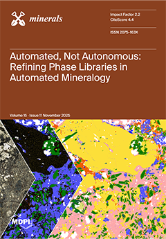
Accurate phase identification is essential for characterizing mineral systems, yet it remains a challenge in SEM-based automated mineralogy (AM) when dealing with compositionally variable rock-forming or accessory minerals. This study presents an integrated approach for refining and validating custom, deposit-specific phase libraries in AM using a Tescan TIMA. By combining iterative, informed library design with complementary microanalytical data (EPMA, LA-ICPMS), the approach improves phase classification accuracy and confidence. This approach is demonstrated through three case studies from complex mineral systems in Queensland, Australia: amphiboles in an IOCG deposit; cobalt-bearing phases in a sediment-hosted Cu–Au–Co deposit; and Li-micas in an LCT pegmatite system. View this paper
- Issues are regarded as officially published after their release is announced to the table of contents alert mailing list.
- You may sign up for e-mail alerts to receive table of contents of newly released issues.
- PDF is the official format for papers published in both, html and pdf forms. To view the papers in pdf format, click on the "PDF Full-text" link, and use the free Adobe Reader to open them.
Previous Issue
Next Issue