
Bioremediation of Uranium- and Nitrate-Contaminated Groundwater after the In Situ Leach Mining of Uranium

1
Xinjiang Key Laboratory of Environmental Pollution and Bioremediation, Xinjiang Institute of Ecology and Geography, Chinese Academy of Sciences, Urumqi 830011, China
2
Desert Engineering Survey and Design Institute, Xinjiang Institute of Ecology and Geography, Chinese Academy of Sciences, Urumqi 830011, China
3
Key Laboratory of Microbial Technology for Industrial Pollution Control of Zhejiang Province, College of Environment, Zhejiang University of Technology, Hangzhou 310014, China
*
Authors to whom correspondence should be addressed.
Water 2021, 13(22), 3188; https://doi.org/10.3390/w13223188
Submission received: 22 October 2021
/
Revised: 8 November 2021
/
Accepted: 8 November 2021
/
Published: 11 November 2021
(This article belongs to the Special Issue Biological Treatment of Mining and Industrial Effluents and Groundwaters)
Abstract
:Uranium and nitrate are common groundwater pollutants near in situ leach uranium mines. However, we still lack techniques that can simultaneously immobilize uranium and reduce nitrate using a single bacterial species. In this study, the potential of simultaneous uranium immobilization and nitrate reduction by a single AFODN (anaerobic Fe(II) oxidizing denitrifier), Clostridium sp. PXL2, was investigated. Clostridium sp. PXL2 showed tolerance to U(VI) concentrations varying from 4.2 µM to 42 µM. The U(VI) immobilization and nitrate reduction rates in groundwater samples inoculated with this bacterium reached up to 75.1% and 55.7%, respectively, under neutral conditions. Exposure to oxidation conditions led to further U(VI) removal but did not show any noticeable effect on nitrate reduction. The U(VI) immobilization rate reached up to 85% with an increased Fe(II) initial concentration, but this inhibited nitrate reduction. SEM (scanning electron microscopy) coupled with EDS (energy dispersive spectroscopy) showed that the U(VI) immobilization was mainly due to sorption to amorphous ferric oxides. U(VI) and nitrate bioremediation by AFODNs, including Clostridium sp. PXL2, may provide a promising method for the treatment of uranium- and nitrate-contaminated groundwater after the in situ leach mining of uranium.
1. Introduction
Uranium pollution has become a serious environmental concern because of extensive uranium mining, leaching, and the poor processing of by-products generated during mining activities [1]. ISL (in situ uranium leaching) techniques were developed in the USA and have also been broadly used in Eastern Europe, including the former Soviet Union, and some parts of Asia. However, the leaching solutions from ISL mines contain high levels of uranium and nitrate, which contaminate nearby aquifers and cause serious pollution to the groundwater. The uranium and nitrate concentrations in leaching solutions at some typical ISL mines are given in Table 1. The uranium concentration in the groundwater in uranium mining and milling sites can range from 1 mM to 210 mM [2,3]. Long-term exposure to uranium-polluted drinking water can cause serious health problems, including renal function damage or kidney failure [4,5]. The permissible value for uranium in drinking water is set as 2.0 mg L−1 by the World Health Organization [6] and is 30 µg L−1 in the USA [7]. Nitrate is another common pollutant produced by uranium leaching and processing due to the use of nitric acid or ammonia [8,9]. Groundwater nitrate levels have been observed to reach up to 130 mM in Oak Ridge, TN [8], and 19.7 mM in mill tailing sites near Tuba City, AZ (USA) [10]. The main health problem associated with nitrate is blue baby syndrome, which is caused by the reaction of nitrate with hemoglobin [11]. The guideline value for nitrate in groundwater is set as 10 mg L−1 in many countries [12].
In recent decades, a number of biotechnologies, including biomineralization [15,16,17], bioreduction [18,19,20,21], biosorption [22,23,24,25], and bioaccumulation [26], have been proposed in order to immobilize uranium in contaminated groundwater. Among the uranium bioremediation techniques available, uranium bioreduction has been studied extensively, where U(VI) is reduced to U(IV) by a variety of bacteria. In situ demonstrations of uranium bioreduction have been conducted successfully at the US DOE Rifle site, Colorado [27], and at the US DOE Oak Ridge site, Tennessee [28,29]. However, the stability of reduced U(IV) depends on protecting it from re-oxidation by nitrate [30,31] and O2 [28]. Even certain intermediates of denitrification products such as nitrite and nitric oxide have been shown to cause the re-oxidation of U(IV) [32]. Therefore, the coexistence of nitrate with uranium poses a challenge when using this technique as a viable uranium bioremediation strategy for polluted groundwater [31,32]. Generally, uranium biomineralization has only been studied at the laboratory scale due to the high cost of organic phosphates and other toxic effects on cell metabolism [9,33].
There is, therefore, an urgent need to find cost-effective biological techniques that can remediate groundwater contaminated with uranium and nitrate. Although uranium and nitrate are frequently detected together in groundwater near uranium mines and milling sites, especially at in situ leaching uranium sites, currently we still lack biotechnologies that can simultaneously remove uranium and nitrate using a single species of bacteria. Bacterial species known as AFODNs have been reported to oxidize Fe(II) to Fe(III) under anoxic conditions [34,35]. AFODNs can simultaneously reduce nitrate to denitrification intermediates such as nitric oxide and nitrogen. They also produce ferric oxides that have a great adsorption capacity for U(VI) [36,37]. These anaerobes may therefore provide a novel solution to concurrently remediate uranium and nitrate contaminated groundwater arising from in situ uranium mining, milling, and leaching sites.
The purpose of this study was to simulate polluted groundwater after the in situ leach mining of uranium and to investigate the potential of the AFODN bacterium, Clostridium sp. PXL2, for the simultaneous removal of uranium and nitrate. The effects of environmental factors such as pH, oxidation, and Fe(II) on uranium immobilization and nitrate reduction by this anaerobe were assessed. SEM-EDS analysis was applied to characterize the adsorption of uranium by the biogenic iron oxides.
2. Materials and Methods
2.1. Sample Collection and Processing
Groundwater samples were collected from a uranium rich coal mining site in Xinjiang, China. The groundwater was pumped up by an electric well water pump from a depth of about 150 m and collected in 100 L plastic containers, which were completely filled and capped tightly in order to avoid further oxidation. Some basic physical and chemical parameters of the groundwater are listed in Table 2.
2.2. Culture of Clostridium sp. PXL2
The anaerobic bacterium was originally isolated from activated sludge collected from the sewage management station of Miquan in Urumqi, China, and identified as Clostridium sp. PXL2 [12]. The bacterium was incubated at 30 °C in a medium of the following composition (L−1 deionized water): 0.28 g L−1 NH4CI, 0.25 g L−1 KH2PO4, 0.1 g L−1 MgSO4, 0.1 g L−1 yeast extract, 0.5 g L−1 KNO3, 0.5 g L−1 FeCI2·4H2O. The growing culture was used for further experiments after 1 week of incubation at 30 °C. The initial optical density (OD600) of the experimental bacterium was adjusted to 0.2.
2.3. Groundwater Treatments
Groundwater was amended with 0.3 g L−1 of yeast extract and then sterilized using 0.22 µm hydrophilic polyestersulfone membrane filters. The sterile groundwater was placed in an anaerobic chamber overnight in order to remove dissolved oxygen. The pH was adjusted with 0.1 M sodium hydroxide solution or 0.1 M hydrochloric acid. The groundwater was supplemented with filter-sterilized 1.0 g L−1 U(VI) stock solution and 130 g L−1 sodium nitrate solution to provide initial concentrations of 1.5 mg L−1 U(VI) and 1050 mg L−1 nitrate, respectively. Various Fe(II) concentrations were obtained using filter-sterilized 35.2 g L−1 Fe(II) (FeCI2·4H2O) stock solution. All manipulations and experiments were conducted in 250 mL serum bottles in an anaerobic workstation at 30 °C. A 2% (v/v) Clostridium PXL2 culture was inoculated into the test samples after supplementing with the other chemicals. The Fe(II), U(VI), nitrate, and nitrite concentrations were measured every 24 h. All experiments were conducted in triplicate.
2.4. Measurement of Growth
Clostridium sp. PXL2 was inoculated in the above-mentioned selective media in 100 mL serum bottles containing various U(VI) concentrations (0, 4.2, 10.5, 21.0, 42.0 µM) and cultivated at 30 °C in the DG250 anaerobic workstation. Samples without U(VI) were used as controls. Using a UV2800 spectrophotometer (Unico, Shanghai, China), we measured the bacterial growth at 600 nm (OD600) at exposure to U(VI) for 12, 24, 36, 48, 60, 72, and 84 h.
2.5. Analytical Methods
The concentrations of Fe(II), U(VI), nitrate, and nitrite were measured by spectrophotometric methods using a UV2800 spectrophotometer. The Fe(II) concentration was determined by a modified phenanthroline method at 510 nm [39]. Instead of using a 25% HCI solution, 40 mM of sulfamic acid was used, as this reacts quickly with nitrite and prevents Fe(II) oxidation by the nitrite at acidic pH values [40]. U(VI) concentration was monitored using an optimized photometric Arsenazo-III method at 651 nm [41]. Briefly, several milliliters of prefiltered (0.22 µm) groundwater samples were heated slowly to near dryness followed by the addition of 0.007% Arsenazo-III solution. A total of 0.007% Arsenazo-III solution was prepared by dissolving Arsenazo-III in 3 mol L−1 of HClO4 (w/v). The final volume was made up to 5.0 mL in a volumetric flask by using the same Arsenazo-III solution. The absorbance was measured at 651 nm against the reagent blank after thorough mixing. The nitrate concentration was measured at 220 and 275 nm. Certain amounts of prefiltered groundwater samples were added to 50 mL colorimetric tubes and diluted to 50 mL with distilled water followed by the addition of 1 mL of 1 mol L−1 hydrochloric acid. An amount of 50 mL of distilled water plus 1 mL of hydrochloric acid was used as reference. A quartz cuvette of optical length 10 mm was used for the measurement [38]. The nitrite concentration was determined to be 540 nm. Certain amounts of prefiltered groundwater samples were added to 50 mL colorimetric tubes and diluted to 50 mL with distilled water. A total of 1 mL of chromogenic agent (N-(a-naphthyl)-ethylenediamine) was added and thoroughly mixed. Distilled water was used as a reference. The samples were measured after resting for 20 min [42].
Biogenic Fe(II) oxidation sediments and bacterial cells were examined by SEM-EDS. Sediment samples for SEM-EDS analysis were prepared as follows. First, the samples were centrifuged for 5 min at 8000 rpm and supernatants were abandoned. Pellets were washed with distilled water. This process was repeated 3 times. Afterwards, the washed pellets were frozen at −80 °C for 4 h before drying at −60 °C for 48 h. A scanning electron microscope (Zeiss Super 55VP, Oberkochen, Germany) was used for analyzing our samples. Then, we used an energy dispersive X-ray spectrometer (Bruker XFlash 5010, Karlsruhe, Germany) to confirm the elemental composition of the sediments.
2.6. Chemicals
U(VI) was applied in the form of analytical-grade uranyl nitrate hexahydrate [UO2(NO3)2·6H2O] (Hubei Chushengwei Chemistry Co., Ltd., Wuhan, China). Initially, a 1.0 g L−1 stock U(VI) solution was prepared in deionized water and stored at 4 °C. Other chemicals included FeCl2·4H2O (analytical grade, Beijing Kangpu Huiwei Technology Co., Ltd., Beijing, China) and sodium nitrate (analytical grade, Tianjin Yongsheng Chemical Co., Ltd., Tianjin, China).
2.7. Statistical Methods
The Mann–Whitney–Wilcoxon method was applied to determine whether there were significant differences between the subgroups. The t-test statistical method was applied to determine whether there were significant differences between the values of this study and mean (or average) values of other studies.
3. Results and Discussion
3.1. Growth in the Presence of U(VI)
Figure 1 shows the growth of Clostridium sp. PXL2 in the presence of various concentrations of U(VI). The cell growth in all U(VI) concentrations showed a trend of steady increase over the 96 h of incubation. Compared to the control, the cell growth was not significantly affected by U(VI) exposure to up to 42 µΜ (p > 0.05), implying that U(VI) has no significant toxic effects on this bacterium in an aquatic environment. It is also noticeable from Figure 1 that there was a slight but not significant increase (p > 0.05) in the cell growth with the increase in U(VI) concentrations. This was possibly due to the precipitation of U(VI) with phosphate ions which were released from NB media during the bacterial cell growth.
3.2. Influence of pH
Figure 2 shows the changes in Fe(II), U(VI), nitrate, and nitrite concentrations in the control and inoculated (pH 5.5 and pH 7.0) cultures over the 96 h of incubation under anoxic conditions. We observed a decline in the concentrations of Fe(II), U(VI), and nitrate, whereas the nitrite concentration increased (Figure 2a,b). After the 96 h of incubation, the Fe(II) concentrations in the inoculated groundwater samples at initial pH 5.5 and pH 7.0 were reduced by 27.2% and 65.4%, from an initial concentration of 2.35 mM and 1.30 mM, respectively (Figure 2a). The lower Fe(II) oxidation rate at pH 5.5 was possibly due to the fact that Fe(II) is more resistant to oxidation in acidic conditions than at neutrality. Moreover, neutral conditions are more favorable for the growth of Clostridium sp. PXL2 than alkaline or acidic conditions [12]. The U(VI) concentrations in the inoculated pH 5.5 and pH 7.0 samples dropped by 70.0% and 75.1%, respectively (Figure 2a). The estimated molar ratios of immobilized U(VI) and oxidized Fe(II) at pH 5.5 and pH 7.0 were 0.0067 and 0.0047, respectively. The higher effective U(VI) immobilization at pH 5.5 was due to aqueous U(VI) being strongly adsorbed to ferric oxides at low pH [43,44]. In higher pH environments (pH ≥ 7), UO22+ formed strong aqueous complexes with CO32−, which greatly enhanced the solubility of U(VI) in carbonate-containing aquatic environments [45,46]. During the 96 h of incubation, the nitrate concentrations at pH 5.5 and pH 7.0 rapidly declined with 43.5% and 55.7% levels of removal, respectively (Figure 2b). The higher nitrate reduction at pH 7.0 was due to the better bacterial growth in a neutral environment [12]. Concurrently, 4.32 mM and 5.72 mM of nitrite were produced at pH 5.5 and pH 7.0 after 96 h, which are lower values than the stoichiometric loss of 7.24 mM and 9.49 mM nitrate. This indicates that nitrite may be further reduced to nitrogen, ammonia, or NO [12]. Comparatively, only 11.9% of the Fe(II) and U(VI) and 8.0% of the nitrate were removed in the control sample at pH 7.0, while 1.7%, 4.7%, and 2.4% of the Fe(II), U(VI), and nitrate were lost from the control sample at pH 5.5, respectively. This confirmed that the nitrate reduction and U(VI) immobilization in the groundwater samples were mainly due to the activities of the Clostridium sp. PXL2.
3.3. Influence of Oxidation
Figure 3 shows the changes in Fe(II), U(VI), nitrate, and nitrite concentrations in the groundwater samples over a 2 h oxidation period after 96 h of incubation. The Fe(II) concentrations in the inoculated pH 5.5 and pH 7.0 samples dropped rapidly by 33.6% and 9.2% compared to the 37.3% and 58.9% losses in the pH 5.5 and pH 7.0 control samples over 2 h of oxidation (Figure 3). This means that Fe(II) is much more vulnerable to oxidation by oxygen than to bacterial oxidation. The U(VI) concentrations simultaneously decreased by 30.0% and 9.8% in the pH 5.5 and pH 7.0 inoculated samples, while 42.3% and 60.9% were lost in the pH 5.5 and pH 7.0 control samples (Figure 3). This rapid and efficient immobilization of U(VI) in oxic conditions can be explained by the fact that abiotically produced Fe(III) oxides have a better binding capacity for U(VI) than biogenic Fe(III) oxides [47]. During the 2 h oxidation period, the nitrate concentrations in both the inoculated pH samples decreased slightly (p > 0.05) while the nitrite concentrations slightly increased (p > 0.05) (Figure 3),implying, therefore, that the facultative bacterium Clostridium sp. PXL2 was still active in oxic conditions.
3.4. Effect of Fe(II)
Figure 4 shows the effects of Fe(II) on the changes in the Fe(II), U(VI), nitrate, and nitrite concentrations in the groundwater samples over 96 h of anoxic incubation. The initial Fe(II) concentrations of 1.2 mM and 4.0 mM in the groundwater samples decreased rapidly over the first 48 h before declining slowly to 0.4 mM and 2.5 mM after 96 h, with 66.7% and 37.5% levels of oxidation, respectively (Figure 4a). This low Fe(II) conversion to Fe(III) at a high initial Fe(II) concentration was due to the fact that the bacterial cells tended to settle at the bottom of the reaction vessel with the produced Fe(III) oxides, inhibiting the further oxidation of Fe(II) in the system. Furthermore, it is possible that Fe(III) oxides might have toxic effects on bacterial growth [48]. About 73.7% and 84.5% of the supplied U(VI) were immobilized from the initial concentrations of 6.34 µM and 6.39 µM, respectively (Figure 4a). The molar ratios of U(VI) immobilization to Fe(II) oxidation were 0.0058 and 0.0036, indicating that the U(VI) immobilization was more efficient at lower Fe(II) concentrations. It is also noticeable from Figure 4a that the U(VI) concentration in the control sample without the Fe(II) addition was reduced slightly (p > 0.05), probably due to biosorption to the bacterial cells. Bacterial cell walls contain ligands such as phosphate, carboxyl, hydroxyl, sulfhydryl, and amine groups, which are able to bind metal cations [9]. As can be seen from Figure 4b, the concentration of nitrate within the samples showed a rapid decrease during the initial 48 h but gradually subsided to under 44.3–58.1% reduction in the 96 h from an initial concentration of about 17 mM. It is also noticeable that the nitrate reduction (58.1%) was higher in the control sample than in the 1.2 mM Fe(II) (54.2%) and 4.0 mM Fe(II) (44.3%) samples. The higher nitrate reduction in the 1.2 mM Fe(II) sample than in the 4.0 mM Fe(II) sample further suggests that high Fe(II) levels inhibited the bacterial growth.
3.5. SEM-EDS Analysis
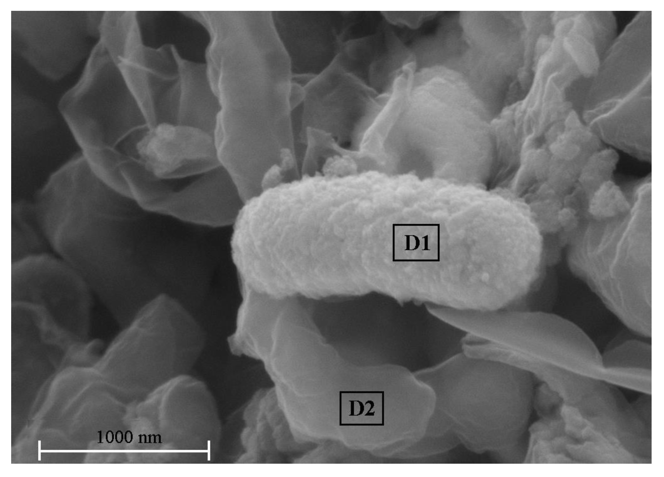
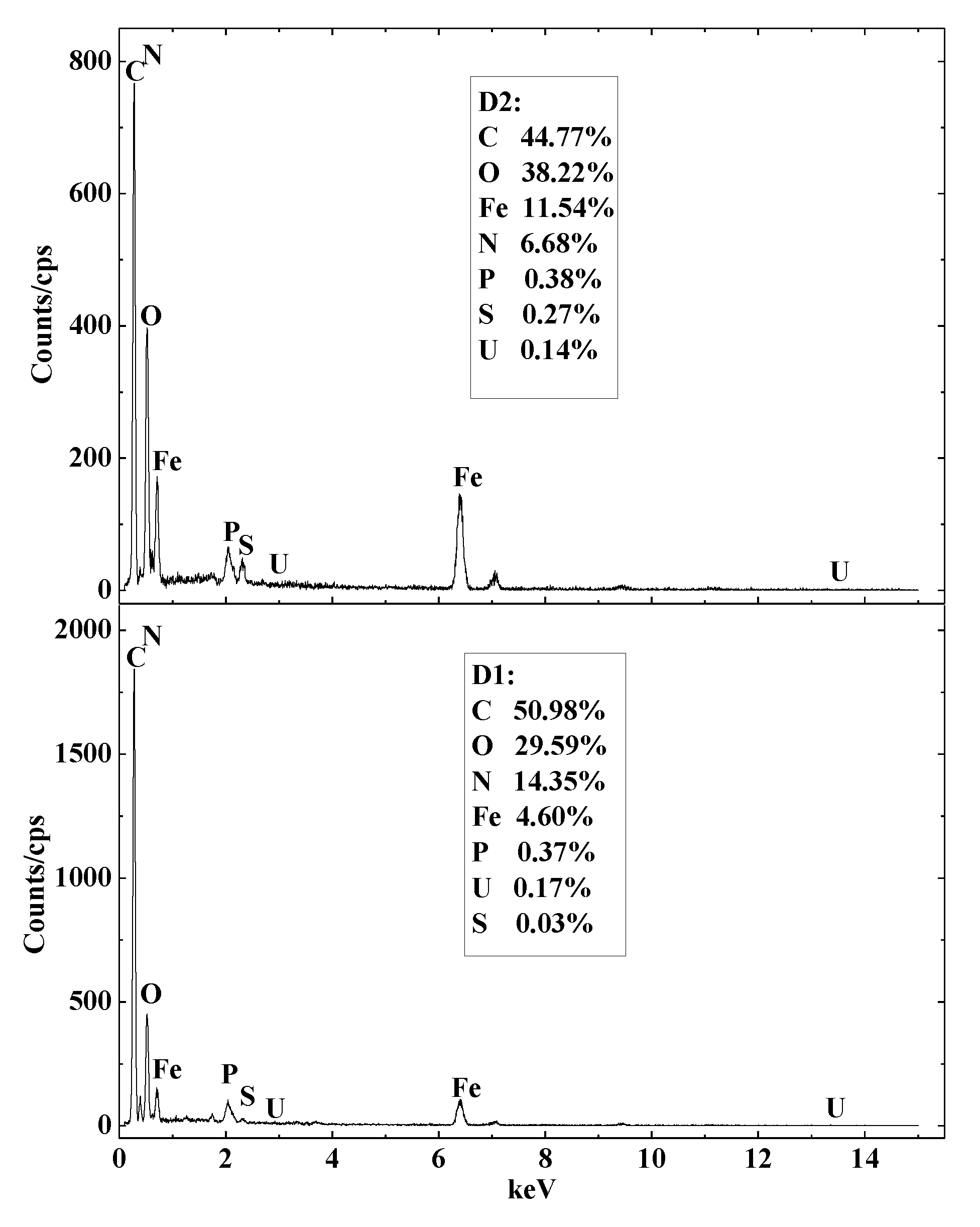
Figure 5 shows Clostridium sp. PXL2 and sediments containing various concentrations of Fe(II), U(VI), and other elements. The Clostridium sp. PLX2 cells were approximately 1.8 μm in length. Amorphous iron oxides were observed on the cell surfaces and crystalline iron oxides were not observed. The formation of amorphous iron oxides by some other AFODN species has also been reported [49,50]. Figure 6 shows the elemental composition of the sediments around the bacteria cells. The following, including C, O, N, Fe, and a small of proportion of P, U, and S based on their weight, primarily formed the sediments’ composition. The U(VI) percentages were 0.17% and 0.14% for D1 and D2 spots, indicating that the U(VI) was mainly immobilized due to adsorption on iron oxides produced by Clostridium sp. PXL2. The slightly higher content of U(VI) in D1 could be attributed to biosorption by bacterial cells [9].
4. Conclusions
Our results demonstrated that the AFODN bacterium Clostridium sp. PXL2 could simultaneously remove U(VI) and nitrate from polluted groundwater. The removal efficiency of these two pollutants was affected by environmental factors such as pH, redox conditions, and initial Fe(II) concentration. SEM-EDS analysis showed that the U(VI) was immobilized mainly by sorption to poorly crystalline ferric oxides that were produced by Clostridium sp. PXL2. Therefore, nitrate-dependent Fe(II) oxidizing bacteria including Clostridium sp. PXL2 are effective microbes for the bioremediation of uranium- and nitrate-contaminated groundwater after the in situ leach mining of uranium.
Author Contributions
X.P., W.L. and R.W. conceived the ideas and designed this study; R.W., J.D. and J.F. interpreted the data of spatial distribution and structure characteristics and analysis; R.W. wrote the manuscript; X.P. reviewed the manuscript. All authors have read and agreed to the published version of the manuscript.
Funding
This research was supported by Science and Technology Department of Xinjiang Uyghur Autonomous Region (2017B03014-1, 2, 3, 2018D04013), the West Light Foundation of Chinese Academy of Sciences (2018-XBQNXZ-B-014, 2017-XBQNXZ-B-011).
Institutional Review Board Statement
This study does not involve humans and animals.
Informed Consent Statement
This study does not involve any humans.
Data Availability Statement
This study did not report any dada.
Acknowledgments
The authors are grateful to the experimenters at the center Laboratory of Xinjiang Institute of Ecology and Geography for their assistance during the experiment. We also thank Friday U. Ochege for editing the English text of a draft of this manuscript.
Conflicts of Interest
The authors declare that they have no known competing financial interest or personal relationships that could have appeared to influence the work reported in this paper.
References
- OECD-NEA; IAEA. Environmental Activities in Uranium Mining and Milling; OECD Publishing: Paris, France, 1999; p. 173. [Google Scholar]
- Wu, W.; Carley, J.; Fienen, M.; Mehlhorn, T.; Lowe, K.; Nyman, J.; Luo, J.; Gentile, M.; Rajan, R.; Wagner, D. Pilot-scale in situ bioremediation of uranium in a highly contaminated aquifer. 1. Conditioning of a treatment zone. Environ. Sci. Technol. 2006, 40, 3978–3985. [Google Scholar] [CrossRef] [PubMed]
- Wufuer, R.; Song, W.; Zhang, D.; Pan, X.; Gadd, G.M. A survey of uranium levels in urine and hair of people living in a coal mining area in Yili, Xinjiang, China. J. Environ. Radioact. 2018, 189, 168–174. [Google Scholar] [CrossRef] [PubMed] [Green Version]
- ATSDR. Toxicological Profile for Uranium; Agency for Toxic Substances and Disease Registry: Atlanta, GA, USA, 1999; p. 462. [Google Scholar]
- Choy, C.C.; Korfiatis, G.P.; Meng, X. Removal of depleted uranium from contaminated soils. J. Hazard. Mater. 2006, 136, 53–60. [Google Scholar] [CrossRef] [PubMed]
- WHO. Guidelines for Drinking Water Quality; The World Health Organisation: Geneva, Switzerland, 1998. [Google Scholar]
- USEPA. Drinking Water Contaminants; 1999. Available online: http://www.epa.gov/safewater/contaminants/index.html (accessed on 25 December 2015).
- Istok, J.D.; Senko, J.M.; Krumholz, L.R.; Watson, D.; Bogle, M.A.; Peacock, A.; Chang, Y.-J.; White, D.C. In Situ Bioreduction of Technetium and Uranium iii a Nitrate-Contaminated Aquifer. Environ. Sci. Technol. 2004, 38, 468–475. [Google Scholar] [CrossRef]
- Lloyd, J.R.; Macaskie, L.E. Bioremediation of radionuclide-containing wastewaters. In Environmental Microbe-Metal Interactions; ASM Press: Washington, DC, USA, 2000; pp. 277–327. [Google Scholar]
- Abdelouas, A.; Lutze, W.; Gong, W.; Nuttall, E.H.; Travis, B.J. Biological reduction of uranium in groundwater and subsurface soil. Sci. Total Environ. 2000, 250, 21–35. [Google Scholar] [CrossRef]
- Fan, A.M.; Steinberg, V.E. Health Implications of Nitrate and Nitrite in Drinking Water: An Update on Methemoglobinemia Occurrence and Reproductive and Developmental Toxicity. Regul. Toxicol. Pharmacol. Rtp 1996, 23, 35. [Google Scholar] [CrossRef]
- Li, B.; Pan, X.; Zhang, D.; Lee, D.J.; Al-Misned, F.A.; Mortuza, M.G. Anaerobic nitrate reduction with oxidation of Fe(II) by Citrobacter Freundii strain PXL1–a potential candidate for simultaneous removal of As and nitrate from groundwater. Ecol. Eng. 2015, 77, 196–201. [Google Scholar] [CrossRef]
- Mudd, G.M. Critical review of acid in situ leach uranium mining: 1. USA and Australia. Environ. Geol. 2001, 41, 390–403. [Google Scholar] [CrossRef]
- Mudd, G.M. Critical review of acid in situ leach uranium mining: 2. Soviet Block and Asia. Environ. Geol. 2001, 41, 404–416. [Google Scholar] [CrossRef]
- Beazley, M.J.; Martinez, R.J.; Webb, S.M.; Sobecky, P.A.; Taillefert, M. The effect of pH and natural microbial phosphatase activity on the speciation of uranium in subsurface soils. Geochim. Cosmochim. Acta 2011, 75, 5648–5663. [Google Scholar] [CrossRef]
- Macaskie, L.E.; Bonthrone, K.M.; Yong, P.; Goddard, D.T. Enzymically mediated bioprecipitation of uranium by a Citrobacter sp.: A concerted role for exocellular lipopolysaccharide and associated phosphatase in biomineral formation. Microbiology 2000, 146, 1855–1867. [Google Scholar] [CrossRef] [Green Version]
- Kuippers, G.; Morris, K.; Townsend, L.T.; Bots, P.; Lloyd, J.R. Biomineralization of Uranium-Phosphates Fueled by Microbial Degradation of Isosaccharinic Acid (ISA). Environ. Sci. Technol. 2021, 55, 4597–4606. [Google Scholar] [CrossRef]
- Lovley, D.R.; Phillips, E.J.; Gorby, Y.A.; Landa ER Lovley, D.R.; Phillips, E.J.; Gorby, Y.A.; Landa, E.R. Microbial reduction of uranium. Nature 1991, 350, 413. [Google Scholar] [CrossRef]
- Williams, K.H.; Long, P.E.; Davis, J.A.; Wilkins, M.J.; N’Guessan, A.L.; Steefel, C.I.; Yang, L.; Newcomer, D.; Spane, F.A.; Kerkhof, L.J. Acetate availability and its influence on sustainable bioremediation of uranium-contaminated groundwater. Geomicrobiol. J. 2011, 28, 519–539. [Google Scholar] [CrossRef]
- Wang, G.; Yang, S.; Zhou, Y.; Yuan, H.; Shiyou, L.I.; Yijin, L.; Xie, S. Research Progress on the Bioremediation of Groundwater Polluted by Uranium via Bio-reduction. Environ. Sci. Technol. 2019, 42, 47–53. [Google Scholar]
- You, W.; Peng, W.; Tian, Z.; Zheng, M. Uranium bioremediation with U(VI)-reducing bacteria. Sci. Total Environ. 2021, 798, 1–15. [Google Scholar] [CrossRef]
- Gadd, G.M. Biosorption: Critical review of scientific rationale, environmental importance and significance for pollution treatment. J. Chem. Technol. Biotechnol. 2010, 84, 13–28. [Google Scholar] [CrossRef]
- Schiewer, S.; Volesky, B. Biosorption processes for heavy metal removal. Environ. Microbe-Met. Interact. 2000, 14, 329–362. [Google Scholar]
- Suzuki, Y.; Banfield, J.F. Geomicrobiology of uranium. Rev. Minera. Geochem. 1999, 38, 393–432. [Google Scholar]
- Coelho, E.; Reis, T.A.; Cotrim, M.; Rizzutto, M.; Corrêa, B. Bioremediation of water contaminated with uranium using Penicillium piscarium. Biotechnol. Prog. 2020, 36, e30322. [Google Scholar] [CrossRef]
- Choudhary, S.; Sar, P. Uranium biomineralization by a metal resistant Pseudomonas aeruginosa strain isolated from contaminated mine waste. J. Hazard. Mater. 2011, 186, 336–343. [Google Scholar] [CrossRef]
- Anderson, R.T.; Vrionis, H.A.; Ortiz-Bernad, I.; Resch, C.T.; Long, P.E.; Dayvault, R.; Karp, K.; Marutzky, S.; Metzler, D.R.; Peacock, A. Stimulating the in situ activity of geobacter species to remove uranium from the groundwater of a uranium-contaminated aquifer. Appl. Environ. Microbiol. 2003, 69, 5884–5891. [Google Scholar] [CrossRef] [Green Version]
- Duff, M.C.; Hunter, D.B.; Bertsch, P.M.; Amrhein, C. Factors influencing uranium reduction and solubility in evaporation pond sediments. Biogeochemistry 1999, 45, 95–114. [Google Scholar] [CrossRef]
- Wu, W.M.; Carley, J.; Gentry, T.; Ginder-Vogel, M.A.; Criddle, C.S. Pilot-scale in situ bioremedation of uranium in a highly contaminated aquifer. 2. Reduction of u(VI) and geochemical control of u(VI) bioavailability. Environ. Sci. Technol. 2006, 40, 3986–3995. [Google Scholar] [CrossRef] [PubMed]
- Moon, H.S.; Komlos, J.; Jaffe, P.R. Uranium reoxidation in previously bioreduced sediment by dissolved oxygen and nitrate. Environ. Sci. Technol. 2007, 41, 4587–4592. [Google Scholar] [CrossRef] [PubMed]
- Senko, J.M.; Mohamed, Y.; Dewers, T.A.; Krumholz, L.R. Role for Fe(III) minerals in nitrate-dependent microbial U(IV) oxidation. Environ. Sci. Technol. 2005, 39, 2529–2536. [Google Scholar] [CrossRef] [PubMed]
- Senko, J.M.; Istok, J.D.; Suflita, J.M.; Krumholz, L.R. In-situ evidence for uranium immobilization and remobilization. Environ. Sci. Technol. 2002, 36, 1491–1496. [Google Scholar] [CrossRef] [PubMed]
- Benzerara, K.; Miot, J.; Morin, G.; Ona-Nguema, G.; Skouri-Panet, F.; Ferard, C. Significance, mechanisms and environmental implications of microbial biomineralization. Comptes Rendus-Géoscience 2011, 343, 160–167. [Google Scholar] [CrossRef]
- Straub, K.L.; Schonhuber, W.A.; Buchholz-Cleven, B.; Schink, B. Diversity of ferrous iron-oxidizing, nitrate-reducing bacteria and their involvement in oxygen-independent iron cycling. Geomicrobiol. J. 2004, 21, 371–378. [Google Scholar] [CrossRef]
- Weber, K.A.; Urrutia, M.M.; Churchill, P.F.; Kukkadapu, R.K.; Roden, E.E. Anaerobic redox cycling of iron by freshwater sediment microorganisms. Environ. Microbiol. 2010, 8, 100–113. [Google Scholar] [CrossRef] [Green Version]
- Means, J.L.; Crerar, D.A.; Borcsik, M.P.; Duguid, J.O. Radionuclide adsorption by manganese oxides and implications for radioactive waste disposal. Nature 1978, 274, 44–47. [Google Scholar] [CrossRef]
- Means, J.L.; Crerar, D.A.; Borcsik, M.P.; Duguid, J.O. Adsorption of Co and selected actinides by Mn and Fe oxides in soils and sediments. Geochim. Cosmochim. Acta 1978, 42, 1763–1773. [Google Scholar] [CrossRef]
- Slanina, J.; Lingerak, W.A.; Bergman, L. A fast determination of nitrate in rain and surface waters by means of UV spectrophotometry. Fresenius Z. Für Anal. Chem. 1976, 280, 365–368. [Google Scholar] [CrossRef]
- Gendel, Y.; Lahav, O. Accurate determination of Fe(II) concentrations in the presence of a very high soluble Fe(III) background. Appl. Geochem. 2008, 23, 2123–2129. [Google Scholar] [CrossRef]
- Klueglein, N.; Kappler, A. Abiotic oxidation of Fe(II) by reactive nitrogen species in cultures of the nitrate-reducing Fe(II) oxidizer Acidovorax sp. BoFeN1–questioning the existence of enzymatic Fe(II) oxidation. Geobiology 2013, 11, 180–190. [Google Scholar] [CrossRef]
- Jauberty, L.; Drogat, N.; Decossas, J.L.; Delpech, V.; Gloaguen, V.; Sol, V. Optimization of the arsenazo-III method for the determination of uranium in water and plant samples. Talanta 2013, 115, 751–754. [Google Scholar] [CrossRef]
- Geng, Y.J.; Qi, W.; Muszynski, M.; Hansson, G.K.; Libby, P. Apoptosis of vascular smooth muscle cells induced by in vitro stimulation with interferon-γ, tumor necrosis factor–α, and interleukin-1β. Arterioscler. Thromb. 2015, 7, 19–27. [Google Scholar] [CrossRef]
- Han, R.; Zou, W.; Wang, Y.; Lu, Z. Removal of uranium(VI) from aqueous solutions by manganese oxide coated zeolite: Discussion of adsorption isotherms and pH effect. J. Environ. Radioact. 2007, 93, 127–143. [Google Scholar] [CrossRef]
- Waite, T.D.; Davis, J.A.; Payne, T.E.; Waychunas, G.A.; Xu, N. Uranium(VI) adsorption to ferrihydrite: Application of a surface complexation model. Geochim. Et Cosmochim. Acta 1994, 58, 5465–5478. [Google Scholar] [CrossRef]
- Langmuir, D. Aqueous Environmental Geochemistry; Prentice Hall: Hoboken, NJ, USA, 1997. [Google Scholar]
- Ulrich, K.U.; Ilton, E.S.; Veeramani, H.; Sharp, J.O.; Bernier-Latmani, R.; Schofield, E.J.; Bargar, J.R.; Giammar, D.E. Comparative dissolution kinetics of biogenic and chemogenic uraninite under oxidizing conditions in the presence of carbonate. Geochim. Cosmochim. Acta 2009, 73, 6065–6083. [Google Scholar] [CrossRef]
- Lack, J.G.; Chaudhuri, S.K.; Kelly, S.D.; Kemner, K.M.; O’Connor, S.M.; Coates, J.D. Immobilization of Radionuclides and Heavy Metals through Anaerobic Bio-Oxidation of Fe(II). Appl. Environ. Microbiol. 2002, 68, 2704–2710. [Google Scholar] [CrossRef] [Green Version]
- Benckiser, G.; Santiago, S.; Neue, H.U.; Watanabe, I.; Ottow, J.C.G. Effect of fertilization on exudation, dehydrogenase activity, iron-reducing populations and Fe++ formation in the rhizosphere of rice (Oryza sativa L.) in relation to iron toxicity. Plant Soil 1984, 79, 305–316. [Google Scholar] [CrossRef]
- Kappler, A. Geomicrobiological Cycling of Iron. Rev. Mineral. Geochem. 2005, 59, 85–108. [Google Scholar] [CrossRef]
- Li, B.; Tian, C.; Zhang, D.; Pan, X. Anaerobic Nitrate-Dependent Iron (II) Oxidation by a Novel Autotrophic Bacterium, Citrobacter freundii Strain PXL1. Geomicrobiology 2014, 31, 138–144. [Google Scholar] [CrossRef]
Figure 1.
Growth of Clostridium sp. PXL2 at various U(VI) concentrations. ■, control; ●, 4.2 µM; ▲, 10.5 µM; ▼, 21.0 µM; ★, 42.0 µM. Data are presented as means ± standard deviations (SD) of triplicate measurements.
Figure 1.
Growth of Clostridium sp. PXL2 at various U(VI) concentrations. ■, control; ●, 4.2 µM; ▲, 10.5 µM; ▼, 21.0 µM; ★, 42.0 µM. Data are presented as means ± standard deviations (SD) of triplicate measurements.
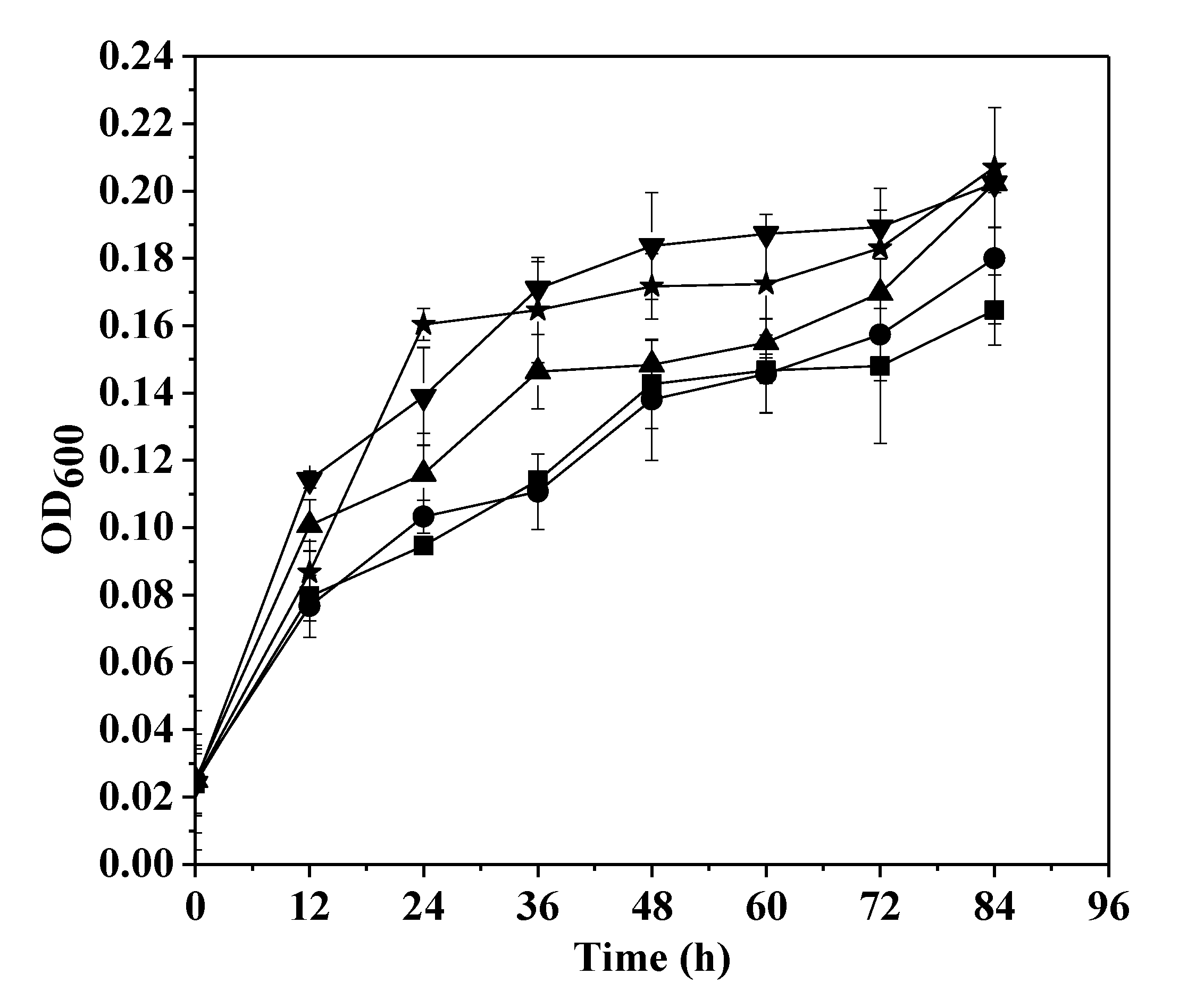
Figure 2.
The effects of pH on changes in Fe(II), U(VI), nitrate, and nitrite concentrations during the growth of Clostridium sp. PXL2. ■ and □, pH 5.5; ▼ and ▽, pH 5.5 (non-inoculated control ①); ● and ○, pH 7.0; ▲ and △, pH 7.0 (uninoculated control ②). Filled symbols, Fe(II) and nitrate; open symbols, U(VI) and nitrite. (a), Fe(II) and U(VI); (b), nitrate and nitrite. Data shown are means ± SD (n = 3).
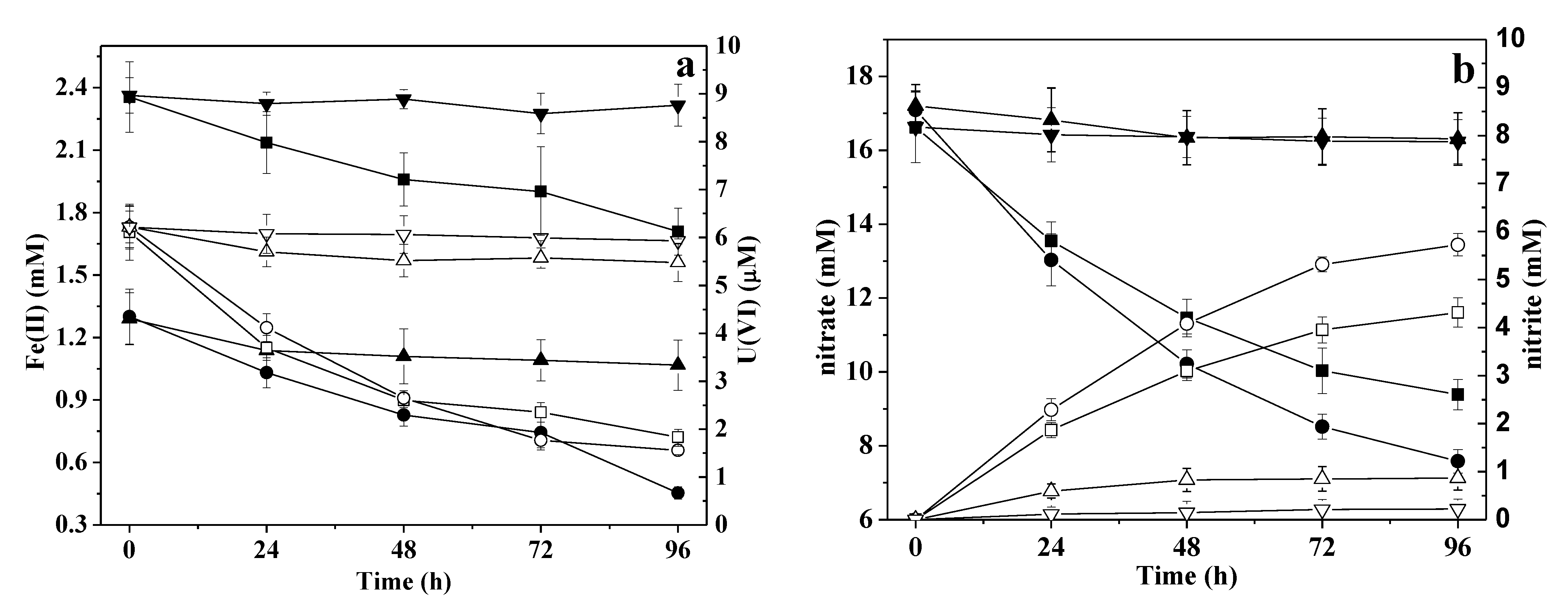
Figure 3.
Changes in concentrations of Fe(II), U(VI), nitrate, and nitrite after oxidation for 2 h following 96 h of anaerobic incubation. Data shown are means ± SD (n = 3).
Figure 3.
Changes in concentrations of Fe(II), U(VI), nitrate, and nitrite after oxidation for 2 h following 96 h of anaerobic incubation. Data shown are means ± SD (n = 3).
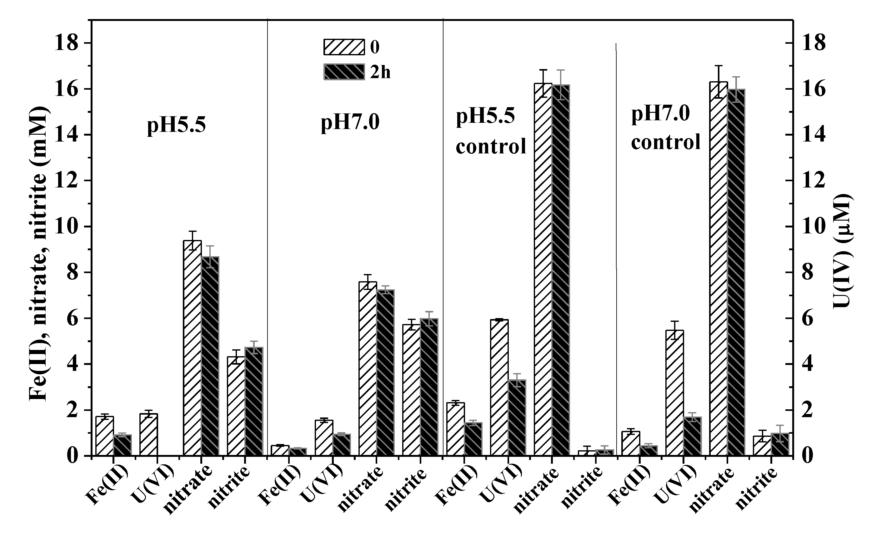
Figure 4.
The effects of Fe(II) on changes in Fe(II), U(VI), nitrate, and nitrite concentrations during the growth of Clostridium sp. PXL2. ■ and □, 0 mM Fe(II) (control); ● and ○, 1.2 mM Fe(II); ▲ and △, 4.0 mM Fe(II); ▼ and ▽, 1.2 mM Fe(II) (uninoculated). Filled symbols, Fe(II) and nitrate; open symbols, U(VI) and nitrite. (a), Fe(II) and U(VI); (b), nitrate and nitrite. Data shown are means ± SD (n = 3).

Figure 5.
SEM image of sediment from the Clostridium sp. PXL2 cultures. D1 and D2 are typical spot locations for EDS analysis on and around the bacterial cells. Typical images are shown from one of triplicate examinations.
Figure 5.
SEM image of sediment from the Clostridium sp. PXL2 cultures. D1 and D2 are typical spot locations for EDS analysis on and around the bacterial cells. Typical images are shown from one of triplicate examinations.

Figure 6.
EDS analysis of the Clostridium sp. PXL2 sediment (corresponding to spots D1 and D2 in Figure 5). Typical spectra are shown from one of triplicate determinations.
Figure 6.
EDS analysis of the Clostridium sp. PXL2 sediment (corresponding to spots D1 and D2 in Figure 5). Typical spectra are shown from one of triplicate determinations.


Table 1.
Leaching solution quality in some in situ leaching mines (mg L−1).
| Czech Rep. | Uzbekistan | Yining, China | |
|---|---|---|---|
| nitrate | 200–1400 | 750 | - |
| U(VI) | 20–500 | 86 | 75 |
Table 2.
Physical and chemical parameters of groundwater sample.
| pH | U(VI) | Conductivity | Cl− | SO42− | Ca2+ | K+ | Mg2+ | Na+ | CO32− | HCO3− | Nitrate |
|---|---|---|---|---|---|---|---|---|---|---|---|
| µg L−1 | us cm−1 | mg L−1 | mg L−1 | mg L−1 | mg L−1 | mg L−1 | mg L−1 | mg L−1 | mg L−1 | mg L−1 | |
| 7.74 | 8.71 | 1238 | 113.1 | 357.9 | 175.6 | 3.4 | 36.6 | 116.0 | 0 | 55.9 | 49.5 |
The pH was measured using a pH meter (Mettler SevenEasy); conductivity was measured by a conductivity detector (DDS-307); Cl− and SO42+ were measured by ion chromatography (ICS-5000 Dionex, Waltham, MA, USA); Ca2+, K+, Mg2+, and Na+ were measured by ICP-OES (735, Agilent, Santa Clara, CA, USA); HCO3− and CO32− were measured by an automatic potentiometric titrator (G20, Mettler); U(VI) concentration was measured by ICP-MS (8800, Agilent, USA); nitrate was measured by the photometric method of Slanina et al. [38].
Publisher’s Note: MDPI stays neutral with regard to jurisdictional claims in published maps and institutional affiliations. |
© 2021 by the authors. Licensee MDPI, Basel, Switzerland. This article is an open access article distributed under the terms and conditions of the Creative Commons Attribution (CC BY) license (https://creativecommons.org/licenses/by/4.0/).
Share and Cite
MDPI and ACS Style
Wufuer, R.; Duo, J.; Li, W.; Fan, J.; Pan, X. Bioremediation of Uranium- and Nitrate-Contaminated Groundwater after the In Situ Leach Mining of Uranium. Water 2021, 13, 3188. https://doi.org/10.3390/w13223188
AMA Style
Wufuer R, Duo J, Li W, Fan J, Pan X. Bioremediation of Uranium- and Nitrate-Contaminated Groundwater after the In Situ Leach Mining of Uranium. Water. 2021; 13(22):3188. https://doi.org/10.3390/w13223188
Chicago/Turabian StyleWufuer, Rehemanjiang, Jia Duo, Wenfeng Li, Jinglong Fan, and Xiangliang Pan. 2021. "Bioremediation of Uranium- and Nitrate-Contaminated Groundwater after the In Situ Leach Mining of Uranium" Water 13, no. 22: 3188. https://doi.org/10.3390/w13223188
Note that from the first issue of 2016, this journal uses article numbers instead of page numbers. See further details here.