Article

Diverse Horizontally-Acquired Gene Clusters Confer Sucrose Utilization to Different Lineages of the Marine Pathogen Photobacterium damselae subsp. damselae

Departamento de Microbioloxía e Parasitoloxía, Instituto de Acuicultura, Universidade de Santiago de Compostela, 15782 Santiago de Compostela, Spain
*
Author to whom correspondence should be addressed.
Genes 2020, 11(11), 1244; https://doi.org/10.3390/genes11111244
Submission received: 17 September 2020 / Accepted: 19 October 2020 / Published: 22 October 2020
(This article belongs to the Special Issue Genes at Ten)

Abstract

:
The ability to metabolize sucrose is a variable trait within the family Vibrionaceae. The marine bacterium Photobacterium damselae subsp. damselae (Pdd), pathogenic for marine animals and humans, is generally described as negative for sucrose utilization (Scr). Previous studies have reported sucrose-utilizing isolates (Scr+), but the genetic basis of this variable phenotype remains uncharacterized. Here, we carried out the genome sequencing of five Scr+ and two Scr Pdd isolates and conducted a comparative genomics analysis with sixteen additional Pdd genomes sequenced in previous studies. We identified two different versions of a four-gene cluster (scr cluster) exclusive of Scr+ isolates encoding a PTS system sucrose-specific IIBC component (scrA), a fructokinase (scrK), a sucrose-6-phosphate hydrolase (scrB), and a sucrose operon repressor (scrR). A scrA deletion mutant did not ferment sucrose and was impaired for growth with sucrose as carbon source. Comparative genomics analyses suggested that scr clusters were acquired by horizontal transfer by different lineages of Pdd and were inserted into a recombination hot-spot in the Pdd genome. The incongruence of phylogenies based on housekeeping genes and on scr genes revealed that phylogenetically diverse gene clusters for sucrose utilization have undergone extensive horizontal transfer among species of Vibrio and Photobacterium.

1. Introduction

The family Vibrionaceae includes a diverse and large number of bacterial species that are ubiquitous in marine habitats. Some species are recognized as pathogens for marine animals and for humans [1,2]. Also, their ecological importance in aquatic ecosystems and their high genetic plasticity have fuelled a major research effort on Vibrio, Photobacterium, and other genera of this family [3,4,5,6,7]. Most species exhibit a rapid growth in culture and use a large variety of nutrient sources, and the considerable inter- and intraspecies variability in metabolic and nutritional patterns is explained in part by horizontal transfer of gene functions [8,9,10,11]. Historically, carbon source utilization patterns have been extensively used to differentiate species within this family, and this knowledge has inspired the development of selective and differential culture media to enrich for specific taxa and for fast discrimination of some species. The thiosulfate citrate bile-salt sucrose (TCBS) agar, is a classical medium originally developed for the isolation of Vibrio parahaemolyticus [12] and is suitable for the isolation of many Vibrio and Photobacterium species. In addition to compounds that provide selective properties, TCBS contains 2% sucrose and pH indicators, allowing the differentiation between sucrose fermenters that produce yellow colonies (Scr+ phenotype) and non-fermenters that produce green colonies (Scr phenotype). The sucrose fermentation pattern on TCBS is a widespread taxonomic tool for species of the family Vibrionaceae [1]. However, very much as a contradiction, the genetic basis of sucrose utilization in species of this family has received scarce attention. Pioneering studies reported the cloning and characterization of sucrose utilization genes in Vibrio alginolyticus [13,14,15,16], and later studies reported some aspects on genetics of sucrose utilization in Vibrio cholerae [17,18,19,20], whereas no studies have been conducted in species of the genus Photobacterium to date.
Understanding the genetic basis for sucrose utilization in different species of the Vibrionaceae is essential to elucidate the evolutionary history of the acquisition of this metabolic capability. Sucrose utilization ability exhibits interspecies and intraspecies variability within this family which suggests that sucrose utilization genes are prone to be acquired by horizontal gene transfer. However, the phylogenetic relationships of genes for sucrose metabolism among Photobacterium and Vibrio species has not been analyzed so far. In addition, much remains to be learned about the role of sucrose utilization genes in the ecology of Vibrios. Sucrose, a disaccharide of glucose and fructose, is one of the most abundant sugars in terrestrial environments and a carbon source used by many bacterial taxa [21]. However, the availability of sucrose in marine environments has received little attention, and of what use is sucrose utilization in the marine environment for bacteria is a question that remains largely unanswered.
The marine pathogen Photobacterium damselae subsp. damselae (hereafter Pdd), formerly known as Vibrio damsela, is a member of the family Vibrionaceae that thrives in marine and estuarine environments and is a generalist pathogen that causes high financial losses in cultures of marine fish and crustaceans [22]. Moreover, it can cause opportunistic infections in humans that may evolve into a fatal outcome [23]. Pdd is considered a genetically diverse subspecies, and horizontal gene transfer largely contributes to the generation of such diversity [22,24,25,26]. Pdd typically forms green colonies on TCBS medium, but early studies pointed out that 5% of the isolates produced yellow colonies on TCBS [27]. Since then, a number of studies have described Pdd strains that produce yellow colonies on TCBS [24,28,29,30,31], but the genetic basis of such phenotypic variability remains uncharacterized.
In the present study, we accomplished the de novo genome sequencing of 7 Pdd isolates, 5 that tested positive for sucrose utilization, and 2 strains that were negative for this trait. Retrieval of additional 16 Pdd genome sequences from GenBank database allowed us to conduct an extensive comparative genomics analysis of Scr+ and Scr isolates that unveiled the presence of two distinct genetic variants of a four-gene cluster (scr cluster) that is necessary for sucrose utilization as a carbon source in Pdd. Notably, the majority of the scr clusters appear to be inserted into a hot-spot for recombination of variable DNA sequences within the Pdd genome. Based on the phylogenetic analyses of the scr clusters of Pdd and other species of the family Vibrionaceae, and on the reconstruction of species phylogenies by using the sequences of eight conserved housekeeping genes, we propose that sucrose utilization in Pdd arose from the horizontal acquisition by different Pdd lineages, of at least two distinct versions of gene clusters from species of the Vibrio and Photobacterium genera. It is also proposed that the exchange of scr clusters among species of the family Vibrionaceae has been extensive.

2. Materials and Methods

2.1. Bacterial Strains, Plasmids, and Culture Conditions

Pdd strains used here are described in Table 1. Genetically-modified Pdd strains, Escherichia coli strains, and plasmids used and constructed in this study are described in Table 2. Pdd cells were routinely grown at 25 °C on tryptic soy agar and broth supplemented with 1% NaCl (TSA-1 and TSB-1, respectively), supplemented with antibiotics when appropriate. E. coli strains were routinely grown at 37 °C in LB broth and LB agar supplemented with antibiotics when appropriate. Antibiotics were used at the following final concentrations: Kanamycin (Km) at 50 µg mL−1, ampicillin at 50 µg mL−1, and gentamicin at 15 µg mL−1. For sucrose fermentation phenotypical tests, Pdd strains were grown on TSA-1 plates overnight at 25 °C, and isolated colonies were seeded on thiosulfate-citrate-bile salts-sucrose agar plates (TCBS) and incubated at 25 °C for 18–24 h. Alternatively, API-20E galleries (Biomérieux, Marcy l’Etoile, France) were used for sucrose fermentation assays, following manufacturer’s recommendations.

2.2. PCR Assays

All primers used in this study are described in Table 3. For PCR template preparation, isolated single colonies of each strain were picked with sterile tips, resuspended in 20 µL sterile water, and 1 µL suspension was added as template to the PCR tube containing the NZYTaq II 2× green master mix (NZYTech, Lisbon, Portugal), followed by a cell lysis and denaturation step of 5 min at 95 °C. PCR conditions were standardized as follows: 94 °C denaturation for 30 s, primer annealing at 56 °C for 30 s, elongation at 72 °C for 30 s per kilobase. After 30 cycles of amplification, a final elongation step at 72 °C for 1 min was added.

2.3. Genome Sequencing

The draft genome sequences of 5 Scr+ (DK32, TW250/03, 162bp-OG4A, 189bp-OG7B and 125dy-OG11) and of 2 Scr Pdd strains (CDC-1421-81 and 82dy-OG8) were determined in the present study. High-purity genomic DNA was extracted using the G NOME DNA Kit (MPBio, Irvine, CA, USA). For sequencing libraries preparation, purified DNA was mechanically sheared using an ultrasonicator (Covaris Ltd., Brighton, UK), ends were enzymatically repaired and adaptors (Illumina, San Diego, C, USA) were ligated. Libraries were sequenced using Illumina MiSeq platform (2 × 150-bp paired-end reads). The reads were assembled with SPAdes 3.6 [45]. Genomes were annotated using the NCBI Prokaryotic Genome Annotation Pipeline [46].

2.4. Construction of a scrA Mutant in Pdd DK32

A nonpolar deletion of the scrA gene was constructed in the Scr+ Pdd parental strain DK32 using PCR amplification of the ca. 2000 bp sequences upstream and downstream the scrA coding sequence, which, when fused together, would result in an in-frame deletion of more than 90% of the coding sequence. Amplification was carried out with primer pair scrA 1-2 to get the 5′ fragment, and with primer pair scrA 3-4 to get the 3′ fragment (Table 3). The PCR products were cut with suitable restriction enzymes and sequentially ligated into the multiple cloning site of pKWS30 plasmid. This cloned fragment involving the deleted region was excised with NotI and ApaI and then ligated into the Kmr suicide vector pNidKan containing the sacB gene, which confers sucrose sensitivity, and R6K ori, which requires the pir gene product for replication. The pNidKan plasmid construct containing the deleted allele was transferred from E. coli S17-1-λpir into parental Pdd strain DK32. After conjugation for 48 h on TSA plates prepared with seawater, cells were scrapped off the plate and suspended in TSB-1. Next, 100 μL aliquots of serial decimal dilutions were spread on TCBS agar supplemented with kanamycin to select for Pdd clones that have undergone a first recombination event, as growth of E. coli donors is inhibited on TCBS agar. Insertion of the suicide vector into the Pdd genome by homologous recombination results in kanamycin resistance, and in sucrose sensitivity due to sacB gene. Kanamycin resistant colonies were subsequently cultured in TSB-1 without antibiotic selection, and serial decimal dilutions were spread on TSA-1 plates supplemented with sucrose (15% (wt/vol)) to select for a second recombination event. Reisolated colonies were tested by PCR using primer pair scrA-mutant-test (Table 3) to identify recombinants in which the mutant allele of the scrA gene replaced the parental allele. This led to mutant strain DK32 ΔscrA (SSS165) (Table 2).

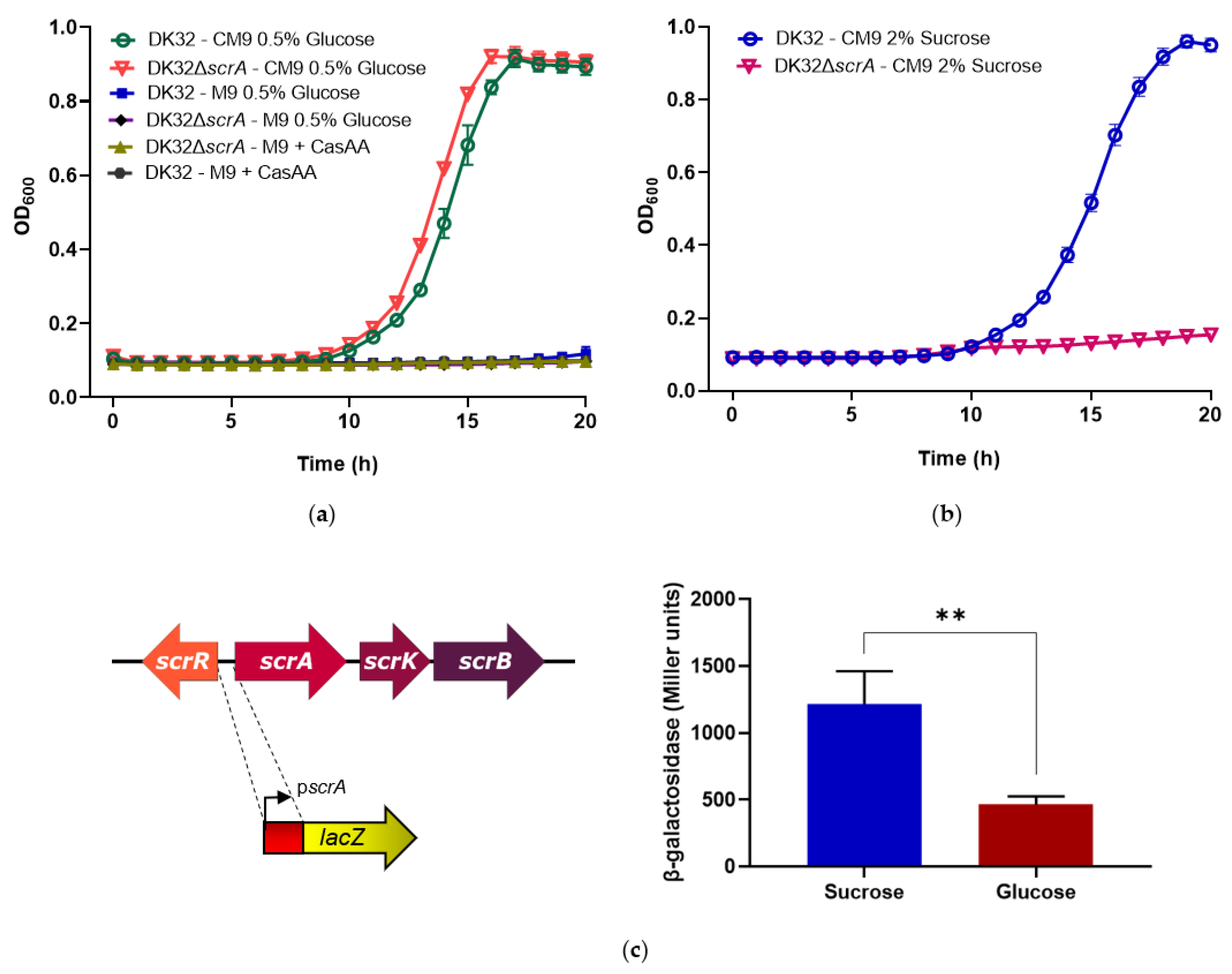
2.5. Growth Assays with Sucrose as a Carbon Source

Parental DK32, and DK32 ΔscrA strain were streaked on a TSA-1 plate and incubated overnight at 25 °C. A loopful of cells was resuspended in phosphate buffered saline (PBS) to achieve an optical density of 0.3 at 600 nm (OD600:0.3). For the assay, 1 μL of the bacterial cell suspensions was aliquoted into the wells of a 96-well plate containing 100 μL of M9 minimal medium [47] supplemented, when necessary, with 0.2% (wt/vol) Casamino Acids (Thermo Fisher Scientific Inc, Waltham, MA, USA) (CM9), and with a sugar (0.5% glucose or 2% sucrose (wt/vol)) depending on the aim of the experiment. Final concentration of NaCl was adjusted to 1% in all the assays. For each assay, OD600 values were recorded every 10 min for 20 h. This experiment was automated using the spectrophotometer Epoch2 microplate reader (BioTek, Winooski, VT, USA). The 96-well plates were continuously incubated at 25 °C in the plate reader with shaking. Three replicates were performed per assayed condition and strain. Mean values are reported and error bars represent the standard deviations.

2.6. Construction of a Transcriptional Fusion of scrA Promoter to a lacZ Reporter Gene, and β-Galactosidase Assays

The putative scrA gene promoter was PCR amplified with primer pair scrA-promoter (Table 3) and fused to a promotorless lacZ gene in the low-copy-number reporter plasmid pHRP309. The plasmid with the transcriptional fusion construct pscrA::lacZ (pSSS250), was mobilized from E. coli S17-1-λpir into Pdd parental strain DK32 by conjugation. After conjugation for 24 h on TSA plates prepared with seawater, cells were scrapped off the plate, suspended in TSB-1, and 100 μL aliquots of serial decimal dilutions were spread on TCBS agar supplemented with gentamicin (resistance provided by pHRP309). DK32 transformants harboring pSSS250 plasmid (DK32 pscrA::lacZ) were grown in CM9 medium supplemented either with 1% glucose or with 1% sucrose, and β-galactosidase activities were measured as previously described and expressed in Miller units [48]. Three independent experiments with 3 replicates each were conducted. Mean values are reported and error bars represent the standard deviations. The statistical analysis of the gene expression data was carried out with Mann–Whitney test.

2.7. Comparative Genomics and Molecular Phylogeny Analyses

Comparative genomics analysis to search for genes specific of Scr+ strains was conducted with RAST [49]. Pfam database was used for predictions of protein domains [50]. Easyfig v.2.2.3. [51], was used for comparative analysis and visualization of gene architecture of scr clusters of Photobacterium and Vibrio species. The Genbank accession numbers of Vibrio and Photobacterium genomes used in the comparative genomics and in the phylogenetic analyses are listed in Table 4. A phylogenetic tree of 23 Pdd complete genomes was constructed using the guide tree obtained by MAUVE genome alignment program (progressive Mauve option) [52,53].
The species tree was generated using the concatenated amino acid sequences of the proteins encoded by 8 housekeeping genes, ftsZ (cell division protein FtsZ), gapA (glyceraldehyde 3-phosphate dehydrogenase), gyrB (DNA gyrase subunit B), mreB (rod shape-determining protein MreB), recA (RecA recombinase A), pyrH (uridylate kinase, uridine monophosphate kinase), topA (DNA topoisomerase I), and toxR (transmembrane transcription regulator). These housekeeping sequences were selected on the basis of their demonstrated value for fine-tuned discrimination of taxa within the family Vibrionaceae [9,54,55]. The sucrose genes tree was constructed using the concatenated amino acid sequences of the proteins encoded by the 4 sucrose cluster genes scrRAKB. The Mesquite 3.61 program was used to concatenate the protein sequences [56].
Evolutionary analyses were conducted in MEGA X [57]. The evolutionary history of the strains was inferred using the Neighbor-Joining method [58]. The percentage of replicate trees in which the associated taxa clustered together in the bootstrap test (1000 replicates) is shown next to the branches. The evolutionary distances were computed using the Maximum Composite Likelihood method [59], and are in the units of the number of base substitutions per site.

2.8. Database Submission

The draft genome sequences of the 7 Pdd strains determined in this study are available from Genbank under accession numbers listed in Table 5.

3. Results

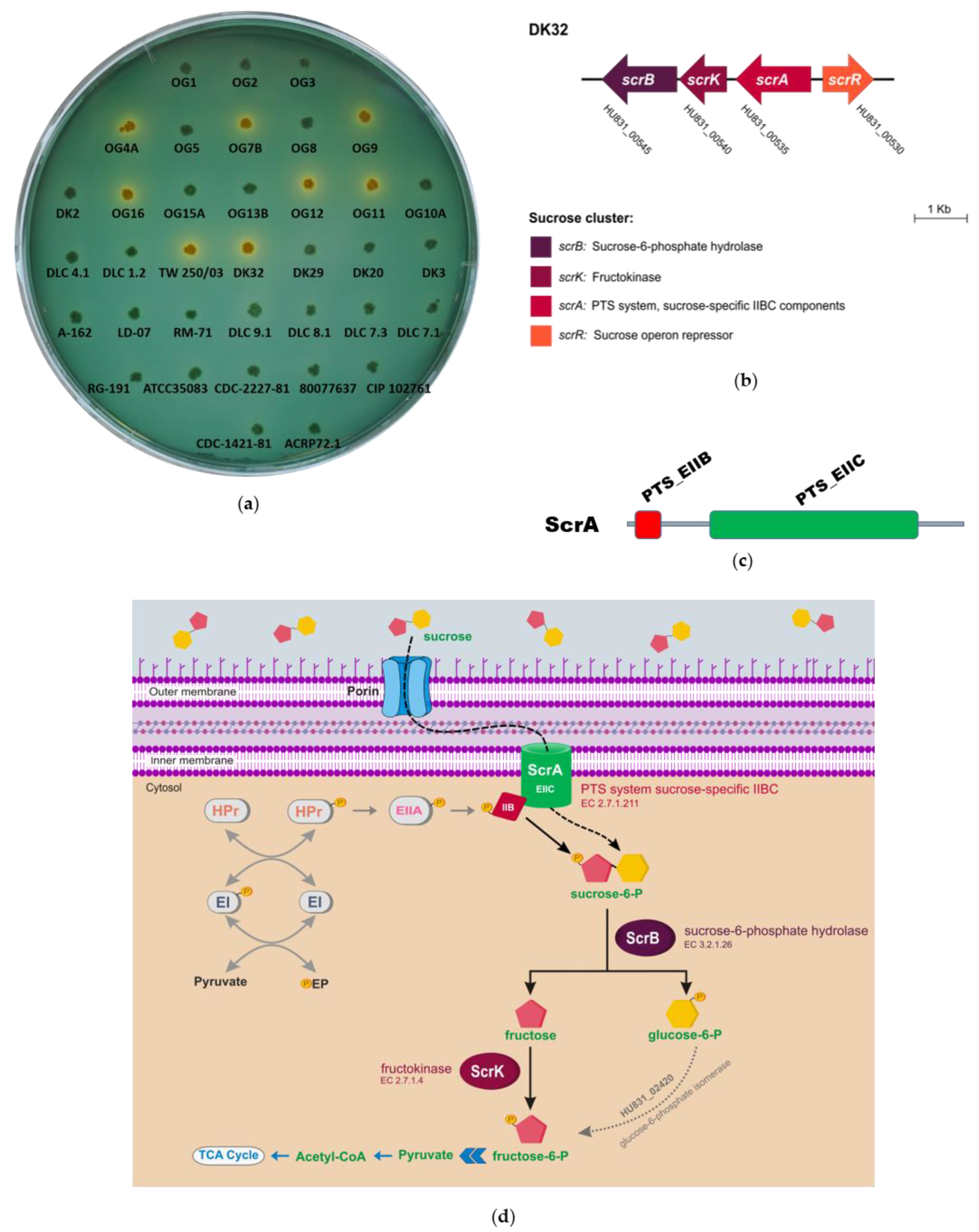
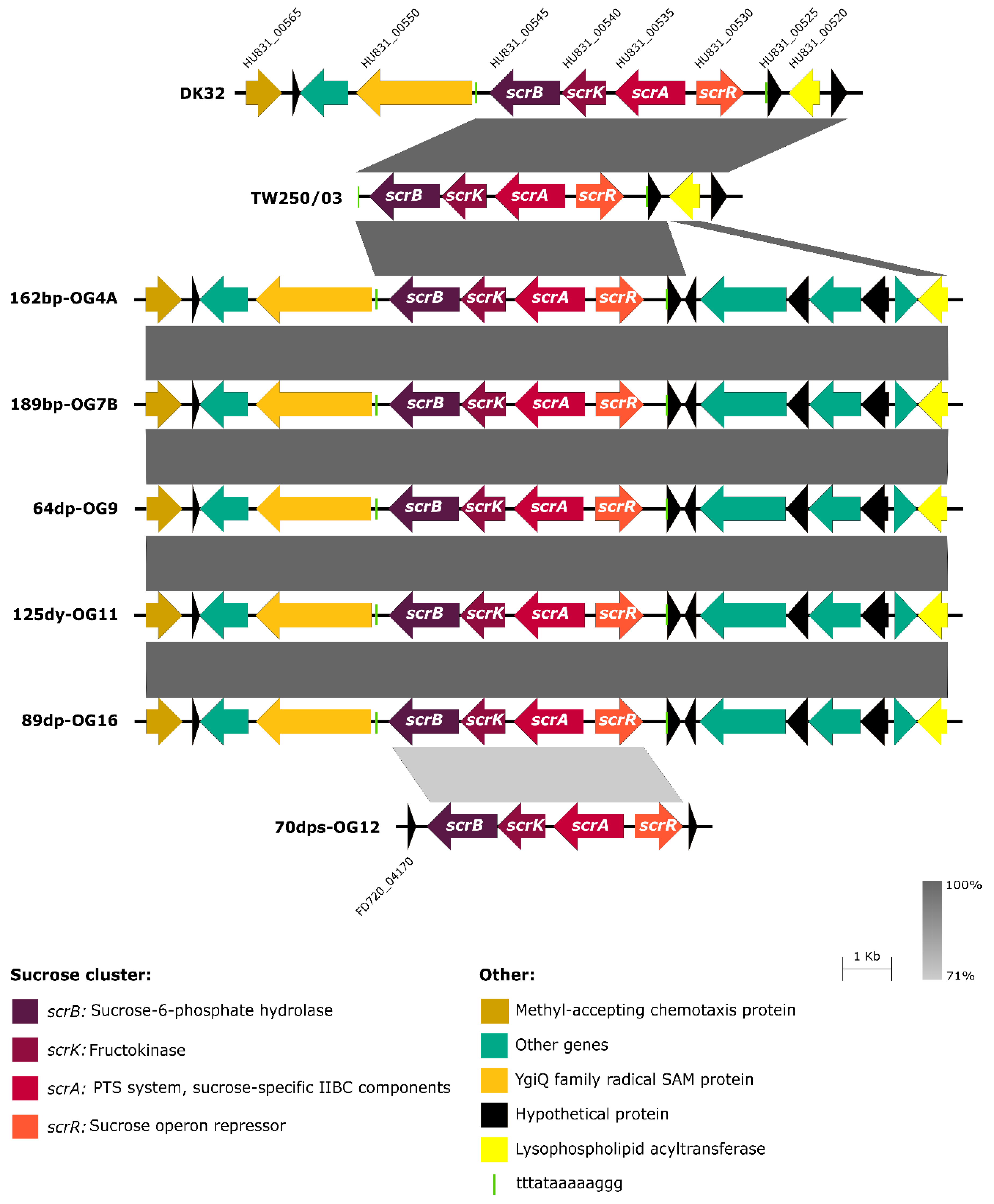
3.1. Identification of Two Different Genetic Variants of a Four Gene Cluster Encoding Functions for Sucrose Uptake and Catabolism in Pdd Strains

In order to study sucrose utilization in this marine pathogen, we screened a collection of 36 Pdd isolates from diverse geographical origins and isolation sources, for their ability to utilize sucrose on the selective and differential medium TCBS. This screening revealed that 8 strains grew as yellow colonies, thus exhibiting a Scr+ phenotype (Figure 1a; Table 1). To gain an insight into the genetic basis of sucrose utilization, we here obtained the de novo genome sequences of 5 Scr+ strains, namely DK32, TW250/03, 162bp-OG4A, 189bp-OG7B and 125dy-OG11, and 2 Scr Pdd isolates, CDC-1421-81 and 82dy-OG8. The general features of the 7 Pdd genomes sequenced in the present study are described in Table 5. In addition, we retrieved from GenBank database the genomes of 3 Scr+ strains (64bp-OG9, 70dps-OG12, and 89dp-OG16) that have been sequenced in our laboratory in a recent study focused on the presence of large multidrug resistance plasmids in this subspecies [26] (Table 1), as well as 13 genomes of Scr Pdd strains obtained in previous studies in our laboratory (Table 1). Pdd genomes were compared using RAST tool [49], searching for genes present in Scr+ strains and absent from Scr strains. Interestingly, the comparative genomics analysis unveiled two distinct versions of a 4-gene scr cluster, and the versions differed in their nucleotide sequence in a 30% (Figure 2). The majority version, hereafter version 1, was present in all the Scr+ strains except in OG12, that contained the version 2 of the cluster instead. The locus tags of the four genes comprising the scr cluster in each of the 8 Scr+ genomes are detailed in Table 6. These genes were organized in two divergently transcribed putative operons (Figure 1b and Figure 2). On the one side, scrR encodes a putative sucrose operon repressor of the LacI family. The other three genes are transcribed from the opposite strand, and encode, based on their similarity to the V. alginolyticus and V. cholerae sucrose utilization genes, a PTS system sucrose-specific IIBC component (EC 2.7.1.211) (scrA), a fructokinase (EC 2.7.1.4) (scrK), and a sucrose-6-phosphate hydrolase (EC 3.2.1.26) (scrB), respectively [14,15,16,18].
Protein domain predictions by Pfam database, and similarity search analysis by BLAST, indicated that the Pdd scrA gene encodes a EIIBC-domain containing protein, of the phosphoenolpyruvate-dependent phosphotransferase system (PTS) (Figure 1c). According to the in silico analysis of these four genes and their predicted protein products, it is hypothesized that Pdd utilizes sucrose via the PTS mechanism, following the scheme depicted in Figure 1d. The conserved PTS system is one of the most common mechanisms for high affinity uptake of sugars in bacteria. It consists of a phosphotransfer cascade that catalyzes the transfer of a phosphate group from phosphoenolpyruvate (PEP) to a carbohydrate substrate concomitant with its transport into the cytoplasm [60,61]. The PTS system includes three major components: Enzyme I (EI), EII (EIA-C) and the histidine-containing phosphocarrier protein (HPr). EI and HPr are shared by all PTS transporters. EI autophosphorylates in the presence of PEP and transfers the phosphate group to HPr. Subsequently, HPr passes the phosphoryl group to a sugar-specific EIIA protein, EIIA passes it to its cognate EIIB, and the latter transfers the phosphoryl group to a sugar bound to the cognate, inner membrane-spanning EIIC element, facilitating sugar import. Thus, the EII complexes consist of three domains, EIIA, EIIB, and EIIC, that can either be part of the same protein (an EIIABC protein), or can be encoded by distinct proteins. The EIIC domain defines the carbohydrate specificity within these transport systems [60]. Once sucrose is taken up by the EIIC component of ScrA, it yields intracellular sucrose-6-phosphate that is hydrolyzed to glucose-6-phosphate and fructose by the sucrose-6-phosphate hydrolase (scrB). Fructose is phosphorylated to fructose-6-phosphate by the fructokinase encoded by srcK, and the two monosaccharides would enter the glycolytic pathway (Figure 1d).
On the light of the comparative genomics of 23 Pdd strains for which genome sequences are available (Table 1), there is a 100% correlation between presence of the scr cluster, either version 1 or version 2, and the ability to ferment sucrose on TCBS. A previous study reported that a naturally-occurring, single nucleotide insertion in scrA gene rendered V. cholerae strain IEC224 unable to ferment sucrose [19]. We thus wanted to assess whether the Scr phenotype of Pdd strains could be due in some instances to point mutations in the sucrose operon. To this aim we selected 13 Pdd strains which are phenotypically Scr and whose genome sequences are not available (Table 1), and conducted a PCR screening with two primer pairs targeted to sequences conserved in the scrB and scrR genes of the two versions of scr clusters described in this study. As a result, it was found that all the Scr isolates tested negative in the PCR tests, whereas all the Scr+ strains tested positive for scrB and scrR genes (Table 1).
The genetic context upstream and downstream the scr cluster was found to be highly conserved in six of the eight Scr+ strains (Figure 2), where this cluster is invariably flanked by genes encoding a YgiQ family protein (locus HU831_00550) and a hypothetical protein (locus HU831_00525) (loci tags refer to the DK32 draft genome). The gene homologous to HU831_00550 in the closed genome sequence of the Pdd type strain CIP102761 (locus VDA_001759) maps to chromosome I, which suggests that the version 1 of scr cluster is chromosome I-borne in Pdd. For strains 70dps-OG12 and TW250/3 the flanking regions could not be identified with precision due to gaps in draft genome assembly, likely caused by the existence of repeated sequences, as insertion sequence elements. In any case, the context of sucrose genes in 70dps-OG12 was unrelated to the context of the other 7 Scr+ strains. The observation of a highly conserved context in most Scr+ strains suggest that the scr cluster was inserted into the genome by means of some mechanism of DNA recombination. Notably, a duplicated 13-mer sequence (TTTATAAAAAGGG) was found flanking both sides of the scr cluster in all the strains with the exception of 70dps-OG12 (Figure 2).

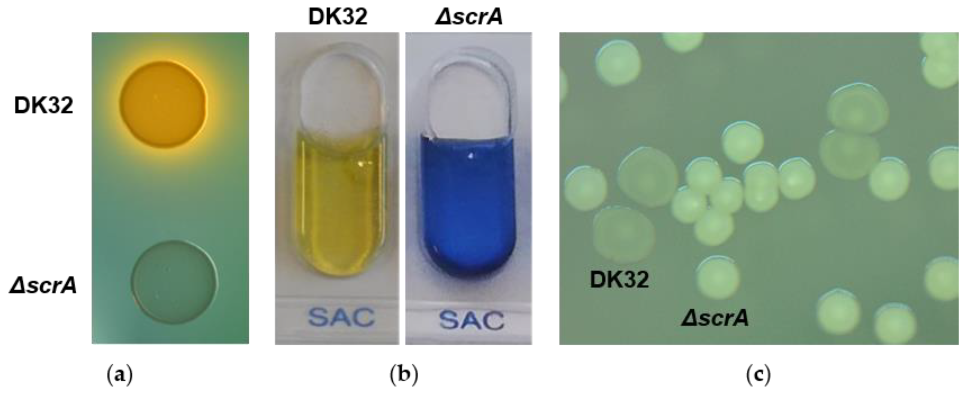
3.2. Deletion of scrA in the scr+ Strain DK32 Causes Green Colonies on TCBS Medium, and Abolishes Growth with Sucrose as Carbon Source

As described above, scrA encodes a EIIBC-domain containing protein with a predicted role in sucrose utilization. In order to investigate this, a non-polar, unmarked deletion mutant for scrA gene was constructed in the Pdd wild type strain DK32 by allelic exchange, a process that removed 90% of the scrA coding sequence without disrupting the reading frame. As a result, it was observed that DK32 ΔscrA produced green colonies on TCBS (Figure 3a), suggesting that this mutant is unable to transport sucrose into the cell. In addition, DK32 ΔscrA produced a negative result for sucrose fermentation in the API-20E gallery, which is routinely used in laboratories for the identification of fish pathogenic bacteria (Figure 3b). Unexpectedly, mutation of scrA caused a change in the colony phenotype compared to the wild type colonies, when the strains were cultured on TSA-1 supplemented with 15% (wt/vol) sucrose, the sugar concentration used to select for a second recombination step during the allelic exchange process (see methods). DK32 wt colonies appeared flat and translucent while DK32 ΔscrA colonies appeared convex and whitish (Figure 3c). We thus hypothesized that the presence of the sucrose fermenting gene cluster in the bacterial genome might be responsible for causing such phenotype when the strain is growing in presence of high concentrations of sucrose. In order to test this, we analyzed the colony morphologies of the naturally Scr+ and Scr strains 89dp-OG16 and LD-07, respectively. Strains containing the scr cluster yielded flat and translucent colonies, whereas Scr strains yielded convex and whitish colonies. The colony phenotypes of Scr+ and Scr strains were indistinguishable on TSA-1 without added sucrose (Supplementary Figure S1).
We next studied how deletion of scrA impacted Pdd DK32 growth with sucrose as a carbon source. To this aim, it was first necessary to optimize the conditions for culturing Pdd in minimal medium. It is pertinent to highlight that no detectable growth was achieved neither by the parental strain nor by the ΔscrA mutant when cultured in minimal medium M9 with glucose 0.5%, suggesting that Pdd DK32 cannot efficiently synthesize all the necessary amino acids in a mineral minimal medium with glucose as carbon source (Figure 4a). Similarly, the minimal medium M9 supplemented with casamino acids as sole carbon source in the absence of glucose, did not support growth within 20 h (Figure 4a), suggesting that Pdd does not efficiently use amino acids as carbon source, and needs a sugar for optimal growth. In support of these hypotheses, growth of DK32 and DK32 ΔscrA strains was completed to the stationary phase within 20 h when casamino acids were added to M9 with glucose 0.5% (CM9 medium) (Figure 4a). These results demonstrate that deletion of scrA does not cause a fitness cost in the growth of Pdd with glucose as carbon source in presence of casamino acids. However, when 2% sucrose was added to CM9 medium in substitution of glucose, the DK32 ΔscrA mutant was drastically impaired for growth, whereas the parental strain achieved full growth values as with glucose (Figure 4b).
We also analyzed whether the transcriptional activity of the promoter upstream scrA was affected by growth in presence of sucrose. To this aim, the scrA promoter sequence (pscrA) was fused to a promoterless lacZ gene in plasmid pHRP309, that was further mobilized to Pdd parental strain DK32, yielding DK32 pscrA::lacZ. This indicator strain was cultured in CM9 medium supplemented with either 1% sucrose or 1% glucose. The measure of β-galactosidase activities demonstrated that scrA promoter exhibited basal transcription levels in presence of glucose as carbon, and promoter activity was increased 2.5-fold in the presence of sucrose as carbon source (Figure 4c). Collectively, the growth assay data and transcriptional fusion analysis, demonstrate that scrA is necessary for sucrose utilization as carbon source in Pdd and its transcription is increased in presence of sucrose.

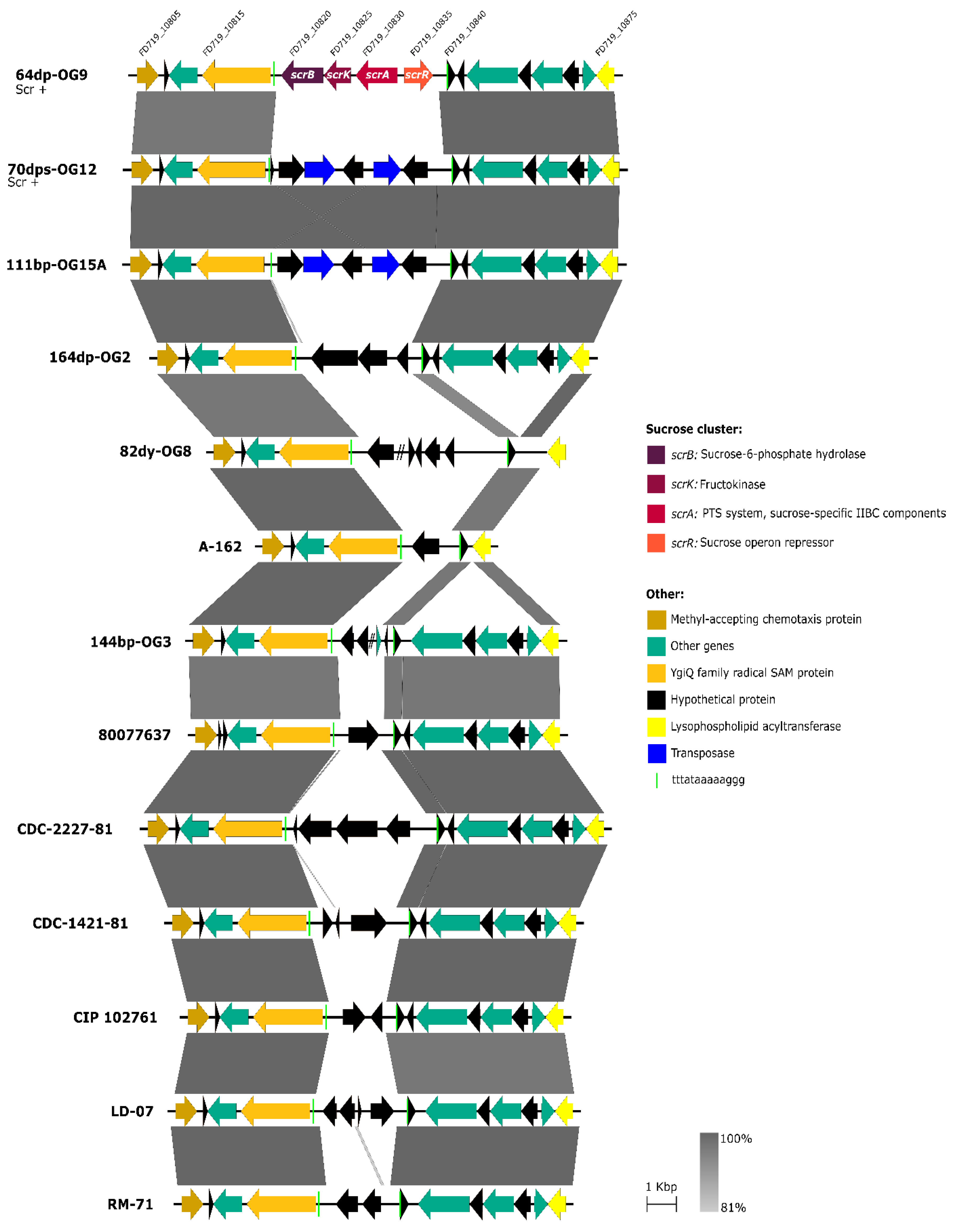
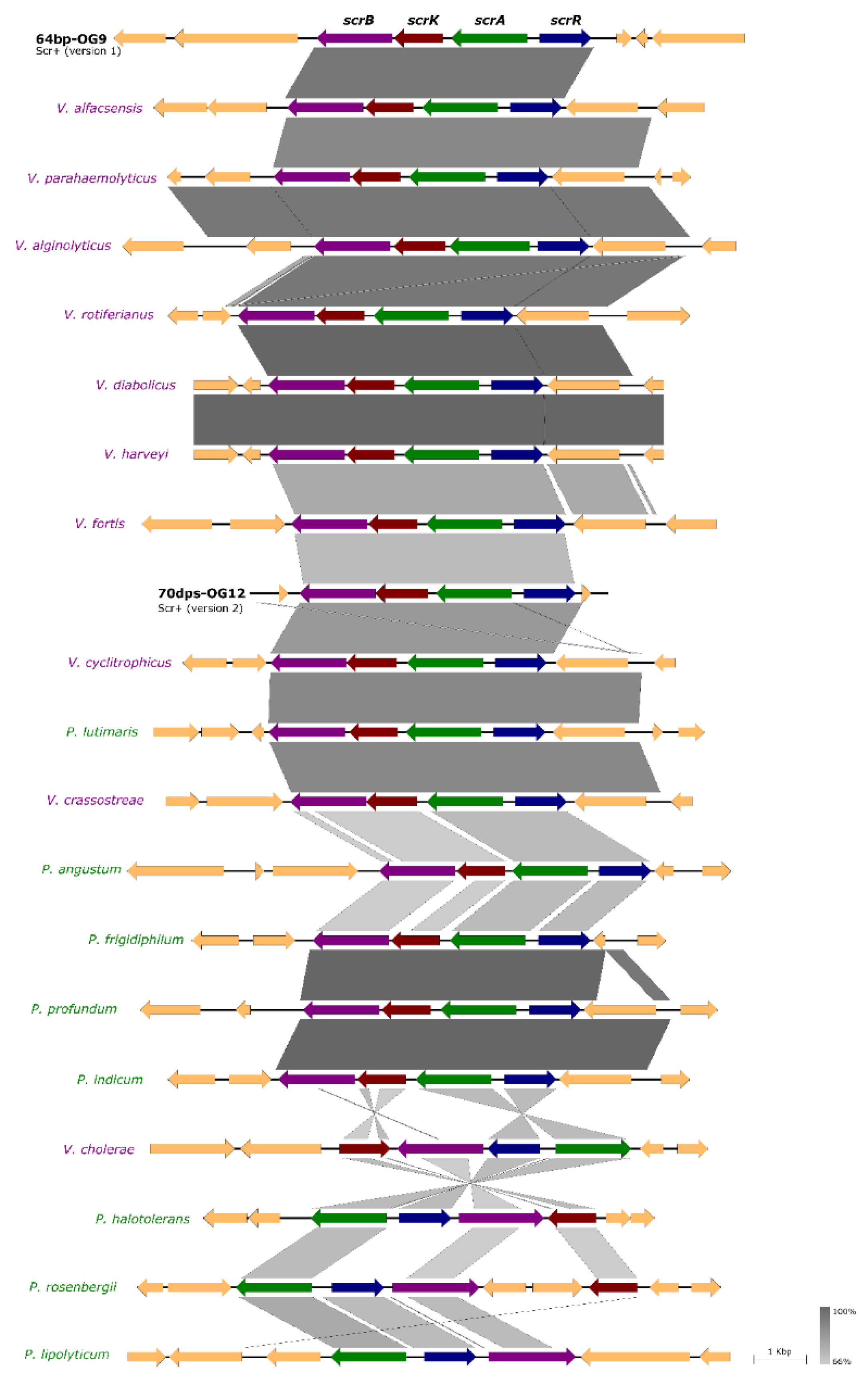
3.3. Scr Clusters Are Inserted into Putative Hot-Spots for DNA Acquisition in Pdd Genomes

We hypothesized that the genome region flanked by the genes encoding the YgiQ family protein and the lysophospholipid acyltransferase, i.e., the DNA region harboring the scr cluster in the majority of Scr+ Pdd strains, represents a hot-spot for events of DNA acquisition and loss. In order to gain an insight into this, we conducted a comparative analysis of the gene composition within this putative hot-spot in eleven Scr Pdd strains, which included two strains newly sequenced in the present study (CDC-1421-81 and 82dy-OG8) and 9 additional Scr strains whose draft genomes were retrieved from GenBank database (Table 1). We found that 70dps-OG12, the Scr+ strain whose context of the scr cluster remains unknown, contained a number of hypothetical proteins and transposases in this region, and was identical to the gene composition of the homologous region in the Scr strain 111bp-OG15A (Figure 5). Notably, analysis of the 11 Scr genomes unveiled the existence of 11 unique gene combinations, i.e., each strain contained genes with no counterparts in the other Pdd genomes. These observations provide strong evidence that this genome region is highly prone to events of DNA acquisition and can be considered as a recombination hot-spot for acquisition of DNA. Of note, the duplicated 13-mer sequence TTTATAAAAAGGG was found in most strains at exactly the same point (Figure 5).

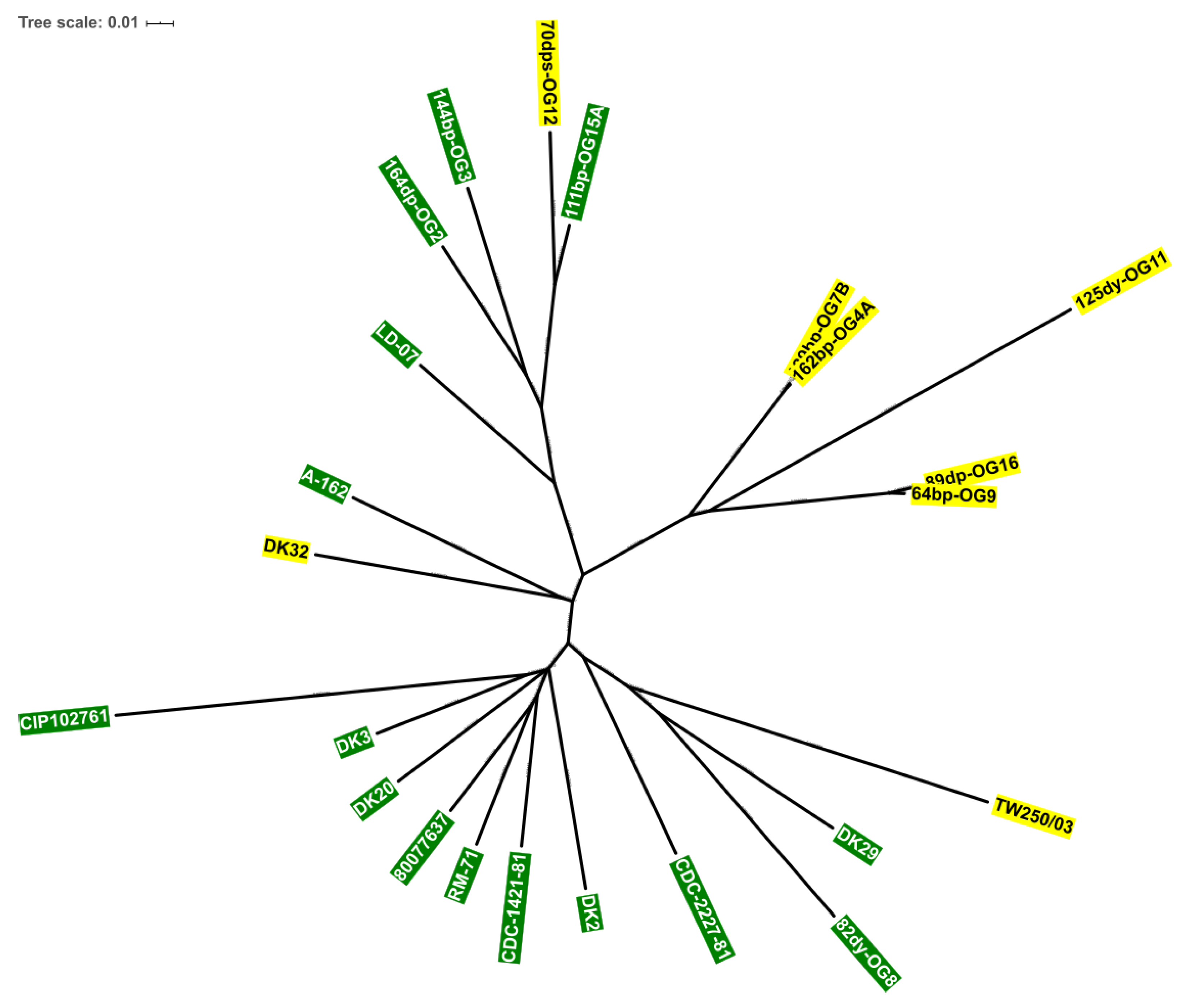
3.4. Scr Clusters Occur in Different Genetic Lineages of Pdd

In this study, 8 Pdd strains contained a scr gene cluster. Of these, six were isolated from European seabass in the Black Sea [30], one from diseased rainbow trout in Denmark [24,37] and one from gilthead seabream from unknown geographical origin (Table 1). In order to ascertain whether the sucrose positive strains conform a clonal entity or whether the scr clusters have been acquired by different genetic lineages of Pdd, we carried out a comparative analysis of 23 complete Pdd genomes. As shown in Figure 6, five Scr+ strains isolated from the Black Sea group together within a clade, and among these, the pairs OG7B/OG4A and OG9/OG16 likely correspond to two clonal lines respectively. However, 125dy-OG11 is distantly related from the aforementioned four strains, and clearly represents an evolutionary line that has diverged from the other strains. Of note, the three remaining Scr+ strains are unevenly distributed with other Scr genomes in different branches of the phylogenetic tree. It is noticeable that strain DK32, isolated in the coast of Denmark in 2006, harbors a scr cluster 99.9% identical at the nucleotide sequence level to the scr cluster of 125dy-OG11, isolated in the Black Sea in 2011, despite these two strains being distantly located in the phylogenetic tree (Figure 6). All these results provide strong evidence that the Scr+ isolates are not clonal derivatives (with the exception of the pairs OG7B/OG4A and OG9/OG16), and support the hypothesis that scr clusters have been acquired by horizontal transfer by different genetic lineages of Pdd, as a result of independent events of DNA acquisition.

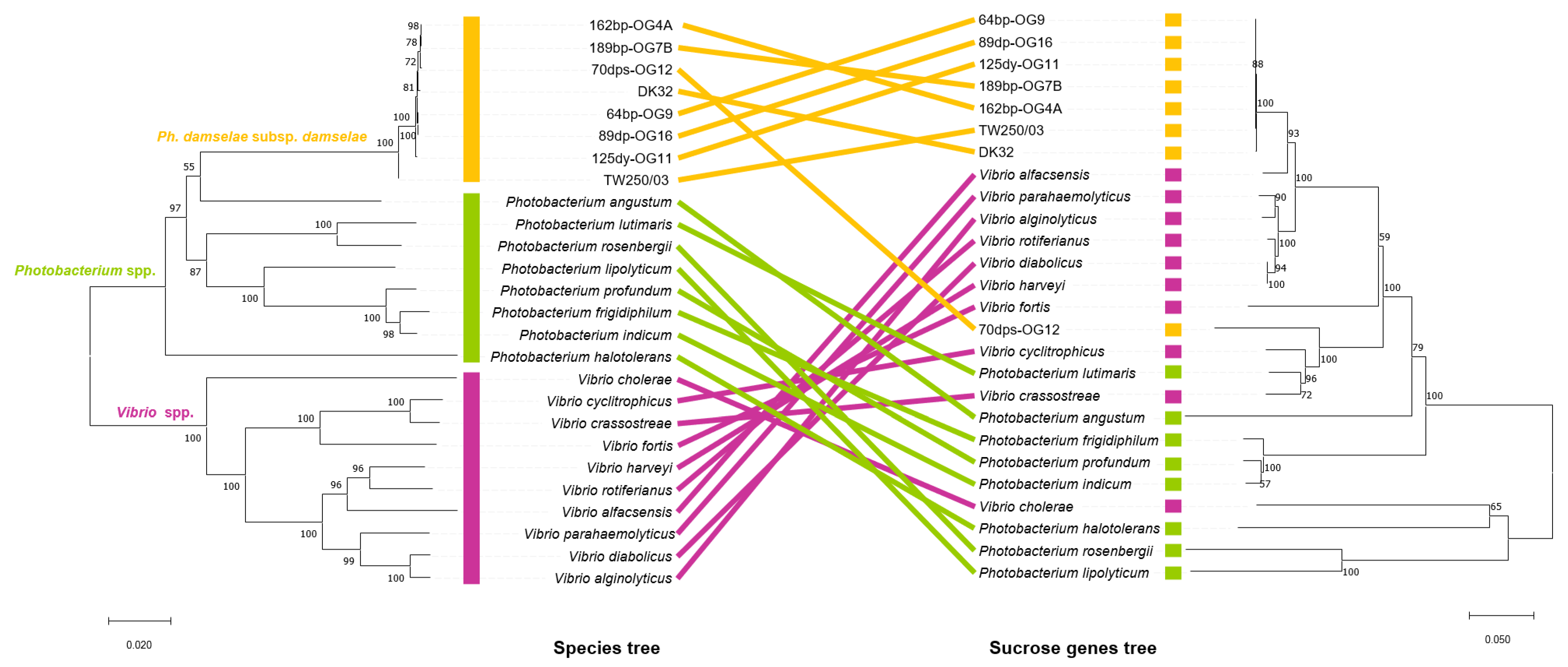
3.5. Incongruences between the Species Tree and the Sucrose Genes Tree Reveal Extensive Horizontal Transfer of scr Genes among Species of Vibrio and Photobacterium

The high percentages of sequence identity and of operon architecture in scr cluster genes among 7 of the 8 Pdd strains analyzed in the present study, clearly indicate a common evolutionary origin of sucrose cluster genes in these strains. In addition, the observation that the scr cluster in strain 70dps-OG12 exhibits 30% sequence divergence with respect to the other 7 Pdd strains, suggests that scr clusters have been gained in this subspecies by horizontal gene transfer from different donor species. Although sucrose utilization is a widespread trait in many species within the family Vibrionaceae, the phylogenetic relationships among sucrose utilization clusters from different species remains largely uncharacterized. Here, we conducted a Genbank database search to retrieve gene clusters encoding sucrose utilization genes in species of Photobacterium and Vibrio (Accession numbers are listed in Table 4) that were homologous to the Pdd clusters. A schematic analysis of the two representative Pdd clusters (64bp-OG9 and 70dps-OG12) and of 18 Photobacterium and Vibrio clusters revealed that gene architecture is largely conserved, with the exception of V. cholerae, P. halotolerans, P. rosenbergii, and P. lipolyticum that showed different gene arrangements (Figure 7).
In order to determine the evolutionary history of sucrose uptake and catabolism genes, we reconstructed the phylogeny of ScrB, ScrK, ScrA and ScrR proteins among Photobacterium and Vibrio species. Proteins representing homologues within these two genera were aligned using ClustalW and phylogenetic trees were constructed by the neighbor-joining method using MEGA X [57]. This analysis demonstrated that the scr genes of 7 out of the 8 Pdd strains (version 1 cluster), are closely related to the clusters found in Vibrio species. On the contrary, the cluster of Pdd 70dps-OG12 (version 2 cluster) is related to clusters found in species of the genus Photobacterium and shares the same branch with V. cyclitrophicus and V. crassostreae (Figure 8). These observations clearly suggest that scr clusters have undergone horizontal gene transfer among species of the two genera. In order to clarify this, we conducted in parallel a phylogenetic analysis of the same Photobacterium and Vibrio species, by analyzing the concatenated amino acid sequences of eight housekeeping genes conserved in all the species, and producing a “species tree” that was compared to the “sucrose genes tree”. The analysis of these housekeeping genes generated a phylogenetic tree with three distinct clades that include the 8 Pdd strains, the rest of Photobacterium species, and the Vibrio species, respectively. The noticeable incongruence between sucrose operon tree and species tree clearly indicates that sucrose genes found in 7 Pdd strains (version 1 cluster) are closely related to clusters of Vibrio species, being V. alfacsensis the closest relative, while they are more distantly related to the clusters of Photobacterium species. This suggests that Pdd sucrose cluster version 1 may have been acquired by Pdd strains from a Vibrio-like donor. The scr cluster of Pdd 70dps-OG12 (version 2) has a different evolutionary history than the other 7 Pdd clusters, and is more similar to both Vibrio and Photobacterium clusters. Notably, the close evolutionary distance between sucrose clusters of P. lutimaris and V. crassostreae contrasts with the placement of these two species in two different clades in the species tree. All these observations provide strong evidence that horizontal gene transfer of sucrose genes is extensive among Photobacterium and Vibrio species.

4. Discussion

Pdd (formerly known as V. damsela), is an important and emerging pathogen for marine animals and for humans [22]. An early review on pathogenic Vibrio species already highlighted that a 5% of Pdd isolates were sucrose fermenters on TCBS agar [27]. Since then, various studies have unveiled the existence of Scr+ Pdd isolates at varying frequencies. An extensive study that analyzed 71 Pdd isolates from fish in Spain, reported that 4% were sucrose-fermenters [28]. In another study with Pdd strains from disease outbreaks in marine rainbow trout, one isolate out of 31 tested positive for sucrose fermentation [24]. A study reported the simultaneous isolation on a TCBS agar plate of yellow and green Pdd colonies from different organs of the same fish [29]. Notably, in a recent study, of 14 Pdd strains isolated from diseased seabass in the Turkish coast of the Black Sea, 6 were able to ferment sucrose [30], which accounts for 40% of strains positive for this trait. Thus, albeit sucrose fermentation can be considered as an infrequent phenotype in this subspecies, the intraspecies variability of this metabolic trait needs to be taken into consideration, in order to avoid misidentification of potential Pdd strains in veterinary, clinical and environmental studies.
Even though sucrose fermentation tests are routinely used for identification and classification of species of the Vibrionaceae, the genetic basis of sucrose utilization in species of this family has received scarce attention. Homologues of the scr cluster genes described in the present study in Pdd, were reported for the first time in V. alginolyticus in pioneering studies in the family Vibrionaceae [14,15,16]. Later, different investigations reported some data on sucrose utilization genes in V. cholerae [17,18,19,20] whereas, to the best of our knowledge, no functional and evolutionary analyses have been conducted in species of the genus Photobacterium so far. In this study, we describe two variants of a gene cluster that is required for the utilization of sucrose by Pdd. Mutational analysis has demonstrated that scrA gene encoding the predicted EIIBC component of the PTS sucrose transport system is essential for sucrose utilization as carbon source by Pdd. In a previous study, the systematic genetic dissection of PTS systems in V. cholerae demonstrated that single deletion of VCA0563, a gene homologous to the Pdd scrA, was sufficient to abolish sucrose utilization [20], indicating that no redundant PTS functions existed in the V. cholerae genome capable of conferring the ability to take up sucrose. In contrast, V. cholerae showed to encode redundant PTS functions for the uptake of glucose, mannose and fructose [20]. We have here found that single deletion of scrA abolished sucrose utilization in Pdd DK32, demonstrating that this strain does not encode redundant PTS functions for the uptake of this disaccharide.
The scr clusters in some species of Enterobacteriaceae are known to be negatively controlled by the repressor encoded by scrR gene and thus genes encoding enzymes for sucrose catabolism are inducible by presence of sucrose in the medium [62,63]. The Pdd scr cluster also contains a homologue of scrR gene, but so far, no experimental evidence on the sucrose-inducible nature of scr clusters in the Photobacterium genus was available. We here have shown that growth of Pdd DK32 with sucrose as carbon source increased in 2.5-fold the transcriptional activity of the scrA promoter compared to growth in presence of glucose as carbon source, suggesting that sucrose metabolism in Pdd is subjected to a negative regulation similar to that described in some enterobacteria.
It was observed that presence of scr cluster genes imparted a unique colony phenotype to Pdd strains when grown in presence of high sucrose concentrations (15%). The mechanisms underlying this phenotype await further and promising studies in Pdd. Interestingly, a recent study pointed out that the V. cholerae VCA0653 gene encoding the sucrose-specific PTS component ScrA, was 30-fold downregulated on the phase variation switch from wild-type-colony phenotype to a rugose-colony phenotype, associated with advanced biofilm architecture [64]. This suggests that the ability to utilize sucrose would impair biofilm formation, and thus transition to a rugose phenotype correlates with a strong downregulation of sucrose uptake. Similarly, Lactobacillus strains grown on sucrose were found to produce dextrane, and it influenced self-aggregation and biofilm formation [65].
Carbohydrates constitute a main carbon source in bacteria [66]. Horizontal gene transfer enables bacteria to acquire new gene clusters that provide the recipient with novel metabolic activities to confront ecological changes [67,68]. Acquisition by horizontal transfer of genes for transport and catabolism of carbohydrates in members of the Vibrionaceae is being increasingly reported, as is the case of genes for cellobiose utilization [69], for degradation of algal polysaccharides [8], and genes of D-galactose metabolism [11], among others. Sucrose utilization clusters are not an exception to this, and previous studies have pointed out that sucrose gene clusters have been extensively exchanged among Eubacteria [21]. In the present study, the incongruence of phylogenies based on 8 representative housekeeping genes on the one side, and on the four scr genes on the other side, clearly revealed that genes for sucrose metabolism have undergone extensive horizontal transfer among genera and species within the family Vibrionaceae. Of the two scr cluster versions reported in the present study, the majority version 1 is likely chromosome I-borne considering that the conserved genome context upstream and downstream of sucrose genes corresponds to chromosome I genes in well studied Scr Pdd genomes. In addition, the seven scr clusters of version 1 are invariably flanked by a duplicated 13-mer sequence that is a putative candidate to play a role in DNA acquisition. A recent study has reported that some regions of the Pdd genome constituted hot-spots for DNA acquisition, and such hypervariable regions were flanked by repeated sequences in tandem [24]. Similarly, sucrose utilization genes in enteropathogenic E. coli strains were found to be located within variable chromosomal regions rich in repeated sequences dubbed iap sequences [70]. The genetic context of version 2 represented by Pdd strain 70dps-OG12 is uncertain, and it might be either chromosome- or plasmid borne. It is known that genes for sucrose metabolism in Enterobacteria are either chromosome-borne [71,72] or plasmid-borne [73,74,75]. Further studies are prompted in order to ascertain the existence of plasmid-borne scr clusters in members of the family Vibrionaceae.
The acquisition of sucrose metabolism genes by species of Vibrionaceae raises the question about what is the role of sucrose utilization in the ecology of Vibrios. Sucrose is the most abundant disaccharide on earth because of its origin in higher plant tissues. Early studies reported that green microalgae of the genus Trebouxia secreted sucrose to the culture medium [76], and nowadays it is widely known that green algae, cyanobacteria and purple bacteria synthesize sucrose [77]. Microalgae and cyanobacteria species accumulate sucrose as compatible osmolyte under osmotic stress, a process that has been mainly studied in freshwater environments [78,79,80,81,82,83]. A recent study reported the production of sucrose by marine species of Pyrocystis and Nannochloropsis genera [84]. Thus, either secreted by live cells or released upon cell lysis, sucrose is expected to be available as a carbon source in marine ecosystems. In support of this idea, a recent metabolomics study has reported the detection of abundant sugars, which included sucrose and trehalose, in marine ecosystems [85], and sucrose has also been detected as a major nutrient available in seagrass (Posidonia oceanica) meadows ecosystems [86]. It is also expected that the digestive tracts of marine herbivores as sea urchins and algae grazers among others, constitute a sucrose-rich niche for bacteria. Of note, a previous study has demonstrated that sucrose is a potent chemoattractant for Vibrio furnissii and this chemotaxis was suggested to be dependent on presence of an intact PTS system [87]. Hence, it is hypothesized that the scr gene clusters would confer advantage to the bacterial cells to use sucrose available in marine environment niches as a carbon source. Studies to test this hypothesis are currently under way.

Supplementary Materials

The following are available online at https://www.mdpi.com/2073-4425/11/11/1244/s1, Figure S1: Colony morphologies of Scr+ and Scr Pdd strains grown in either plain TSA-1 or in TSA-1 supplemented with 15% sucrose. Strains harboring the scr gene cluster form flat and translucent colonies in presence of high concentrations of sucrose, while strains lacking a functional scr cluster form convex and whitish colonies. The colony phenotypes of Scr+ and Scr strains are indistinguishable on TSA-1 without added sucrose.

Author Contributions

Conceptualization, S.A., A.V., A.V.B., and C.R.O.; Methodology: S.A., A.V., A.V.B., and C.R.O.; Software: A.V.; Formal Analysis: S.A., A.V., A.V.B. and C.R.O.; Investigation: S.A., A.V., A.V.B.; Resources: A.V., and C.R.O.; Data Curation: S.A., A.V., A.V.B., and C.R.O.; Writing—Original Draft Preparation: S.A.; Writing—Review and Editing: A.V., and C.R.O.; Supervision: A.V. and C.R.O.; Project Administration: C.R.O.; Funding Acquisition: C.R.O. All authors have read and agreed to the published version of the manuscript.

Funding

This work was supported by the Agencia Estatal de Investigación (AEI) of Spain co-funded by the FEDER Programme from the European Union (grants no. AGL2016-79738-R and PID2019-110558RB-I00) and by Xunta de Galicia (Spain) (grant no. ED431C 2018/18).

Conflicts of Interest

The authors declare no conflict of interest. The funders had no role in the design of the study; in the collection, analyses, or interpretation of data; in the writing of the manuscript, or in the decision to publish the results.

References

  1. Thompson, F.L.; Iida, T.; Swings, J. Biodiversity of vibrios. Microbiol. Mol. Biol. Rev. 2004, 68, 403–431. [Google Scholar] [CrossRef] [Green Version]
  2. Baker-Austin, C.; Oliver, J.D.; Alam, M.; Ali, A.; Waldor, M.K.; Qadri, F.; Martinez-Urtaza, J. Vibrio spp. infections. Nat. Rev. Dis. Prim. 2018, 4, 8. [Google Scholar] [CrossRef] [PubMed]
  3. Takemura, A.F.; Chien, D.M.; Polz, M.F. Associations and dynamics of Vibrionaceae in the environment, from the genus to the population level. Front. Microbiol. 2014, 5, 38. [Google Scholar] [CrossRef] [PubMed] [Green Version]
  4. Reen, F.J.; Almagro-Moreno, S.; Ussery, D.; Boyd, E.F. The genomic code: Inferring Vibrionaceae niche specialization. Nat. Rev. Microbiol. 2006, 4, 697–704. [Google Scholar] [CrossRef] [PubMed]
  5. Thompson, C.C.; Vicente, A.C.P.; Souza, R.C.; Vasconcelos, A.T.R.; Vesth, T.; Alves, N.; Ussery, D.W.; Iida, T.; Thompson, F.L. Genomic taxonomy of vibrios. BMC Evol. Biol. 2009, 9, 258. [Google Scholar] [CrossRef] [Green Version]
  6. Urbanczyk, H.; Ast, J.C.; Dunlap, P.V. Phylogeny, genomics, and symbiosis of Photobacterium. FEMS Microbiol. Rev. 2011, 35, 324–342. [Google Scholar] [CrossRef]
  7. Le Roux, F.; Wegner, K.M.; Baker-Austin, C.; Vezzulli, L.; Osorio, C.R.; Amaro, C.; Ritchie, J.M.; Defoirdt, T.; Destoumieux-Garzón, D.; Blokesch, M.; et al. The emergence of Vibrio pathogens in Europe: Ecology, evolution, and pathogenesis (Paris, 11–12 March 2015). Front. Microbiol. 2015, 6, 830. [Google Scholar]
  8. Hehemann, J.-H.; Arevalo, P.; Datta, M.S.; Yu, X.; Corzett, C.H.; Henschel, A.; Preheim, S.P.; Timberlake, S.; Alm, E.J.; Polz, M.F. Adaptive radiation by waves of gene transfer leads to fine-scale resource partitioning in marine microbes. Nat. Commun. 2016, 7, 1–10. [Google Scholar] [CrossRef]
  9. Machado, H.; Gram, L. Comparative genomics reveals high genomic diversity in the genus Photobacterium. Front. Microbiol. 2017, 8, 1204. [Google Scholar] [CrossRef] [Green Version]
  10. Le Roux, F.; Blokesch, M. Eco-evolutionary dynamics linked to horizontal gene transfer in vibrios. Annu. Rev. Microbiol. 2018. [Google Scholar] [CrossRef]
  11. Regmi, A.; Boyd, E.F. Carbohydrate metabolic systems present on genomic islands are lost and gained in Vibrio parahaemolyticus. BMC Microbiol. 2019, 19, 112. [Google Scholar] [CrossRef] [PubMed]
  12. Kobayashi, T.; Enomoto, S.; Sakazaki, R.; Kuwahara, S. A new selective isolation medium for pathogenic vibrios: TCBS agar. Jap. J. Bact. 1963, 18, 387–391. [Google Scholar] [CrossRef]
  13. Scholle, R.R.; Coyne, V.E.; Maharaj, R.; Robb, F.T.; Woods, D.R. Expression and regulation of a Vibrio alginolyticus sucrose utilization system cloned in Escherichia coli. J. Bacteriol. 1987, 169, 2685–2690. [Google Scholar] [CrossRef] [Green Version]
  14. Scholle, R.R.; Robb, S.M.; Robb, F.T.; Woods, D.R. Nucleotide sequence and analysis of the Vibrio alginolyticus sucrase gene (scrB). Gene 1989, 80, 49–56. [Google Scholar] [CrossRef]
  15. Blatch, G.L.; Scholle, R.R.; Woods, D.R. Nucleotide sequence and analysis of the Vibrio alginolyticus sucrose uptake-encoding region. Gene 1990, 95, 17–23. [Google Scholar] [CrossRef]
  16. Blatch, G.L.; Woods, D.R. Nucleotide sequence and analysis of the Vibrio alginolyticus scr repressor-encoding gene (scrR). Gene 1991, 101, 45–50. [Google Scholar] [CrossRef]
  17. Houot, L.; Chang, S.; Absalon, C.; Watnick, P.I. Vibrio cholerae phosphoenolpyruvate phosphotransferase system control of carbohydrate transport, biofilm formation, and colonization of the germfree mouse intestine. Infect. Immun. 2010, 78, 1482–1494. [Google Scholar] [CrossRef] [Green Version]
  18. Wang, D.; Wang, H.; Zhou, Y.; Zhang, Q.; Zhang, F.; Du, P.; Wang, S.; Chen, C.; Kan, B. Genome sequencing reveals unique mutations in characteristic metabolic pathways and the transfer of virulence genes between V. mimicus and V. cholerae. PLoS ONE 2011, 6, e21299. [Google Scholar] [CrossRef] [Green Version]
  19. Garza, D.R.; Thompson, C.C.; Loureiro, E.C.B.; Dutilh, B.E.; Inada, D.T.; Junior, E.C.S.; Cardoso, J.F.; Nunes, M.R.T.; de Lima, C.P.S.; Silvestre, R.V.D. Genome-wide study of the defective sucrose fermenter strain of Vibrio cholerae from the Latin American cholera epidemic. PLoS ONE 2012, 7, e37283. [Google Scholar] [CrossRef]
  20. Hayes, C.A.; Dalia, T.N.; Dalia, A.B. Systematic genetic dissection of PTS in Vibrio cholerae uncovers a novel glucose transporter and a limited role for PTS during infection of a mammalian host. Mol. Microbiol. 2017, 104, 568–579. [Google Scholar] [CrossRef] [Green Version]
  21. Reid, S.J.; Abratt, V.R. Sucrose utilisation in bacteria: Genetic organisation and regulation. Appl. Microbiol. Biotechnol. 2005, 67, 312–321. [Google Scholar] [CrossRef] [PubMed]
  22. Osorio, C.R.; Vences, A.; Matanza, X.M.; Terceti, M.S. Photobacterium damselae subsp. damselae, a generalist pathogen with unique virulence factors and high genetic diversity. J. Bacteriol. 2018, 15, e00002-18. [Google Scholar] [CrossRef] [PubMed] [Green Version]
  23. Hundenborn, J.; Thurig, S.; Kommerell, M.; Haag, H.; Nolte, O. Severe wound infection with Photobacterium damselae ssp. damselae and Vibrio harveyi, following a laceration injury in marine environment: A case report and review of the literature. Case Rep. Med. 2013, 2013, 610632. [Google Scholar] [CrossRef] [PubMed] [Green Version]
  24. Terceti, M.S.; Vences, A.; Matanza, X.M.; Dalsgaard, I.; Pedersen, K.; Osorio, C.R. Molecular epidemiology of Photobacterium damselae subsp. damselae outbreaks in marine rainbow trout farms reveals extensive horizontal gene transfer and high genetic diversity. Front. Microbiol. 2018, 9, 2155. [Google Scholar] [CrossRef] [PubMed]
  25. Osorio, C.R. Photobacterium damselae: How horizontal gene transfer shaped two different pathogenic lifestyles in a marine bacterium. In Horizontal Gene Transfer: Breaking Borders between Living Kingdoms; Villa, T.G., Viñas, M., Eds.; Springer: Cham, Switzerland, 2019; pp. 175–199. [Google Scholar]
  26. Vences, A.; Abushattal, S.; Matanza, X.M.; Dubert, J.; Uzun, E.; Ogut, H.; Osorio, C.R. Highly transferable pAQU-related plasmids encoding multidrug resistance are widespread in the human and fish pathogen Photobacterium damselae subsp. damselae in aquaculture areas in the Black Sea. Microb. Ecol. 2020. [Google Scholar] [CrossRef] [PubMed]
  27. Farmer, J.J., III; Hickman-Brenner, F.W.; Kelly, M.T. Vibrio. In Manual of Clinical Microbiology, 4th ed.; Lennette, E.H., Balows, A., Hausler, W.J., Shadomy, H.J., Eds.; American Society for Microbiology: Washington, DC, USA, 1985; pp. 282–301. [Google Scholar]
  28. Botella, S.; Pujalte, M.; Macián, M.; Ferrús, M.; Hernández, J.; Garay, E. Amplified fragment length polymorphism (AFLP) and biochemical typing of Photobacterium damselae subsp. damselae. J. Appl. Microbiol. 2002, 93, 681–688. [Google Scholar] [CrossRef] [PubMed]
  29. Zhao, D.H.; Sun, J.J.; Liu, L.; Zhao, H.H.; Wang, H.F.; Liang, L.Q.; Liu, L.B.; Li, G.F. Characterization of two phenotypes of Photobacterium damselae subsp. damselae isolated from diseased juvenile Trachinotus ovatus reared in cage mariculture. J. World Aquac. Soc. 2009, 40, 281–289. [Google Scholar] [CrossRef]
  30. Terceti, M.S.; Ogut, H.; Osorio, C.R. Photobacterium damselae subsp. damselae, an emerging fish pathogen in the Black Sea: Evidence of a multiclonal origin. Appl. Environ. Microbiol. 2016, 82, 3736–3745. [Google Scholar] [CrossRef] [Green Version]
  31. Tao, Z.; Shen, C.; Zhou, S.-M.; Yang, N.; Wang, G.-L.; Wang, Y.-J.; Xu, S.-L. An outbreak of Photobacterium damselae subsp. damselae infection in cultured silver pomfret Pampus argenteus in Eastern China. Aquaculture 2018, 492, 201–205. [Google Scholar] [CrossRef]
  32. Love, M.; Teebken-Fisher, D.; Hose, J.E.; Farmer, J.J., 3rd; Hickman, F.W.; Fanning, G.R. Vibrio damsela, a marine bacterium, causes skin ulcers on the damselfish Chromis punctipinnis. Science 1981, 214, 1139–1140. [Google Scholar] [CrossRef]
  33. Fouz, B.; Larsen, J.L.; Nielsen, B.; Barja, J.L.; Toranzo, A.E. Characterization of Vibrio damsela strains isolated from turbot Scophthalmus maximus in Spain. Dis. Aquat. Organ. 1992, 12, 155–166. [Google Scholar] [CrossRef]
  34. Vences, A.; Rivas, A.J.; Lemos, M.L.; Husmann, M.; Osorio, C.R. Chromosome-encoded hemolysin, phospholipase, and collagenase in plasmidless isolates of Photobacterium damselae subsp. damselae contribute to virulence for fish. Appl. Environ. Microbiol. 2017, 83, e00401-17. [Google Scholar] [CrossRef] [PubMed] [Green Version]
  35. Vera, P. First isolation of Vibrio damsela from sea bream (Sparus aurata). Bull. Eur. Assoc. Fish Pathol. 1991, 11, 112. [Google Scholar]
  36. Pedersen, K.; Dalsgaard, I.; Larsen, J.L. Vibrio damsela associated with diseased fish in Denmark. Appl. Environ. Microbiol. 1997, 63, 3711–3715. [Google Scholar] [CrossRef] [Green Version]
  37. Pedersen, K.; Skall, H.F.; Lassen-Nielsen, A.M.; Bjerrum, L.; Olesen, N.J. Photobacterium damselae subsp. damselae, an emerging pathogen in Danish rainbow trout, Oncorhynchus mykiss (Walbaum), mariculture. J. Fish Dis. 2009, 32, 465–472. [Google Scholar] [CrossRef]
  38. Kreger, A.S. Cytolytic activity and virulence of Vibrio damsela. Infect. Immun. 1984, 44, 326–331. [Google Scholar] [CrossRef] [Green Version]
  39. Grimes, D.J.; Colwell, R.R.; Stemmler, J.; Hada, H.; Maneval, D.; Hetrick, F.M.; May, E.B.; Jones, R.T.; Stoskopf, M. Vibrio species as agents of elasmobranch disease. Helgoländer Meeresunters 1984, 37, 309–315. [Google Scholar] [CrossRef] [Green Version]
  40. Matanza, X.M.; Osorio, C.R. Exposure of the opportunistic marine pathogen Photobacterium damselae subsp. damselae to human body temperature is a stressful condition that shapes the transcriptome, viability, cell morphology, and virulence. Front. Microbiol. 2020, 11, 1771. [Google Scholar] [CrossRef]
  41. Herrero, M.; de Lorenzo, V.; Timmis, K.N. Transposon vectors containing non-antibiotic resistance selection markers for cloning and stable chromosomal insertion of foreign genes in gram-negative bacteria. J. Bacteriol. 1990, 172, 6557–6567. [Google Scholar] [CrossRef] [Green Version]
  42. Parales, R.E.; Harwood, C.S. Construction and use of a new broad-host-range lacZ transcriptional fusion vector, pHRP309, for Gram− bacteria. Gene 1993, 133, 23–30. [Google Scholar] [CrossRef]
  43. Wang, R.F.; Kushner, S.R. Construction of versatile low-copy-number vectors for cloning, sequencing and gene expression in Escherichia coli. Gene 1991, 100, 195–199. [Google Scholar] [CrossRef]
  44. Mouriño, S.; Osorio, C.R.; Lemos, M.L. Characterization of heme uptake cluster genes in the fish pathogen Vibrio anguillarum. J. Bacteriol. 2004, 186, 6159–6167. [Google Scholar] [CrossRef] [PubMed] [Green Version]
  45. Nurk, S.; Bankevich, A.; Antipov, D.; Gurevich, A.A.; Korobeynikov, A.; Lapidus, A.; Prjibelski, A.D.; Pyshkin, A.; Sirotkin, A.; Sirotkin, Y.; et al. Assembling single-cell genomes and mini-metagenomes from chimeric MDA products. J. Comput. Biol. 2013, 20, 714–737. [Google Scholar] [CrossRef] [Green Version]
  46. Tatusova, T.; DiCuccio, M.; Badretdin, A.; Chetvernin, V.; Nawrocki, E.P.; Zaslavsky, L.; Lomsadze, A.; Pruitt, K.D.; Borodovsky, M.; Ostell, J. NCBI Prokaryotic Genome Annotation Pipeline. Nucleic Acids Res. 2016, 44, 6614–6624. [Google Scholar] [CrossRef]
  47. Miller, J.H. Experiments in Molecular Genetics; Cold Spring Harbor Laboratory: Cold Spring Harbor, NY, USA, 1972. [Google Scholar]
  48. Miller, J.H. A Short Course in Bacterial Genetics; Cold Spring Harbor Laboratory Press: Cold Spring Harbor, NY, USA, 1992. [Google Scholar]
  49. Aziz, R.K.; Bartels, D.; Best, A.A.; DeJongh, M.; Disz, T.; Edwards, R.A.; Formsma, K.; Gerdes, S.; Glass, E.M.; Kubal, M.; et al. The RAST Server: Rapid annotations using subsystems technology. BMC Genom. 2008, 9, 75. [Google Scholar] [CrossRef] [Green Version]
  50. El-Gebali, S.; Mistry, J.; Bateman, A.; Eddy, S.R.; Luciani, A.; Potter, S.C.; Qureshi, M.; Richardson, L.J.; Salazar, G.A.; Smart, A.; et al. The Pfam protein families database in 2019. Nucleic Acids Res. 2019, 47, D427–D432. [Google Scholar] [CrossRef]
  51. Sullivan, M.J.; Petty, N.K.; Beatson, S.A. Easyfig: A genome comparison visualizer. Bioinformatics 2011, 27, 1009–1010. [Google Scholar] [CrossRef]
  52. Darling, A.C.; Mau, B.; Blattner, F.R.; Perna, N.T. Mauve: Multiple alignment of conserved genomic sequence with rearrangements. Genome Res. 2004, 14, 1394–1403. [Google Scholar] [CrossRef] [Green Version]
  53. Darling, A.E.; Mau, B.; Perna, N.T. progressiveMauve: Multiple genome alignment with gene gain, loss and rearrangement. PLoS ONE 2010, 5, e11147. [Google Scholar] [CrossRef] [Green Version]
  54. Pérez-Cataluña, A.; Lucena, T.; Tarazona, E.; Arahal, D.R.; Macián, M.C.; Pujalte, M.J. An MLSA approach for the taxonomic update of the Splendidus clade, a lineage containing several fish and shellfish pathogenic Vibrio spp. Syst. Appl. Microbiol. 2016, 39, 361–369. [Google Scholar] [CrossRef]
  55. Pascual, J.; Macián, M.C.; Arahal, D.R.; Garay, E.; Pujalte, M.J. Multilocus sequence analysis of the central clade of the genus Vibrio by using the 16S rRNA, recA, pyrH, rpoD, gyrB, rctB and toxR genes. Int. J. Syst. Evol. Microbiol. 2010, 60, 154–165. [Google Scholar] [CrossRef] [Green Version]
  56. Maddison, W.P.; Maddison, D.R. Mesquite: A Modular System for Evolutionary Analysis. Version 3.61. Available online: http://www.mesquiteproject.org (accessed on 29 February 2020).
  57. Kumar, S.; Stecher, G.; Li, M.; Knyaz, C.; Tamura, K. MEGA X: Molecular evolutionary genetics analysis across computing platforms. Mol. Biol. Evol. 2018, 35, 1547–1549. [Google Scholar] [CrossRef] [PubMed]
  58. Saitou, N.; Nei, M. The neighbor-joining method: A new method for reconstructing phylogenetic trees. Mol. Biol. Evol. 1987, 4, 406–425. [Google Scholar]
  59. Tamura, K.; Nei, M.; Kumar, S. Prospects for inferring very large phylogenies by using the neighbor-joining method. Proc. Natl. Acad. Sci. USA 2004, 101, 11030–11035. [Google Scholar] [CrossRef] [PubMed] [Green Version]
  60. Deutscher, J.; Aké, F.M.D.; Derkaoui, M.; Zébré, A.C.; Cao, T.N.; Bouraoui, H.; Kentache, T.; Mokhtari, A.; Milohanic, E.; Joyet, P. The bacterial phosphoenolpyruvate: Carbohydrate phosphotransferase system: Regulation by protein phosphorylation and phosphorylation-dependent protein-protein interactions. Microbiol. Mol. Biol. Rev. 2014, 78, 231–256. [Google Scholar] [CrossRef] [Green Version]
  61. McCoy, J.G.; Ren, Z.; Stanevich, V.; Lee, J.; Mitra, S.; Levin, E.J.; Poget, S.; Quick, M.; Im, W.; Zhou, M. The structure of a sugar transporter of the glucose EIIC superfamily provides insight into the elevator mechanism of membrane transport. Structure 2016, 24, 956–964. [Google Scholar] [CrossRef] [Green Version]
  62. Jahrels, K.; Lengeler, J.W. Molecular analysis of two ScrR repressors and of a ScrR–FruR hybrid repressor for sucrose and D-fructose specific regulons from enteric bacteria. Mol. Microbiol. 1993, 9, 195–209. [Google Scholar] [CrossRef]
  63. Bogs, J.; Geider, K. Molecular analysis of sucrose metabolism of Erwinia amylovora and influence on bacterial virulence. J. Bacteriol. 2000, 182, 5351–5358. [Google Scholar] [CrossRef] [Green Version]
  64. Lambert, B.; Dassanayake, M.; Oh, D.-H.; Garrett, S.B.; Lee, S.-Y.; Pettis, G.S. A novel phase variant of the cholera pathogen shows stress-adaptive cryptic transcriptomic signatures. BMC Genom. 2016, 17, 914. [Google Scholar] [CrossRef] [Green Version]
  65. Nácher-Vázquez, M.; Iturria, I.; Zarour, K.; Mohedano, M.L.; Aznar, R.; Pardo, M.Á.; López, P. Dextran production by Lactobacillus sakei MN1 coincides with reduced autoagglutination, biofilm formation and epithelial cell adhesion. Carbohydr. Polym. 2017, 168, 22–31. [Google Scholar] [CrossRef]
  66. Durica-Mitic, S.; Goepel, Y.; Görke, B. Carbohydrate utilization in bacteria: Making the most out of sugars with the help of small regulatory RNAs. Microbiol. Spectr. 2018, 6. [Google Scholar] [CrossRef]
  67. Ochman, H.; Lawrence, J.G.; Groisman, E.A. Lateral gene transfer and the nature of bacterial innovation. Nature 2000, 405, 299–304. [Google Scholar] [CrossRef]
  68. Hazen, T.H.; Pan, L.; Gu, J.-D.; Sobecky, P.A. The contribution of mobile genetic elements to the evolution and ecology of Vibrios. FEMS Microbiol. Ecol. 2010, 74, 485–499. [Google Scholar] [CrossRef] [PubMed] [Green Version]
  69. Adin, D.M.; Visick, K.L.; Stabb, E.V. Identification of a cellobiose utilization gene cluster with cryptic beta-galactosidase activity in Vibrio fischeri. Appl. Environ. Microbiol. 2008, 74, 4059–4069. [Google Scholar] [CrossRef] [Green Version]
  70. Treviño-Quintanilla, L.G.; Escalante, A.; Caro, A.D.; Martínez, A.; González, R.; Puente, J.L.; Bolívar, F.; Gosset, G. The phosphotransferase system-dependent sucrose utilization regulon in enteropathogenic Escherichia coli strains is located in a variable chromosomal region containing iap sequences. J. Mol. Microbiol. Biotechnol. 2007, 13, 117–125. [Google Scholar] [CrossRef]
  71. Alaeddinoglu, N.G.; Charles, H.P. Transfer of a gene for sucrose utilization into Escherichia coli k12, and consequent failure of expression of genes for D-serine utilization. Microbiology 1979, 110, 47–59. [Google Scholar] [CrossRef] [Green Version]
  72. Bockmann, J.; Heuel, H.; Lengeler, J.W. Characterization of a chromosomally encoded, non-PTS metabolic pathway for sucrose utilization in Escherichia coli EC3132. Mol. Gen. Genet. MGG 1992, 235, 22–32. [Google Scholar] [CrossRef]
  73. Wohlhieter, J.A.; Lazere, J.R.; Snellings, N.J.; Johnson, E.M.; Synenki, R.M.; Baron, L.S. Characterization of transmissible genetic elements from sucrose-fermenting Salmonella strains. J. Bacteriol. 1975, 122, 401–406. [Google Scholar] [CrossRef] [Green Version]
  74. Schmid, K.; Ebner, R.; Altenbuchner, J.; Schmitt, R.; Lengeler, J.W. Plasmid-mediated sucrose metabolism in Escherichia coli K12: Mapping of the scr genes of pUR400. Mol. Microbiol. 1988, 2, 1–8. [Google Scholar] [CrossRef]
  75. Hardesty, C.; Ferran, C.; DiRienzo, J.M. Plasmid-mediated sucrose metabolism in Escherichia coli: Characterization of scrY, the structural gene for a phosphoenolpyruvate-dependent sucrose phosphotransferase system outer membrane porin. J. Bacteriol. 1991, 173, 449–456. [Google Scholar] [CrossRef] [Green Version]
  76. Maruo, B.; Hattori, T.; Takahashi, H. Excretion of ribitol and sucrose by green algae into the culture medium. Agric. Biol. Chem. 1965, 29, 1084–1089. [Google Scholar] [CrossRef]
  77. Lunn, J.E. Evolution of sucrose synthesis. Plant Physiol. 2002, 128, 1490–1500. [Google Scholar] [CrossRef] [PubMed] [Green Version]
  78. Hagemann, M. Molecular biology of cyanobacterial salt acclimation. FEMS Microbiol. Rev. 2011, 35, 87–123. [Google Scholar] [CrossRef]
  79. Bremauntz, M.; Torres-Bustillos, L.G.; Cañizares-Villanueva, R.; Duran-Paramo, E.; Fernández-Linares, L. Trehalose and sucrose osmolytes accumulated by algae as potential raw material for bioethanol. Nat. Resour. 2011, 2, 173. [Google Scholar] [CrossRef] [Green Version]
  80. Du, W.; Liang, F.; Duan, Y.; Tan, X.; Lu, X. Exploring the photosynthetic production capacity of sucrose by cyanobacteria. Metab. Eng. 2013, 19, 17–25. [Google Scholar] [CrossRef]
  81. Kolman, M.A.; Nishi, C.N.; Perez-Cenci, M.; Salerno, G.L. Sucrose in cyanobacteria: From a salt-response molecule to play a key role in nitrogen fixation. Life 2015, 5, 102–126. [Google Scholar] [CrossRef]
  82. Kolman, M.A.; Salerno, G.L. Sucrose in bloom-forming cyanobacteria: Loss and gain of genes involved in its biosynthesis. Environ. Microbiol. 2016, 18, 439–449. [Google Scholar] [CrossRef]
  83. Kirsch, F.; Klähn, S.; Hagemann, M. Salt-regulated accumulation of the compatible solutes sucrose and glucosylglycerol in cyanobacteria and its biotechnological potential. Front. Microbiol. 2019, 10, 2139. [Google Scholar] [CrossRef] [Green Version]
  84. Carrasco-Reinado, R.; Escobar, A.; Carrera, C.; Guarnizo, P.; Vallejo, R.A.; Fernández-Acero, F.J. Valorization of microalgae biomass as a potential source of high-value sugars and polyalcohols. LWT-Food Sci. Technol. 2019, 114, 108385. [Google Scholar] [CrossRef]
  85. Sogin, E.M.; Puskas, E.; Dubilier, N.; Liebeke, M. Marine metabolomics: A method for nontargeted measurement of metabolites in seawater by gas chromatography–mass spectrometry. mSystems 2019, 4. [Google Scholar] [CrossRef] [Green Version]
  86. Martínez-Crego, B.; Vizzini, S.; Califano, G.; Massa-Gallucci, A.; Andolina, C.; Gambi, M.C.; Santos, R. Resistance of seagrass habitats to ocean acidification via altered interactions in a tri-trophic chain. Sci. Rep. 2020, 10, 1–13. [Google Scholar] [CrossRef] [PubMed] [Green Version]
  87. Yu, C.; Bassler, B.L.; Roseman, S. Chemotaxis of the marine bacterium Vibrio furnissii to sugars. A potential mechanism for initiating the chitin catabolic cascade. J. Biol. Chem. 1993, 268, 9405–9409. [Google Scholar] [PubMed]
Figure 1. (a) Sucrose fermentation phenotypes of 36 Pdd strains cultured on thiosulfate-citrate-bile salts-sucrose agar plates (TCBS) agar. Sucrose-fermenting strains (Scr+) form yellow colonies whereas non-sucrose fermenters (Scr) form green colonies; (b) scr cluster of Pdd strain DK32 identified in this study, depicting the conserved gene architecture in this subspecies; (c) Pfam domain prediction for the Pdd ScrA protein, showing the presence of the two domains PTS_EIIB and PTS_EIIC within the same protein; (d) A model for sucrose utilization pathway in Pdd.
Genes 11 01244 g001
Figure 2. Representation of the scr clusters identified in the eight sucrose-degrading Pdd strains characterized in the present study, depicting the majority version 1, and the version 2 unique to strain 70dps-OG12. The nucleotide sequence of the cluster of strain 70dps-OG12 differs in 30% from the clusters of the other 7 strains. The genetic context revealed to be highly conserved in 5 out of the 8 strains, with some differences in the context of DK32. For 70dps-OG12 and TW250/03 the flanking regions could not be identified with precision due to difficulties in draft genome assembly, likely motivated by the existence of repeated sequenced regions. With the exception of 70dps-OG12, we identified a duplicated 13-mer sequence (TTTATAAAAAGGG) flanking the scr cluster in all the strains, that is denoted by vertical green bars.
Genes 11 01244 g002
Figure 3. Phenotypical analysis of the scrA mutant in Pdd DK32. Deletion of scrA produces green colonies on TCBS agar (a), yields a negative result in the sucrose fermentation test in API-20E (b), and causes changes in colony morphology compared to the parental strain when grown on TSA agar plates supplemented with 15% sucrose (c).
Genes 11 01244 g003
Figure 4. (a) Growth curves of Pdd DK32 parental and ΔscrA strains in minimal medium M9 and in M9 supplemented with casamino acids (CasAA) (CM9), with or without glucose. Pdd is unable to grow in minimal medium unless casamino acids and sugar (glucose) are provided. No differences in growth are detected between parental and ΔscrA strains; (b) Substitution of 0.5% glucose by 2% sucrose in CM9 sustains growth of DK32 parental strain, whereas ΔscrA mutant growth is abolished; (c) A transcriptional fusion of scrA promoter to a reporter lacZ gene reveals that promoter activity is 2.5-fold upregulated when sucrose substitutes glucose as carbon source. Mean values ± SE; n = 3; **, p ≤ 0.01. Statistical significance was determined by an unpaired two-tailed Mann–Whitney test.
Genes 11 01244 g004
Figure 5. Version 1 Pdd scr clusters are inserted within a genome region that shows high genetic plasticity among Pdd strains. The scr cluster of strain 64bp-OG9 is shown as reference. Note that the 11 depicted Scr strains, as well as Scr+ strain 70dps-OG12 (that harbors a scr cluster in another genomic location) each contain a unique gene repertoire in this genomic region, that is proposed to be a hot-spot for recombination of horizontally acquired DNA. The variable DNA is in most cases flanked by a direct repeat of the 13-mer sequence TTTATAAAAAGGG.
Genes 11 01244 g005
Figure 6. Dendrogram of Scr+ (yellow) and Scr (green) Pdd strains based on the MAUVE genome alignment program, showing that scr clusters occur in different genetic lineages of this subspecies.
Genes 11 01244 g006
Figure 7. Comparative analysis of the sucrose degradation gene clusters in different species of Vibrio and Photobacterium, including the Pdd strains 64bp-OG9 (representative of version 1 of scr cluster) and 70dps-OG12 (version 2).
Genes 11 01244 g007
Figure 8. Comparison of the sucrose operon tree (right side) with the species tree (left side) for Scr+ Pdd strains, and additional Photobacterium and Vibrio species. The sucrose operon tree was constructed based on the amino acid sequence alignment of the concatenated proteins ScrB, ScrK, ScrA, and ScrR. The species tree was constructed based on the amino acid sequence alignment of 8 concatenated proteins encoded by housekeeping genes ftsZ, gapA, gyrB, mreB, recA, pyrH, top, and toxR. Species and strains are connected with different colors in both trees: Pdd strains (orange), Photobacterium spp. (green) and Vibrio spp. (purple). The noticeable incongruence between sucrose operon and species tree indicates that sucrose genes of Pdd are evolutionarily closely related to sucrose clusters of Vibrio species, while more distantly related to the sucrose clusters found in the majority of Photobacterium species. Bootstrap support values are displayed on the nodes.
Genes 11 01244 g008
Table 1. General information, and results of the phenotypic tests for sucrose utilization and of PCR detection of scr genes, for the 36 Pdd strains used in this study.
StrainIsolation SourceStrain ReferenceScr Phenotype aPresence of Sucrose Genes bGenbank Acc. No. cGenome Reference
154dp-OG1European sea bass, Turkey[30]NANA
164dp-OG2European sea bass, Turkey[30]VAUU00000000[26]
144bp-OG3European sea bass, Turkey[30]VAND00000000[26]
162bp-OG4AEuropean sea bass, Turkey[30]++JABWTP000000000This study
158dp-OG5European sea bass, Turkey[30]NANA
189bp-OG7BEuropean sea bass, Turkey[30]++JABXOP000000000This study
82dy-OG8European sea bass, Turkey[30]JABXOQ000000000This study
64bp-OG9European sea bass, Turkey[30]++VANE00000000[26]
156dp-OG10AEuropean sea bass, Turkey[30]NANA
125dy-OG11European sea bass, Turkey[30]++JACFTX000000000This study
70dps-OG12European sea bass, Turkey[30]++VANF00000000[26]
164dpbuy-OG13BEuropean sea bass, Turkey[30]NANA
111bp-OG15AEuropean sea bass, Turkey[30]VANG00000000[26]
89dp-OG16European sea bass, Turkey[30]++VANH00000000[26]
CIP 102761Damselfish, United Estates[32]ADBS00000000Unpublished
TW250/03Gilthead seabreamLaboratory collection++JABXOR000000000This study
RM-71Turbot, Spain[33]LYBT00000000[34]
A-162Eel, BelgiumNALZFN00000000[34]
LD-07Gilthead seabream, Spain[35]LYBU00000000[34]
DK2Rainbow trout, Denmark[24,36]PVXF00000000[24]
DK3Rainbow trout, Denmark[24,36]PVXG00000000[24]
DK20Rainbow trout, Denmark[24,37] PVXH00000000[24]
DK29Rainbow trout, Denmark[24,37]PVXI00000000[24]
DK32Rainbow trout, Denmark[24,37]++JABWTO000000000This study
CDC-1421-81Fish, Senegal[38]JABXYE000000000This study
RG-191Turbot, Spain[33]NANA
ATCC35083Brown shark, United States[39]NANA
CDC-2227-81Human, United States[38]VZUQ00000000[40]
80077637Human, Australia[23]WAEO00000000[40]
ACRP-72.1Turbot, PortugalLaboratory collectionNANA
DLC 1.2Gilthead seabream, SpainLaboratory collectionNANA
DLC 4.1Gilthead seabream, SpainLaboratory collectionNANA
DLC 7.1Gilthead seabream, SpainLaboratory collectionNANA
DLC 7.3Gilthead seabream, SpainLaboratory collectionNANA
DLC 8.1Gilthead seabream, SpainLaboratory collectionNANA
DLC 9.1Gilthead seabream, SpainLaboratory collectionNANA
a Scr phenotype evaluated by growth on TCBS agar: +, Sucrose degrading strain producing yellow colonies on TCBS; −, non-sucrose degrading strain producing green colonies. b Presence of scr cluster genes for those strains whose genome sequence is not available was assessed by PCR. c NA: Not available.
Table 2. Strains and plasmids used and constructed in this study.
Strain or PlasmidDescription aReference/Source
Strains
P. damselae subsp. damselae
DK32Isolated from rainbow trout; ferments sucrose (Scr+)[24,37]
SSS165DK32 ΔscrA. Does not ferment sucrose (Scr)This study
SSS250DK32 with plasmid pSSS250This study
E. coli
DH5αCloning strainLaboratory stock
S17-1-λpirRP4-2(Km::Tn7, Tc::Mu-1) pro-82 λpir recA1 endA1 thiE1 hsdR17 creC510[41]
Plasmids
pHRP309lacZ reporter plasmid, mob Gmr[42]
pSSS250pHRP309 with a transcriptional fusion of scrA promoter to lacZThis study
pWKS30Low-copy-number cloning vector; Apr[43]
pNidKanSuicide vector derived from pCVD442; Kmr[44]
a Gmr: Gentamicin resistant; Apr: Ampicillin resistant; Kmr: Kanamycin resistant.
Table 3. Primer pair combinations used in this study.
Name of Primer PairOligonucleotide Sequence 1Amplicon Size (bp)
scrA-1-2F: 5’-GCTCTAGAGCCATTCGCACAACACTTTG-3’
R: 5’-GCGGATCCGTTCGCTAGATCAGTCAATC-3’
2106
scrA-3-4F: 5’-GCGGATCCTCAAGGTGCTGCCGCTTTAG-3’
R: 5’-GCGAATTCAGGACCTTTATGCTGCCACG-3’
2122
scrA-mutant-testF: 5’-GGCTCAGGCATAGTAAACCA-3’
R: 5’-CCGCGATAAATGGGTAACGT-3’
1024
scrA-promoterF: 5’-GCTCTAGAACATCATGCAGACTCGCCAT-3’
R: 5’-GCGGATCCCTCTTTAGCTACTGCCGGAT-3’
281
scrKF: 5’-TTACGCGACTCACCTCGACA-3’
R: 5’-ATCGGTCGCGCAGAACAAAC-3’
373
scrBF: 5’-GACCAAGACTACGATTCACA-3’
R: 5’-ACACTCCCACATGTACCCAA-3’
372
1 Underlined sequences denote recognition sites for restriction enzymes.
Table 4. GenBank accession numbers of the Vibrio and Photobacterium genomes whose sucrose degrading cluster genes (scr genes) were used in the comparative genomics and phylogenetic analyses in this study.
SpeciesAccession Number
Vibrio alfacsensisCP032093.1
V. parahaemolyticusQPIY01000005
V. alginolyticusAAPS01000004
Vibrio rotiferianusNZ_KV861318
Vibrio diabolicusCP014133
Vibrio harveyiCP014038
Vibrio fortisNZ_JFFR01000009
Vibrio cyclitrophicusVUKB01000001
Photobacterium lutimarisNZ_SNZO01000003
Vibrio crassostreaeNZ_AJZB02000137
Photobacterium angustumNZ_PYOK01000006
Photobacterium frigidiphilumNZ_PYMJ01000001
Photobacterium profundumNZ_PYOD01000001
Photobacterium indicumNZ_PYOC01000002
V. choleraeNZ_VTLI01000001
Photobacterium halotoleransNZ_AULG01000013
Photobacterium rosenbergiiNZ_PYMB01000001
Photobacterium lipolyticumNZ_PYMC01000002
Table 5. General features of the 7 Pdd genomes sequenced in this study.
Attribute162bp-OG4A (Scr+)189bp-OG7B (Scr+)82dy-OG8 (Scr)125dy-OG11 (Scr+)DK32 (Scr+)TW250/03 (Scr+)CDC-1421-81 (Scr)
Accession no.JABWTP000000000JABXOP000000000JABXOQ000000000JACFTX000000000JABWTO000000000JABXOR000000000JABXYE000000000
Genome size (bp)4,306,101 bp4,302,857 bp4,627,325 bp4,450,948 bp4,248,331 bp4,695,503 bp4,432,211 bp
Contigs133127138147106164981
% GC40.70%40.70%40.70%40.70%40.60%39.80%40.40%
Genes (total)3809380740964089375050893916
CDSs 3756375840383879369950413860
Table 6. Locus tags of the scr gene clusters in the 8 Scr+ Pdd genomes analyzed in this study.
Strain162bp-OG4A189bp-OG7B64bp-OG9125dy-OG1170dps-OG1289dp-OG16DK32TW250/03
Accession no.JABWTP000000000JABXOP000000000VANE00000000JACFTX000000000VANF00000000VANH00000000JABWTO000000000JABXOR000000000
Sucrose operon genes (locus_tag)
scrAHU985_14400HVV26_09235FD719_10830H3N34_00965FD720_04185FD722_11585HU831_00535HWA77_17195
scrBHU985_14390HVV26_09225FD719_10820H3N34_00955FD720_04175FD722_11575HU831_00545HWA77_17185
scrKHU985_14395HVV26_09230FD719_10825H3N34_00960FD720_04180FD722_11580HU831_00540HWA77_17190
scrRHU985_14405HVV26_09240FD719_10835H3N34_00970FD720_04190FD722_11590HU831_00530HWA77_17200
Publisher’s Note: MDPI stays neutral with regard to jurisdictional claims in published maps and institutional affiliations.

