Article

Mechanism of Hydrothermal Zeolite Crystallization from Kaolin in Concentrated NaOH Solutions (1–5 M): Formation of NaP1, NaP2, Analcime, Sodalite and Cancrinite

1
Department of Chemical, Physical, Mathematical and Natural Sciences, University of Sassari, 07100 Sassari, Italy
2
Department of Chemistry, University of Bari “Aldo Moro”, 70125 Bari, Italy
3
Italian Clay Association, Bari, Italy
4
Instituto Andaluz de Ciencias de la Tierra (IACT-CSIC), 18100 Granada, Spain
*
Authors to whom correspondence should be addressed.
Crystals 2025, 15(11), 980; https://doi.org/10.3390/cryst15110980
Submission received: 9 October 2025 / Revised: 31 October 2025 / Accepted: 6 November 2025 / Published: 14 November 2025

Abstract

Kaolin from the Donigazza deposit (NW Sardinia, Italy) was used to investigate the mechanisms of zeolite crystallization under alkaline hydrothermal conditions. The starting material, composed mainly of kaolinite and opal-CT with minor quartz and low iron content, was reacted with NaOH solutions (1–5 mol L−1) at 100 °C for 12–168 h. XRD analyses revealed the formation of zeolitic and related phases, including NaP1, NaP2, analcime, sodalite, and cancrinite, with zeolite contents reaching up to 100%. The extent of kaolinite dissolution varied with both NaOH concentration and reaction time, with complete transformation occurring at ≥3 mol L−1 and ≥48 h. SEM imaging showed idiomorphic crystals (100 nm–10 μm) and globular nanoparticles (<50 nm), likely Na-Al-Si gels. Phase distribution reflected evolving solution chemistry, particularly changes in the Si/Al ratio due to differential dissolution of opal-CT and kaolinite. Crystallization proceeded via both classical (monomer addition) and non-classical (particle attachment) pathways, influenced by supersaturation, gel composition, and reaction kinetics. The transition from NaP1 to NaP2, and the development of metastable phases, indicate kinetic control consistent with Ostwald’s step rule. These results provide insights into the complex dynamics of zeolite formation from natural aluminosilicate precursors in alkaline environments.

Graphical Abstract

1. Introduction

More than 93 natural zeolites, which are porous tectosilicate minerals, have been identified in nature [1]. In contrast, over 200 different framework types have been synthesized in laboratories [2]. Many of these synthesized zeolites are extensively used in industrial and environmental applications because of their specific properties and tunable characteristics, making them suitable for catalysis, gas sorption, ion exchange, biomedical treatments, and other uses (e.g., [3,4,5,6,7,8,9,10], and references therein). The synthesis of zeolites has been accomplished using various starting materials, including coal fly ash, porcelain, mining wastes, biomass, and clays. Comprehensive information on the current understanding of raw materials, processes, and best practices for synthesizing zeolites is well documented in the literature (e.g., [10,11,12,13,14,15,16], and references therein).
Since R.W. Milton introduced synthesis as an effective method for producing zeolites, kaolin (along with metakaolin) has been extensively used as a raw material [17]. Kaolin is an abundant and inexpensive aluminosilicate that enables the synthesis of a wide variety of zeolites, including those with low Si/Al ratios such as zeolites A, X, and Y [18,19,20,21].
Depending on the origin (natural or anthropogenic), composition (mineralogical and chemical), and state (liquid, gel, or solid) of the starting materials, as well as the specific type of zeolite to be formed, researchers have employed various procedures. The most common methods include hydrothermal, alkali-fusion, and alkali-leaching techniques. Each method has its advantages and disadvantages, as evidenced by numerous studies (see reviews [6,16,22], for example).
Recent advances in alternative hydrothermal methods for zeolite synthesis include using a zeolite as a seed or as a source of silica and alumina. Seed-assisted synthesis involves adding crystals into the mixture to promote nucleation [23,24]. The resulting daughter crystals typically adopt the same framework as the parent zeolite. Interzeolite transformation is used to produce a daughter zeolite with a different structure than the parent [25,26,27,28,29]. These techniques allow more precise control over the active site of the daughter zeolite and enable the design of materials with specific characteristics for specialized applications.
Among natural raw materials used to synthesize zeolites, kaolin, a rock mainly composed of kaolinite, has been widely used due to its low cost. It is often converted into “metakaolin” through thermal activation at temperatures above 600 °C, followed by hydrothermal treatment in alkaline solutions such as NaOH or KOH [17,18,30,31,32,33,34,35,36,37,38,39,40,41,42,43,44,45]. Heating kaolinite helps transform it into a more reactive raw material: within the temperature range of 520 °C to 600 °C, kaolinite undergoes dehydroxylation, its crystalline structure partially collapses, and aluminum coordination shifts from octahedral to tetrahedral, pentahedral, and even some three-fold configurations [46,47,48]. This structural disorder makes metakaolinite highly reactive and suitable not only for the synthesis of zeolites but also for various advanced applications [49]. Although it is generally believed that metakaolinization is necessary to “activate” kaolinite and other clay minerals [21], the synthesis of kaolin-based zeolites can also be achieved without metakaolinization by using a “quasi-solid-phase approach” [50] in an alkaline environment.
Recently, analcime and NaP1 zeolites were synthesized using the clay fraction of kaolin from the Romana mining district (Sardinia, Italy) [51,52]. The kaolin was initially calcined at 650 °C for 2 h, ground, sieved, dispersed in water, sonicated, and centrifuged to obtain the clay fraction, which was then subjected to hydrothermal treatment with NaOH. These findings prompted further investigation into a previous study [53] focused on synthesizing zeolites from Romana kaolin without physical or thermal pre-treatments, using different NaOH concentrations (1, 2, 3, 4, and 5 mol L−1), varying reaction times (12, 24, 48, 72, 96, 168 h), at a 1:5 solid-to-solution ratio at 100 °C. Although the reaction times may be longer, the overall process could be more economically viable.

2. Materials and Methods

A set of kaolin samples was collected near the locality of Donigazza (N 40° 27.700′, E 8° 39.020′; Figure S1) in the Romana district. The Romana deposits in northwest Sardinia, Italy, consist of partially exploited kaolin deposits (see Supplementary Materials). These deposits are located within a thick volcanic rock sequence of the Tertiary calc-alkaline suite. They formed through hydrothermal processes that altered various volcanic rocks, with the richest kaolin derived from pyroclastic rocks due to their high porosity and low crystal content, which enhanced their reactivity. The kaolin exhibits massive features with varying degrees of bleaching, attributed to low-pH, chlorine-rich solutions [54]. These deposits are estimated to contain reserves of 2–3 million tons and mainly consist of kaolinite, cristobalite/tridymite, and quartz. Minor or in trace mineral phases such as alunite/natroalunite, hematite, feldspars, smectite, chlorite, and jarosite can also be found [55,56]. Bulk samples were crushed and finely ground in an agate mortar, then split to obtain a representative sample for use as the starting material in laboratory experiments.
Mineralogical analyses of the kaolin samples were conducted using powder X-ray diffraction (XRD) with a PANalytical X’Pert Pro system (PANalytical B.V., Almelo, The Netherlands), using CuKα radiation (45 kV, 40 mA) and an X’Celerator solid-state detector. The initial material was analyzed by X-ray fluorescence (XRF) spectrometry using a Philips PW 1400 instrument (Philips Analytical, Almelo, The Netherlands) equipped with a Rh tube. Loss on ignition (LOI) was measured by heating the sample in a muffle furnace at 1000 °C for 2 h. The chemical composition of the natural kaolin, labeled as DG1, is shown in Table 1. Crystalline phases were identified using the JCPDS database [57]. A semi-quantitative estimate of mineral abundance was performed with the Reference Intensity Ratio (RIR) method [58,59,60] and PANalytical X’Pert Highscore software V2.0 [61], constrained by the chemical data from XRF analysis. The mineralogical composition of DG1 kaolin (wt.%) is estimated to be 60% kaolinite, 37% opal-CT (hydrated SiO2), less than 3% quartz, and less than 5% smectite (Figure 1).
SEM of the starting material was conducted using a Zeiss DSM 950 (Zeiss, Oberkochen, Germany), with an accelerating voltage of 20 kV and a working distance of 18 mm for SEM observations. A few drops of a 0.1% suspension of the solid sample in water were placed on an Al stub coated with a carbon film to prevent Al interference during chemical analysis and then allowed to dry. The samples were subsequently coated with carbon. Kaolinite appeared as large pseudo-hexagonal platy crystals, forming booklets and vermiform aggregates (Figure S2A,B). The crystals displayed an unusual size, sometimes exceeding 10 µm. Opal was observed as lepidospheres larger than 30 µm, featuring an onion-like structure and a high specific surface area (Figure S2C,D). Additionally, some euhedral quartz and a few pseudocubic jarosite crystals were also detected.
One gram of raw kaolin was aged in 5 mL of NaOH solution (concentration ranging from 1 M to 5 M) in Teflon-lined stainless-steel vessels (Parr Instrument Company, Moline, IL-USA), under autogenous water vapor pressure to prevent evaporation. The treatment was conducted at 100 °C in a thermostatically controlled oven, equipped with a rotating device to improve homogeneity during the process. The kaolin was aged for specific durations (12, 24, 48, 72, 96, and 168 h). After each experiment, the solution was separated from the solid by filtration, and the solid was repeatedly washed with deionized water. No special measures were taken to keep the alkaline solutions in a controlled atmosphere and prevent the diffusion of CO2 during the manipulation of the reactors or during the collection of the solids. The washed solid was then dried at 70 °C for 12 h. Powder XRD patterns were recorded to identify residual and newly formed minerals [57,62,63], and a semi-quantitative estimation of mineral abundance was obtained using the RIR method.
Microscopic observations of 13 selected cured samples were conducted using a field emission scanning electron microscope (FESEM; SUPRA 40, Zeiss, Oberkochen, Germany) operated at an accelerating voltage of 1–2 kV, with a 30 mm aperture and a working distance of 1.5–2 mm. The microscope used an In-Lens secondary electron detector for imaging. Additionally, the FESEM was equipped with an energy-dispersive X-ray spectrometer (EDS, Oxford Inca Energy 350; Oxford Instruments, Abingdon, Oxfordshire, UK) for chemical analysis. Before microscopy, the samples were dispersed in distilled water and sonicated in an ultrasonic bath. The suspended material was then filtered through a Sigma-Aldrich filtration system with a polycarbonate membrane of 0.4 µm porosity. Subsequently, the filtered material was mounted on an aluminum stub and coated with carbon prior to observation.

3. Results and Discussion

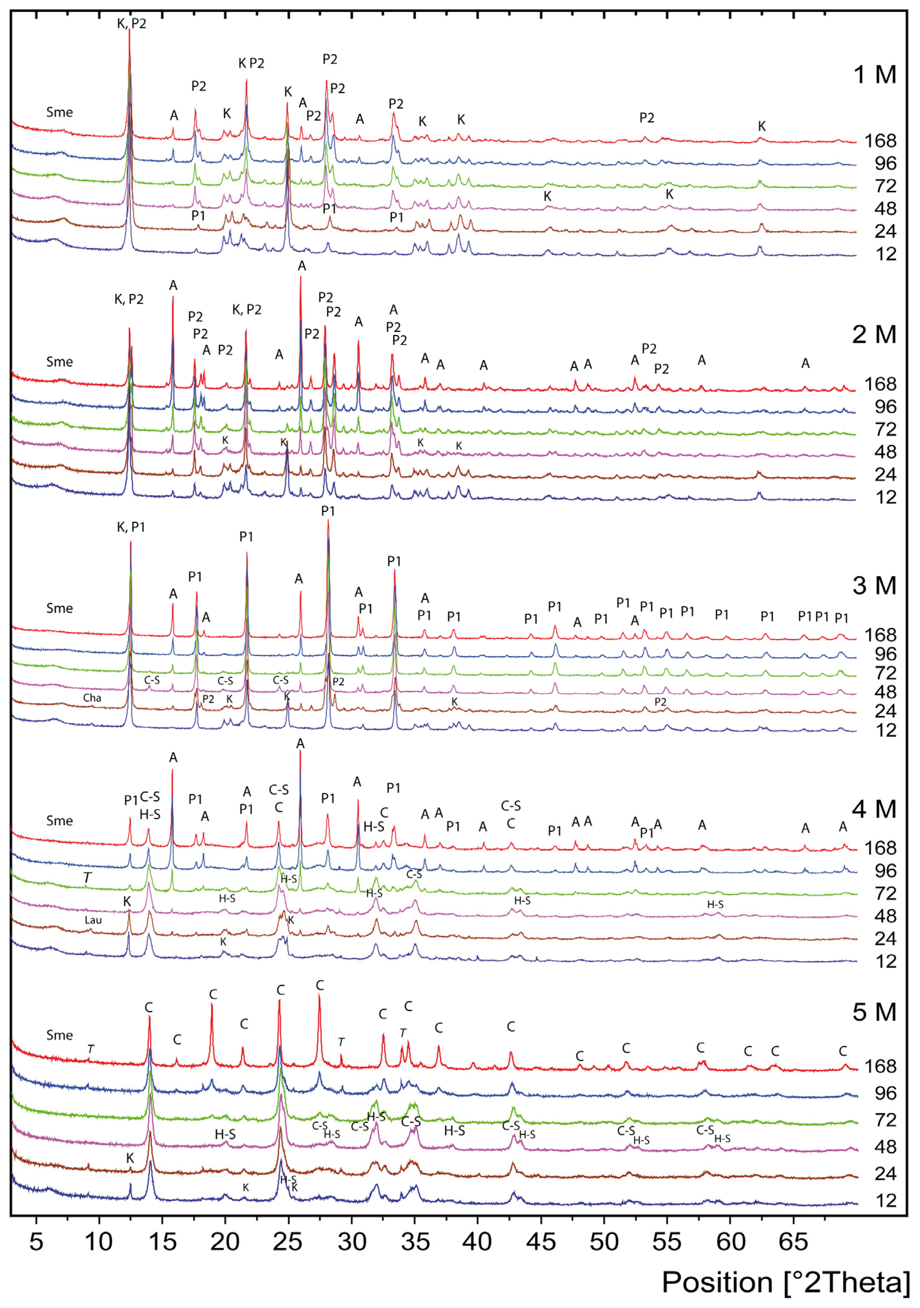
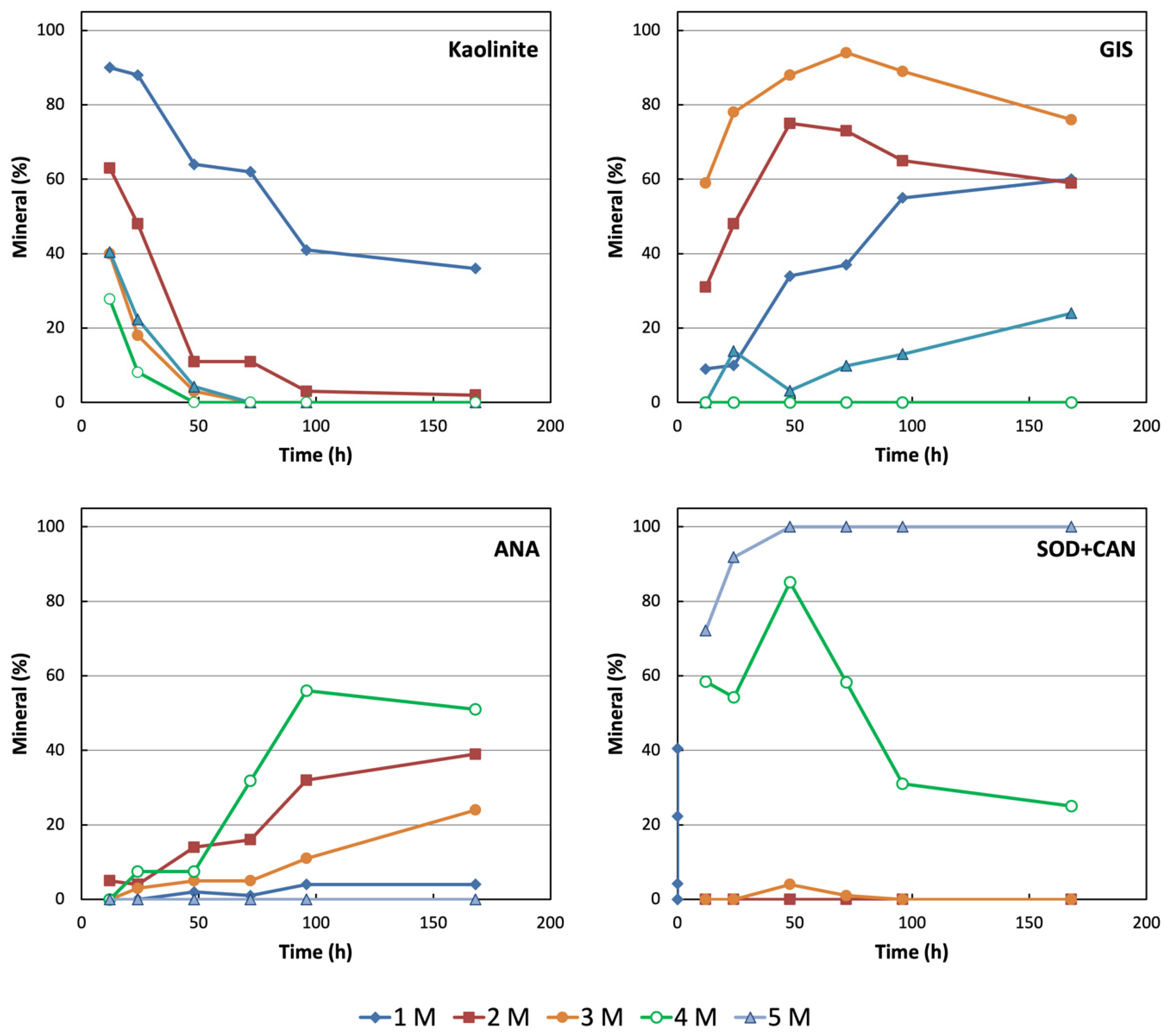
XRD analysis of synthetic samples (Figure 2) revealed the formation of zeolites, such as NaP, analcime, sodalite, and cancrinite, with variations depending on reaction time and NaOH concentration. Detailed mineralogical characterization of each sample is provided in the Supplementary Materials (Table S1). The alkaline hydrothermal treatment resulted in the progressive dissolution of the kaolin-forming minerals, as expected. No opal was identified in the products, suggesting that it dissolved within less than 12 h, regardless of the NaOH concentration. Residual traces of quartz were observed after 12–24 h in 1–2 M NaOH exclusively. The dissolution of kaolinite was expected to be faster in more alkaline solutions [64]. It was still present in samples aged in 1 M NaOH after 168 h, whereas it was nearly completely dissolved in 3–5 M NaOH after 48 h (Figure 3). This is likely due to the unusual size of the kaolinite crystals (>10 µm). The slow dissolution of kaolinite and the rapid dissolution of opal, which, respectively, lead to low Al supersaturation and an excess of Si, explain the absence of zeolite LTA. This zeolite has been reported to act as a precursor for the crystallization of sodalite and cancrinite ([41] and references therein). The different dissolution rates of these two main minerals in kaolin lead to a gradual change in the Si/Al ratio of the solution over time and with varying NaOH concentrations.
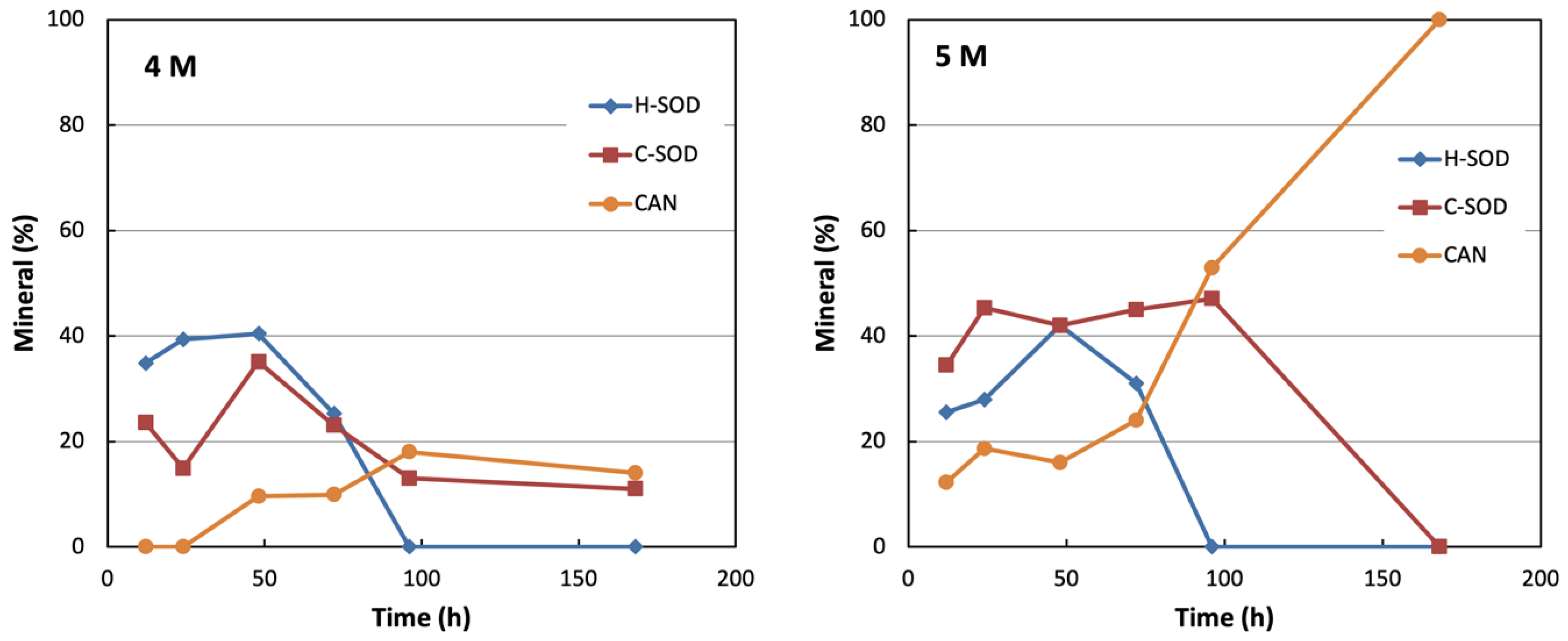
The mineralogy of the synthetic products showed significant complexity, as the relative abundance of minerals changed over time and with alkalinity (Figure 3 and Figure S3). Five main phases dominated across different experimental runs: NaP1 (JCPDS #01-71-0962; based on [2]), NaP2 (JCPDS: #01-80-0700; simulated patterns [65,66]), analcime (ANA; JCPDS #01-76-0143), cancrinite (CAN; JCPDS #01-88-1931), and sodalite (SOD). In solids from 4 and 5 M solutions, two distinct sodalite phases were found, with their quantity and distribution over time shown in Figure 4. The first to crystallize exhibited a hydrate sodalite structure without anions (JCPDS #01-88-1190), denoted as hydrate-sodalite (H-SOD) hereafter. The next phase was identified as carbonate-sodalite (C-SOD) (JCPDS #01-89-9099), characterized by the presence of carbonate anions in the framework, resulting from the incorporation of atmospheric CO2 as CO32− during sample preparation. NaP1, NaP2 (polymorphs of the GIS-type zeolites), and ANA are considered strictly zeolites, while SOD and CAN are feldspathoids, yet they can be classified as zeolites because of their structural features [2,67]. Minor amounts of chabazite and laumontite were found in shorter experiments, specifically in 3 and 4 M NaOH solutions, respectively (Table S1). Trona, a hydrated sodium carbonate and bicarbonate phase, was also detected in products from 4 and 5 M NaOH solutions. This phase likely resulted from the carbonation of highly alkaline solutions during experimental quenching, filtration, and rinsing of solids, and was therefore excluded from mineralogical estimates and discussion. A small amount of smectite, initially present in kaolin, showed a diffuse diffraction band at 12–15 Å (Figure 1). This band appeared in diffraction patterns of some synthetic materials but diminished in longer experiments and with more alkaline solutions. It indicates a minor, poorly crystalline phase and was not discussed further. To focus on the main zeolite phases, they were classified into three groups based on their structure: GIS, ANA, and SOD-CAN. The mineralogical distribution of the zeolite-kaolinite assemblage was recalculated to total 100% (Table 2).
Microscopic observations showed that NaP1 and NaP2 zeolites consistently form subspherical aggregates measuring 8–10 μm, made up of subprismatic stubby crystals, which are often twinned (Figure 5). This distinctive morphology has been previously reported in the literature [49,68,69,70,71,72,73]. However, the size of these aggregates can vary widely, from less than 1 μm to over 10 μm, and appears to be independent of both the initial material and experimental conditions. Since crystals of these two GIS-type polymorphs exhibit similar morphology, they cannot be distinguished if they form in the same experimental run.
Table 2. Mineralogy of solids obtained after hydrothermal treatment of DG1 kaolin (wt.%). Zeolites (see Table S1 in Supplementary Materials) are categorized as gismondine-type (GIS), analcime-type (ANA), and sodalite- and cancrinite-types (SOD-CAN). Kln and Qtz stand for kaolinite and quartz, respectively. The composition was recalculated from Table S1, excluding trona. Mineral abbreviations follow recommendations from The Commission on New Minerals, Nomenclature and Classification (CNMNC) of the International Mineralogical Association (IMA) [74] and The Structure Commission of the International Zeolite Association [62].
Ref.
#
NaOH (M)Time (h)KlnQtzGISANASOD + CAN
111290Tr90-
1612488<3100-
3114864-34<3-
4617262-37Tr-
6119641-554-
76116836-604-
421263Tr315-
1922447-484-
3424811-7514-
4927211-7316-
64296<3-6532-
792168<3-5939-
731240-60Tr-
2232418-78<3-
37348<3-8854
52372--945Tr
67396--8911-
823168--7624-
1041241-0-59
2542423-14755
404484-<3785
55472--103258
70496--135631
854168--245125
1351228---72
285248---92
43548----100
58572----100
73596----100
885168----100
Analcime crystals, showing the characteristic trapezohedral shape (Figure 6A), consistently appeared as individual crystals with sizes ranging from 2 to 8 μm depending on the experimental run. ANA may coexist with NaP phases. In some experiments, these crystals had poorly defined outlines (Figure 6B) and were coated with globular nanoparticles, confirming the potential metastability of the mineral under certain experimental conditions. The coexistence of ANA with NaP1 indicates overlapping metastable fields, and both minerals display well-defined outlines (see Figure 5A and Figure 6A, for example). It is likely that under these chemical conditions, ANA grew from the solution, as indicated by epitaxial growth (Figure 6A). At higher molarities (NaOH > 3 M), it becomes unstable and tends to amorphize (Figure 6B).
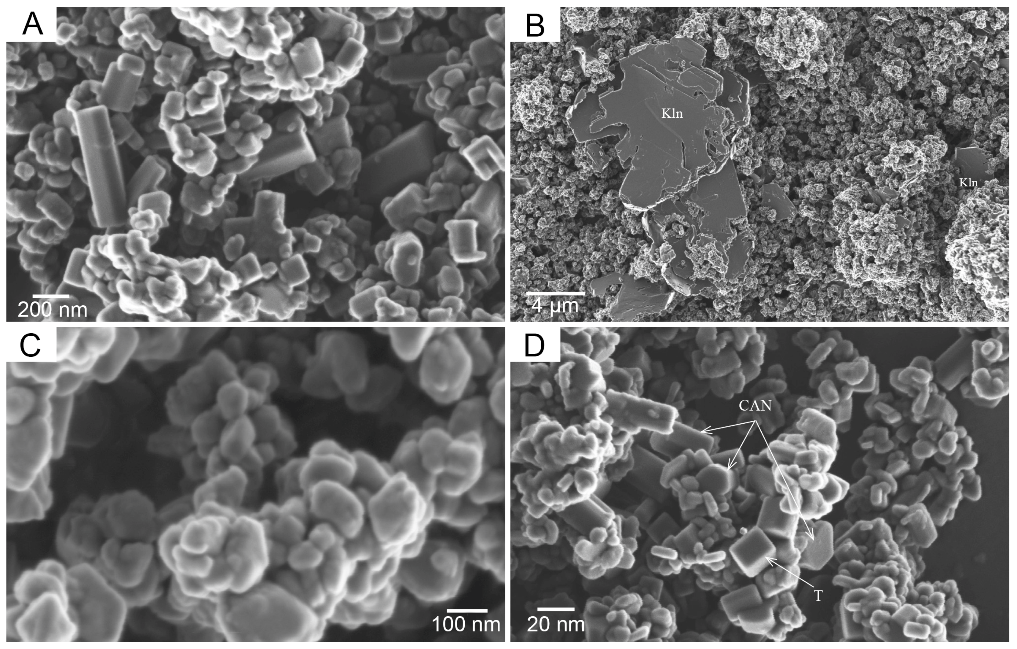
Cancrinite displayed its characteristic hexagonal prismatic morphology (Figure 7A), with sizes ranging from 100 to 600 nm. Microscopic observations revealed shapeless particles measuring from a few nanometers to 100 nm, likely indicating an amorphous nature resulting from the dissolution of pre-existing minerals (Figure 7B). Notably, the material synthesized after 168 h in a 5 M NaOH solution was almost entirely cancrinite. Well-defined sodalite crystals were not observed, even in samples where it was the main mineral (#43: 84%). The nanoparticles with linear outlines (Figure 7D) are probably sodalite crystals, as no other phases besides cancrinite were present, and cancrinite itself was easy to identify. Additionally, pseudocubic crystals of sodium carbonate-bicarbonate hydrate were identified as a byproduct of the treatment (see Figure 7C).
The crystallization of various zeolites was influenced by a competitive process, where the most stable phase formed under specific conditions of alkalinity and silica and alumina concentrations. Time played a crucial role in determining whether each phase crystallized or dissolved: with longer durations, kaolinite dissolved, changing the Si/Al ratio of the solution. During shorter periods (12–24 h) and lower alkalinity (1 M), the Si/Al ratio was mainly driven by the dissolution of opal, resulting in silica-rich solutions. Aluminum was added more slowly than silica because the dissolution of kaolinite was promoted by prolonged exposure to alkalinity. This sequential process suggests kinetic control, aligning with the principles of the Ostwald step rule. The observed progression GIS → ANA → CAN occurred because Si-rich phases, metastable, with lower nucleation barriers form initially, later dissolving and reprecipitating into the thermodynamically stable, Al-rich frameworks. Besides Si and Al, Na significantly influenced the crystallization pathway. At low Na+ molarity, it mainly acts as a charge-balancing cation for tetrahedral AlO4 units, whereas at higher concentrations (4–5 M), the surplus Na+, together with carbonate species, drives the nucleation of sodalite-type cages.
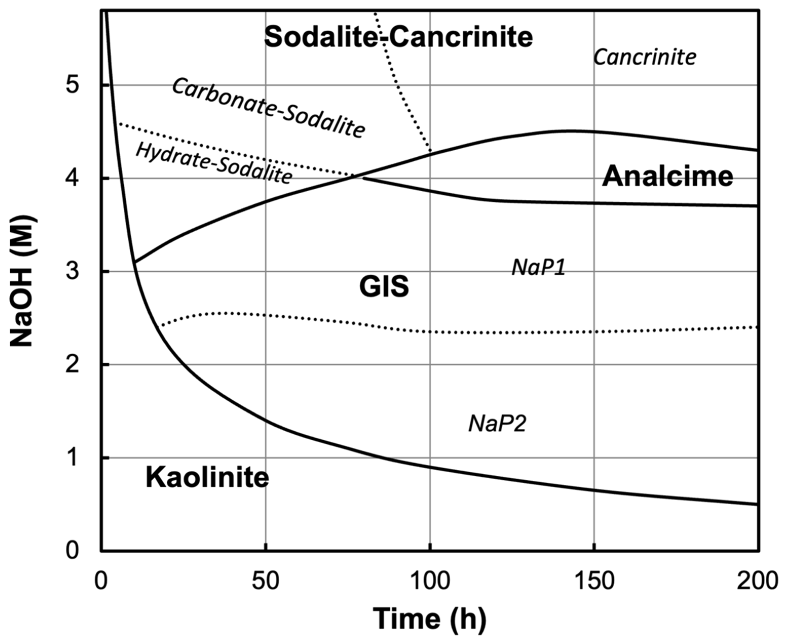
The relative abundance of system-forming minerals can be shown as a function of NaOH concentration and aging time (Figure 8). Hydrothermal crystallization products may exhibit metastable characteristics, where crystallization fields do not necessarily indicate regions of actual thermodynamic stability. Instead, they define compositions under specific experimental conditions where certain products nucleate, crystallize, and can be isolated and preserved as crystals. Investigating a hydrothermal alkaline system starting from kaolin and quartz, a phase diagram of NaOH/[Si + Al] vs. [Si + Al] at 200 °C for 24 h was proposed [75], concluding that the NaOH/[Si + Al] ratio controlled the formation of aluminosilicates. This phase diagram seems to contrast with the experimental findings presented here, likely due to differences in mineralogy (such as the presence of diaspore and the absence of opal-CT) and grain size (specific dimensions of the kaolin components) of the starting materials.

Mechanisms of Crystallization

The mineralogical insights from this research shed light on the crystallization processes of specific zeolites in alkaline environments. A substantial body of literature addresses the mechanisms of nucleation, growth, and crystallization of zeolites, with thorough reviews reflecting the accumulated knowledge over the past 80 years [16,76,77]. There is a broad consensus on the nucleation of zeolitic domains within gels [78] for zeolite-A. This process also accounts for the spherical morphology of kaolinite synthesized from pure aluminosilicate gels under hydrothermal conditions [79], confirmed later by HRTEM images [80]. Zeolitic domains can grow through the continuous transformation of amorphous material into ordered structures or by merging of crystallization centers, leading to protozeolitic minerals. Growth can occur via the addition of monomeric chemical species from solution (classical mechanism) or through attachment of oligomers, nanoparticles, and tetrahedral building blocks (nonclassical mechanism). Predicting nonclassical growth pathways of crystalline materials is highly complex, involving factors such as nanoparticle size, morphology, structure, movement, the properties of the solution, and interfacial forces [81,82]. Recently, a three-dimensional growth mechanism differing from layer-by-layer and crystallization by attached particles was reported for faujasite [83].
In zeolite synthesis, this complexity increases when natural raw materials are used as sources of alumina and silica. Due to their inherent physical and chemical heterogeneities, both classical and nonclassical pathways may occur simultaneously or at different stages in the process. This appears to be true for the zeolites produced in the current study.
The Romana kaolins mainly consisted of kaolinite (60%) and opal-CT (37%). They facilitated zeolite synthesis because other phases like quartz and smectite were present only in trace amounts or dissolved quickly at lower NaOH concentrations. Since opal-CT dissolved completely within the first 12 h regardless of Na concentration, the nucleation and growth of new phases were influenced by kaolinite dissolution, NaOH concentration, and the duration of the experiment. As kaolinite dissolved, globular amorphous nanoparticles (usually less than 50 nm) precipitated to form subspherical micrograins (Figure 7B). The particles’ composition was varied, which is consistent with previous studies (e.g., [48,79,84]). It has been shown that gels precipitated from solutions containing pure aluminosilicates or sodium aluminate and silicate salts, as well as those formed after hydrothermal treatment of fused raw materials, can be chemically inhomogeneous. As a result, crystalline domains may form and grow, remain inert, or dissolve depending on the chemical composition of the supernatant—which constantly changes as the starting material dissolves—and the local composition of the gel.
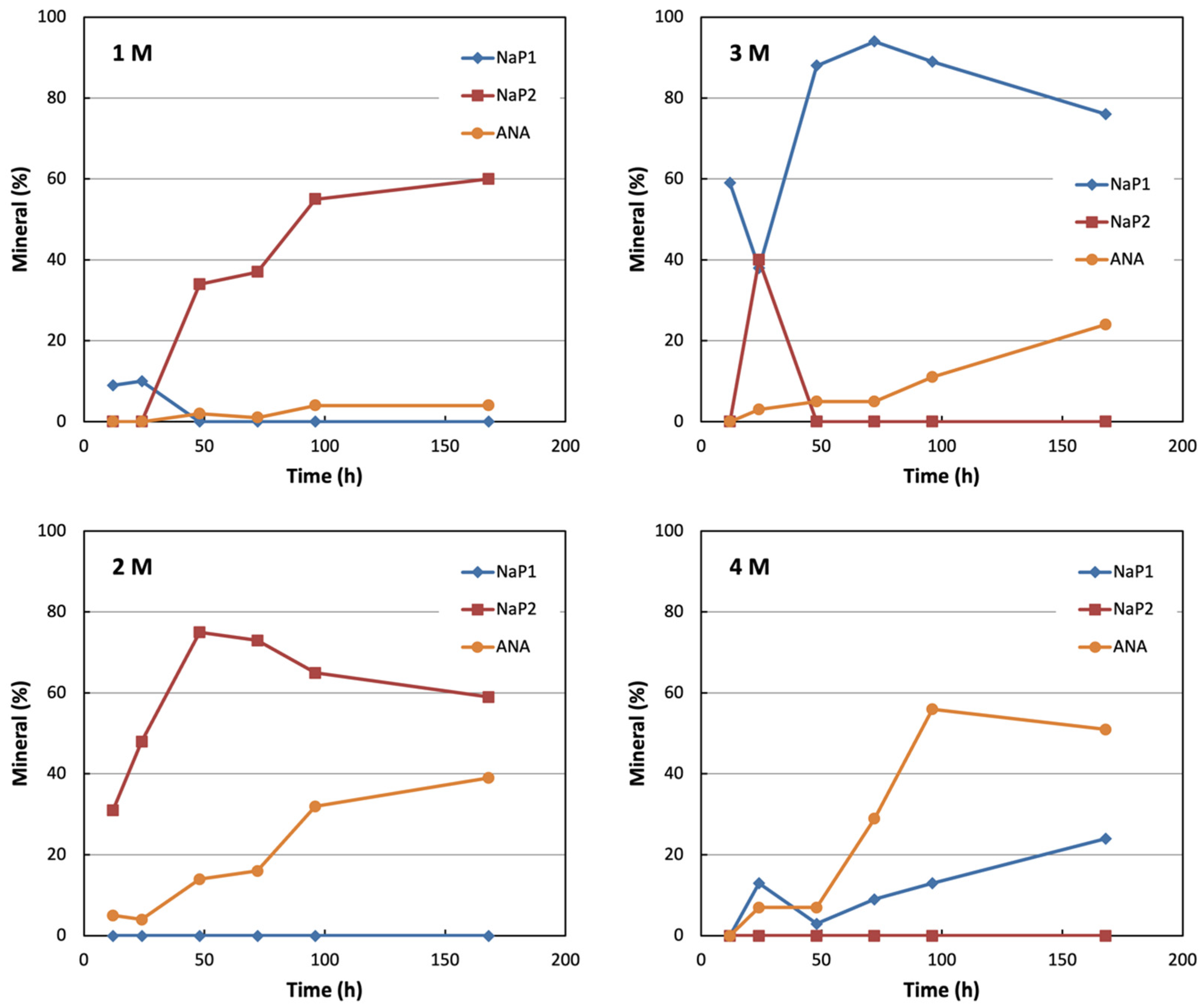
One of the most intriguing findings in this study was the formation and distribution of GIS-type polymorphs (Figure 9). It was observed that NaP1 formed in 1 M NaOH at 12 and 24 h, but it was metastable and transformed into NaP2 after 24 h (up to 60% at 76 h). The size of the aggregates and the crystals remained unchanged over time, indicating no transfer of chemical components from the supernatant to the already formed crystals; instead, new aggregates formed and grew. In a detailed study aimed at determining NaP1 and NaP2 synthesis conditions [66], a kinetic phase diagram using the molar fractions of Si, Al, and NaOH (where Na can be substituted with any M+ alkali metal) was constructed [56]. Starting from such an experimental diagram and considering that the concentrations of Si, Al, and NaOH continuously changed as kaolinite dissolution progressed in the studied system, it can be understood why NaP1 and NaP2 formed at different times in the runs with 1 M NaOH and why the isomorphic transformation from NaP1 to NaP2 occurred. The influence of both NaOH molarity and the concentrations of Si and Al on the stability of the two polymorphs was highlighted by the formation of NaP2 (+ANA) in the 2 M NaOH runs, NaP1 (+ANA) in the 3 M NaOH runs, and the coexistence of NaP1 + NaP2 (+ANA) in sample #22 (3 M NaOH, 24 h). Other notable results include the fluctuation in the amounts of NaP1 in samples #52, #67, and #82 (3 M NaOH), and the increase in NaP1 in samples #55, #70, and #85 (4 M NaOH), despite no detection of kaolinite by XRD. This suggests that the crystallization of GIS phases was influenced by both the gel composition and the chemistry of the growth solution.
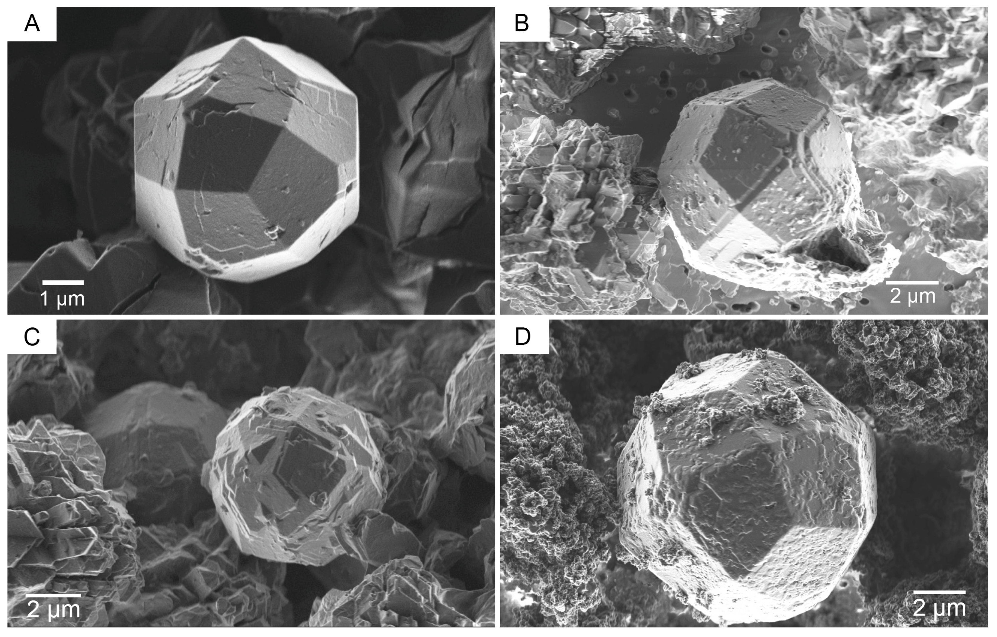
The morphologies of analcime crystals also provide valuable insights into their growth mechanisms. Typically, the crystals measured about 3 µm in size with a euhedral habit. Their surfaces displayed outlines resembling epitaxial growth (Figure 10A), indicating a two-dimensional growth mechanism (classical pathway). Additionally, larger crystals (up to 10 µm) were observed, which showed interpenetrations (Figure 10C) or irregular surfaces (Figure 10B,D). These features probably suggest growth through a nonclassical pathway.
SOD + CAN phases formed in the 4 M and 5 M NaOH sets, except for sample #88 (5 M NaOH; t = 168 h), which contained 91% cancrinite (Table S1), consistent with the effect of increasing NaOH molarity. Higher NaOH concentrations decrease the effective Si/Al ratio of the reactive gel through preferential silica dissolution, thereby promoting the stabilization of lower-silica frameworks (SOD–CAN).
A previous study [85] showed that these phases develop in the following sequence: zeolite-A -> sodalite -> cancrinite. However, under the conditions of this study, zeolite-A did not form (Figure 4), and the phases coexisted with microagglomerates of nanometric particles. SOD crystals were tiny, and FESEM offered limited details (Figure 7D). In comparison, CAN crystals showed euhedral morphologies (Figure 7A) and a relatively consistent size, suggesting their formation involved a solution-mediated mechanism with gel dissolution and phase precipitation.
The comparison of previously published results [52] and those of the present study deserves attention because both experiments used kaolin from the same mining district (Romana), although prepared differently. The authors of [52] used kaolin that was calcined at 650 °C, ground, sieved, dispersed in water, sonicated, and centrifuged to isolate the clay fraction. This fraction was then treated with a NaOH solution (pH ~ 12.8) at 65–100 °C for various durations (8–91 h), resulting in the formation of NaP1 (about 92%) at both 65 °C and 100 °C. In contrast, the present study, under similar experimental conditions (sample #52 in Table 2), observed the crystallization of NaP1 along with a minor amount of ANA (94% and 5%, respectively). The formation of ANA can be attributed to differences in the Si and Al content of the initial material, as their kaolin was richer in silica and poorer in alumina (70.17% vs. 21.34%) [42] than ours (65.22% vs. 23.86%). Another important factor was the duration of the experiment: longer durations resulted in greater amounts of ANA (Table 2, samples #67 and #82). NaP1 crystals showed no signs of dissolution, indicating that ANA formation was likely due to the dissolution of remaining amorphous particles, which dissolved over time and changed the chemical composition of the reacting solution. A slight variation in the Si/Al ratio, as predicted by the aforementioned kinetic phase diagram [66], dictated the co-precipitation of GIS and ANA zeolites.

4. Conclusions

The alkaline hydrothermal treatment of Donigazza kaolin is a one-step process suitable for transforming kaolinite and silica into zeolites at moderate temperatures (around 100 °C) and durations (less than 168 h). Zeolites formed under these experimental conditions mainly include GIS polymorphs, analcime, and sodalite–cancrinite. Alkalinity, Si, and Al determine the mineral association. The NaOH solution concentration governs alkalinity, while the Si and Al levels depend on the relative dissolution rates of silica (opal-CT) and kaolinite. Rapid dissolution of opal-CT (within 12 h), regardless of NaOH concentration, produces Si-rich solutions. In contrast, the slower dissolution of kaolinite supplies Al, and its dependence on NaOH influences the actual zeolite formation under specific conditions. As a result, the Si/Al ratio decreases over time and with increasing alkalinity. At 1–3 M NaOH, the dominant phases include GIS polymorphs NaP1 and NaP2, along with analcime. At 4–5 M NaOH, the main phases shift to SOD-CAN. By adjusting key parameters, it is possible to tailor the experiment to achieve a desired zeolite assemblage, highlighting Donigazza kaolin as a versatile raw material for various industrial and environmental applications.
Our experiments demonstrate that zeolites can form through both classical and non-classical crystallization mechanisms, which coincide. The slow dissolution of well-formed kaolinite gradually releases Al and, to a lesser degree, Si, which results in the formation of nanometer-sized gel-like particles that develop into zeolitic mineral phases through a dissolution-mediated dissolution–coprecipitation process. Additionally, some analcime crystals exhibit growth consistent with the classical model, characterized by the development of growth terraces on the faces of euhedral crystals. The kaolin-NaOH solution behaves as a complex dynamic system, influenced by factors such as alkalinity, temperature, and time.

Supplementary Materials

The following supporting information can be downloaded at: http://hdl.handle.net/10261/401995 (accessed on 5 November 2025), Figure S1: Geological sketch map; Figure S2: Scanning electron images of kaolinite and opal; Table S1: Mineralogical composition of the aged samples; Figure S3: Variation in the composition of the synthetic products [86,87,88,89,90,91,92].

Author Contributions

Conceptualization, P.M., S.F. and F.J.H.; methodology, P.M. and A.M.F.; validation, S.F. and F.J.H.; investigation, P.M. and A.M.F.; writing—original draft preparation, P.M.; writing—review and editing, S.F. and F.J.H.; funding acquisition, P.M., A.M.F. and F.J.H. All authors have read and agreed to the published version of the manuscript.

Funding

This research was supported by Grants PID2020-114355GB-I00 (funded by MCIN/AEI/10.13039/501100011033 and “ERDF A way of making Europe”), and by CGL2017–82331-R (MCIN—Spanish Ministry of Science and Innovation) awarded to F.J.H. P.M. gratefully acknowledges financial support from Fondo Sociale Europeo (FSE) and University of Sassari (Fondo di Ateneo per la Ricerca 2019—Mameli). IMAA (CNR, Italy) and DICATECH (Polytechnic University of Bari, Italy) financed the grant “Synthesis of silicate materials and their environmental applications” awarded to A.M.F.

Data Availability Statement

The supplementary data presented in this study are openly available in DIGITALCSIC at http://hdl.handle.net/10261/401995 (accessed on 5 November 2025). The data have been included either in the main text or in the Supplementary Materials. Further specifications on the methodology and/or results will be provided upon request.

Acknowledgments

A.M.F. is indebted to DICATECH (Polytechnic University of Bari, Italy) for having made FESEM available for the microscopic study. F.J.H., amdg. The authors would like to thank three anonymous reviewers for their comments and suggestions, as well as the academic editor.

Conflicts of Interest

The authors declare that they have no known competing financial interest or personal relationships that could have appeared to influence the work reported in this paper.

Abbreviations

The following abbreviations are used in this manuscript:
ANAAnalcime
CANCancrinite
CNMNECommission on New Minerals, Nomenclature and Classification
EDSEnergy-Dispersive X-ray Spectrometer
FESEMField Emission Scanning Electron Microscope
GISGismondine
HRTEMHigh-Resolution Transmission Electron Microscope
IMAInternational Mineralogical Association
IZAInternational Zeolite Association
JCPDSJoint Committee on Powder Diffraction Standards
LOILoss on Ignition
RIRReference Intensity Ratio
SEMScanning Electron Microscope
SODSodalite
XRDX-ray Diffraction
XRFX-ray Fluorescence

References

  1. Colella, C.; Wise, W.S. The IZA Handbook of Natural Zeolites: A tool of knowledge on the most important family of porous minerals. Micropor. Mesopor. Mater. 2014, 149, 4–10. [Google Scholar] [CrossRef]
  2. IZA (Structure Commission). Database of Zeolite Structures. 2017. Available online: https://www.iza-structure.org/databases/ (accessed on 5 October 2025).
  3. Breck, D.W. Zeolite Molecular Sieves: Structure, Chemistry and Use; John Wiley & Son: New York, NY, USA, 1974; pp. 1–771. [Google Scholar]
  4. Jansen, J.C. The Preparation of Molecular Sieves. In Studies in Surface Science and Catalysis; Flanigen, E.M., Janse, J.C., Eds.; Elsevier: Amsterdam, The Netherlands, 1991; Volume 58, pp. 77–136. [Google Scholar] [CrossRef]
  5. Moshoeshoe, M.; Nadiye-Tabbiruka, M.S.; Obuseng, V. A review of the chemistry, structure, properties and applications of zeolites. Am. J. Mater. Sci. 2017, 7, 196–221. [Google Scholar] [CrossRef]
  6. Khaleque, A.; Alam, M.M.; Hoque, M.; Mondal, S.; Haider, J.B.; Johir, M.A.H.; Karmakar, A.K.; Ahmed, M.B.; Moni, M.A. Zeolite synthesis from low-cost materials and environmental applications: A review. Environ. Adv. 2020, 2, 100019. [Google Scholar] [CrossRef]
  7. Li, Y.; Yu, J. Emerging applications of zeolites in catalysis, separation and host–guest assembly. Nat. Rev. Mater. 2021, 6, 1156–1174. [Google Scholar] [CrossRef]
  8. Pérez-Botella, E.; Valencia, S.; Rey, F. Zeolites in adsorption processes: State of the art and future prospects. Chem. Rev. 2022, 122, 17647–17695. [Google Scholar] [CrossRef] [PubMed]
  9. Dehmani, Y.; Mohammed, B.B.; Oukhrib, R.; Dehbi, A.; Lamhasni, T.; Brahmi, Y.; El-Kordy, A.; Franco, D.S.P.; Georgin, J.; Lima, E.C.; et al. Adsorption of various inorganic and organic pollutants by natural and synthetic zeolites: A critical review. Arab. J. Chem. 2024, 17, 105474. [Google Scholar] [CrossRef]
  10. Izidoro, J.D.C.; Fungaro, D.A.; Cataldo, E. Zeolites synthesized from agro-industrial residues applied in agriculture: A review and future prospects. Soil Use Manag. 2024, 40, e13003. [Google Scholar] [CrossRef]
  11. Zhao, Y.; Zhang, X.; Wang, J.; Liu, J.; Chen, R. Ammonium removal from aqueous solution by zeolite X synthesized from halloysite mineral. Water Sci. Technol. 2010, 62, 937–946. [Google Scholar] [CrossRef] [PubMed]
  12. Jiménez, I.; Pérez, G.; Guerrero, A.; Ruiz, B. Mineral phases synthesized by hydro-thermal treatment from biomass ashes. Int. J. Miner. Process 2017, 158, 8–12. [Google Scholar] [CrossRef]
  13. Belviso, C. State-of-the-art applications of fly ash from coal and biomass: A focus on zeolite synthesis processes and issues. Prog. Energy Combust. Sci. 2018, 65, 109–135. [Google Scholar] [CrossRef]
  14. Yoldi, M.; Fuentes-Ordoñez, E.G.; Korili, S.A.; Gil, A. Zeolite synthesis from industrial wastes. Micropor. Mesopor. Mater. 2019, 287, 183–191. [Google Scholar] [CrossRef]
  15. Chen, X.; Zhang, P.; Wang, Y.; Peng, W.; Ren, Z.; Li, Y.; Chu, B.; Zhu, Q. Research progress on synthesis of zeolites from coal fly ash and environmental applications. Front. Environ. Sci. Eng. 2023, 17, 149. [Google Scholar] [CrossRef]
  16. Mallette, A.J.; Shilpa, K.; Rimer, J.G. The current understanding of mechanistic pathways in zeolite crystallization. Chem. Rev. 2024, 124, 3416–3493. [Google Scholar] [CrossRef]
  17. Barrer, R.M.; Cole, J.F.; Sticher, H. Chemistry of soil minerals. Part V. Low temperature hydrothermal transformations of kaolinite. J. Chem. Soc. A 1968, 2475–2485. [Google Scholar] [CrossRef]
  18. Barrer, R.M. The Hydrothermal Chemistry of Zeolites; Academic Press: London, UK, 1982; pp. 1–360. [Google Scholar]
  19. Occelli, M.L.; Robson, H.E. (Eds.) Zeolite Synthesis, ACS Symposium Series; American Chemical Society: Washington, DC, USA, 1989; pp. 1–650. [Google Scholar] [CrossRef]
  20. Robson, H. (Ed.) Verified Synthesis of Zeolitic Materials; Elsevier: Amsterdam, The Netherlands, 2001; pp. 1–266. [Google Scholar] [CrossRef]
  21. Feng, M.; Kou, Z.; Tang, C.; Shi, Z.; Tong, Y.; Zhang, K. Recent progress in synthesis of zeolite from natural clay. Appl. Clay Sci. 2023, 243, 107087. [Google Scholar] [CrossRef]
  22. Meng, X.; Xiao, F.-S. Green routes for synthesis of zeolites. Chem. Rev. 2014, 114, 1521–1543. [Google Scholar] [CrossRef]
  23. Li, C.; Moliner, M.; Corma, A. Building zeolites from precystallized units: Nanoscale architecture. Angew.Chem. Int. Ed. 2018, 57, 15330–15353. [Google Scholar] [CrossRef]
  24. Jain, R.; Mallete, A.; Rimer, J. Controlling nucleation pathways in zeolite crystallization: Seeding conceptual methodologies for advanced materials design. J. Am. Chem. Soc. 2021, 143, 21446–21460. [Google Scholar] [CrossRef]
  25. Goel, S.; Zones, S.I.; Iglesia, E. Synthesis of zeolites via interzeolite transformations without organic structure-directing agents. Chem. Mater. 2015, 27, 2056–2066. [Google Scholar] [CrossRef]
  26. Devos, J.; Shah, M.A.; Dusselier, M. On the key role of aluminium and other heteroatoms during interzeolite conversion synthesis. RSC Adv. 2021, 11, 26188–26210. [Google Scholar] [CrossRef] [PubMed]
  27. Chun-Jon, C.; Martinez, E.; Bertolazzo, A.A.; Banik, S.; Rimer, J.D.; Sankaranarayanan, S.K.R.S.; Molinero, V. Interzeolite transformation through cross-nucleation: A molecular mechanism for seed-assisted synthesis. J. Am. Chem. Soc. 2024, 146, 33204–33213. [Google Scholar] [CrossRef]
  28. Pornsetmetakul, P.; Coumans, F.J.A.G.; Heinrichs, J.M.J.J.; Zhang, H.; Wattanakit, C.; Hensen, E.J.M. Accelerated synthesis of nanolayered MWW zeolite by interzeolite transformation. Chem. Eur. J. 2024, 30, e202302931. [Google Scholar] [CrossRef] [PubMed]
  29. Liu, Y.; Li, Y.; Chen, Z.; Ji, F.; Liang, X.; Han, L.; Han, P. Synthesis of CHA from MFI by three interzeolite transformation strategies and its application in NH3-SCR reaction. J. Porous Mater. 2025, 32, 727–740. [Google Scholar] [CrossRef]
  30. Barrer, R.M.; Mainwaring, D.E. Chemistry of soil minerals. Part XIII. Reactions of metakaolinite with single and mixed bases. J. Chem. Soc. Dalton 1972, 12, 1254–1259. [Google Scholar] [CrossRef]
  31. Madani, A.; Aznar, A.; Sanz, J.; Serratosa, J.M. 29Si and 27Al NMR study of zeolite formation from alkali-leached kaolinites. Influence of thermal preactivation. J. Phys. Chem. 1990, 94, 760–765. [Google Scholar] [CrossRef]
  32. Murat, M.; Amokrane, A.; Bastide, J.P.; Montanaro, L. Synthesis of zeolites from thermally activated kaolinite. Some observations on nucleation and growth. Clay Miner. 1992, 27, 119–130. [Google Scholar] [CrossRef]
  33. Alberti, A.; Colella, C.; Oggiano, G.; Pansini, M.; Vezzalini, G. Zeolite production from waste kaolin-containing materials. Mater. Eng. 1994, 45, 145–158. [Google Scholar]
  34. Akolekar, D.; Chaffee, A.; Howe, R.F. The transformation of kaolin to low-silica X zeolite. Zeolites 1997, 19, 359–365. [Google Scholar] [CrossRef]
  35. Gualtieri, A.; Norby, P.; Artioli, G.; Hanson, J. Kinetics of formation of zeolite Na–A [LTA] from natural kaolinites. Phys. Chem. Miner. 1997, 24, 191–199. [Google Scholar] [CrossRef]
  36. Chandrasekhar, S.; Pramada, P.N. Investigation on the synthesis of zeolite NaX from Kerala kaolin. J. Porous Mater. 1999, 6, 283–297. [Google Scholar] [CrossRef]
  37. Chandrasekhar, S.; Pramada, P.N. Sintering behaviour of calcium exchanged low silica zeolites synthesized from kaolin. Ceram. Int. 2001, 27, 105–114. [Google Scholar] [CrossRef]
  38. Alkan, M.; Hopa, C.; Yilmaz, Z.; Guler, H. The effect of alkali concentration and solid/liquid ratio on the hydrothermal synthesis of zeolite NaA from natural kaolinite. Micropor. Mesopor. Mater. 2005, 86, 176–184. [Google Scholar] [CrossRef]
  39. Heller-Kallai, L.; Lapides, I. Reactions of kaolinites and metakaolinites with NaOH—Comparison of different samples (Part 1). Appl. Clay Sci. 2007, 35, 99–107. [Google Scholar] [CrossRef]
  40. Youssef, H.; Ibrahim, D.; Komarneni, S. Microwave-assisted versus conventional synthesis of zeolite A from metakaolinite. Micropor. Mesopor. Mater. 2008, 115, 527–534. [Google Scholar] [CrossRef]
  41. Ríos, C.A.; Williams, C.D.; Fullen, M.A. Nucleation and growth history of zeolite LTA synthesized from kaolinite by two different methods. Appl. Clay Sci. 2009, 42, 446–454. [Google Scholar] [CrossRef]
  42. Mackinnon, I.D.R.; Millar, G.J.; Stolz, W. Low temperature synthesis of zeolite N from kaolinites and montmorillonites. Appl. Clay Sci. 2010, 48, 622–630. [Google Scholar] [CrossRef]
  43. Johnson, E.B.G.; Arshad, S.E. Hydrothermally synthesized zeolites based on kaolinite: A review. Appl. Clay Sci. 2014, 97–98, 215–221. [Google Scholar] [CrossRef]
  44. Meftah, M.; Oueslati, W.; Chorfi, N.; Ben Haj Amara, A. Effect of the raw material type and the reaction time on the synthesis of halloysite based Zeolite Na-P1. Results Phys. 2017, 7, 1475–1484. [Google Scholar] [CrossRef]
  45. Rubtsova, M.; Smirnova, E.; Boev, S.; Kotelev, M.; Cherednichenko, K.; Vinokurov, V.; Lvov, Y.; Glotov, A. Nanoarchitectural approach for synthesis of highly crystalline zeolites with a low Si/Al ratio from natural clay nanotubes. Micropor. Mesopor. Mater. 2022, 330, 111622. [Google Scholar] [CrossRef]
  46. Gualtieri, A.; Bellotto, M. Modelling the structure of the metastable phases in the reaction sequence kaolinite-mullite by X-ray scattering experiments. Phys. Chem. Miner. 1998, 25, 442–452. [Google Scholar] [CrossRef]
  47. White, C.E.; Provis, J.L.; Proffen, T.; Riley, D.P.; van Deventer, J.S.J. Density functional modeling of the local structure of kaolinite subjected to thermal dihydroxylation. J. Phys. Chem. A 2010, 114, 4988–4996. [Google Scholar] [CrossRef]
  48. Belviso, C.; Giannossa, L.C.; Huertas, F.J.; Lettino, A.; Mangone, A.; Fiore, S. Synthesis of zeolites at low temperatures in fly ash-kaolinite mixtures. Micropor. Mesopor. Mater. 2015, 212, 35–47. [Google Scholar] [CrossRef]
  49. Moreno-Torres, J.A.; Espejel-Ayala, F.; Ramirez-Bon, R.; Coutino-Gonzalez, E. Sustainable strategies to synthesize small—Pore NaP zeolites using natural minerals. J. Mater. Sci. 2024, 59, 423–434. [Google Scholar] [CrossRef]
  50. Yang, J.; Liu, H.; Diao, H.; Li, B.; Yue, Y.; Bao, X. A quasi-solid-phase approach to activate natural minerals for zeolite synthesis. ACS Sustain. Chem. Eng. 2017, 5, 3233–3242. [Google Scholar] [CrossRef]
  51. Novembre, D.; Gimeno, D. Synthesis and characterization of analcime (ANA) zeolite using a kaolinitic rock. Sci. Rep. 2021, 11, 13373. [Google Scholar] [CrossRef] [PubMed]
  52. Novembre, D.; Gimeno, D.; Del Vecchio, A. Synthesis and characterization of Na-P1 (GIS) zeolite using a kaolinitic rock. Sci. Rep. 2021, 11, 4872. [Google Scholar] [CrossRef] [PubMed]
  53. Mameli, P. Rilevamento e Caratterizzazione Mineralogica del Caolino della Sardegna Settentrionale e Proposta di Impiego in settori non Convenzionali. Ph.D. Thesis, Università di Sassari, Sassari, Italy, 1999; pp. 1–125. [Google Scholar]
  54. Mameli, P. Occurrence of halite in kaolin of NW Sardinia: Genetic implications. Water-Rock Interact. 2001, 10, 729–733. [Google Scholar]
  55. Ligas, P.; Uras, I.; Dondi, M.; Marsigli, M. Kaolinitic materials from Romana (north-west Sardinia, Italy) and their ceramic properties. Appl. Clay Sci. 1997, 12, 145–163. [Google Scholar] [CrossRef]
  56. Oggiano, G.; Mameli, P. Tectonic and litho-stratigraphic controls on kaolin deposits within volcanic successions: Insights from the kaoliniferous district of north-western Sardinia (Italy). Ore Geol. Rev. 2012, 48, 151–164. [Google Scholar] [CrossRef]
  57. PDF—Powder Diffraction Data, release 2003. In JCPDS, Joint Committee on Powder Diffraction Standards; International Centre for Diffraction Data: Newtown Square, PA, USA, 2003.
  58. Chung, F.H.; IUCr. Quantitative interpretation of X-ray diffraction patterns of mixtures. I. Matrix-flushing method for quantitative multicomponent analysis. J. Appl. Crystallogr. 1974, 7, 519–525. [Google Scholar] [CrossRef]
  59. Chung, F.H.; IUCr. Quantitative interpretation of X-ray diffraction patterns of mixtures. II. Adiabatic principle of X-ray diffraction analysis of mixtures. J. Appl. Crystallogr. 1974, 7, 526–531. [Google Scholar] [CrossRef]
  60. Chung, F.H.; IUCr. Quantitative interpretation of X-ray diffraction patterns of mixtures. III. Simultaneous determination of a set of reference intensities. J. Appl. Crystallogr. 1975, 8, 17–19. [Google Scholar] [CrossRef]
  61. Degen, T.; Sadki, M.; Bron, E.; König, U.; Nénert, G. The HighScore suite. Powder Diffr. 2014, 29, S13–S18. [Google Scholar] [CrossRef]
  62. Baerlocher, C.; McCusker, L.B.; Olson, D.H. Atlas of Zeolite Framework Types, 6th revised ed.; Elsevier, B.V.: Amsterdam, The Netherlands, 2007; pp. 1–398. [Google Scholar] [CrossRef]
  63. Treacy, M.M.J.; Higgins, J.B. Collection of Simulated XRD Powder Patterns for Zeolites, 5th ed.; Elsevier, B.V.: Amsterdam, The Netherlands, 2007; pp. 1–485. [Google Scholar] [CrossRef]
  64. Huertas, F.J.; Chou, L.; Wollast, R. Mechanism of kaolinite dissolution at room temperature and pressure Part II: Kinetic study. Geochim. Cosmochim. Acta 1999, 63, 3261–3275. [Google Scholar] [CrossRef]
  65. Hansen, S.; Håkansson, U.; Fälth, L. Structure of synthetic zeolite Na-P2. Acta Cryst. C 1990, 46, 1361–1362. [Google Scholar] [CrossRef]
  66. Oleksiak, M.D.; Ghorbanpour, A.; Conato, M.T.; McGrail, P.; Grabow, L.C.; Motkuri, R.K.; Rimer, J.D. Synthesis strategies for ultrastable zeolite GIS polymorphs as sorbents for selective separations. Chem. Eur. J. 2016, 22, 16078–16088. [Google Scholar] [CrossRef]
  67. Coombs, D.S.; Alberti, A.; Armbruster, T.; Artioli, G.; Colella, C.; Galli, E.; Grice, J.D.; Liebau, F.; Mandarino, J.A.; Minato, H.; et al. Recommended nomenclature for zeolite minerals: Report of the Subcommittee on Zeolites of the International Mineralogical Association, Commission on New Minerals and Mineral Names. Eur. J. Miner. 1998, 10, 1037–1081. [Google Scholar] [CrossRef]
  68. Albert, B.R.; Cheetham, A.K.; Stuart, J.A.; Adams, C.J. Investigations on P zeolites: Synthesis, characterisation, and structure of highly crystalline low-silica NaP. Micropor. Mesopor. Mater. 1998, 21, 133–142. [Google Scholar] [CrossRef]
  69. Zubowa, H.L.; Kosslick, H.; Müller, D.; Richter, M.; Wilde, L.; Fricke, L. Crystallization of phase-pure zeolite NaP from MCM-22-type gel compositions under microwave radiation. Micropor. Mesopor. Mater. 2008, 109, 542–548. [Google Scholar] [CrossRef]
  70. Sánchez-Hernández, R.; López-Delgado, A.; Padilla, I.; Galindo, R.; López-Andrés, S. One-step synthesis of NaP1, SOD and ANA from a hazardous aluminum solid waste. Micropor. Mesopor. Mater. 2016, 226, 267–277. [Google Scholar] [CrossRef]
  71. Le, T.; Wang, Q.; Pan, B.; Ravindra, A.V.; Shaohua, J.; Peng, J. Process regulation of microwave intensified synthesis of Y-type zeolite. Micropor. Mesopor. Mater. 2019, 284, 476–485. [Google Scholar] [CrossRef]
  72. Sharma, P.; Song, J.-S.; Han, M.H.; Cho, C.-H. GIS-NaP1 zeolite microspheres as potential water adsorption material: Influence of initial silica concentration on adsorptive and physical/topological properties. Sci. Rep. 2016, 6, 22734. [Google Scholar] [CrossRef] [PubMed]
  73. Tayraukham, P.; Jantarit, N.; Osakoo, N.; Wittayakun, J. Synthesis of pure phase NaP2 zeolite from the gel of NaY by conventional and microwave-assisted hydrothermal methods. Crystals 2020, 10, 951. [Google Scholar] [CrossRef]
  74. Warr, L.N. IMA–CNMNC approved mineral symbols. Min. Mag. 2021, 85, 291–320. [Google Scholar] [CrossRef]
  75. Tang, L.-J.; Xie, X.-Z.; Huang, Y.-X.; Pan, Y.; Mi, J.-X. Phase diagram for hydrothermal alkali activation of kaolin and quartz: Optimal digestion for the synthesis of zeolites. Mater. Chem. Phys. 2022, 290, 126570. [Google Scholar] [CrossRef]
  76. Cundy, C.S.; Cox, P.A. The hydrothermal synthesis of zeolites:  History and development from the earliest days to the present time. Chem. Rev. 2003, 103, 663–702. [Google Scholar] [CrossRef]
  77. Cundy, C.S.; Cox, P.A. The hydrothermal synthesis of zeolites: Precursors, intermediates and reaction mechanism. Micropor. Mesopor. Mater. 2005, 82, 1–78. [Google Scholar] [CrossRef]
  78. Mintova, S.; Olson, N.H.; Valtchev, V.; Bein, T. Mechanism of zeolite A nanocrystal growth from colloids at room temperature. Science 1999, 283, 958–960. [Google Scholar] [CrossRef]
  79. Fiore, S.; Huertas, F.J.; Huertas, F.; Linares, J. Morphology of kaolinite crystals synthesized under hydrothermal conditions. Clays Clay Miner. 1995, 43, 353–360. [Google Scholar] [CrossRef]
  80. Huertas, F.J.; Fiore, S.; Linares, J. In situ transformation of amorphous gels into spherical aggregates of kaolinite: A HRTEM study. Clay Miner. 2004, 39, 423–431. [Google Scholar] [CrossRef]
  81. Ivanov, V.K.; Fedorov, P.P.; Baranchikov, A.E.; Osiko, V.V. Oriented attachment of particles: 100 years of investigations of non-classical crystal growth. Russian Chem. Rev. 2014, 83, 1204–1222. [Google Scholar] [CrossRef]
  82. De Yoreo, J.J.; Gilbert, P.U.P.A.; Sommerdijk, N.A.J.M.; Penn, R.L.; Whitelam, S.; Joester, D.; Zhang, H.; Rimer, J.D.; Navrotsky, A.; Banfield, J.F.; et al. Crystallization by particle attachment in synthetic, biogenic, and geologic environments. Science 2015, 349, aaa6760. [Google Scholar] [CrossRef]
  83. Jain, R.; Niu, Z.; Choudhary, M.; Bourji, H.; Palmer, J.C.; Rimer, J.D. In situ imaging of faujasite surface growth reveals unique pathways of zeolite crystallization. J. Am. Chem. Soc. 2023, 145, 1155–1164. [Google Scholar] [CrossRef]
  84. Valtchev, V.P.; Bozhilov, K.N. Transmission electron microscopy study of the formation of FAU-type zeolite at room temperature. J. Phys. Chem. B 2004, 108, 15587–15598. [Google Scholar] [CrossRef]
  85. Barnes, M.C.; Addai-Mensah, J.; Gerson, A.R. The mechanism of the sodalite-to-cancrinite phase transformation in synthetic spent Bayer liquor. Micropor. Mesopor. Mater. 1999, 31, 287–302. [Google Scholar] [CrossRef]
  86. Palomba, P.; Padalino, G.; Marchi, M. Industrial mineral occurrences associated with Cenozoic volcanic rocks of Sardinia (Italy): Geological, mineralogical, geochemical features and genetic implications. Ore Geol. Rev. 2006, 29, 118–145. [Google Scholar] [CrossRef]
  87. Peccerillo, A. Sardinia. In Cenozoic Volcanism in the Tyrrhenian Sea Region, 2nd ed.; Advances in Volcanology; Springer: Cham, Switzerland, 2017; pp. 313–338. [Google Scholar] [CrossRef]
  88. Coulon, C.; Dupuy, C. Evolution spatiale des caractères chimiques du volcanisme andésitique de la Sardaigne (Italie). Earth Planet. Sci. Lett. 1975, 25, 170–176. [Google Scholar] [CrossRef]
  89. Beccaluva, L.; Brotzu, P.; Morbidelli, L.; Serri, G.; Traversa, G. Cainozoic tectono-magmatic evolution and inferred mantle in the Sardo-Tyrrhenian area. The lithosphere in Italy. Advances in Earth Science Research. Accad. Naz. Lincei 1987, 229–248. [Google Scholar]
  90. Beccaluva, L.; Bianchini, G.; Mameli, P.; Natali, C. Miocene shoshonite volcanism in Sardinia: Implications for magma sources and geodynamic evolution of the central-western Mediterranean. Lithos 2013, 180–181, 128–137. [Google Scholar] [CrossRef]
  91. Utada, M. Hydrothermal alteration related to igneous acidity in Cretaceous and Neogene formations of Japan. Min. Geol. jpn. Spec. Issue 1980, 8, 67–83. [Google Scholar]
  92. Berger, G.; Velde, B. Chemical parameters controlling the propylitic and argillic alteration process. Eur. J. Mineral. 1992, 4, 1439–1456. [Google Scholar] [CrossRef]
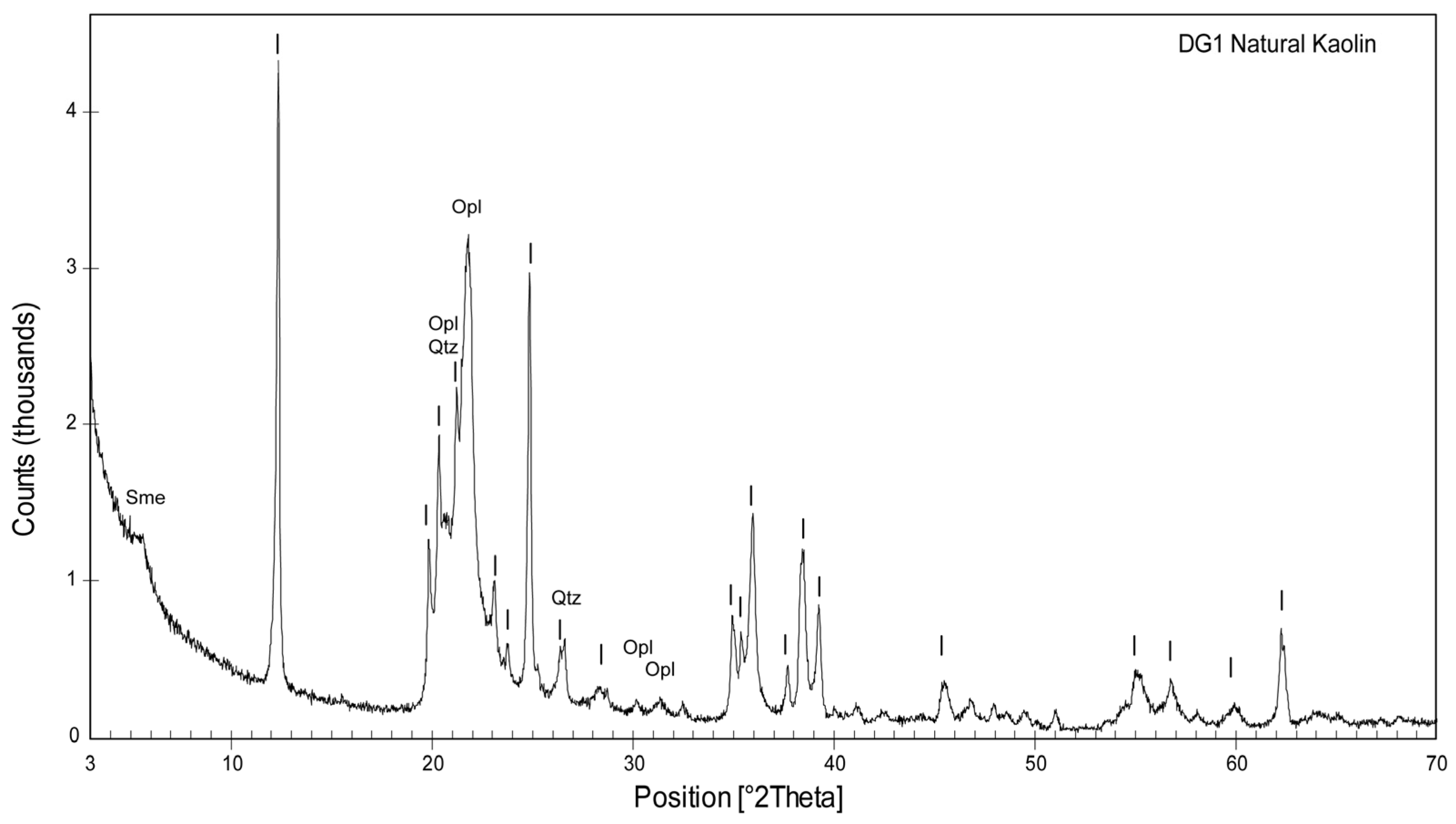
Figure 1. Powder XRD pattern of DG1 natural kaolin. Main reflections of kaolinite are indicated with a vertical line. Sme: smectite; Opl: opal-CT; Qtz: quartz.
Crystals 15 00980 g001
Figure 2. Powder XRD patterns of the solids obtained after hydrothermal treatment of DG1 kaolin, as a function of NaOH concentration and time. Legend: A: analcime; C: cancrinite; C-S: carbonate-sodalite; Cha: chabazite; H-S: hydrate-sodalite; K: kaolinite; Lau: laumontite; P1: NaP1; P2: NaP2; Sme: smectite; T: trona.
Crystals 15 00980 g002
Figure 3. Variation in kaolinite and zeolite content in synthetic products as a function of time and NaOH concentration (1–5 M) in the aging solution.
Crystals 15 00980 g003
Figure 4. Evolution of SOD-CAN phase content with aging time in solids synthesized in 4 and 5 M NaOH solutions. Observe the crystallization sequence H-SOD -> C-SOD -> CAN. H-SOD, hydrate-sodalite; C-SOD, carbonate-sodalite; CAN, cancrinite.
Crystals 15 00980 g004
Figure 5. SEM images of NaP1 and NaP2. (A) Aggregates of NaP1 crystals with a few euhedral crystals of ANA (sample #7, 12 h in 3 M). (B) Crystals, up to 1.2 μm in size, consistently had a radial arrangement to form aggregates 8–10 μm in size and were never observed as single entities. (C) Details of NaP2 crystal aggregates (sample #34, 48 h in 2 M). (D) NaP1 twinned individuals were frequently observed.
Crystals 15 00980 g005
Figure 6. FESEM images of ANA crystals showing very different morphological features. (A) In sample #67 (96 h in 3 M), the crystals (coexisting with NaP1) were fairly uniform in size (about 6 μm), had a “clean” surface, and well-defined edges. (B) In sample #40 (48 h in 4 M), individual crystals are coated with nanometric gel particles and display a jelly-like surface with smooth edges, indicating surface amorphization.
Crystals 15 00980 g006
Figure 7. FESEM images of (A) isolated hexagonal prismatic crystals of cancrinite (sample #88, 196 h in 5 M). (B) Aggregates of amorphous particles surrounding kaolinite particles showing intense signs of dissolution (sample #13, 12 h in 5 M). (C) Nanoparticles with linear outlines, likely sodalite crystals since no other phases besides cancrinite were present (sample #43, 48 h in 5 M). (D) A few trona crystals with pseudocubic morphologies, surrounded by cancrinite crystals (sample #88, 196 h in 5 M).
Crystals 15 00980 g007
Figure 8. Tentative representation of the relative abundance of mineral phases (>35% by XRD semi-quantitative estimation) as a function of NaOH concentration and aging time.
Crystals 15 00980 g008
Figure 9. Evolution of the NaP1, NaP2, and analcime (ANA) phases with aging time, in 1 to 4 M NaOH concentration solutions.
Crystals 15 00980 g009
Figure 10. (A) Surface of a euhedral crystal of ANA showing growth terraces with outlines parallel to the crystal edges (epitaxial growth) (#67, 96 h in 3M). (B) ANA crystal with growth macro-steps and nanometric particles on the faces (#82, 168 h in 3 M). (C) ANA crystals with irregular surfaces due to multiple interpenetration twins (#34, 48 h in 2 M). (D) Large ANA crystals with surfaces coated by very thin particles and gel-like materials, suggesting a nonclassical pathway of growth (#70, 96 h in 4 M).
Crystals 15 00980 g010
Table 1. Chemical composition (wt.%) of the material used for zeolite synthesis.
SiO2Al2O3Fe2O3TiO2MgOCaONa2OK2OP2O5LOI 1
65.2223.860.270.400.150.180.020.020.1110.25
1 LOI: loss on ignition.
Disclaimer/Publisher’s Note: The statements, opinions and data contained in all publications are solely those of the individual author(s) and contributor(s) and not of MDPI and/or the editor(s). MDPI and/or the editor(s) disclaim responsibility for any injury to people or property resulting from any ideas, methods, instructions or products referred to in the content.