Cancers 2023, 15(4), 1107; https://doi.org/10.3390/cancers15041107 - 9 Feb 2023
Cited by 23 | Viewed by 5001
| Correction
Abstract
Colorectal cancer (CRC) is a leading public health concern due to its incidence and high mortality rates, highlighting the requirement of an early diagnosis. Evaluation of circulating extracellular vesicles (EVs) might constitute a noninvasive and reliable approach for CRC detection and for patient
[...] Read more.
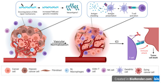
Colorectal cancer (CRC) is a leading public health concern due to its incidence and high mortality rates, highlighting the requirement of an early diagnosis. Evaluation of circulating extracellular vesicles (EVs) might constitute a noninvasive and reliable approach for CRC detection and for patient follow-up because EVs display the molecular features of the cells they originate. EVs are released by almost all cell types and are mainly categorized as exosomes originating from exocytosis of intraluminal vesicles from multivesicular bodies, ectosomes resulting from outward budding of the plasma membrane and apoptotic bodies’ ensuing cell shrinkage. These vesicles play a critical role in intercellular communications during physiological and pathological processes. They facilitate CRC progression and premetastatic niche formation, and they enable transfer of chemotherapy resistance to sensitive cells through the local or remote delivery of their lipid, nucleic acid and protein content. On another note, their stability in the bloodstream, their permeation in tissues and their sheltering of packaged material make engineered EVs suitable vectors for efficient delivery of tracers and therapeutic agents for tumor imaging or treatment. Here, we focus on the physiopathological role of EVs in CRCs, their value in the diagnosis and prognosis and ongoing investigations into therapeutic approaches.
Full article
(This article belongs to the Section Cancer Therapy)
►
Show Figures

Graphical abstract


