Article

The Production of Listeriolysin O and Subsequent Intracellular Infections by Listeria monocytogenes Are Regulated by Exogenous Short Chain Fatty Acid Mixtures

Department of Biology, Integrative Science and Engineering Center, University of Dayton, Dayton, OH 45469, USA
*
Author to whom correspondence should be addressed.
Toxins 2020, 12(4), 218; https://doi.org/10.3390/toxins12040218
Submission received: 27 February 2020 / Revised: 24 March 2020 / Accepted: 26 March 2020 / Published: 30 March 2020
(This article belongs to the Special Issue Toxins and Virulence Factors of Listeria monocytogenes)

Abstract

:
Listeria monocytogenes is a foodborne pathogen capable of secreting listeriolysin O (LLO), a pore-forming toxin encoded by the hly gene. While the functions of LLO have been studied extensively, how the production of LLO is modulated by the intestinal environment, devoid of oxygen and enriched in short chain fatty acids (SCFAs), is not completely understood. Using L. monocytogenes strain 10403s, we found that hly transcription was moderately decreased by aerobic SCFA exposures but significantly increased by anaerobic SCFA exposures. Moreover, aerobic, but not anaerobic, exposure to low levels of SCFAs resulted in a significantly higher LLO activity. These results demonstrated that transcriptional and post-transcriptional regulations of LLO production were separately modulated by SCFAs and were responsive to oxygen levels. Examining isogenic mutants revealed that PrfA and SigB play a role in regulating LLO production in response to SCFAs. Effects of SCFAs were also present in the cardiotropic strain 07PF0776 but distinctly different from those in strain 10403s. For both strains, prior exposures to SCFAs altered intracellular infections in Caco-2 and RAW264.7 cells and the plaque sizes in L fibroblasts, a result confirming the ability of L. monocytogenes to adapt to SCFAs in ways that impact its subsequent infection outcomes.
Key Contribution: Results from this study demonstrate the ability of L. monocytogenes to sense and respond to key environmental factors in the intestinal lumen, namely the lack of oxygen and high levels of short chain fatty acids. These responses, ranging from modifications in membrane fatty acid compositions and altered productions of virulence factors, significantly influenced the outcomes of subsequent cell culture infections. Therefore, considerations over SCFA levels in individuals as well as L. monocytogenes strain variations in SCFA responses may reveal novel preventative and control strategies for deadly L. monocytogenes infections.

1. Introduction

Listeria monocytogenes is a Gram-positive, facultative anaerobe and a foodborne pathogen. While minor gastrointestinal complications arise in infected immunocompetent individuals, symptoms can be more severe in pregnant women, the elderly, and immunocompromised populations. Approximately 1600 people annually are estimated to become infected with L. monocytogenes and hospitalized in the United States, among which as many as one in five patients do not survive, making L. monocytogenes infections contributing to 19% of the overall deaths related to foodborne pathogens [1]. As a result of the high mortality rate, L. monocytogenes is under stringent surveillance in the food industry and is responsible for $2.8 billion of costs associated with recalls and medical expenses annually [2].
Transmission and infection of L. monocytogenes rely on multiple virulence genes contributing to various stages of pathogenesis—surviving and colonizing the host gastrointestinal tract, binding to and entering host cells, growing inside the host cell cytosol, and spreading from one host cell to another. Therefore, to coordinate appropriate expression of virulence genes, L. monocytogenes needs to be well in-tune with its environmental cues. For example, transcription of the hly gene, which encodes listeriolysin O (LLO)—a cholesterol-dependent pore forming toxin that facilitates L. monocytogenes entry into the host cell cytosol [3], is responsive to a multitude of environmental factors, ranging from salt concentrations [4], growth temperature [5], to culture pH [5,6]. We have also shown that anaerobic conditions and propionate, two important environmental signals present in the intestinal lumen, can impact LLO production [7,8,9].
Propionate is one of the common intestinal short chain fatty acids (SCFAs), which are byproducts of bacterial fermentation of non-digestible carbohydrates and dietary fibers in the gut [10,11]. Fermentation of carbohydrates by the endogenous microbes in the gastrointestinal lumen can yield 500–600 mmol of SCFAs daily, most of which are absorbed and serve as an energy source for the host, accounting for 5–15% of human energy requirements [12,13]. SCFAs also regulate colonic pH, colonic epithelial cell volume, and a variety of cellular functions, including proliferation, differentiation, and gene expression [14,15,16,17,18]. Moreover, SCFAs are important modulators of leukocyte function, such as chemotaxis, oxidative burst, phagocytosis, and microbial killing [19,20,21,22]. Therefore, individual variations in SCFA levels may underline important clinical implications and pathogenesis [23]. However, very little is known whether the level of SCFAs plays a role in individual susceptibility to L. monocytogenes infections.
Butyrate, one of the major SCFAs found in the human intestines along with acetate and propionate, exhibits a strong inhibitory activity on LLO production by L. monocytogenes, an activity likely mediated by the loss of branched chain fatty acids (BCFAs) [8]. The membrane fatty acid composition of L. monocytogenes, when grown under standard laboratory conditions, is made up of approximately 90% anteiso-BCFAs, specifically anteiso-C15:0 and C17:0 [8,24,25]. The synthesis of BCFAs is catalyzed mainly by the branched chain alpha-keto acid dehydrogenase complex (BKD), a lipoic acid-dependent enzyme complex responsible in forming the branched chain amino acid-derived substrates for BCFA synthesis [26]. Transposon insertion mutants of BKD have dramatically decreased levels of BCFAs (<40% of WT) and exhibited severely compromised fitness phenotypes and LLO production [27,28], similarly to butyrate-supplemented wildtype bacteria. Together, these reports highlighted the role of BCFAs in L. monocytogenes fitness and pathogenesis and further suggest a potential effect of butyrate on L. monocytogenes during the intestinal phase of infection. However, all the earlier studies were performed under aerobic conditions, which are relevant conditions during L. monocytogenes intracellular infection but are not representative conditions during L. monocytogenes exposure to SCFAs in the anaerobic lumen of the intestines.
In a subsequent study investigating the effects of propionate on L. monocytogenes under aerobic as well as anaerobic conditions identified that the production of LLO was highly sensitive to regulation by exogenous propionate [9]. More specifically, while aerobic propionate exposure resulted in a dose-dependent decrease in LLO production, anaerobic propionate exposure resulted in a dose-dependent increase in LLO production [9]. Given that overall BCFA levels were also decreased in propionate-treated L. monocytogenes under aerobic as well as anaerobic conditions [9], it is clear that the general loss of BCFAs upon propionate or butyrate exposure is not the only mechanism mediating the regulation of LLO production. Moreover, these results highlight a potential opposing effect of individual SCFAs on LLO production and justify a need to consider the effects of SCFAs on L. monocytogenes as mixtures in addition to individual components to identify potential interactions.
In this study we investigated the effects of exposure to SCFA mixtures, at two different combinations (Mlo and Mhi) under aerobic or anaerobic conditions, on growth, membrane fatty acid composition, LLO production, and subsequent infections to gain a better insight into L. monocytogenes behavior in response to SCFAs during intestinal transit. The SCFA mixtures were used to obtain a better understanding of the effects of SCFAs as one integrated, physiologically relevant signal. We also examined the effects of SCFAs on transcription factor mutants with constitutively active PrfA or deletion in sigB to further investigate the regulatory mechanism underlying L. monocytogenes response to SCFAs. Finally, we compared the effects of SCFAs on LLO production as well as subsequent infections in two different strains, 10403s and 07PF0776, to determine the relevance of our observations. We found that, compared to no SCFA controls, supplementation of SCFAs resulted in distinctive changes in the transcriptional and post-transcriptional regulations of LLO production, altering subsequent infection outcomes in a manner sensitive to oxygen levels. These results highlight the ability of L. monocytogenes to detect and respond to SCFAs and suggest the SCFA levels in the intestine might play a role in determining the outcome of L. monocytogenes infections.

2. Results

2.1. High but not Low Levels of SCFAs Reduced In Vitro Growth

The growth of L. monocytogenes under these conditions was evaluated by culture optical density and culture pH measurements. Compared to non-supplemented controls, SCFA supplementations at Mlo or Mhi levels both resulted in a significantly higher culture pH but only supplementation at the Mhi level resulted in a significantly decreased culture optical density under aerobic or anaerobic conditions (Table 1). To ensure the effects of SCFA mixtures on growth was not a result of higher salt levels, we also tested the effects of salt on L. monocytogenes growth and found that supplementations of NaCl at levels as high as 200 mM did not significantly affect L. monocytogenes growth (data not shown). Doubling times were also calculated from growth curves, showing that supplementations of SCFAs at higher (Mhi) but not lower (Mlo) levels resulted in a significantly higher doubling time when compared to no supplementation controls (Table 1). Similar effects of SCFAs on doubling time were also observed in isogenic mutants (Table 1) with a constitutively active virulence regulator PrfA (PrfA*) or a deletion in the stress response sigma factor gene sigBsigB). These results confirmed that higher levels of SCFAs (Mhi) reduced but did not completely inhibit in vitro growth under aerobic or anaerobic conditions.
To determine whether the effects of SCFA supplementations on growth were related to pH variations, we measured overnight culture density and pH in phosphate buffered BHI (Table 2). In the absence of SCFAs, pH optimal for growth in buffered BHI was notably different between aerobic and anaerobic cultures. Whereas BHI buffered at pH = 6.0 yielded the highest culture optical density under aerobic conditions, BHI buffered at pH = 7.0 yielded the highest culture optical density. The addition of SCFAs in general resulted in decrease in growth but the extent of the effects varied among media buffered at different pH values. For example, in BHI buffered at pH = 6.0, addition of Mhi levels of SCFAs resulted in growth inhibition that was not observed in media without buffering or media buffered at PH = 7.0 or 8.0. Significant changes in culture pH by SCFA supplementations were also observed when compared to no SCFA controls under the same oxygen levels. In general, a decrease in growth corresponds to increase in culture pH, but growth decrease was also observed in anaerobic, Mhi SCFA-treated cultures where culture pH was not significantly changed. Therefore, the effects on growth by SCFAs are not mediated exclusively by pH modifications.

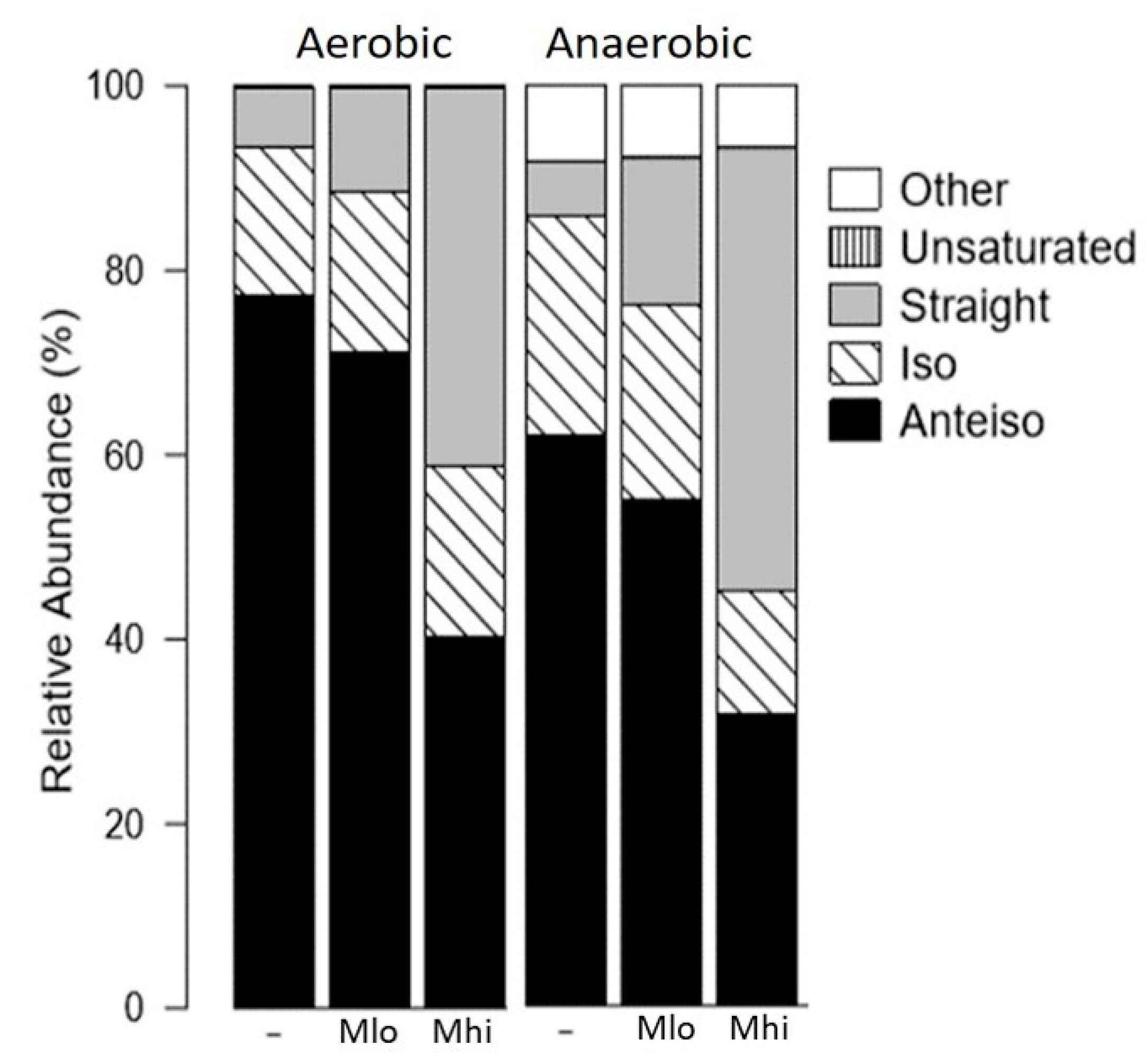
2.2. SCFA Supplementations Altered Membrane Fatty Acid Compositions

Both propionate and butyrate, separately, have been demonstrated to decrease the abundance of branched chain fatty acids (BCFAs) and increase the abundance of straight chain fatty acids in L. monocytogenes [8,9]. Here we investigated how the membrane fatty acid composition was affected by supplementation of SCFAs in mixtures and whether there was an oxygen-dependent alteration of fatty acid composition in response to SCFA exposure. L. monocytogenes from overnight cultures, grown aerobically or anaerobically with or without SCFA mixtures, were collected, frozen, and sent for FAME analysis by Microbial ID (Newark, DE). In general, BCFAs represented the most abundant fatty acids in untreated samples (Figure 1, Table 3 and Table 4) with the production of modified fatty acids only present in anaerobically grown L. monocytogenes (Table 4). Moreover, the proportions of straight chain fatty acids were increased by SCFA supplementations under aerobic or anaerobic conditions (Figure 1). More specifically under aerobic conditions (Table 3), the proportion of total BCFAs was reduced to 58.5% in L. monocytogenes treated with Mhi SCFAs, compared to 93.42% for bacteria without SCFA supplementations or 88.41% for bacteria with Mlo SCFA supplementations. Similarly, under anaerobic conditions (Table 4), the proportion of total BCFAs in cultures supplemented with SCFAs at the Mhi level samples was reduced to 45.2%, compared to 85.93% for no supplementation or 76.1% for Mlo-supplemented samples.
While the reduction of total BCFAs was observed in both aerobic and anaerobic conditions, the specific changes in different BCFAs appeared to respond to the presence or absence of oxygen. Under aerobic conditions, the loss of BCFAs was attributed entirely to the loss of anteiso-BCFAs, decreasing from 77.39% in control samples to 71.11% and 40.41% in cultures supplemented with SCFAs at the Mlo or Mhi levels, respectively (Table 3). The proportion of odd number iso-BCFAs exhibited a minimal, if any, decrease while the proportion of even number iso-BCFAs exhibited a small increase in both Mlo- and Mhi-supplemented samples (Table 3). Under anaerobic conditions, in contrast, the loss of total BCFAs was attributed to the loss of anteiso-BCFAs as well as odd number iso-BCFAs (Table 4). The proportion of even number iso-BCFAs exhibited a small increase in Mlo-supplemented samples but a decrease in Mhi-supplemented samples (Table 4).
Similarly, the accompanying general increase in the proportion of total straight chain fatty acids in response to SCFA supplementations was observed under both aerobic and anaerobic conditions but with distinct differences in the subtype composition. Supplementations with SCFAs at the Mhi level resulted in the most increase in total straight chain fatty acids, from 6.655% to 41.085% under aerobic conditions and 5.85% to 48.11% under anaerobic conditions (Table 3 and Table 4). However, the enrichment of straight chain fatty acids was more apparent in anaerobic samples than aerobic samples. Under aerobic conditions, supplementations of Mlo and Mhi SCFA mixtures resulted in a 1.73- and 6.17-fold increase in total straight chain fatty acids, respectively. Under anaerobic conditions, in contrast, supplementation with Mlo and Mhi SCFA mixtures resulted in a 2.75- and 8.22-fold increase in total straight chain fatty acids, respectively. These results indicate that L. monocytogenes contains the ability to utilize SCFAs and as a result modifies its membrane composition. Also, substrate utilization for fatty acid synthesis is likely modulated by oxygen.

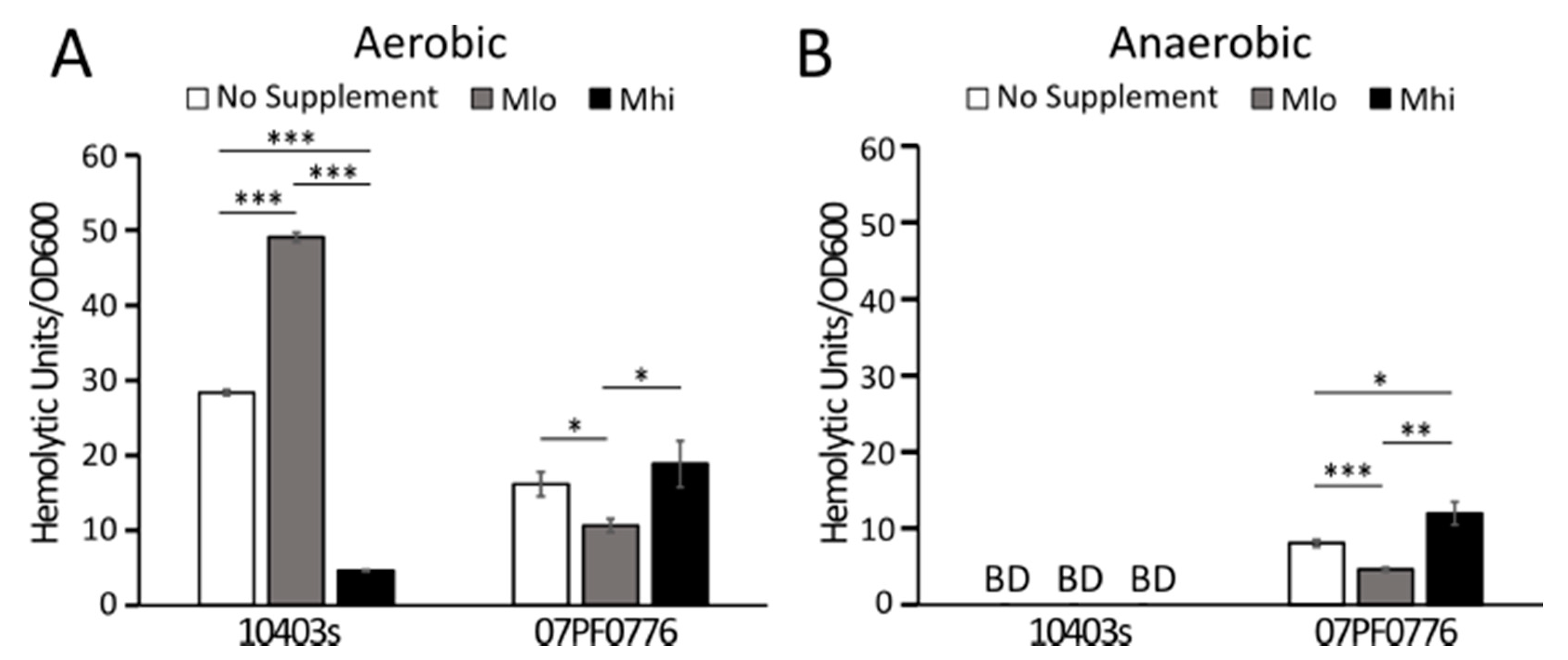
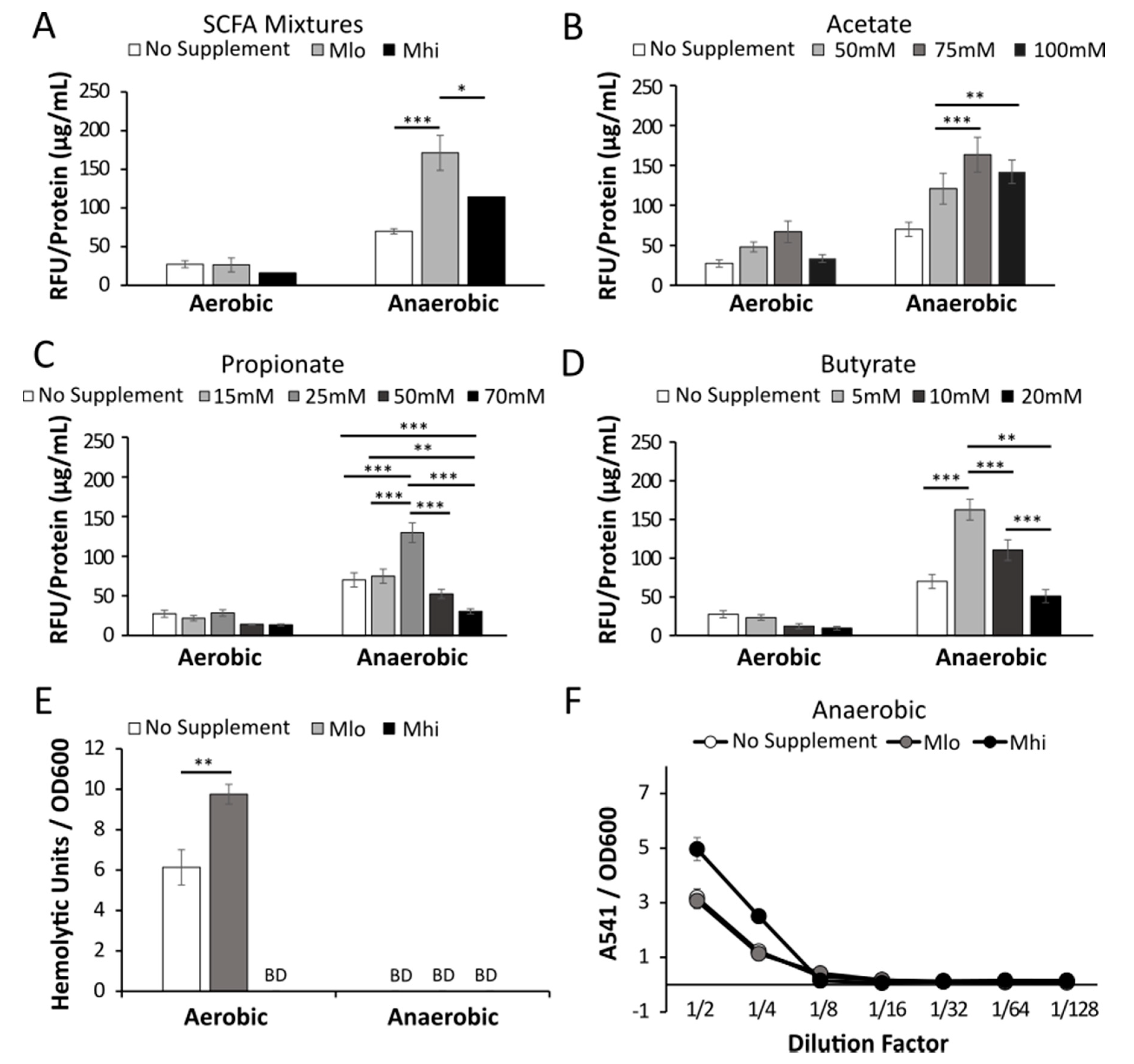
2.3. Anaerobic Transcription of Hly was Enhanced by SCFAs

The effects of SCFAs on L. monocytogenes listeriolysin O (LLO) production were tested at the transcriptional level using a hly transcriptional reporter strain. The reporter strain was cultured overnight in BHI supplemented with low (Mlo) or high (Mhi) SCFA mixtures [29] (Figure 2A) or individual concentrations of SCFAs in ranges encompassing those in the SCFA mixtures to isolate the contributing SCFA and determine potential additive effects of these SCFAs (Figure 2B,C). Under aerobic conditions, supplementations of SCFA mixtures resulted a notable but not significant decrease in hly transcription (Figure 2A). In contrast, under anaerobic conditions, SCFA supplementations at the low Mlo levels resulted in a significantly elevated hly transcription (Figure 2A). Effects of individual SCFA supplementation resulted in a similar pattern. Under aerobic conditions, after averaging 3 independent experiments each performed with triplicates, supplementations of acetate, propionate, or butyrate individually did not significantly alter hly transcription, although an increasing trend with acetate and a decreasing trend with propionate and butyrate were noted (Figure 2B–D). Under anaerobic conditions, supplementations of individual SCFAs resulted in a significantly elevated hly transcription (Figure 2B–D). However, the relationship between concentrations of individual SCFAs and hly transcription levels was not linear. The enhancing effects of individual SCFAs under anaerobic conditions peaked at 75 mM for acetate, 25 mM for propionate, and 5 mM for butyrate over the tested range.
To assess potential post-transcriptional effects of SCFAs on LLO production, we measured the activity of secreted LLO in culture supernatant as a proxy for the overall LLO production. Under aerobic condition, supplementations with Mlo SCFAs resulted in a significant increase in LLO activity while supplementations with Mhi SCFAs resulted in a compromised LLO activity (Figure 2E). Because LLO activity in anaerobically grown cultures was too low to calculate hemolytic activity, we also showed the results in a dilution curve and noted an elevated LLO activity in cultures supplemented with Mhi SCFAs (Figure 2F). These results together indicate that the activity of LLO by L. monocytogenes can be enhanced after exposure to SCFAs with the effects highly dependent on the presence of absence of oxygen.
To explore potential mechanisms that result in altered LLO production in response to SCFAs, we examined the effects of SCFAs on LLO production in isogenic mutants with constitutive active PrfA (PrfA*) or deficient SigB (ΔsigB). Both PrfA and SigB are known transcriptional regulators that can influence LLO production [30] but their roles in L. monocytogenes sensing SCFAs have not been investigated prior to this study. Under aerobic conditions, supplementation of SCFAs resulted in a significant, dose-dependent decrease of LLO activity in both the PrfA* and the ΔsigB mutants (Figure 3). This was in direct contrast to the enhancing effect of aerobic Mlo SCFA exposure on the wildtype bacteria (Figure 2E). Under anaerobic conditions, supplementations of Mlo, not Mhi, resulted in a significant increase in supernatant LLO activity. These results indicate that while the regulation of LLO production in these mutants remain sensitive to perturbations by SCFAs under aerobic or anaerobic conditions, the sensitivity to different levels of SCFAs appears to be altered in the presence of constitutively active PrfA or in the absence of SigB.

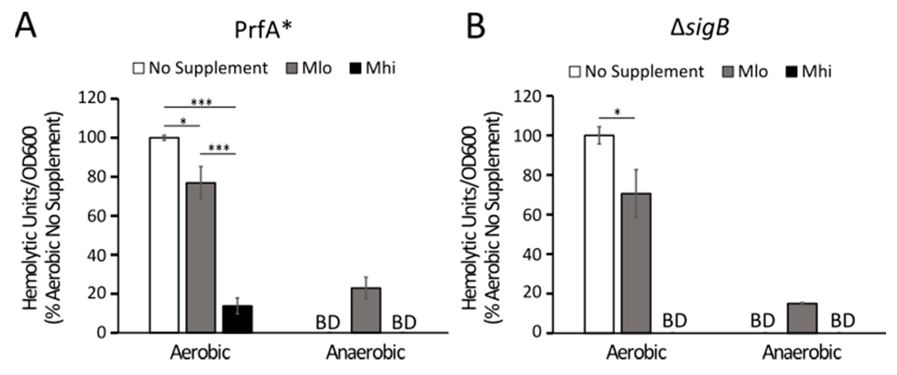
2.4. Production of LLO in Strain 07PF0776 Was Sensitive to Regulation by SCFAs

To establish the relevance of our observations in strain 10403s, we also investigated the effects of SCFA exposure on LLO production in the cardiotropic strain 07PF0776 (serotype 4b) [31] to identify any strain-dependent responses to SCFAs. Strain 07PF0776 in general exhibited lower supernatant LLO activities under aerobic conditions (Figure 4A) but higher LLO activities under anaerobic conditions (Figure 4B) than strain 10403s. There were also distinctive differences in responses to SCFA mixtures. Under aerobic conditions (Figure 4A), while supplementations of Mlo SCFAs resulted in a significantly increased LLO activity in strain 10403s, they resulted in a significantly decreased LLO activity in strain 07PF0776. Under anaerobic conditions, supplementations of Mlo SCFAs resulted in a significant increase in LLO activity in strain 07PF0776 (Figure 4B), similarly to strain 10403s (Figure 2F). However, anaerobic supplementations of Mlo SCFAs resulted in a significant decrease in LLO activity in strain 07PF0776 (Figure 4B), compared to the lack of effect of Mlo SCFAs in strain 10403s (Figure 2E). These results indicate that while the effects of SCFAs on strain 10403s are more sensitive to perturbations by the presence or absence of oxygen than those on strain 07PF0776, both strains are capable of sensing and responding to SCFAs by altering LLO production.

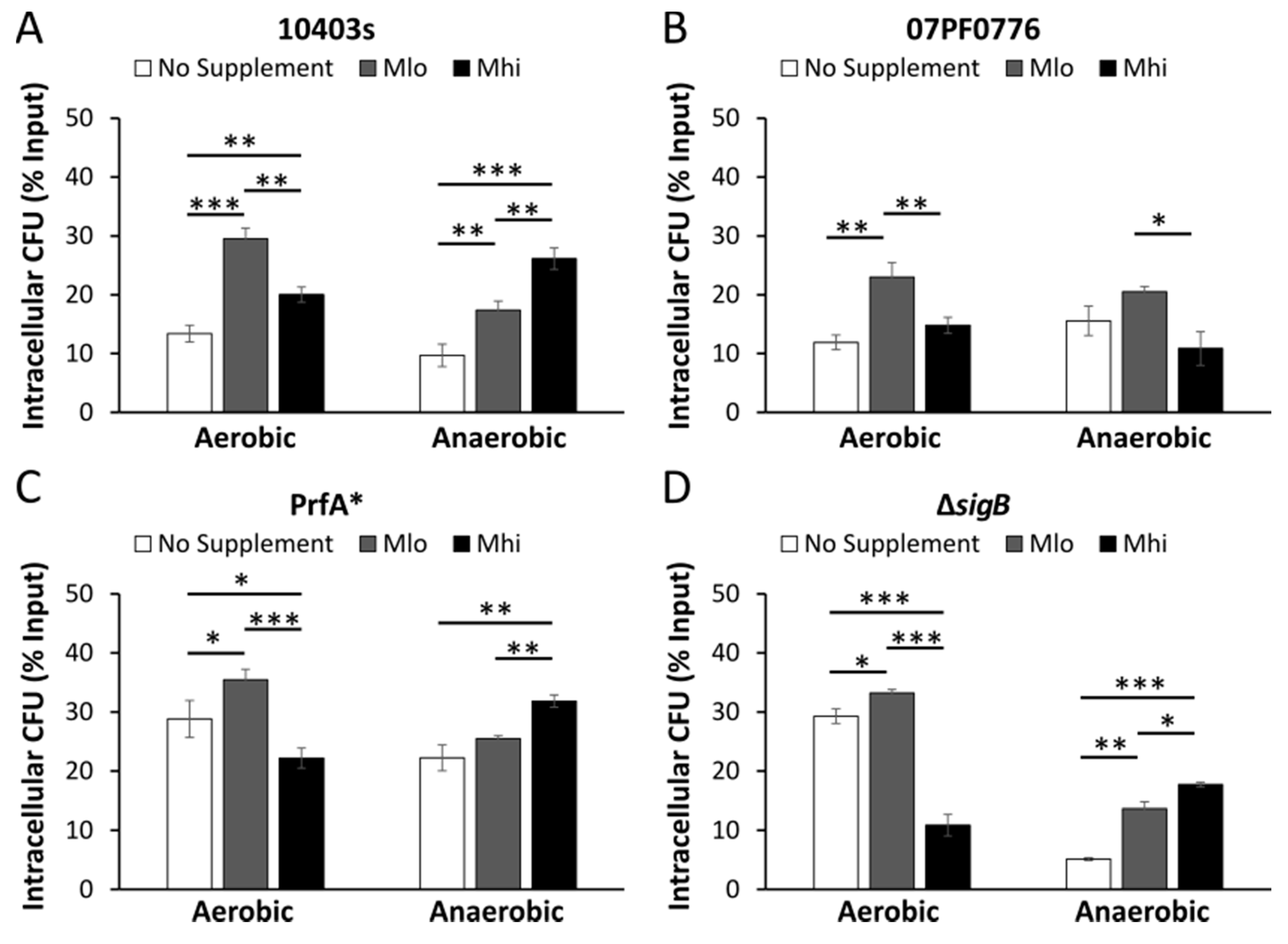
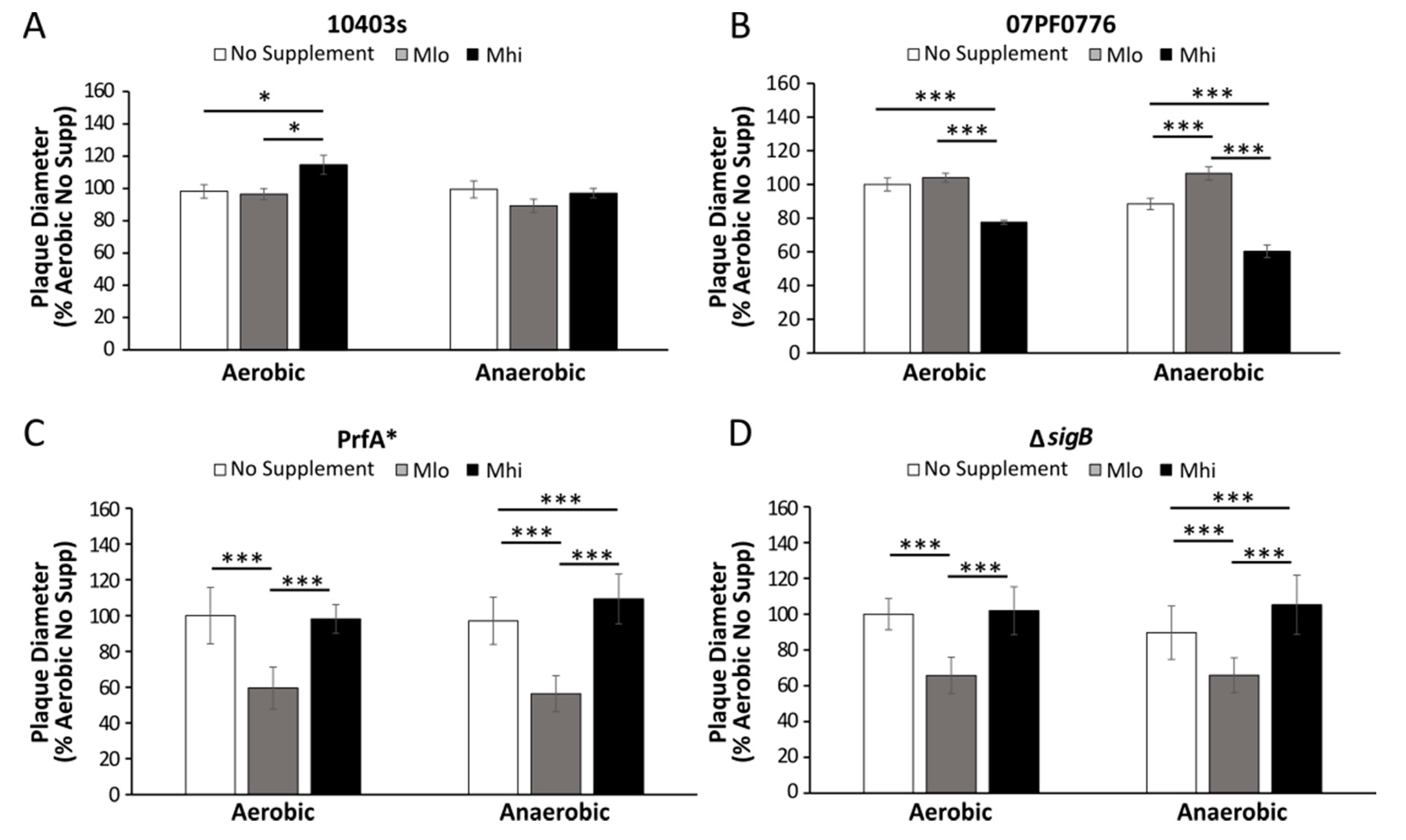
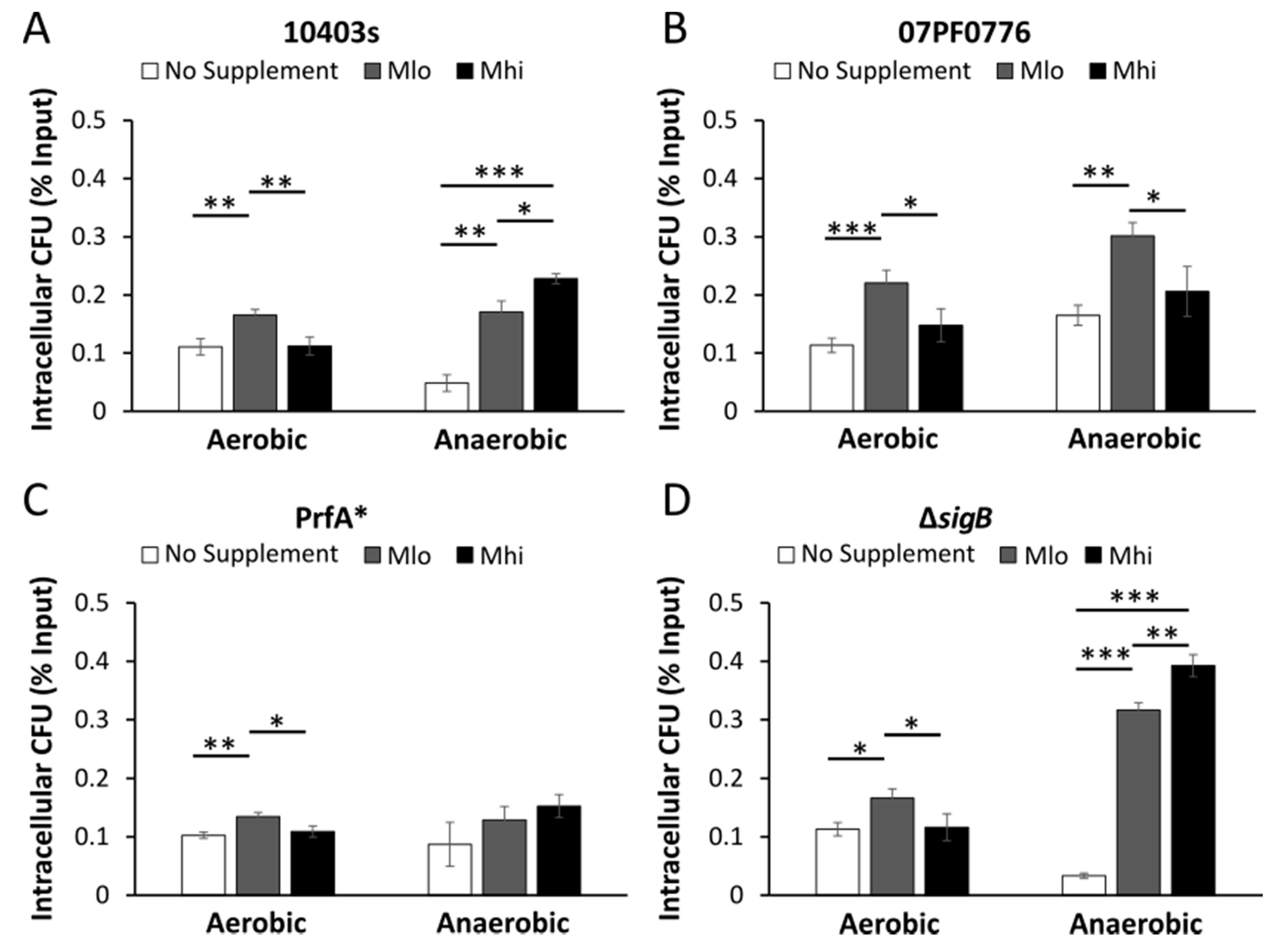
2.5. Prior SCFA Exposure Altered Subsequent Infection Outcomes in Tissue Culture Infections

The regulatory effects of SCFAs on LLO production suggest a potential impact on subsequent intracellular infections after SCFA exposure. Therefore, we infected Caco-2 intestinal epithelial cells and RAW264.7 macrophages with L. monocytogenes grown with or without supplementations of Mlo or Mhi SCFA mixtures under aerobic or anaerobic conditions and assessed subsequent intracellular infections in a standard gentamicin protection assay. It is important to point out that in these experiments, the host cells were never exposed to exogenous SCFAs prior to infection. SCFAs were never added to infected cells throughout infections. Any effects from SCFA exposure were the result of L. monocytogenes adaptations to SCFAs and/or the host responses to differentially adapted L. monocytogenes.
In non-polarized Caco-2 cells infected by aerobically grown bacteria, for all four strains tested, prior bacterial exposure to Mlo SCFA mixtures resulted in a significant increase in the percentages of intracellular bacteria (Figure 5), an observation indicative of an enhancement of invasion by aerobic exposure to low levels of SCFA mixtures regardless of genotype. In contrast, in non-polarized Caco-2 cells infected by anaerobically grown bacteria, supplementation of SCFAs in general showed a dose-dependent increase in the percentages of intracellular WT and isogenic mutants of strain 10403s but not strain 07PF0776 (Figure 5). Anaerobic exposure to high levels of SCFA mixtures significantly enhanced invasion by strain 10403s but not strain 07PF0776. Similar phenotypes were also observed in the infected RAW264.7 macrophages (Figure 6) where the effects of SCFA exposure on subsequent infections were similar among WT and isogenic mutants of strain 10403s (Figure 6A,C,D). Also, the effects of anaerobic, but not aerobic, exposure to high levels of SCFA mixtures on subsequent infections were distinctively different between strains 10403s and 07PF0776 (Figure 6A,B).
To further investigate the consequences of SCFA exposure on L. monocytogenes pathogenesis, an L-cell plaque assay was performed with strains 10403s and 07PF0776 to evaluate the effects of SCFA exposure and adaptation on subsequent infection outcomes. Overnight cultures of L. monocytogenes grown with or without SCFAs under aerobic or anaerobic conditions were washed to remove any residual SCFAs and used to infect monolayers of L fibroblast cells. It is important to point out that SCFAs were not included throughout the infection from seeding the cells to measuring plaque sizes at 2 days post infections. Therefore, plaque sizes represent the effects of prior SCFA exposure on the ability of L. monocytogenes to establish an intracellular life cycle in subsequent infections. For strain 10403s, aerobic pre-exposures to Mhi SCFA mixture, but not other conditions tested, resulted in a significant increase in plaque diameters (Figure 7A). However, this was not seen in plaques created by the isogenic mutants (Figure 7C,D), indicating that normal PrfA and SigB activities may play a role in cell-to-cell spread in adaptations to Mlo SCFAs and oxygen exposure that are relevant for long-term infection success. In contrast, aerobic pre-exposures to Mhi SCFAs by strain 07PF0776 resulted in a significant decrease in plaque diameters (Figure 7B). In cells infected by anaerobically grown strain 07PF0776, pre-exposures to Mlo SCFA mixture prior to infections resulted in a significant increase in plaque sizes compared to no SCFA controls (Figure 7B). However, pre-exposures to Mhi SCFA mixture prior to infections resulted in a significant decrease in plaque sizes compared to no SCFA controls (Figure 7B). These results confirm the presence of strain variations in L. monocytogenes response to SCFAs and suggest the possibility of SCFAs in the intestinal lumen influencing L. monocytogenes infection outcome in a strain-dependent manner.

3. Discussion

In this study we reported that membrane fatty acid composition and production of LLO were sensitive to regulation by SCFA mixtures in a manner that was modulated by oxygen availability in L. monocytogenes strain 10403s. The response to SCFAs was not exclusive to strain 10403s because the effects of SCFAs on LLO production were also observed in strain 07PF0776 with distinct differences. Most notably, pre-exposures to SCFAs prior to infections resulted in a sustained effect on plaque sizes two days post infection despite the lack of SCFAs throughout the infection. By using SCFA mixtures as an integrated signal, these results together suggest for the first time that the intestinal environment, such as levels of SCFAs and oxygen availability, may play a key role in determining strain variations in virulence or the individual susceptibility to L. monocytogenes infections.

3.1. SCFA Metabolism and Fatty Acid Synthesis

SCFAs tested in this study are structurally related compounds differing only by the length of the carbon chain. However, L. monocytogenes is capable of distinguishing between these three SCFAs—with the highest sensitivity to butyrate, followed by propionate and acetate, and responding to these SCFAs by altering hly transcription and LLO production. To begin to dissect the likely complex regulatory mechanism, we explored SCFA metabolism as a potential component in mediating the effects of SCFAs on the regulation of LLO production. The pathways for SCFA metabolism all converge at fatty acid synthesis. While acetate may be directly incorporated into the central metabolism through acetyl-CoA, butyrate and propionate are likely being converted to butyryl- and propionyl-CoA, respectively, first through phosphorylation by kinases (LMRG_00611 and LMRG_00820), followed by the activity of phosphotransbutyrylase (LMRG_00819) that catalyzes the transfer of C3-C5 acyl chain to CoA molecules [32], forming precursors for the synthesis of straight chain fatty acids. As a result, exposure to SCFAs resulted in a dose-dependent increase in straight chain fatty acids (Table 3 and Table 4).
Membrane fatty acid composition is highly sensitive to fluctuations in environmental conditions such as temperature [33] and can influence important biological functions. L. monocytogenes membrane fatty acids are dominated by BCFAs, including anteiso-, odd iso-, and even-iso BCFAs. Growth at temperatures below 10 °C results in an enrichment of anteiso-C15:0 branched chain fatty acids (BCFAs) [24,28,34,35,36,37,38], contributing to homeoviscous adaptation to low temperatures [34,39]. Response and adaptation to pH [25] or adherent growth [40] have also been reported to involve modifications in the fatty acid composition. We showed here that oxygen levels similarly served as an environmental determinant in altering fatty acid composition. While the decreased level of total branched chain fatty acids after SCFA exposure was observed under aerobic and anaerobic conditions, anaerobic modification of membrane fatty acid by SCFAs encompasses a decrease in anteiso-BCFA but an increase in iso-BCFAs, resulting in an overall decrease in the ratio of anteiso- to iso-BCFA of 2.6, a much lower value than the ratio of 4.8 observed under aerobic conditions. With anteiso-BCFAs conferring a higher degree of membrane fluidity than iso-BCFAs [26], it is likely that anaerobically grown L. monocytogenes exhibits a reduced membrane fluidity in comparison to aerobically grown bacteria upon exposure to SCFAs despite the similar growth temperature (37 °C). The extent by which SCFA exposure alters membrane fluidity under aerobic versus anaerobic conditions remains to be experimentally determined. Nevertheless, a reduction in membrane fluidity can render L. monocytogenes more susceptible to antimicrobial peptides [8] and suggests that reducing oxygen levels, such as modified atmosphere packaging [41], might be key in compromising L. monocytogenes fitness and environmental transmission. Moreover, although SCFA supplementations resulted in a dramatic change in L. monocytogenes fatty acid composition, supporting the utilization of SCFAs as substrates for fatty acid synthesis, whether the metabolism of these SCFAs is required for regulating LLO production remains to be determined.

3.2. Transcriptional and Post-Transcriptional Regulations of LLO Production by SCFAs

The transcriptional regulation of LLO production has been extensively investigated, establishing PrfA and SigB as two major transcription factors that can influence LLO production. PrfA is the main transcription activator for many virulence genes in L. monocytogenes [42,43,44,45] while SigB is a stress response sigma factor [46] that can also influence prfA expression [47,48,49,50]. Because the extremely low level of LLO production in a PrfA-deficient mutant, we utilized a mutant with constitutively activated PrfA [51] to investigate the role of PrfA in regulating LLO production in response to SCFAs. Previous work from our lab has demonstrated that in both PrfA* and ΔsigB mutants, LLO activities in anaerobic culture supernatants remained significantly lower than those in aerobic culture supernatants [7], a result supporting that the compromised LLO protein production takes place independently of the normal PrfA or SigB activities. In this study, both PrfA* and ΔsigB mutants, regardless of oxygen levels, exhibited an altered response to low, but not high, levels of SCFA mixtures (Figure 4). These observations are indicative of PrfA and SigB contributing, at least partially, to the regulation of LLO production in response to SCFAs. Moreover, they strongly suggest that the effects of SCFAs on LLO production are not linear and are likely mediated by different mechanisms at low versus high levels.
Our results also reveal that these environmentally important signal—oxygen availability and SCFA levels, exert a separate and often opposing effect on the transcriptional and post-transcriptional regulations of LLO production in strain 10403s. Under anaerobic conditions where LLO activity was low (Figure 2E,F), the hly transcription level remained high (Figure 2A–D). The lack of correlation between transcription and protein activity suggests that while anaerobicity in general results in higher hly transcription, LLO protein production is dependent on the presence of oxygen. In other words, the anaerobic signal exerts an opposing effect on the transcriptional and post-transcriptional regulation of LLO production. In the presence of low levels of SCFAs under aerobic conditions, the lack of transcriptional response (Figure 2A) surprisingly led to a significantly higher supernatant activity (Figure 2E). In contrast, low levels of SCFAs under anaerobic conditions induced a strong transcriptional upregulation without the corresponding increase in supernatant activities. These results confirm that LLO protein production is likely an oxygen dependent process or there exists a strong inhibitory signal on LLO protein production under anaerobic conditions. The specific underlying mechanisms remain to be determined but may reveal novel insight into the virulence regulation in response to physiologically relevant signals.

3.3. Strain-Dependent Variations in SCFA Responses

Based on phylogenetic analyses, L. monocytogenes has multiple evolutionary lineages, with most isolates that cause human disease belonging to lineages I and II [52]. Alternatively, L. monocytogenes is also classified by serotyping somatic and flagellar antigens, in which serotypes 1/2a, 1/2b, 1/2c, and 4b are responsible for majority of the human listeriosis cases [53,54]. Variations in pathogenicity among isolates from different lineages and serotypes have been investigated and established through experimental infections as well as in vitro and in silico comparisons. For example, through in vitro hemolytic assays, it is clear that there exists a strain-dependent variation in the level of LLO production [55,56,57,58,59]. Additionally, as demonstrated through animal models of infections, there are strain-dependent variations in establishing infections in different organs [60,61]. These comparative studies are important for basic phylogenetic and phenotypic comparisons as well as epidemiological analyses that can help predict and prevent deadly outbreaks.
To determine whether the ability to sense and respond to SCFAs is a relevant phenotype for common virulent strains of L. monocytogenes, we compared the effects of SCFAs between the lab strain 10403s, serotype 1/2a, and strain 07PF0776, a cardiotropic strain of L. monocytogenes belonging to serotype 4b [31,59]. Direct comparisons between these two strains have demonstrated that strain 07PF0776 established a significantly higher bacterial burden in the hearts of female ND4 Swiss Webster mice than strain 10403s after intravenous infection [59]. Using a rat cardiac myoblast cell line (H9c2) as a host model, although the intracellular growth curves were similar for the two stains, strain 07PF0776 exhibited a higher invasion rate than strain 10403s at 2 hours post infection [59]. Extending the comparisons to 9 additional strains revealed that the low versus high cardiac infections in mice correlated strongly with the internalin A (InlA) amino acid sequences [59]. In our study, while both strains are capable of altering LLO production in response to SCFAs, the direction of the effects were vastly different (Figure 4). The short-term effects of anaerobic SCFA exposure (Figure 5 and Figure 6) as well as long-term effects of SCFA exposure (Figure 7) on subsequent infections were also distinctively different between the strains. Because no SCFAs were included during infections, these strain-dependent variations are indicative of different adaptations to SCFAs prior to infections that ultimately affect subsequent intracellular success. While testing additional strains is needed to obtain a more complete lineage- or serotype-delineation of SCFA responses, it is possible that host physiology in the intestinal lumen may exert a selective force in L. monocytogenes-host co-evolution.

3.4. Impact of SCFA Exposure on Subsequent Cell Culture Infections

Many enteric pathogens can sense and respond to SCFAs [62] although the mechanisms underlying SCFA responses vary from organism to organism. How these diverse SCFA responses manifest during aerobic versus anaerobic conditions is mostly unknown. While earlier work showed an enhancement of L. monocytogenes infections after exposure to anaerobic conditions [7,63,64], how SCFA exposure might influence the anaerobic enhancement of infections were unclear. Here, we used Caco-2 intestinal epithelial cell lines and RAW264.7 macrophages as infection models to study short term intracellular survival and invasion, respectively, and used L-cells to examine long term cell-cell spread—together to obtain a better understanding of the impact of SCFA exposure on the different stages of L. monocytogenes intracellular infections. It is important to note that because no SCFAs were included during infections so any SCFA treatment effects reflected bacterial adaptations and responses to SCFAs beyond the exposure period.
Curiously, while anaerobic enhancement of invasion was a well-established phenomenon in L. monocytogenes infections [7,65,66], we did not observe similar behavior in our study (Figure 5). We believe this difference was caused by the utilization of 96-well tissue culture plates in this study compared to the 24-well tissue culture plates used in previous studies. It is likely that there were differences in oxygen diffusion rates naturally existing in these cell culture plates. In a 96-well plate, with a typical infection volume of 100 µL per well and a well diameter of 6.4 mm, there is an approximate 5 mm of liquid in height. In contrast, in a 24-well plate, with a typical infection volume of 500 µL and a well diameter of 15.6 mm, the liquid volume is approximately at 3 mm in height. Therefore, even though the multiplicity of infection was consistent, the infected cells were potentially exposed to different oxygen levels. If this variation in oxygen availability was the cause of the different infection outcomes, our results highlight yet another point during L. monocytogenes-host interactions where oxygen levels play an important role in influencing infection outcomes.
Strain 10403s and the isogenic mutants all showed similar trends in invasion and intracellular survival after aerobic or anaerobic exposure to SCFAs. Thus, regular PrfA and SigB activities likely do not play a role in facilitating responses to SCFA pre-treatments that are important for epithelial cell invasion or intracellular survival in macrophages. However, the isogenic mutants behaved very differently from the wildtype strain 10403s in the fibroblast infections (Figure 7). Prior exposure to Mlo concentration of SCFAs in the mutants resulted in significantly smaller plaque sizes compared to no SCFA pre-treatments. This indicates that PrfA and SigB play a role in L. monocytogenes adaptations to Mlo SCFAs that are relevant to success during prolonged infection. Additional studies are required to dissect the specific contributions each regulator might play in L. monocytogenes adaptations to SCFAs.
As the effects of SCFA treatments on infection outcomes did not align with in vitro effects on LLO production, it is likely that additional adaptations by L. monocytogenes play a strong role in survival and growth in host cells. LLO is vital for L. monocytogenes to escape the entry vesicles and grow within the cytosol. Thus, the level of LLO activity should be one determining factor in the success of intracellular L. monocytogenes at the early stage of the infection. At later stages of infections, intracellular L. monocytogenes was found to induce macrophage intracellular production of branched chain amino acids [65], which serve as the precursors for the synthesis of branched chain fatty acids in L. monocytogenes that could potentially counteract the effects of prior SCFA treatments. Whether SCFA-treated L. monocytogenes is equally efficient in stimulating macrophage branched chain amino acid production is unclear. Future studies on the signaling events by which macrophages respond to SCFA-adapted L. monocytogenes will provide more insight into the role of host response in the effects of SCFAs on host-pathogen interactions.

3.5. Potential Implications for Transmission

The observed impact of SCFAs and oxygen levels on L. monocytogenes membrane fatty acid composition in this study, together with reported evidence of modifications in membrane fatty acid composition leading to compromised pathogenesis [8,27], support the importance of prior SCFA adaptations in the success of subsequent infections. Considering the spatial oxygen gradient L. monocytogenes likely experiences during transmission from the food matrices to the intestinal lumen and the intestinal epithelium, L. monocytogenes may undergo major membrane fatty acid modifications that can impact the initial host-pathogen interactions, leading to altered infection outcomes. Similarly, the presence of SCFAs, both as food preservatives [66,67,68] and as products of endogenous gut microbiota, may also alter L. monocytogenes membrane fatty acid composition to further influence subsequent infections.
The active role of SCFAs in modulating L. monocytogenes membrane fatty acid composition highlights a potential explanation for individual variations in susceptibility to L. monocytogenes infections. It is commonly accepted that individuals such as the elderly and neonates are more susceptible to L. monocytogenes infections because of the weaker immune defenses. Curiously, the SCFA production likely declines during aging [69,70,71], a phenomenon suggesting that if a significant correlation between SCFA concentrations and susceptibility to L. monocytogenes infections existed, it might be possible to strengthen the defense in these high risk individuals against L. monocytogenes infections through non-invasive strategies such as an increase in dietary intake of fiber or probiotic organisms [72,73]. Moreover, the sustained, long-term effects of SCFAs on subsequent infections in the absence of SCFAs (Figure 5, Figure 6, Figure 7) suggest that the use of SCFAs in food storage may also be exploited to minimize the infection potential of L. monocytogenes upon accidental consumption. The ability of L. monocytogenes to sense and response to SCFAs provides a potential target to develop novel preventative or treatment strategies against L. monocytogenes at multiple stages of transmission.
In addition to membrane fatty acid modifications, the impact of SCFAs on LLO transcription and activity reported here also suggests potential effects on L. monocytogenes interactions with the intestinal epithelium by regulating LLO production. Several studies have shown that exogenously provided LLO is capable of activating mucin production and subsequent inhibition of L. monocytogenes internalization [74,75,76,77]. If exposures to SCFAs, likely in the anaerobic lumen of the intestines, could increase LLO production at the transcriptional level without immediate upregulation of the LLO activity (Figure 2), they may potentiate L. monocytogenes for subsequent intracellular infections, likely under conditions with higher levels of oxygen, without stimulating production of mucin that can negatively impact invasion. As L. monocytogenes moves toward the aerobic epithelium with reduced levels of SCFAs, the production and secretion of LLO resumes to promote infections. Extracellular LLO has been demonstrated to increase Ca2+ efflux in hepatocytes and promote subsequent internationalization of L. monocytogenes [78,79,80,81]. Therefore, the elevated level of LLO production upon aerobic exposure and the loss of inhibitory signals from SCFAs near the intestinal epithelium may promote L. monocytogenes invasion of the epithelial barrier. Future investigations into the activity of secreted LLO under physiologically relevant conditions in polarized epithelial cells may reveal novel insights on the intestinal phase of L. monocytogenes infections to better prevent L. monocytogenes from breaching the intestinal barrier and establishing lethal infections in peripheral organs.

4. Materials and Methods

4.1. Strains

The L. monocytogenes strains used in this study include the wild-type strain 10403s and the isogenic mutants either containing a constitutively active PrfA transcriptional regulator [51] or a deletion in the gene encoding the stress response sigma factor SigB (ΔsigB) [82]. To quantify hly transcription, an hly transcriptional reporter strain (Phly-gus-neo, NF-L695, provided by Dr. Nancy Freitag at The University of Illinois College of Medicine) with the hly promoter driving the expression of β-glucuronidase (GUS) [83] was used. A cardiotropic strain 07PF0776 (provided by Dr. Nancy Freitag at The University of Illinois College of Medicine) [31] was also used in this study. Bacteria were streaked weekly on brain heart infusion (BHI) agar to provide fresh colonies for experiments. The hly reporter strain was grown on BHI agar containing neomycin.

4.2. Growth Conditions

L. monocytogenes cultures were grown overnight in filter-sterilized brain heart infusion media (BHI; Fisher Scientific #211059, Waltham, MA, USA) at 37 °C either aerobically with shaking at 250 rpm or anaerobically without shaking in an anaerobic chamber (Coy Laboratory, Type A, Grass Lake, MI, USA). The chamber contains a nitrogenous atmosphere and ~2% hydrogen. Sterilization of media was accomplished through filter sterilization to ensure consistency between batches. Buffered BHI was made with 100 mM sodium monobasic and dibasic phosphate.

4.3. Culture Supplementation

Separate stock solutions (1 M) of acetate (sodium acetate trihydrate; Fisher Scientific #121-09-3), propionate (propionic acid, sodium salt; Acros Organics #137-40-6), and butyrate (butyric acid, sodium salt; Acros Organics #156-54-7) were filter-sterilized and frozen in aliquots at −20 °C until needed. The salt forms of SCFAs were used to avoid acidification of culture pH by SCFA supplementations. Mixtures of SCFAs were prepared according to an earlier study [29] and included a low level of SCFA mixture (Mlo: 25.5 mM acetate, 2.25 mM propionate, 2.25 mM butyrate) and a high level of SCFA mixture (Mhi: 110 mM acetate, 70 mM propionate, 20 mM butyrate). These concentrations were chosen to ensure two different mixture levels were tested that fall within known ranges of SCFA levels [84,85].

4.4. GUS Reporter Assay

Cultures of the hly reporter strain were grown in BHI containing desired SCFA concentrations overnight at 37 °C aerobically or anaerobically. Bacteria from overnight cultures (1 mL) were harvested by centrifugation, washed in sterile PBS, and resuspended in sterile PBS (110 μL). Each sample was then sonicated for 30 s (kept on ice when not being sonicated) and centrifuged to remove cell debris and intact cells. A portion (10 μL) of the supernatant lysate was used to determine protein concentration by a BCA assay kit (Thermo Scientific #23227, Waltham, MA, USA) following the manufacturer’s protocol. The remaining lysate (100 μL) was placed into a 96-well flat bottom plate where 20 μL of a 4-methylumbelliferyl-β-D-glucuronide (Fisher Scientific #AAB21190MD) stock solution (0.35 mg/ml) was added in the dark. The plates were covered with foil and incubated at 37 °C for 10 min. Stop solution (10 μL of sodium carbonate at 0.2 M) was then added to the well in the dark and fluorescence (excitation 365 nm, emission 460 nm) was determined using a microplate reader (Biotek Synergy 4).

4.5. Hemolytic Assay

Bacterial cultures were grown in 1 mL of BHI containing the desired SCFA concentration overnight at 37 °C aerobically or anaerobically. After measuring culture optical density at 600 nm, bacteria from overnight cultures (500 μL) were pelleted by centrifugation. Supernatant (100 μL) containing the secreted LLO was incubated at room temperature with 5 μL of 0.1 M dithiothreitol (DTT) for 15 min in a round bottom 96 well plate. The DTT-treated samples were serially diluted (1:2) with 100 μL of hemolytic assay buffer, containing 125 mM sodium chloride (Ameresco #0241-5KG, Framingham, MA, USA) and 35 mM sodium phosphate dibasic (Fisher 7558-79-4) at pH 5.5. Defibrinated sheep blood was prepared at 2% hematocrit in hemolytic assay buffer and was added to each well (100 μL per well), followed by incubation at 37 °C for 30 min. Plates were centrifuged at 2000 rpm for 5 min to pellet intact red blood cells and supernatant (120 μL) was transferred to a flat-bottom 96-well plate for absorbance reading at 541 nm using a microplate reader. Hemolytic unit was calculated by using the inverse dilution factor at half lysis normalized by OD.

4.6. FAME Analysis

Bacterial samples for fatty acid methyl esterification (FAME) analysis were harvested from overnight cultures (20 mL) grown at 37 °C by centrifugation at 10,000 rpm for 10 min. The pellet was then resuspended in 1 mL of sterile water and transferred to a microcentrifuge tube and spun again at 10,000 rpm for 3 min. The supernatant was removed, and the pellet was frozen in an ethanol-dry ice bath and sent to Microbial ID (Newark, DE, USA). The fatty acid composition was then determined using methyl esterification and gas chromatography.

4.7. Tissue Culture Infection

RAW264.7 macrophages and Caco-2 cells were cultured in Dulbecco’s Modified Eagle Medium (DMEM; Corning 10-013-CV, Corning, NY, USA) containing 10% FBS (Corning 35-010-CV) and penicillin/streptomycin (BioWhittaker 17-603E, Rockland, ME, USA) at 37 °C with 5% CO2. Cells were seeded into 96-well plates at 6 × 10⁶ cells per plate and incubated overnight. For infections, overnight L. monocytogenes cultures were centrifuged at 10,000 rpm for 3 min, washed once, and resuspended with sterile PBS. Culture OD was used to calculate the volume of bacterial culture suspension needed to achieve a multiplicity of infection of 10. After 30 min of infection in RAW264.7 and 1 h in Caco-2 cells, each well was washed twice with 1× DPBS (WorldWide Life Sciences, 61211088). DMEM with gentamicin (10 µg/mL) was added to each well to remove any extracellular L. monocytogenes. At 2 h post infection (hpi), adherent macrophages were lysed using filter-sterilized 0.1% Triton-X and the lysate was plated on LB plates. All LB plates were incubated at 37 °C for two days and colonies were counted using an aCOLyte 3 plate reader (Synbiosis). Percent input was calculated by dividing the number of intracellular colony forming units (CFU) at 2 hpi by the number of CFU in the inoculum and multiplying by 100%.

4.8. Fibroblast Plaque Assay

Murine fibroblast L-cells (ATCC CRL-2648) were grown in Dulbecco’s Modified Eagle Medium (DMEM) containing 10% Fetal Bovine Serum (FBS) in a flask. For experimentation, cells were seeded into six-well plates and given 24 h to form a monolayer. Overnight L. monocytogenes cultures were normalized by culture OD, washed, and resuspended in 100 µL of DPBS. Fibroblast monolayers were washed three times and infected with 1 mL of fresh DMEM containing 6 µL of L. monocytogenes suspension at no dilution, 1:10, and 1:100 dilution. At 1 h post infection, cells were covered with 3 mL of an overlay of DMEM containing 10 µg/mL gentamicin and 0.7% agarose. Plates were incubated at 37 °C with 5% CO2 incubator for 48 h. Following incubation, wells were stained with 1 mL of filter sterilized neutral red (0.33% (w/v) in DMEM) for one hour followed by three washes with DPBS. Plaques developed overnight in a 5% CO2 incubator and the plaque diameters were measured using the Gimp software (version 2.10.12).

4.9. Statistics

Statistical analyses include Student’s t-tests, which were performed using Microsoft Excel for pair-wise sample comparisons. Standard error of the mean (SEM) was used when the average of three independent experiments, each with three replicates, was calculated. Using SEM in these instances accounts for natural variation between experiments. Standard deviation (SD) was used when averages of three replicates from one independent experiment were used.

Author Contributions

Conceptualization, E.R. and Y.S.; methodology, E.R. and Y.S.; validation, E.R.; formal analysis, E.R., J.C., and Y.S.; investigation E.R., J.C., and Y.S.; writing—original draft preparation, E.R.; writing—review and editing, E.R. and Y.S.; visualization, E.R., J.C., and Y.S.; supervision, Y.S.; project administration, Y.S.; funding acquisition, Y.S. All authors have read and agreed to the published version of the manuscript.

Funding

Erica Rinehart was supported by in part by the University of Dayton Office for Graduate Academic Affairs through the Graduate Student Summer Fellowship Program. Dr. Sun is supported by the University of Dayton Research Council, Hanley Sustainability Institute, Department of Biology, and American Heart Association (16GRNT27260219).

Acknowledgments

We would like to acknowledge Nancy Freitag for sharing the strain 07PF0776 with us.

Conflicts of Interest

The authors declare no conflict of interest. The founding sponsors had no role in the design of the study; in the collection, analyses, or interpretation of data; in the writing of the manuscript, and in the decision to publish the results.

References

  1. Scallan, E. Foodborne Illness Acquired in the United States—Major Pathogens. Emerg. Infect. Dis. J. 2011, 17, 7. [Google Scholar] [CrossRef]
  2. USDA Economic Research Service—Cost Estimates of Foodborne Illnesses. Available online: http://www.ers.usda.gov/data-products/cost-estimates-of-foodborne-illnesses.aspx (accessed on 28 March 2015).
  3. Schnupf, P.; Portnoy, D.A. Listeriolysin O: A phagosome-specific lysin. Microbes Infect. 2007, 9, 1176–1187. [Google Scholar] [CrossRef]
  4. Myers, E.R.; Dallmier, A.W.; Martin, S.E. Sodium chloride, potassium chloride, and virulence in Listeria monocytogenes. Appl. Environ. Microbiol. 1993, 59, 2082–2086. [Google Scholar] [CrossRef] [Green Version]
  5. Datta, A.R.; Kothary, M.H. Effects of glucose, growth temperature, and pH on listeriolysin O production in Listeria monocytogenes. Appl. Environ. Microbiol. 1993, 59, 3495–3497. [Google Scholar] [CrossRef] [Green Version]
  6. Buncic, S.; Avery, S.M.; Rogers, A.R. Listeriolysin O production and pathogenicity of non-growing Listeria monocytogenes stored at refrigeration temperature. Int. J. Food Microbiol. 1996, 31, 133–147. [Google Scholar] [CrossRef]
  7. Wallace, N.; Newton, E.; Abrams, E.; Zani, A.; Sun, Y. Metabolic determinants in Listeria monocytogenes anaerobic listeriolysin O production. Arch. Microbiol. 2017, 199, 827–837. [Google Scholar] [CrossRef] [PubMed] [Green Version]
  8. Sun, Y.; Wilkinson, B.J.; Standiford, T.J.; Akinbi, H.T.; O’Riordan, M.X.D. Fatty acids regulate stress resistance and virulence factor production for Listeria monocytogenes. J. Bacteriol. 2012, 194, 5274–5284. [Google Scholar] [CrossRef] [Green Version]
  9. Rinehart, E.; Newton, E.; Marasco, M.A.; Beemiller, K.; Zani, A.; Muratore, M.K.; Weis, J.; Steinbicker, N.; Wallace, N.; Sun, Y. Listeria monocytogenes Response to Propionate Is Differentially Modulated by Anaerobicity. Pathog. Basel Switz. 2018, 7, 60. [Google Scholar] [CrossRef] [Green Version]
  10. Koh, A.; De Vadder, F.; Kovatcheva-Datchary, P.; Bäckhed, F. From Dietary Fiber to Host Physiology: Short-Chain Fatty Acids as Key Bacterial Metabolites. Cell 2016, 165, 1332–1345. [Google Scholar] [CrossRef] [Green Version]
  11. Natarajan, N.; Pluznick, J.L. From microbe to man: The role of microbial short chain fatty acid metabolites in host cell biology. Am. J. Physiol. Cell Physiol. 2014, 307, C979–C985. [Google Scholar] [CrossRef] [Green Version]
  12. McNeil, N.I. The contribution of the large intestine to energy supplies in man. Am. J. Clin. Nutr. 1984, 39, 338–342. [Google Scholar] [CrossRef] [PubMed]
  13. Parekh, P.J.; Arusi, E.; Vinik, A.I.; Johnson, D.A. The Role and Influence of Gut Microbiota in Pathogenesis and Management of Obesity and Metabolic Syndrome. Front. Endocrinol. 2014, 5, 47. [Google Scholar] [CrossRef] [PubMed] [Green Version]
  14. Dzierzewicz, Z.; Orchel, A.; Weglarz, L.; Latocha, M.; Wilczok, T. Changes in the cellular behaviour of human colonic cell line Caco-2 in response to butyrate treatment. Acta Biochim. Pol. 2002, 49, 211–220. [Google Scholar] [CrossRef] [PubMed] [Green Version]
  15. Hatayama, H.; Iwashita, J.; Kuwajima, A.; Abe, T. The short chain fatty acid, butyrate, stimulates MUC2 mucin production in the human colon cancer cell line, LS174T. Biochem. Biophys. Res. Commun. 2007, 356, 599–603. [Google Scholar] [CrossRef] [PubMed]
  16. Peng, L.; He, Z.; Chen, W.; Holzman, I.R.; Lin, J. Effects of butyrate on intestinal barrier function in a Caco-2 cell monolayer model of intestinal barrier. Pediatr. Res. 2007, 61, 37–41. [Google Scholar] [CrossRef] [PubMed] [Green Version]
  17. Gibson, P.R.; Rosella, O.; Wilson, A.J.; Mariadason, J.M.; Rickard, K.; Byron, K.; Barkla, D.H. Colonic epithelial cell activation and the paradoxical effects of butyrate. Carcinogenesis 1999, 20, 539–544. [Google Scholar] [CrossRef] [PubMed] [Green Version]
  18. Boren, J.; Lee, W.-N.P.; Bassilian, S.; Centelles, J.J.; Lim, S.; Ahmed, S.; Boros, L.G.; Cascante, M. The stable isotope-based dynamic metabolic profile of butyrate-induced HT29 cell differentiation. J. Biol. Chem. 2003, 278, 28395–28402. [Google Scholar] [CrossRef] [Green Version]
  19. Vinolo, M.A.R.; Rodrigues, H.G.; Nachbar, R.T.; Curi, R. Regulation of Inflammation by Short Chain Fatty Acids. Nutrients 2011, 3, 858–876. [Google Scholar] [CrossRef] [Green Version]
  20. Vinolo, M.A.R.; Rodrigues, H.G.; Hatanaka, E.; Sato, F.T.; Sampaio, S.C.; Curi, R. Suppressive effect of short-chain fatty acids on production of proinflammatory mediators by neutrophils. J. Nutr. Biochem. 2011, 22, 849–855. [Google Scholar] [CrossRef]
  21. Liu, T.; Li, J.; Liu, Y.; Xiao, N.; Suo, H.; Xie, K.; Yang, C.; Wu, C. Short-chain fatty acids suppress lipopolysaccharide-induced production of nitric oxide and proinflammatory cytokines through inhibition of NF-κB pathway in RAW264.7 cells. Inflammation 2012, 35, 1676–1684. [Google Scholar] [CrossRef]
  22. Singh, N.; Thangaraju, M.; Prasad, P.D.; Martin, P.M.; Lambert, N.A.; Boettger, T.; Offermanns, S.; Ganapathy, V. Blockade of dendritic cell development by bacterial fermentation products butyrate and propionate through a transporter (Slc5a8)-dependent inhibition of histone deacetylases. J. Biol. Chem. 2010, 285, 27601–27608. [Google Scholar] [CrossRef] [PubMed] [Green Version]
  23. Neish, A.S. Microbes in gastrointestinal health and disease. Gastroenterology 2009, 136, 65–80. [Google Scholar] [CrossRef] [PubMed] [Green Version]
  24. Annous, B.A.; Becker, L.A.; Bayles, D.O.; Labeda, D.P.; Wilkinson, B.J. Critical role of anteiso-C-15:0 fatty acid in the growth of Listeria monocytogenes at low temperatures. Appl. Environ. Microbiol. 1997, 63, 3887–3894. [Google Scholar] [CrossRef] [PubMed] [Green Version]
  25. Giotis, E.S.; McDowell, D.A.; Blair, I.S.; Wilkinson, B.J. Role of Branched-Chain Fatty Acids in pH Stress Tolerance in Listeria monocytogenes. Appl. Environ. Microbiol. 2007, 73, 997–1001. [Google Scholar] [CrossRef] [PubMed] [Green Version]
  26. Kaneda, T. Iso- and anteiso-fatty acids in bacteria: Biosynthesis, function, and taxonomic significance. Microbiol. Rev. 1991, 55, 288–302. [Google Scholar] [CrossRef] [Green Version]
  27. Sun, Y.; O’Riordan, M.X.D. Branched-chain fatty acids promote Listeria monocytogenes intracellular infection and virulence. Infect. Immun. 2010, 78, 4667–4673. [Google Scholar] [CrossRef] [Green Version]
  28. Zhu, K.; Bayles, D.O.; Xiong, A.; Jayaswal, R.K.; Wilkinson, B.J. Precursor and temperature modulation of fatty acid composition and growth of Listeria monocytogenes cold-sensitive mutants with transposon-interrupted branched-chain α-keto acid dehydrogenase. Microbiology 2005, 151, 615–623. [Google Scholar] [CrossRef] [Green Version]
  29. Lawhon, S.; Maurer, R.; Suyemoto, M.; Altier, C. Intestinal short-chain fatty acids alter Salmonella typhimurium invasion gene expression and virulence through BarA/SirA RID G-5147-2011. Mol. Microbiol. 2002, 46, 1451–1464. [Google Scholar] [CrossRef]
  30. Kazmierczak, M.J.; Wiedmann, M.; Boor, K.J. Contributions of Listeria monocytogenes σB and PrfA to expression of virulence and stress response genes during extra- and intracellular growth. Microbiology 2006, 152, 1827–1838. [Google Scholar] [CrossRef] [Green Version]
  31. McMullen, P.D.; Gillaspy, A.F.; Gipson, J.; Bobo, L.D.; Skiest, D.J.; Freitag, N.E. Genome sequence of Listeria monocytogenes 07PF0776, a cardiotropic serovar 4b strain. J. Bacteriol. 2012, 194, 3552. [Google Scholar] [CrossRef] [Green Version]
  32. Sirobhushanam, S.; Galva, C.; Sen, S.; Wilkinson, B.J.; Gatto, C. Broad substrate specificity of phosphotransbutyrylase from Listeria monocytogenes: A potential participant in an alternative pathway for provision of acyl CoA precursors for fatty acid biosynthesis. Biochim. Biophys. Acta-Mol. Cell Biol. Lipids 2016, 1861, 1102–1110. [Google Scholar] [CrossRef] [PubMed] [Green Version]
  33. Suutari, M.; Laakso, S. Microbial fatty acids and thermal adaptation. Crit. Rev. Microbiol. 1994, 20, 285–328. [Google Scholar] [CrossRef] [PubMed]
  34. Singh, A.K.; Zhang, Y.-M.; Zhu, K.; Subramanian, C.; Li, Z.; Jayaswal, R.K.; Gatto, C.; Rock, C.O.; Wilkinson, B.J. FabH selectivity for anteiso branched-chain fatty acid precursors in low-temperature adaptation in Listeria monocytogenes. FEMS Microbiol. Lett. 2009, 301, 188–192. [Google Scholar] [CrossRef] [PubMed] [Green Version]
  35. Mastronicolis, S.K.; German, J.B.; Megoulas, N.; Petrou, E.; Foka, P.; Smith, G.M. Influence of cold shock on the fatty-acid composition of different lipid classes of the food-borne pathogen Listeria monocytogenes. Food Microbiol. 1998, 15, 299–306. [Google Scholar] [CrossRef]
  36. Püttmann, M.; Ade, N.; Hof, H. Dependence of fatty acid composition of Listeria spp. on growth temperature. Res. Microbiol. 1993, 144, 279–283. [Google Scholar]
  37. Juneja, V.K.; Davidson, P.M. Influence of Temperature on the Fatty Acid Profile of Listeria Monocytogenes. J. Rapid Methods Autom. Microbiol. 1993, 2, 73–81. [Google Scholar] [CrossRef]
  38. Nichols, D.S.; Presser, K.A.; Olley, J.; Ross, T.; McMeekin, T.A. Variation of Branched-Chain Fatty Acids Marks the Normal Physiological Range for Growth in Listeria monocytogenes. Appl. Environ. Microbiol. 2002, 68, 2809–2813. [Google Scholar] [CrossRef] [Green Version]
  39. Saunders, L.P.; Sen, S.; Wilkinson, B.J.; Gatto, C. Insights into the Mechanism of Homeoviscous Adaptation to Low Temperature in Branched-Chain Fatty Acid-Containing Bacteria through Modeling FabH Kinetics from the Foodborne Pathogen Listeria monocytogenes. Front. Microbiol. 2016, 7, 1386. [Google Scholar] [CrossRef] [Green Version]
  40. Dubois-Brissonnet, F.; Trotier, E.; Briandet, R. The Biofilm Lifestyle Involves an Increase in Bacterial Membrane Saturated Fatty Acids. Front. Microbiol. 2016, 7, 1673. [Google Scholar] [CrossRef] [Green Version]
  41. Kader, A.A.; Zagory, D.; Kerbel, E.L. Modified atmosphere packaging of fruits and vegetables. Crit. Rev. Food Sci. Nutr. 1989, 28, 1–30. [Google Scholar] [CrossRef]
  42. Bohne, J.; Sokolovic, Z.; Goebel, W. Transcriptional regulation of prfA and PrfA-regulated virulence genes in Listeria monocytogenes. Mol. Microbiol. 1994, 11, 1141–1150. [Google Scholar] [CrossRef] [PubMed]
  43. de las Heras, A.; Cain, R.J.; Bielecka, M.K.; Vázquez-Boland, J.A. Regulation of Listeria virulence: PrfA master and commander. Curr. Opin. Microbiol. 2011, 14, 118–127. [Google Scholar] [CrossRef] [PubMed]
  44. Freitag, N.E.; Rong, L.; Portnoy, D.A. Regulation of the prfA transcriptional activator of Listeria monocytogenes: Multiple promoter elements contribute to intracellular growth and cell-to-cell spread. Infect. Immun. 1993, 61, 2537–2544. [Google Scholar] [CrossRef] [PubMed] [Green Version]
  45. Scortti, M.; Monzó, H.J.; Lacharme-Lora, L.; Lewis, D.A.; Vázquez-Boland, J.A. The PrfA virulence regulon. Microbes Infect. Inst. Pasteur 2007, 9, 1196–1207. [Google Scholar] [CrossRef]
  46. Hecker, M.; Pané-Farré, J.; Uwe, V. SigB-Dependent General Stress Response in Bacillus subtilis and Related Gram-Positive Bacteria. Annu. Rev. Microbiol. 2007, 61, 215–236. [Google Scholar] [CrossRef]
  47. Nadon, C.A.; Bowen, B.M.; Wiedmann, M.; Boor, K.J. Sigma B contributes to PrfA-mediated virulence in Listeria monocytogenes. Infect. Immun. 2002, 70, 3948–3952. [Google Scholar] [CrossRef] [Green Version]
  48. Ollinger, J.; Bowen, B.; Wiedmann, M.; Boor, K.J.; Bergholz, T.M. Listeria monocytogenes σB Modulates PrfA-Mediated Virulence Factor Expression. Infect. Immun. 2009, 77, 2113–2124. [Google Scholar] [CrossRef] [Green Version]
  49. Schwab, U.; Bowen, B.; Nadon, C.; Wiedmann, M.; Boor, K.J. The Listeria monocytogenes prfAP2 promoter is regulated by sigma B in a growth phase dependent manner. FEMS Microbiol. Lett. 2005, 245, 329–336. [Google Scholar] [CrossRef]
  50. Rauch, M.; Luo, Q.; Müller-Altrock, S.; Goebel, W. SigB-Dependent In Vitro Transcription of prfA and Some Newly Identified Genes of Listeria monocytogenes Whose Expression Is Affected by PrfA In Vivo. J. Bacteriol. 2005, 187, 800–804. [Google Scholar] [CrossRef] [Green Version]
  51. Bruno, J.C.; Freitag, N.E. Constitutive Activation of PrfA Tilts the Balance of Listeria monocytogenes Fitness Towards Life within the Host versus Environmental Survival. PLoS ONE 2010, 5, e15138. [Google Scholar] [CrossRef]
  52. Orsi, R.H.; den Bakker, H.C.; Wiedmann, M. Listeria monocytogenes lineages: Genomics, evolution, ecology, and phenotypic characteristics. Int. J. Med. Microbiol. 2011, 301, 79–96. [Google Scholar] [CrossRef] [PubMed]
  53. McLauchlin, J.; Mitchell, R.T.; Smerdon, W.J.; Jewell, K. Listeria monocytogenes and listeriosis: A review of hazard characterisation for use in microbiological risk assessment of foods. Int. J. Food Microbiol. 2004, 92, 15–33. [Google Scholar] [CrossRef]
  54. Jacquet, C.; Gouin, E.; Jeannel, D.; Cossart, P.; Rocourt, J. Expression of ActA, Ami, InlB, and Listeriolysin O in Listeria monocytogenes of Human and Food Origin. Appl. Environ. Microbiol. 2002, 68, 616–622. [Google Scholar] [CrossRef] [PubMed] [Green Version]
  55. Tabouret, M.; De Rycke, J.; Audurier, A.; Poutrel, B. Pathogenicity of Listeria monocytogenes isolates in immunocompromised mice in relation to listeriolysin production. J. Med. Microbiol. 1991, 34, 13–18. [Google Scholar] [CrossRef]
  56. Erdenlig, S.; Ainsworth, A.J.; Austin, F.W. Pathogenicity and Production of Virulence Factors by Listeria monocytogenes Isolates from Channel Catfish. J. Food Prot. 2000, 63, 613–619. [Google Scholar] [CrossRef]
  57. Ripio, M.-T.; Domínguez-Bernal, G.; Suárez, M.; Brehm, K.; Berche, P.; Vázquez-Boland, J.-A. Transcriptional activation of virulence genes in wild-type strains of Listeria monocytogenes in response to a change in the extracellular medium composition. Res. Microbiol. 1996, 147, 371–384. [Google Scholar] [CrossRef]
  58. Lemes-Marques, E.G.; Yano, T. Influence of environmental conditions on the expression of virulence factors by Listeria monocytogenes and their use in species identification. FEMS Microbiol. Lett. 2004, 239, 63–70. [Google Scholar] [CrossRef]
  59. Alonzo, F.; Bobo, L.D.; Skiest, D.J.; Freitag, N.E. Evidence for subpopulations of Listeria monocytogenes with enhanced invasion of cardiac cells. J. Med. Microbiol. 2011, 60, 423–434. [Google Scholar] [CrossRef]
  60. Hasebe, R.; Nakao, R.; Ohnuma, A.; Yamasaki, T.; Sawa, H.; Takai, S.; Horiuchi, M. Listeria monocytogenes serotype 4b strains replicate in monocytes/macrophages more than the other serotypes. J. Vet. Med. Sci. 2017, 79, 962–969. [Google Scholar] [CrossRef] [Green Version]
  61. Menudier, A.; Bosiraud, C.; Nicolas, J.-A. Virulence of Listeria monocytogenes Serovars and Listeria spp. in Experimental Infection of Mice. J. Food Prot. 1991, 54, 917–921. [Google Scholar] [CrossRef]
  62. Sun, Y.; O’Riordan, M.X.D. Regulation of bacterial pathogenesis by intestinal short-chain Fatty acids. Adv. Appl. Microbiol. 2013, 85, 93–118. [Google Scholar] [PubMed] [Green Version]
  63. Burkholder, K.M.; Kim, K.-P.; Mishra, K.K.; Medina, S.; Hahm, B.-K.; Kim, H.; Bhunia, A.K. Expression of LAP, a SecA2-dependent secretory protein, is induced under anaerobic environment. Microbes Infect. 2009, 11, 859–867. [Google Scholar] [CrossRef] [PubMed]
  64. Andersen, J.B.; Roldgaard, B.B.; Christensen, B.B.; Licht, T.R. Oxygen restriction increases the infective potential of Listeria monocytogenes in vitro in Caco-2 cells and in vivo in guinea pigs. BMC Microbiol. 2007, 7, 55. [Google Scholar] [CrossRef] [PubMed] [Green Version]
  65. Gesbert, G.; Ramond, E.; Tros, F.; Dairou, J.; Frapy, E.; Barel, M.; Charbit, A. Importance of Branched-Chain Amino Acid Utilization in Francisella Intracellular Adaptation. Infect. Immun. 2015, 83, 173–183. [Google Scholar] [CrossRef] [PubMed] [Green Version]
  66. CFR—Code of Federal Regulations Title 21. Available online: https://www.accessdata.fda.gov/scripts/cdrh/cfdocs/cfcfr/CFRSearch.cfm?fr=184.1081 (accessed on 16 June 2019).
  67. Affairs, O. of R. CPG Sec. 562.100 Acetic Acid—Use in Foods—Labeling of Foods in Which Used. Available online: http://www.fda.gov/regulatory-information/search-fda-guidance-documents/cpg-sec-562100-acetic-acid-use-foods-labeling-foods-which-used (accessed on 20 December 2019).
  68. CFR—Code of Federal Regulations Title 21. Available online: https://www.accessdata.fda.gov/scripts/cdrh/cfdocs/cfcfr/cfrsearch.cfm?fr=582.60 (accessed on 16 June 2019).
  69. Claesson, M.J.; Jeffery, I.B.; Conde, S.; Power, S.E.; O’Connor, E.M.; Cusack, S.; Harris, H.M.B.; Coakley, M.; Lakshminarayanan, B.; O’Sullivan, O.; et al. Gut microbiota composition correlates with diet and health in the elderly. Nature 2012, 488, 178–184. [Google Scholar] [CrossRef] [PubMed]
  70. Hippe, B.; Zwielehner, J.; Liszt, K.; Lassl, C.; Unger, F.; Haslberger, A.G. Quantification of butyryl CoA:acetate CoA-transferase genes reveals different butyrate production capacity in individuals according to diet and age. FEMS Microbiol. Lett. 2011, 316, 130–135. [Google Scholar] [CrossRef] [Green Version]
  71. Mathers, J.C.; Kennard, J.; James, O.F. Gastrointestinal responses to oats consumption in young adult and elderly rats: Digestion, large bowel fermentation and crypt cell proliferation rates. Br. J. Nutr. 1993, 70, 567–584. [Google Scholar] [CrossRef] [Green Version]
  72. Cuervo, A.; Salazar, N.; Ruas-Madiedo, P.; Gueimonde, M.; González, S. Fiber from a regular diet is directly associated with fecal short-chain fatty acid concentrations in the elderly. Nutr. Res. N. Y. N 2013, 33, 811–816. [Google Scholar] [CrossRef]
  73. Arboleya, S.; Watkins, C.; Stanton, C.; Ross, R.P. Gut Bifidobacteria Populations in Human Health and Aging. Front. Microbiol. 2016, 7, 1204. [Google Scholar] [CrossRef] [Green Version]
  74. Coconnier, M.H.; Lorrot, M.; Barbat, A.; Laboisse, C.; Servin, A.L. Listeriolysin O-induced stimulation of mucin exocytosis in polarized intestinal mucin-secreting cells: Evidence for toxin recognition of membrane-associated lipids and subsequent toxin internalization through caveolae. Cell. Microbiol. 2000, 2, 487–504. [Google Scholar] [CrossRef]
  75. Liévin-Le Moal, V.; Servin, A.L.; Coconnier-Polter, M.-H. The increase in mucin exocytosis and the upregulation of MUC genes encoding for membrane-bound mucins induced by the thiol-activated exotoxin listeriolysin O is a host cell defence response that inhibits the cell-entry of Listeria monocytogenes. Cell. Microbiol. 2005, 7, 1035–1048. [Google Scholar]
  76. Coconnier, M.H.; Dlissi, E.; Robard, M.; Laboisse, C.L.; Gaillard, J.L.; Servin, A.L. Listeria monocytogenes stimulates mucus exocytosis in cultured human polarized mucosecreting intestinal cells through action of listeriolysin O. Infect. Immun. 1998, 66, 3673–3681. [Google Scholar] [CrossRef] [PubMed] [Green Version]
  77. Liévin-Le Moal, V.; Huet, G.; Aubert, J.-P.; Bara, J.; Forgue-Lafitte, M.-E.; Servin, A.L.; Coconnier, M.-H. Activation of mucin exocytosis and upregulation of MUC genes in polarized human intestinal mucin-secreting cells by the thiol-activated exotoxin listeriolysin O. Cell. Microbiol. 2002, 4, 515–529. [Google Scholar]
  78. Vadia, S.; Arnett, E.; Haghighat, A.-C.; Wilson-Kubalek, E.M.; Tweten, R.K.; Seveau, S. The pore-forming toxin listeriolysin O mediates a novel entry pathway of L. monocytogenes into human hepatocytes. PLoS Pathog. 2011, 7, e1002356. [Google Scholar] [CrossRef] [PubMed] [Green Version]
  79. Vadia, S.; Seveau, S. Fluxes of Ca2+ and K+ are required for the listeriolysin O-dependent internalization pathway of Listeria monocytogenes. Infect. Immun. 2014, 82, 1084–1091. [Google Scholar] [CrossRef] [PubMed] [Green Version]
  80. Lam, J.G.T.; Vadia, S.; Pathak-Sharma, S.; McLaughlin, E.; Zhang, X.; Swanson, J.; Seveau, S. Host cell perforation by listeriolysin O (LLO) activates a Ca2+-dependent cPKC/Rac1/Arp2/3 signaling pathway that promotes Listeria monocytogenes internalization independently of membrane resealing. Mol. Biol. Cell 2018, 29, 270–284. [Google Scholar] [CrossRef] [PubMed]
  81. Phelps, C.C.; Vadia, S.; Arnett, E.; Tan, Y.; Zhang, X.; Pathak-Sharma, S.; Gavrilin, M.A.; Seveau, S. Relative Roles of Listeriolysin O, InlA, and InlB in Listeria monocytogenes Uptake by Host Cells. Infect. Immun. 2018, 86, e00555-18. [Google Scholar] [CrossRef] [Green Version]
  82. Ferreira, A.; O’Byrne, C.P.; Boor, K.J. Role of sigma(B) in heat, ethanol, acid, and oxidative stress resistance and during carbon starvation in Listeria monocytogenes. Appl. Environ. Microbiol. 2001, 67, 4454–4457. [Google Scholar] [CrossRef] [Green Version]
  83. Behari, J.; Youngman, P. Regulation of hly expression in Listeria monocytogenes by carbon sources and pH occurs through separate mechanisms mediated by PrfA. Infect. Immun. 1998, 66, 3635–3642. [Google Scholar] [CrossRef] [Green Version]
  84. Cummings, J.H.; Pomare, E.W.; Branch, W.J.; Naylor, C.P.; Macfarlane, G.T. Short chain fatty acids in human large intestine, portal, hepatic and venous blood. Gut 1987, 28, 1221–1227. [Google Scholar] [CrossRef] [Green Version]
  85. Macfarlane, G.T.; Gibson, G.R.; Cummings, J.H. Comparison of fermentation reactions in different regions of the human colon. J. Appl. Bacteriol. 1992, 72, 57–64. [Google Scholar] [PubMed]
Figure 1. Fatty acid composition (in percentages) varies in L. monocytogenes strain 10403s grown in BHI supplemented with or without Mlo or Mhi SCFA mixtures. Averages of 2 independent replicates were plotted. “Straight” indicates straight chain fatty acids. “Iso” and “Anteiso” indicate iso- and anteiso-branched chain fatty acids. Details of specific fatty acids are listed in Table 3 and Table 4.
Toxins 12 00218 g001
Figure 2. Supplementations of SCFAs alter hly transcription levels and supernatant LLO activity. Using an hly transcriptional reporter, relative fluorescence units (RFU) were obtained from GUS reporter assays and normalized to sample protein concentrations (µg/mL) to assess the level of hly transcription in bacteria grown in BHI supplemented with or without SCFA mixtures (A), acetate (B), propionate (C), or butyrate (D). Averages of 9 replicates from three independent experiments were plotted with error bars representing SEM. Significant differences between samples are indicated by: * p < 0.05, ** p < 0.01, *** p < 0.001. The impact of aerobic or anaerobic SCFA supplementations on supernatant listeriolysin O (LLO) activity (E,F). “BD” represents values below detection or a level lower than half complete lysis, thus unable to calculate hemolytic units. For anaerobic cultures, hemolytic assay absorbance values, normalized by culture optical densities, were shown to highlight differences among samples (F). Averages of three replicates were plotted with error measurements indicating SD. Results were representative of at least three independent experiments. Significant difference between samples are indicated by: * p < 0.05, ** p < 0.01, *** p < 0.001.
Toxins 12 00218 g002
Figure 3. Supplementations of SCFAs alter supernatant LLO activity in the PrfA* (A) and ΔsigB (B) mutants of strain 10403s. The impact of aerobic or anaerobic SCFA supplementations on supernatant LLO activity was determined in overnight cultures. Hemolytic units were normalized by overnight culture optical densities and then compared to the no supplementation control in each strain. “BD” represents values below detection or a level lower than half complete lysis, where hemolytic units could not be calculated. Averages of three independent experiments (n = 9) were plotted with error measurements indicating SEM. Significant difference between samples are indicated by: * p < 0.05, ** p < 0.01, *** p < 0.001.
Toxins 12 00218 g003
Figure 4. Supplementations of SCFA mixtures alter supernatant LLO activity in strains 10403s and 07PF0776. The impact of aerobic (A) or anaerobic (B) SCFA supplementations on supernatant LLO activity was determined in both strain 10403s and the cardiotropic strain 07PF0776. Hemolytic units were normalized by culture optical densities. “BD” represents values below detection or a level lower than half complete lysis, where hemolytic units could not be calculated. Averages of three replicates were plotted with error measurements indicating SD. * p < 0.05, ** p < 0.01, *** p < 0.001. BD signifies below detection. Results represent 3 independent experiments.
Toxins 12 00218 g004
Figure 5. Supplementations of SCFA mixtures alter L. monocytogenes invasion in non-polarized Caco-2 intestinal epithelial. The impact of aerobic or anaerobic pre-exposure to SCFAs on L. monocytogenes epithelial cell invasion was determined for strain 10403s (A), the cardiotropic strain 07PF0776 (B), and the mutants PrfA* (C) and ΔsigB (D). PrfA* and ΔsigB are isogenic strains of wildtype strain 10403s. Cells were infected with a MOI of 10 and lysed at 2 hpi to determine L. monocytogenes invasion. Averages of three replicates were plotted with error measurements indicating SD. * p < 0.05, ** p < 0.01, *** p < 0.001. BD signifies below detection. Results represent 2 independent experiments.
Toxins 12 00218 g005
Figure 6. Supplementations of SCFA mixtures alter L. monocytogenes intracellular survival in RAW264.7 macrophages. The impact of aerobic or anaerobic pre-exposure to SCFAs on L. monocytogenes intracellular survival in macrophages was determined for strain 10403s (A), the cardiotropic strain 07PF0776 (B), and the mutants PrfA* (C) and ΔsigB (D). PrfA* and ΔsigB are isogenic strains of wildtype strain 10403s. Cells were infected with a MOI of 10 and lysed at 2 hpi to determine L. monocytogenes early intracellular survival. Averages of three replicates were plotted with error measurements indicating SD. * p < 0.05, ** p < 0.01, *** p < 0.001. BD signifies below detection. Results represent 3 independent experiments.
Toxins 12 00218 g006
Figure 7. SCFA pre-exposures by L. monocytogenes prior to infections alter plaque sizes in infected L fibroblasts. The impact of aerobic or anaerobic SCFA pre-exposures by L. monocytogenes on plaque sizes in subsequent infections in L-cell monolayers was determined for both strains 10403s (A) and 07PF0776 (B) and 10430s isogenic mutants PrfA* (C) and ΔsigB (D). Plaques were visualized two days post infection. Percent plaque sizes were calculated based on comparisons to the average plaque sizes by cells infected by bacteria without SCFA pre-exposures. Averages of 30 plaques were plotted with error measurements indicating SD. Results represent 3 independent experiments. * p < 0.05, ** p < 0.01, *** p < 0.001.
Toxins 12 00218 g007
Table 1. Growth characteristics of L. monocytogenes strain 10403s in unbuffered brain heart infusion (BHI) with or without Mlo or Mhi short chain fatty acid (SCFA) mixtures. Asterisks, *, denote samples that showed a significant difference from the no supplementation control under the same oxygen levels where * p < 0.05, ** p < 0.01, *** p < 0.001. Averages of triplicates ± standard deviations are listed.
SCFAODpHDoubling Time (Minutes)
WTWTWTPrfA*ΔsigB
AerobicNo0.96 ± 0.025.30 ± 0.0274.73 ± 4.0167.02 ± 1.4974.02 ± 2.21
Mhi0.90 ± 0.065.75 ± 0.01*77.69 ± 4.1170.01 ± 2.8875.12 ± 3.34
Mlo0.60 ± 0.01 *6.16 ± 0.04 *95.88 ± 5.18 *96.00 ± 12.60 *91.41 ± 4.93 **
AnaerobicNo0.39 ± 0.024.78 ± 0.0373.99 ± 2.0380.39 ± 3.5880.54 ± 2.28
Mhi0.41 ± 0.015.20 ± 0.03 *77.69 ± 3.5183.17 ± 11.0783.84 ± 2.43
Mlo0.32 ± 0.03*5.79 ± 0.07 *108.87 ± 3.35 ***119.61 ± 15.34 ***126.59 ± 8.51 ***
Table 2. Overnight culture optical density (OD) and pH values in buffered BHI with or without Mlo or Mhi SCFA mixtures. Asterisks, *, denote samples that showed a significant difference from the no supplementation control under the same oxygen levels where * p < 0.05, ** p < 0.01, *** p < 0.001. NG represents cultures with no growth. Averages of triplicates ± standard deviations are listed.
SCFABuffered BHI
6.07.08.0
ODpHODpHODpH
Aerobicno0.87 ± 0.015.26 ± 0.030.74 ± 0.046.65 ± 0.020.38 ± 0.087.31 ± 0.01
Mlo0.52 ± 0.07 **5.67 ± 0.07 **0.70 ± 0.036.73 ± 0.040.33 ± 0.077.32 ± 0.01
MhiNG6.09 ± 0.0 ***0.57 ± 0.0 **6.72 ± 0.0 **0.26 ± 0.05 **7.36 ± 0.01 **
Anaerobicno0.19 ± 0.025.66 ± 0.020.40 ± 0.036.67 ± 0.010.11 ± 0.037.84 ± 0.02
Mlo0.11 ± 0.035.78 ± 0.03 *0.34 ± 0.036.67 ± 0.040.23 ± 0.027.90 ± 0.12
MhiNG6.12 ± 0.03 ***0.20 ± 0.2 **6.68 ± 0.060.15 ± 0.01 **7.55 ± 0.06 **
Table 3. Fatty acid composition (in percentages) in L. monocytogenes grown under aerobic conditions in BHI at 37 °C. Values represent averages of two independent replicates.
Aerobic BHI
NoneMloMhi
13 anteiso0.060.070.07
15 anteiso40.9040.1129.03
17 anteiso36.2630.8211.31
19 anteiso0.170.120
Anteiso Total77.3971.1140.41
13 iso0.0600.09
15 iso9.019.289.79
17 iso3.933.582.31
Odd Iso Total12.9912.8612.18
14 iso0.480.832.11
16 iso2.573.624.11
18 iso000
Even Iso Total3.044.456.22
Branched Total93.4288.4158.8
11 straight000.16
12 straight0.110.170.65
13 straight00.071.78
14 straight0.301.185.45
16 straight01.413.17
17 straight4.547.2115.67
18 straight00.241.35
20 straight1.721.252.87
Straight Total6.65511.5141.085
16:1 w7c000
18.1 w9c0.080.080.11
Unsaturated Total0.080.080.11
Branched to Straight Ratio14.047.681.43
Culture OD0.685 ± 0.100.74 ± 0.020.480 ± 0.01
Table 4. Fatty acid composition (in percentages) in L. monocytogenes grown under anaerobic conditions in BHI at 37 °C. Values represent averages of two independent replicates. DMA, dimethyl acetate; OD, optical density.
Anaerobic BHI
No SuppMloMhi
13 anteiso000
15 anteiso36.8533.8623.68
17 anteiso24.8621.068.12
19 anteiso0.170.130
Anteiso Total61.8855.0531.80
13 iso000
15 iso13.2210.897.38
17 iso5.254.321.91
Odd Iso Total18.4715.219.29
14 iso1.271.411.53
16 iso4.34.422.57
18 iso000
Even Iso Total5.575.834.10
Branched Total85.9376.1045.20
11 straight000.1
12 straight0.190.270.35
13 straight00.122.33
14 straight0.562.235.08
15 Straight02.81525.39
16 straight4.259.1411.34
17 straight00.412.11
18 straight0.841.071.39
20 straight000
Straight Total5.8516.0748.11
16:1 w7c000
18.1 w9c000
Unsaturated Total000
15:0 ISO aldehyde 0.290.20.1
unknown 13.493 000
14:0 2OH 000
14:0 DMA 000
15:0 iso DMA 1.040.710.31
15:0 DMA 00.211.89
16:0 aldehyde 00.190.30
UN 16.107 1.080.980.50
16:0 DMA 0.441.081.39
un 17.103 0.730.620.33
17:0 anteiso DMA 4.663.841.90
Other Total8.247.836.72
Branched to Straight Ratio14.674.730.94
Culture OD0.524 ± 0.010.485 ± 0.000.301 ± 0.00

