


Abstract
Background: The aim of this study was to investigate the relationship between surgical timing and treatment outcomes in calcaneal fractures treated with the sinus tarsi approach. Methods: Eighty-eight patients with displaced intra-articular calcaneal fractures who were operated on between March 2015 and March 2022 were included in the study. The patients were divided into two groups: those operated on within 3 days post-injury (Gearly, n: 32) and those operated on past 3 days post-injury (Glate, n: 56) with respect to admission year. The groups were compared in terms of Bohler angle (BA), Gissane angle (GA), calcaneal length (CL), calcaneal height (CH), complications, and American Orthopedic Foot and Ankle Society (AOFAS) score. In addition, we made comparisons based on fracture type. Patients with Sanders type 2 and type 3 fractures were included in Group S2–3 (n: 58; median age: 45.2), and those with Sanders type 4 fractures were included in Group S4 (n: 30; median age: 46.5). Results: The median follow-up period of our patients was 62.5 (min: 24; max: 100) months. No statistically significant differences were observed between the groups in terms of comorbidities, age, gender, mechanism of injury, Sanders fracture type classification, and operative and follow-up times. There were no statistically significant differences between the groups regarding postoperative AOFAS scores. The main complication was screw head irritation on the posterior foot while wearing shoes in both the early and delayed fixation groups. There were no statistically significant differences between the groups regarding soft tissue complications and postoperative radiological parameters. Conclusions: The sinus tarsi approach is safe for the early fixation of intra-articular calcaneal fractures and can even be safely applied for intra-articular Sanders type 4 fractures.
1. Introduction
Calcaneal fractures account for 2% of all fractures [1,2]. Calcaneal fractures are the most frequently occurring among tarsal bone fractures, comprising 60% of all such injuries [3]. Most calcaneal fractures occur at a young age and are more common in men [4]. Approximately 75% of these fractures are displaced and involve the joint [5]. These fractures usually result from high-energy trauma, but can also happen due to low-energy injuries, particularly in women over 60 years old [2]. Patients usually present with painful and swollen heels and arches. Significant swelling may result in blistering of the skin. Approximately 15% of all calcaneal fractures are open wounds, and 5% to 10% involve bilateral fractures [6].
Treatment can be conservative or operative [7,8]. The advantages of operative treatment include restoration of the calcaneal anatomy and accurate reduction, particularly of the posterior facet, to obtain better functional outcomes [7,8,9,10]. Operative treatment also decreases the likelihood of posttraumatic osteoarthritis [7,9]. The extended lateral approach is the most frequently utilized technique for surgically treating displaced intra-articular calcaneal fractures [7,8,9,10,11]. However, this approach can cause significant wound complications [7]. A meta-analysis comparing operative treatment using the extended lateral approach with conservative treatment showed that the benefits of surgery are negatively affected by the increased incidence of postoperative wound complications [7]. Therefore, it is recommended that surgery be delayed to avoid surgical site complications [8,9,10]. Ho et al. reported a study focusing on the surgical timing of displaced intra-articular calcaneus fractures. They concluded that when performed by experienced surgeons, open reduction within three days may not impact postoperative infection rates in carefully selected patients without risk factors for wound complications [12].
Less invasive approaches, such as the sinus tarsi approach (STA), are becoming popular today [8,13,14,15]. This approach utilizes a smaller incision and minimizes tissue manipulation, potentially leading to fewer wound healing complications and allowing for earlier surgical intervention [8,13,14]. Thanks to this approach, patients can be operated on earlier, which allows them to be discharged earlier [8,14,15]. Thus, their length of stay can be reduced, and they can return to work earlier [8,14,15]. Because of these advantages of STA over open reduction with respect to soft tissues, we wondered if early surgical treatment is suitable for Sanders type 2 to type 4 fractures, even in risky populations.
The aim of this study is to investigate the relationship between surgical timing and wound complications in calcaneal fractures managed via the sinus tarsi approach. The secondary aim of the study is to investigate the association between fracture type and functional and radiological outcomes in calcaneal fractures managed via the sinus tarsi approach. In this study, we hypothesized that the sinus tarsi approach is a safe approach for the early fixation (≤3 days) of intra-articular calcaneal fractures and that it can even be safely applied for intra-articular Sanders type 4 fractures.
2. Materials and Methods
2.1. Study Design and Patient Characteristics
This retrospective study was conducted at a single level-1 trauma center in an urban setting. This study was conducted on patients who underwent surgery for displaced intra-articular calcaneal fractures (DIACF) in the Orthopedics and Traumatology Department, starting from the approval date of the Medical Research Ethics Committee (2023/82). Participants’ informed consent was obtained.
One hundred and fifty-seven patients with DIACF who were operated on between March 2015 and March 2022. Eight patients were excluded from the study due to open fractures, 7 patients due to undergoing surgery with an extensile approach, 14 patients because they had bilateral fractures, 15 patients due to multiple trauma, and 25 patients because of inadequate follow-up. Therefore, the remaining 88 patients were included. The median follow-up duration of our patients was 62.5 months (min: 24; max: 100). The study included patients with a minimum two-year follow-up. Between 2020 and 2022, 32 patients were operated on within 3 days (0–3). This group was marked as Gearly (n: 32; median age: 47 years). The remaining 56 patients were operated on between 2015 and 2019 past 3 days post-injury (>3). This group was named Glate (n: 56; median age: 44.8 years). In addition, the patients were divided into two groups according to fracture type: Group S2–3 for patients with Sanders type 2 and type 3 fractures (n: 58, median age: 45.2 years) and Group S4 for those with Sanders type 4 fractures (n: 30, median age: 46.5 years).
2.2. Assessment of the Parameters Among Groups
Sanders classifications of calcaneal fractures, as well as preoperative and postoperative calcaneus bone measurements, were preoperatively recorded using computed tomography scans and preoperative and postoperative X-rays.
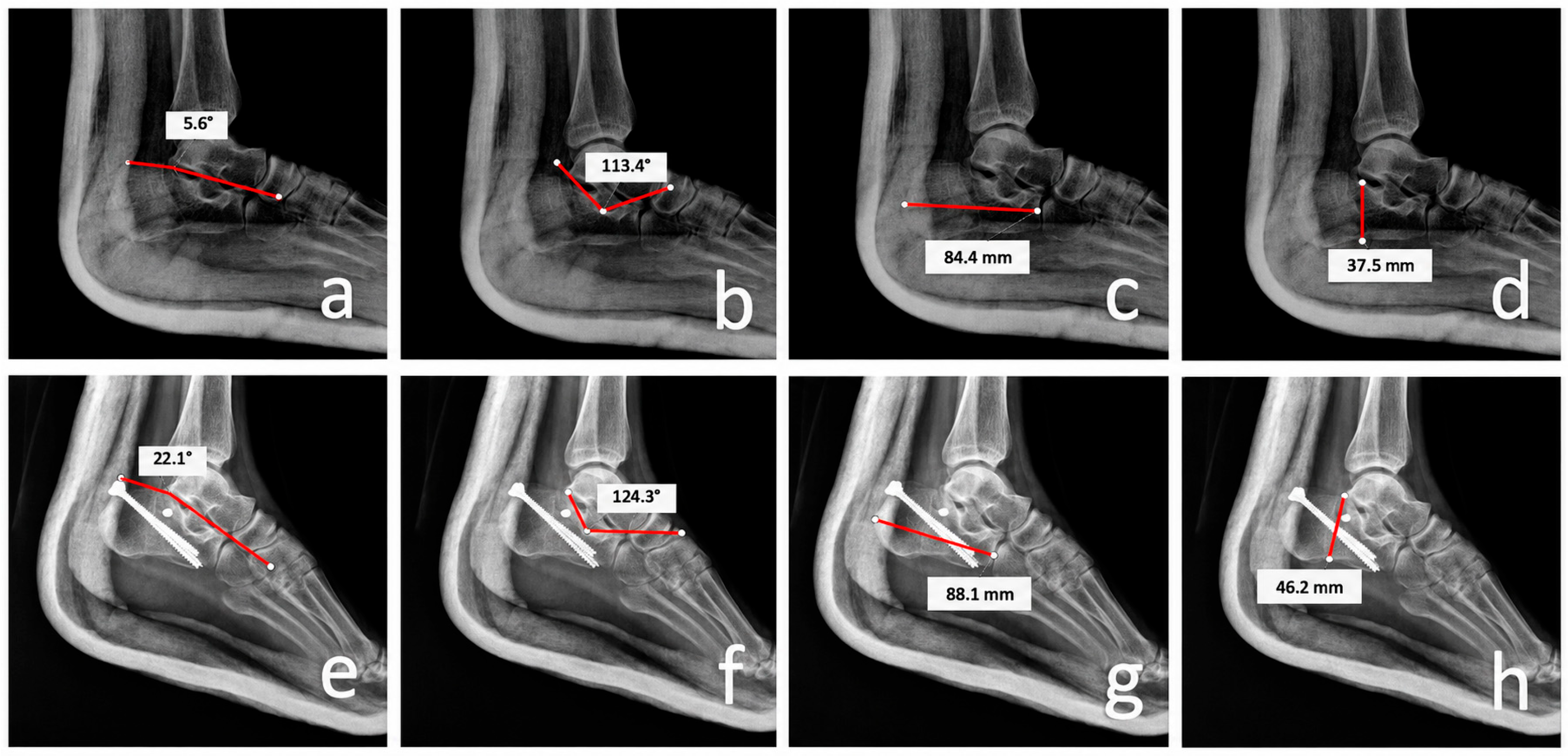
All patients were followed up regularly with clinical and radiological evaluation. Böhler angle (BA), Gissane angle (GA), calcaneal length (CL), and calcaneal height (CH) taken immediately preoperatively, one day postoperatively, and at the two-year follow-up were retrospectively compared. Complications and American Orthopedic Foot and Ankle Society (AOFAS) scores were compared between the groups at the two-year follow-up. Radiological assessments of all patients were compared, and these measurements were made by three experienced orthopedic surgeons involved in the study (Figure 1). All radiological images were assessed in a blinded manner.
Figure 1.
Bohler angle (BA) measurement preoperative (a), early postoperative (e), Gissane angle (GA) measurement preoperative (b), early postoperative (f), calcaneal length (CL) measurement preoperative (c), early postoperative (g), calcaneal height (CH) measurement preoperative (d), and early postoperative (h) in Sanders type-3 calcaneal fracture.
2.3. Surgery
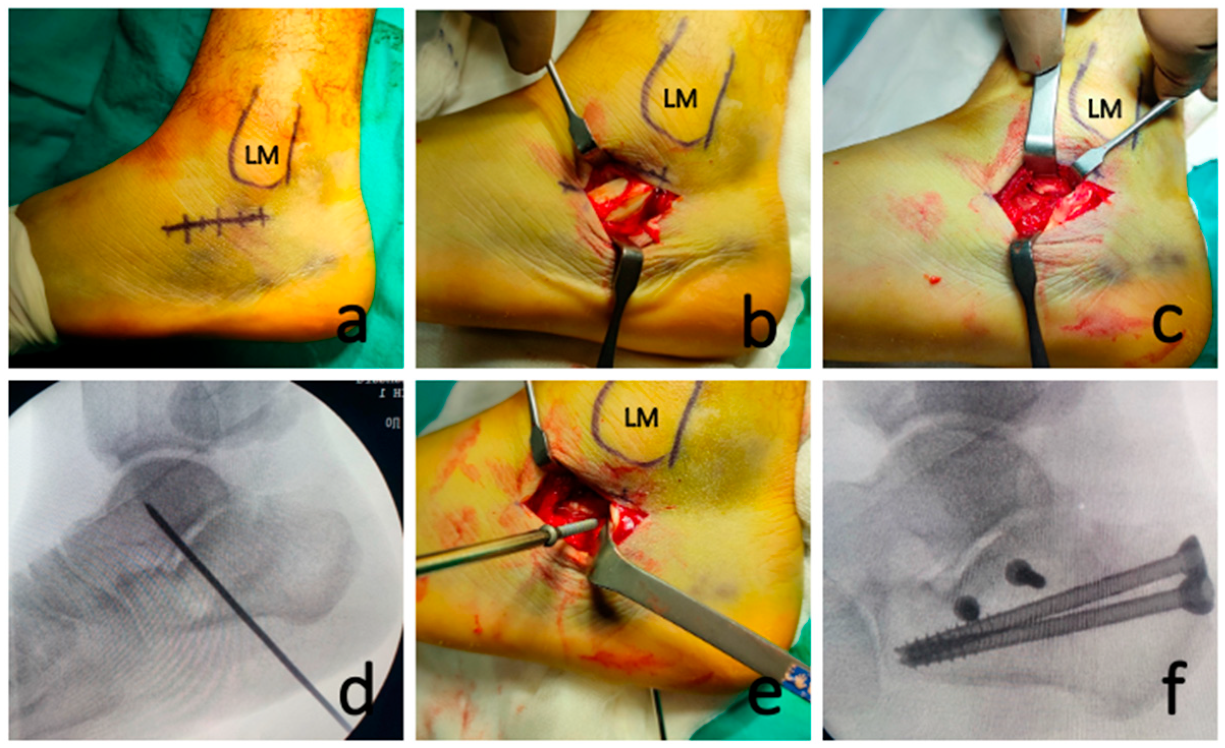
The patient is placed on a radiolucent table in the lateral decubitus position, and surgery is performed under spinal or general anesthesia with a tourniquet wrapped around the thigh. An approximately 5 cm long incision is made 1 cm distal and posterior to the fibula to protect the sural nerve (Figure 2a). The sheath of the peroneal tendons is opened and protected with retractors (Figure 2b). The soft tissues within the sinus tarsi are excised, and the facet joints are exposed (Figure 2c). Debris, tissues, and fracture hematoma are removed from the fracture ends to improve the evaluation of fracture reduction. The displaced posterior facet is reduced through an elevation–derotation maneuver using a spatula and fixed temporarily with Kirschner wires (Figure 2d). After a satisfactory reduction is achieved, one or two 3.5 mm cortical screws are inserted from the lateral wall of the posterior facet to the sustentaculum tali (Figure 2e). Then, under C-arm fluoroscopy, two cancellous cannulated screws are placed from the tuber calcanei to the anterior of the calcaneus, aiming at the calcaneocuboid joint under the cortical screw to create a rafting effect (Figure 2f). After the radiological controls under fluoroscopy, the surgery is ended as usual. The patient receives 24 h IV antibiotic prophylaxis. After two weeks of resting in a short leg splint, active–passive ankle movements are initiated. Partial weight-bearing exercises are recommended six weeks after surgery; the patient is instructed to bear one fourth of their body weight using scales with crutches. After 10 weeks, full weight-bearing was permitted.
Figure 2.
Approximately 5 cm long incision was made 1 cm distal and posterior to the fibula (a). The sheath of the peroneal tendons was opened and protected with retractors (b). The soft tissues within the sinus tarsi were excised and the facet joints were exposed (c). The displaced fragments were reduced with a spatula and fixed temporarily with Kirschner wire (d). A 3.5 mm cortical screw was inserted from the posterior facet joint of the calcaneus bone to the sustentaculum tali (e). Two cancellous cannulated screws were placed from posterior to anterior of the calcaneus, under the cortical screw, to create a rafting effect (f). LM, lateral malleolus.
2.4. Statistical Analysis
Student’s t-test for independent samples, chi-squared tests, and the Friedman test were used to analyze the data. A p-value of 0.05 was regarded as statistically significant. Categorical variables are presented as counts and percentages, while continuous variables are summarized using the median and interquartile range. Data are presented as mean ± standard deviation. Statistical analyses were performed using IBM SPSS Statistics for Windows, version 23.0 (IBM Corp., Armonk, NY, USA). Interrater reliability was evaluated at each measurement time point using the intraclass correlation coefficient (ICC; two-way mixed effects model with absolute agreement and three raters).
The study was approved by the Medical Research Ethics Committee of the University of Health Sciences, İzmir Bozyaka Training and Research Hospital (protocol code: 2023/82; date of approval: 14 June 2023).
3. Results
Eighty-eight patients with DIACF who were operated on between March 2015 and March 2022 were included in the study. The median follow-up duration of our patients was 62.5 months (min: 24; max: 100). The median age of our patients was 45.6 (min: 18; max: 82) years.
No statistical difference was seen between the groups in terms of age, gender, mechanism of injury, comorbidities, Sanders classification of the fracture types, and follow-up times (Table 1).
Table 1.
Demographic and clinical characteristics of the two groups.
An infection was detected in six of our patients (6.8%), with two cases occurring in Gearly and four in Glate. Superficial wound infections around skin incision were observed in patients with Sanders type 3 fractures in both Gearly (n = 1) and Glate (n = 1). Both were treated with oral antibiotics and wound care. The major complication was screw head irritation at the posterior heel skin while wearing shoes. Seventeen patients suffered from skin irritation (19%) (seven from Gearly, 10 in Glate). Four of them had superficial infection around a screw head (one from Gearly, 3 from Glate). All cases with skin irritation underwent surgery for implant removal after bony healing was completed. The mean surgery time for implant removal was 13 months (9–23). There was no statistically significant difference between the two groups regarding infection development (p = 0.8748) (Table 2). Furthermore, wound dehiscence was observed in one patient with a Sanders type 4 fracture who was operated on early-term. That case was treated with wound care alone without antibiotics at an outpatient clinic. The wound healed completely 4 weeks after the operation. No nonunion or malunion was detected. We found that using a 3.5 mm screw fixation does not lead to irritation of the peroneal tendons or cause subfibular impingement. Sural nerve neuritis was observed in five cases (5.6%), two in Gearly and three in Glate. All cases spontaneously resolved without treatment. Subtalar arthrosis was observed in 13 cases (14.8%): four in Gearly and nine in Glate. No cases underwent subtalar arthrodesis due to advanced subtalar arthrosis. There were no significant differences between the groups, which included patients operated on within 3 days post-injury and those operated on past 3 days post-injury, in terms of wound complications (p = 0.1875) (Table 2).
Table 2.
Complications in the two groups.
Satisfactory agreement was reached among the three physicians when they independently performed radiological assessments for all patients.
The results of the radiological evaluations are shown in Table 3. Table 3 shows that BA, GA, CL, and CH were significantly restored after the operation (p < 0.0001) in groups with respect to operation time. In addition, there were no significant differences between Gearly and Glate in terms of preoperative and postoperative BA, two-year follow-up BA, correction loss in BA, preoperative and postoperative GA, two-year follow-up GA, correction loss in GA, preoperative and postoperative CL, two-year follow-up CL, correction loss in CL, preoperative and postoperative CH, two-year follow-up CH, and correction loss in CH (Table 3).
Table 3.
Radiological parameters and functional scores of two groups with regard to operation time.
We observed significant differences between Group S2–3 and Group S4 with regard to preoperative BA and CH (p < 0.0001 and p = 0.0364, respectively) (Table 4). However, there were no significant differences between Group S2–3 and Group S4 regarding postoperative BA, two-year follow-up BA, correction loss in BA, preoperative and postoperative GA, two-year follow-up GA, correction loss in GA, preoperative and postoperative CL, two-year follow-up CL, correction loss in CL, postoperative CH, two-year follow-up CH, and correction loss in CH.
Table 4.
Radiological parameters and functional scores of two groups with regard to fracture type.
4. Discussion
In our study, we analyzed the postoperative complications and radiographic results of intra-articular calcaneal fractures treated with the minimally invasive sinus tarsi approach in patients, with a follow-up period of at least two years. The patients were divided into two groups: those operated on within 3 days post-injury (n: 32; median age: 47) and those operated on past 3 days post-injury (n: 56; median age: 44.8). Superficial wound infection was observed in two patients who were operated on within 3 days post-injury and in four patients who were operated on past 3 days post-injury. Wound necrosis was observed in one patient with a Sanders type 4 fracture who was operated on within 3 days post-injury. When patients with DIACFs who were operated on with the STA in the early and late periods were compared, no differences were found in terms of complications, radiological values, and functional scores in the postoperative period and during follow-up. In addition, the patients were categorized into two groups based on the type of fracture. Patients with Sanders type 2 and type 3 fractures were included in Group S2–3 (n: 58; median age: 45.2), and those with Sanders type 4 fractures were included in Group S4 (n: 30; median age: 46.5). These groups were compared postoperatively and during follow-up periods, and no differences were found in terms of radiological values, functional scores, or complications. Thus, we concluded that STA can be used safely in multicomponent intra-articular fractures.
Currently, the indications for conservative treatment are limited to patients with low preoperative functional demands, poor soft tissue healing potential, and minimal comminution or displacement [16]. DIACFs continue to be a source of debate regarding the best surgical treatment to be applied for these injuries. A surgeon’s treatment decision is influenced by various factors, including the type of calcaneal fracture, the condition of the skin, the cause of the injury, and any existing comorbidities [16,17].
Surgical options include extensile lateral, medial, combined medial and lateral, percutaneous, posterior, and sinus tarsi approaches [17,18,19,20,21,22]. All of these techniques aim to minimize soft tissue damage while ensuring consistent fracture reduction and fixation. Therefore, it has been proposed that minimally invasive approaches for these fractures can help reduce soft tissue complications while still achieving the desired fixation outcomes. It is believed that delaying the surgical fixation of DIACF may help lower the risk of wound complications [19,21]. However, recent studies have not demonstrated this advantage [23,24]. In a study involving 53 patients treated for DIACF with the extensile lateral approach (ELA), Ho et al. investigated the relationship between the timing of surgery and the rate of wound complications. Despite the fact that their 30 patients were operated on within 72 h of injury, they only recorded one postoperative infection that required surgical debridement and implant removal. They found that early surgery may be safe for appropriately selected patients [12]. They excluded patients with insulin-dependent diabetes mellitus and fracture blisters on the incision line from their study. However, in our study, we included all diabetes mellitus cases, even if they were insulin-dependent or had fracture blisters on the foot—even on the incision line—regardless of age. The sinus tarsi approach yielded satisfactory results, even for high-risk patients.
In a study of 70 patients, Joseph et al. found that intra-articular calcaneus fractures could be treated acutely within 72 h of injury using the STA, resulting in minimal wound complications and no adverse effect on short-term functional outcomes [25]. They found that only a psychiatric history is a risk factor for worse clinical results, whereas diabetes mellitus, smoking, Sanders classification, open fractures, and the number of days until the operation were not. They reported that four cases underwent arthrodesis (5.7%), and 10 cases underwent implant removal (14.3%). However, in our series, no cases underwent arthrodesis, but 17 cases underwent implant removal (19.3%). For this reason, we changed our implant choice from 6.5 mm cancellous screws to 6 mm headless screws.
Kwon et al. found that postponing surgery increased the risk of wound complications in patients treated with both the ELA and STA [26]. Additionally, they found that patients who underwent STA surgical fixation more than two weeks after the injury were three times more likely to experience wound complications compared to those who had surgery within the first week [26]. Similar to these studies, our study showed that the STA is a safe approach for the early fixation of intra-articular calcaneus fractures.
Poor outcomes, such as abnormal subtalar contact forces, malleolar impingement, and subtalar osteoarthritis, have been associated with inadequate restoration of the calcaneal facet, height, and length, as well as improper axial alignment [27,28,29]. In comparison to the ELA, the STA offers sufficient exposure to the calcaneocuboid and posterior subtalar joints, while achieving a similar quality of reduction [30,31,32]. In their meta-analysis comparing the STA with the ELA, Bai et al. found no significant difference in AOFAS scores and no change in Bohler angle between the groups [33]. In our cohort, the radiological parameters of Sanders type 4 fractures were mildly inferior compared to those of Sanders type 2 and type 3 fractures. However, there were no statistically significant differences.
The choice of surgical technique for Sanders type 4 fractures is another issue. Among the surgical approaches, the ELA has the advantage of excellent exposure. However, soft tissue complications have made surgeons seek more biologic procedures. In a study involving 69 patients with Sanders type 4 displaced intra-articular calcaneal fractures, Lin et al. demonstrated that similar clinical and radiographic outcomes can be achieved using the STA and ELA approaches [34]. O’Driscoll et al. reported that their nine cases with Sanders type 4 fractures treated via STA showed no collapse in Böhler angles, unlike the results of Richter et al., because of restricted weight-bearing in the first 6 weeks. It was revealed that controlled weight-bearing after 6 weeks does not cause a collapse of radiological parameters while avoiding osteopenia. In our study, we encouraged the patients to bear weight after 6 weeks of outpatient control. They were instructed to bear ¼ of their body weight using scales. Ten weeks after operation, all patients were permitted to bear their full weight. The last control radiological parameters showed that the mean loss in Böhler angles was less than 2°, even in Sanders type 4 fractures. Late demonstration of symptomatic arthritis requiring arthrodesis was reported by Joseph et al. in four cases, three of which were Sanders type 4 fractures [25]. Moreover, Sanders type 3 and type 4 fractures had higher rates of complications, such as arthritis, independent of the surgical approach [32].
Achieving anatomic reconstruction of the calcaneal body and articular surface can lead to favorable long-term functional outcomes [33,34]. Sanders et al. investigated 108 DIACF patients who underwent the ELA surgical treatment and had an average AOFAS score of 75 after a minimum of ten years of follow-up. After an average postoperative follow-up of 44 months, painful arthritis requiring subtalar arthrodesis developed in only 29% (n = 31) of surgically treated patients [35]. In our study, after a two-year minimum follow-up period, 13 cases suffered arthritis, but none needed arthrodesis yet (14.8%).
Hardware removal rates have been found to range between 10% and 40% in studies evaluating the sinus tarsi lateral approach due to a variety of complications [36]. In the current study, 17 (19%) patients underwent implant removal surgery. Due to insufficient soft tissue support, the tuber calcanei was irritated by the screw heads.
This study has some limitations that should be mentioned, including its retrospective design, relatively short-term follow-up, and limited number of patients examined. Conducting larger-scale studies is needed to further evaluate the sinus tarsi approach for the early fixation of intra-articular calcaneal fractures.
5. Conclusions
Early fixation within 3 days via the sinus tarsi approach is a safe treatment alternative for displaced intra-articular Sanders type 2, type 3, and even type 4 fractures.
Author Contributions
Conceptualization, C.K. and O.S.; methodology, C.K., O.S. and A.A.K.; software, O.S. and M.U.; validation, C.K., O.S. and A.A.K.; formal analysis, O.S. and M.U.; investigation, O.S., M.U., R.S.E. and F.C.; resources, C.K., R.S.E., F.C. and A.A.K.; data curation, O.S., M.U. and R.S.E.; writing—original draft preparation, C.K. and O.S.; writing—review and editing, C.K., O.S., M.U., R.S.E., F.C. and A.A.K.; visualization, O.S. and M.U.; supervision, C.K. and A.A.K.; project administration, C.K. and O.S.; funding acquisition, not applicable. All authors have read and agreed to the published version of the manuscript.
Funding
This research received no external funding.
Institutional Review Board Statement
The study was conducted in accordance with the Declaration of Helsinki and approved by the Medical Research Ethics Committee of the University of Health Sciences, İzmir Bozyaka Training and Research Hospital (protocol code: 2023/82; date of approval: 14 June 2023).
Informed Consent Statement
Informed consent was obtained from all subjects involved in the study. The consent included permission for the use of anonymized clinical and radiological data for research, scientific publication, and presentation purposes. No identifiable patient information is presented in this manuscript.
Data Availability Statement
The raw data supporting the conclusions of this article will be made available by the authors upon request.
Conflicts of Interest
The authors declare no conflicts of interest.
References
- Mitchell, M.J.; McKinley, J.C.; Robinson, C.M. The epidemiology of calcaneal fractures. Foot 2009, 19, 197–200. [Google Scholar] [CrossRef] [Scilit]
- Rammelt, S.; Zwipp, H. Calcaneus fractures: Facts, controversies and recent developments. Injury 2004, 35, 443–461. [Google Scholar] [CrossRef] [Scilit] [PubMed]
- De Boer, A.S.; Van Lieshout, E.M.; Den Hartog, D.; Weerts, B.; Verhofstad, M.H.J.; Schepers, T. Functional outcome and patient satisfaction after displaced intra-articular calcaneal fractures: A comparison among open, percutaneous, and nonoperative treatment. J. Foot Ankle Surg. 2015, 54, 298–305. [Google Scholar] [CrossRef] [Scilit] [PubMed]
- Ibrahim, T.; Rowsell, M.; Rennie, W.; Brown, A.; Taylor, G.; Gregg, P. Displaced intra-articular calcaneal fractures: 15-year follow-up of a randomised controlled trial of conservative versus operative treatment. Injury 2007, 38, 848–855. [Google Scholar] [CrossRef] [Scilit] [PubMed]
- Sanders, R. Displaced intra-articular fractures of the calcaneus. J. Bone Jt. Surg. 2000, 82, 225–250. [Google Scholar] [CrossRef] [Scilit] [PubMed]
- Veltman, E.S.; Doornberg, J.N.; Stufkens, S.A.; Luitse, J.S.; Bekerom, M.P.v.D. Longterm Outcomes of 1730 Calcaneal Fractures: Systematic Review of the Literature. J. Foot Ankle Surg. 2013, 52, 486–490. [Google Scholar] [CrossRef] [Scilit] [PubMed]
- Zhang, W.; Lin, F.; Chen, E.; Xue, D.; Pan, Z. Operative Versus Nonoperative Treatment of Displaced Intra-Articular Calcaneal Fractures: A Meta-Analysis of Randomized Controlled Trials. J. Orthop. Trauma 2016, 30, e75–e81. [Google Scholar] [PubMed]
- Nosewicz, T.; Knupp, M.; Barg, A.; Maas, M.; Bolliger, L.; Goslings, J.C.; Hintermann, B. Mini-Open Sinus Tarsi Approach with Percutaneous Screw Fixation of Displaced Calcaneal Fractures: A Prospective Computed TomographyBased Study. Foot Ankle Int. 2012, 33, 925–933. [Google Scholar] [CrossRef] [Scilit] [PubMed]
- Buckley, R.; Tough, S.; McCormack, R.; Pate, G.; Leighton, R.; Petrie, D.; Galpin, R. Operative compared with nonoperative treatment of displaced intra-articular calcaneal fractures: A prospective, randomized, controlled multicenter trial. J. Bone Jt. Surg. Am. 2002, 84, 1733–1744. [Google Scholar]
- Kurozumi, T.; Jinno, Y.; Sato, T.; Inoue, H.; Aitani, T.; Okuda, K. Open reduction for intraarticular calcaneal fractures: Evaluation using computed tomography. Foot Ankle Int. 2003, 24, 942–948. [Google Scholar] [CrossRef] [Scilit] [PubMed]
- Pastor, T.; Gradl, G.; Klos, K.; Ganse, B.; Horst, K.; Andruszkow, H.; Hildebrand, F.; Pape, H.; Knobe, M. Displaced intraarticular calcaneal fractures: Is there a consensus on treatment in Germany? Int. Orthop. 2016, 40, 2181–2190. [Google Scholar] [CrossRef] [Scilit] [PubMed]
- Ho, C.J.; Huang, H.T.; Chen, C.H.; Chen, J.C.; Cheng, Y.M.; Huang, P.J. Open reduction and internal fixation of acute intra-articular displaced calcaneal fractures: A retrospective analysis of surgical timing and infection rates. Injury 2013, 44, 1007–1010. [Google Scholar] [CrossRef] [Scilit] [PubMed]
- Bahaeddini, M.R.; Konjkav, A.R.; Aminian, A.; Tabrizian, P.; Gravand, S.N.; Amiri, S.; Mirjalily, M.S.; Tayyebi, H.; Mazhar, F.N. A simple modified technique for screw fixation of displaced intra-articular calcaneus fracture through a sinus tarsi approach: A comparison with plate fixation. BMC Musculoskelet. Disord. 2024, 25, 750. [Google Scholar] [CrossRef] [Scilit] [PubMed]
- Guo, C.; Xu, Y.; Li, C.; Li, X.; Wang, Z.; Cai, M.; Xu, X. Comparing less invasive plate fixation versus screw fixation of displaced intra-articular calcaneus fracture via sinus tarsi approach. Int. Orthop. 2020, 45, 2231–2237. [Google Scholar] [CrossRef] [Scilit] [PubMed]
- Sato, K.; Yorimitsu, M.; Uehara, T.; Fukuoka, S.; Hata, T.; Saiga, K.; Noda, T.; Ozaki, T. Comparison of screw versus locking plate fixation via sinus tarsi approach for displaced intra-articular calcaneal fractures. Foot Ankle Surg. 2022, 29, 97–102. [Google Scholar] [CrossRef] [Scilit] [PubMed]
- Schleunes, S.; Lobos, E.; Saltrick, K. Current Management of Intra-Articular Calcaneal Fractures. Clin. Podiatr. Med. Surg. 2024, 41, 473–490. [Google Scholar] [CrossRef] [Scilit] [PubMed]
- Benirschke, S.K.; Sangeorzan, B.J. Extensive intraarticular fractures of the foot: Surgical management of calcaneal fractures. Clin. Orthop. 1993, 292, 128–134. [Google Scholar]
- Burdeaux, B.D. The medial approach for calcaneal fractures. Clin. Orthop. Relat. Res. 1993, 290, 96–107. [Google Scholar] [CrossRef] [Scilit]
- Stephenson, J.R. Surgical treatment of displaced intraarticular fractures of the calcaneus: A combined lateral and medial approach. Clin. Orthop. 1993, 290, 68–75. [Google Scholar]
- Tomesen, T.; Biert, J.; Frolke, J.P.M. Treatment of displaced intra-articular calcaneal fractures with closed reduction and percutaneous screw fixation. J. Bone Jt. Surg. 2011, 93, 920–928. [Google Scholar] [CrossRef] [Scilit] [PubMed]
- Park, I.; Song, K.W.; Shin, S.; Lee, J.Y.; Kim, T.G.; Park, R.S. Displaced intra-articular calcaneal fracture treated surgically with limited posterior incision. Foot Ankle Int. 2000, 21, 195–205. [Google Scholar] [CrossRef] [Scilit] [PubMed]
- Carr, J.B. Mechanism and pathoanatomy of the intraarticular calcaneal fracture. Clin. Orthop. Relat. Res. 1993, 290, 36–40. [Google Scholar] [CrossRef] [Scilit]
- Court-Brown, C.M.; Schmidt, M.; Schutte, B.G. Factors affecting infection after calcaneal fracture fixation. Injury 2009, 40, 1313–1315. [Google Scholar] [CrossRef] [Scilit] [PubMed]
- Folk, J.W.; Starr, A.J.; Early, J.S. Early wound complications of operative treatment of calcaneus fractures: Analysis of 190 fractures. J. Orthop. Trauma 1999, 13, 369–372. [Google Scholar] [CrossRef] [Scilit] [PubMed]
- Joseph, N.M.; Benedick, A.; McMellen, C.; Napora, J.; Wetzel, R.; Sontich, J.; Ochenjele, G. Acute Fixation of Displaced Intra-articular Calcaneus Fractures Is Safe Using the Sinus Tarsi Approach. J. Orthop. Trauma 2021, 35, 289–295. [Google Scholar] [CrossRef] [Scilit] [PubMed]
- Kwon, J.Y.; Guss, D.; Lin, D.E.; Abousayed, M.; Jeng, C.; Kang, S.; Ellington, J.K. Effect of delay to definitive surgical fixation on wound complications in the treatment of closed, intra-articular calcaneus fractures. Foot Ankle Int. 2015, 36, 508–517. [Google Scholar] [CrossRef] [Scilit] [PubMed]
- Makki, D.; Alnajjar, H.M.; Walkay, S.; Ramkumar, U.; Watson, A.J.; Allen, P.W. Osteosynthesis of displaced intra-articular fractures of the calcaneum: A long-term review of 47 cases. J. Bone Jt. Surg. Br. 2010, 92, 693–700. [Google Scholar]
- Ozturk, A.M.; Ozer, M.A.; Suer, O.; Derin, O.; Govsa, F.; Aktuglu, K. Evaluation of the effects of using 3D—Patient specific models of displaced intra—Articular calcaneal fractures in surgery. Injury 2022, 53, S40–S51. [Google Scholar] [CrossRef] [Scilit] [PubMed]
- Agren, P.H.; Wretenberg, P.; Sayed-Noor, A.S. Operative versus nonoperative treatment of displaced intra-articular calcaneal fractures: A prospective, randomized, controlled multicenter trial. J. Bone Jt. Surg. 2013, 95, 1351–1357. [Google Scholar]
- Schepers, T. The sinus tarsi approach in displaced intra-articular calcaneal fractures: A systematic review. Int. Orthop. 2011, 35, 697–703. [Google Scholar] [CrossRef] [Scilit] [PubMed]
- Kayalı, C.; Ozan, F.; Altay, T.; Karahan, H.G.; Yamak, K.; Özdemir, S. Efficacy of calcium phosphate cementing in the surgical treatment of Sanders Type II and III calcaneal fractures using screw fixation with sinus tarsi approach. Acta Orthop. Traumatol. Turc. 2021, 55, 265–270. [Google Scholar] [CrossRef] [Scilit] [PubMed]
- Joseph, N.M.; Sinkler, M.A.; Magister, S.; Bacharach, A.; Pang, A.; Boes, K.; Vallier, H.A.; Hirschfeld, A.G.; Ochenjele, G. Fixation of displaced intra-articular calcaneus fractures is safe using either sinus tarsi or extensile lateral approach. Injury 2023, 54, 1824–1830. [Google Scholar] [CrossRef] [Scilit] [PubMed]
- Bai, L.; Hou, Y.L.; Lin, G.H.; Zhang, X.; Liu, G.Q.; Yu, B. Sinus tarsi approach (STA) versus extensile lateral approach (ELA) for treatment of closed displaced intra-articular calcaneal fractures (DIACF): A meta-analysis. Orthop. Traumatol. Surg. Res. 2018, 104, 239–244. [Google Scholar] [CrossRef] [Scilit] [PubMed]
- Lin, J.; Xie, C.; Chen, K.; Sun, S.; Zhou, K.; Zhou, C.; Shui, X.; Kong, J. Comparison of sinus tarsi approach versus extensile lateral approach for displaced intra-articular calcaneal fractures Sanders type IV. Int. Orthop. 2019, 43, 2141–2149. [Google Scholar] [CrossRef] [Scilit] [PubMed]
- Sanders, R.; Vaupel, Z.M.; Erdogan, M.; Downes, K. Operative treatment of displaced intraarticular calcaneal fractures: Long-term (10–20 years) results in 108 fractures using a prognostic CT classification. J. Orthop. Trauma 2014, 28, 551–563. [Google Scholar] [PubMed]
- Spagnolo, R.; Bonalumi, M.; Pace, F.; Capitani, D. Calcaneus fractures, results of the sinus tarsi approach: 4 years of experience. Eur. J. Orthop. Surg. Traumatol. 2009, 20, 37–42. [Google Scholar] [CrossRef] [Scilit]

Disclaimer/Publisher’s Note: The statements, opinions and data contained in all publications are solely those of the individual author(s) and contributor(s) and not of MDPI and/or the editor(s). MDPI and/or the editor(s) disclaim responsibility for any injury to people or property resulting from any ideas, methods, instructions or products referred to in the content. |
© 2026 by the authors. Published by MDPI on behalf of the American Podiatric Medical Association (APMA). Licensee MDPI, Basel, Switzerland. This article is an open access article distributed under the terms and conditions of the Creative Commons Attribution (CC BY) license.