Article

Anti-Inflammatory and Neuroprotective Effect of the Anti-Obesity Dietary Supplement Dekosilhue® in an In Vitro Model of Neuroinflammation

by
Vittoria Borgonetti
1,
Lorenzo Cenci
2 and
Nicoletta Galeotti
1,*
1
Department of Neuroscience, Psychology, Drug Research and Child Health (NEUROFARBA), Section of Pharmacology and Toxicology, University of Florence, Viale G. Pieraccini 6, 50139 Florence, Italy
2
Department of Biomedical Sciences, University of Padua, Via Marzolo, 3, 35131 Padua, Italy
*
Author to whom correspondence should be addressed.
Nutraceuticals 2022, 2(1), 22-31; https://doi.org/10.3390/nutraceuticals2010002
Submission received: 6 December 2021 / Revised: 24 January 2022 / Accepted: 28 January 2022 / Published: 8 February 2022
(This article belongs to the Special Issue Current State of the Art—Nutraceutical Components of Foods)

Abstract

:
Obesity is now recognized as a global health problem and has reached epidemic proportions, affecting both developed and developing countries. This condition is associated with an increased incidence in central nervous system (CNS) disorders. The intake of foods or supplements with anti-inflammatory activity could potentially lead to a lower impact of obesity on the functionality of CNS. Therefore, in this work we tested the effect of a dietary supplement, DEKOSILHUE® (DSK), containing polyphenols enriched herbal extracts and successfully used for improving the control of body weight, in an in vitro inflammatory model. LPS-stimulated immortalized microglia BV2 cells have been used as a model of neuroinflammation. The stimulation with LPS increased the release and activation of several neuroinflammatory mediators, such as IKB⍺, HDAC-1, and IL-1ß. DSK reduced the levels and the activation of all these pro-inflammatory markers. In addition, DSK increased the expression of the anti-inflammatory cytokine IL-10, promoting an anti-inflammatory response. Finally, we showed that the anti-inflammatory effect of DSK can prevent the neurotoxicity related to microglia hyperactivation. In conclusion, DSK could potentially help in controlling neuroinflammation CNS, reducing microglia pro-inflammatory activation.

1. Introduction

Obesity is now recognized as a global health problem and has reached epidemic proportions, affecting both developed and developing countries. The average global body mass index (BMI) has increased from 21.7 in 1975 to 24.2 in 2014. If this trend continues, it will exceed the upper limit of normal (BMI = 24.9) to overweightness (BMI = 25.0–29.9), then to obesity (>30.0), and the prevalence of global obesity is expected to be around 20% by 2025 [1]. Although genetic obesity can be found in some patients, it remains relatively rare [2] and predominantly results from environmental factors, such as diet type or physical activity level. This condition is associated with an increased incidence in central nervous system (CNS) disorders, such as dementia, stroke, depression, and Alzheimer’s disease [3]. In particular, the type of diet correlates with major disorders in obese individuals. For example, following a “Western diet” alters glucose transporter function at the hippocampal level with severe cognitive deficits and increased anxiety levels [4], while a diet high in sugar predisposes to the development of depression. Although these diseases have different etiologies and pathophysiological manifestations, they all share a neuroinflammatory component [5] and obese patients represent a population that is more prone to develop central disorders [6]. Increased adipose tissue affects the normal function of many organs, but more importantly, it promotes the release of substances with proinflammatory activity. Obesity causes increased levels of circulating fatty acids, endotoxins (e.g., LPS), and inflammatory mediators, altering the normal function of many organs. This inflammation can also affect the CNS leading to the appearance of neuroinflammation, resulting from hyperactivity of microglial cells (CNS resident immune cells), mainly affecting the hippocampus, amygdala, and cerebral cortex, leading to severe cognitive deficits and major alterations in mood [7]. The intake of foods or supplements with anti-inflammatory activity could potentially lead to a lower impact of obesity on the functionality of peripheral organs and higher nervous centers, with undoubted benefits in counteracting cognitive decline and states of anxiety and depression [8]. Among the substances of natural origin, those rich in polyphenols, such as green tea and coffee, have proved to be among the most promising in counteracting neuroinflammation and memory disorders [9]. Therefore, a product used for weight control and able to hamper neuroinflammatory processes, could help prevent CNS disorders related to obesity. On these bases, in this work, we tested the effect on an in vitro model of neuroinflammation of a dietary supplement, namely DEKOSILHUE®, based on aqueous extracts of herbal extracts used to promote the metabolism of carbohydrates and lipids for improving the control of body weight and sense of hunger.

2. Materials and Methods

2.1. Chemicals

DEKOSILHUE® (DSK) food supplement, containing aqueous extracts of cinnamon (Cinnamomum zeylanicum Blume) bark, Orthosiphon (Orthosiphon stamineus Benth) leaf, green tea (Camellia sinensis (L.) Kuntze) leaf, mate (Ilex paraguariensis A.St.Hill) leaf, Gymnema (Gymnema sylvestre R. Br.) leaf, bean (Phaseolus vulgaris L.) pod, pineapple (Ananas comosus (L.) Merr.) stem, common gromwell (Lithospermum officinale L.) seeds, horsetail (Equisetum arvense L.) herb, curly dock (Rumex crispus L.) root, asparagus (Asparagus officinalis L.) root, fennel (Foeniculum vulgare Miller) fruit, and birch (Betuta pendolo Roth.) leaf, was provided by Gianluca Mech S.p.A. (Vicenza, Italy) (Table 1). DSK was obtained by a decoction process. Briefly, the raw material is root, leaf, or other part of the plant which is then cooked several times in water at different temperatures. For the in vitro test, DSK was diluted in distilled water and diluted in media at different concentrations: 1, 10, 100, and 1000 µg/mL. Bacterial lipopolysaccharide (LPS) from Gram-negative Salmonella enteridis was purchased from Sigma-Aldrich.(Milan, Italy).

2.2. Cell Culture

BV-2 (Tema Ricerca, Genova, Italy), a microglial cell line was used for this study. Cells were thawed and cultured in a 75 cm2 flask (Sarstedt, Nümbrecht, Germany) in a medium containing RPMI with the addition of 10% of heat-inactivated (56 °C, 30 min) fetal bovine serum (FBS, Gibco®, Milan, Italy), 1% glutamine (Sigma-Aldrich, Milan, Italy), and 1% penicillin-streptomycin solution (Sigma-Aldrich). Cells were grown at 37 °C and 5% CO2 with daily medium change. A human neuroblastoma cell line SH-SY5Y was kindly donated by Prof. Lorenzo Corsi (University of Modena and Reggio Emilia, Italy), was cultured in DMEM and F12 Ham’s nutrients mixture (Sigma-Aldrich), containing 10% heat-inactivated FBS (Sigma-Aldrich), 1% L-glutamine (Sigma-Aldrich), and 1% penicillin-streptomycin solution (Sigma-Aldrich) until confluence (70–80%). Cells were grown in a humidified atmosphere with 5% CO2 at 37 °C. EDTA-trypsin solution (Sigma-Aldrich) was used for detaching the cells from flasks, and cell counting was performed using a hemocytometer by Trypan blue staining as previously reported [10].

2.3. Sulforhodamine B (SRB) Assay

Cell viability was assessed by SRB assay. BV2 (2 × 10⁵ cells in 200 μL) were seeded in 96-well plates, each well corresponding to a different treatment: control (CTRL), LPS, DSK 1, 10, 100, and 1000 µg/mL with or not LPS. The CTRL group was BV2 cells no treated with LPS. Serum-free medium was added to the wells followed by trichloroacetic acid, used as fixative (250 mg/mL TCA, Sigma-Aldrich). The plate was then incubated at 4 °C for 1 h. This was followed by 3–5 washes with double sterile water. The day after, a solution of SRB dissolved in 1% acetic acid was added to each well and incubated for 30 min at room temperature. Then, 3–5 washes with acetic acid were performed. Then, TRIS HCl solution (pH = 10) was added and incubated for 5 min. Finally, the absorbance was recorded at a wavelength of 570 nm. The treatments were performed in six replicates in three independent experiments, and cell viability was calculated by normalizing the values to the control’s mean [11].

2.4. Cell Counting Kit (CCK-8) Assay

BV2 cells were seeded in 6-well plates (3 × 10⁵ cells/well), pre-treated with DSK for 4 h, and then stimulated with LPS 250 ng/mL for 24 h. The neuroprotective effect of DSK was evaluated by assessing the cell viability of SH-SY5Y cells treated with the LPS-conditioned BV2 medium for 24 h. Unstimulated BV2 medium was used as control. Cell viability was performed using the CCK-8 kit (Sigma-Aldrich) according to the manufacturer’s instructions. The absorbance was measured at 450 nm using a MP96 microplate reader spectrophotometer (Benchmark, Biorad). The treatments were replicated three times in six independent experiments, and cell viability was calculated by normalizing the values to the control’s mean [12].

2.5. Preparation of Cell Lysate

BV2 cells were seeded in 6-well plates (3 × 10⁵ cells/well), pre-treated with DSK for 4 h, and then stimulated with LPS 250 ng/mL for 24 h. Then, the conditioned BV2 medium was collected and centrifuged (1000× g for 10 min, 37 °C). The pellet was discarded, and the supernatant was stored at −80 °C for cytokines dosages or used immediately to treat SH-SY5Y cells. Cell lysates were prepared using RIPA buffer (50 mM Tris-HCl pH 7.4, 150 mM NaCl 1% sodium deoxycolate, 1% Tryton X-100, 2 mM PMSF) (Sigma-Aldrich) and the insoluble pellet was separated by centrifugation (12,000 × g for 30 min, 4 °C). The total protein concentration in the supernatant was measured using the Bradford colorimetric method (Sigma-Aldrich) [10].

2.6. Supernatant Protein Precipitation

TCA (25 µL) was added to 50 µL of medium and incubated at 4 °C for 10 min. This was followed by centrifugation at 14,000 rpm for 5 min. The supernatant was then removed, the pellet was washed three times by resuspending in cold acetone, and centrifuging again at 14,000 rpm for 5 min. At this point the microtubes were placed in a heated chamber, favoring the evaporation of the acetone and the drying of the pellets. Finally, the pellet was resuspended in loading buffer 4x and the samples were stored at −20 °C.

2.7. Western Blotting

Protein samples (40 µg/sample) were separated by 10% SDS-PAGE. Proteins were then blotted onto nitrocellulose membranes (120 min at 100 V) using standard procedures. Membranes were blocked in PBST containing 5% non-fat dry milk for 120 min and incubated overnight at 4 °C with primary antibodies anti-IKBα (1:1000, sc-1643), anti-HDAC1 (1:1000, sc-81598), anti-IL10 (1:500, bs-0698R), and anti-IL-1β (1:1000, bs6319R). The blots were rinsed three times with PBST and incubated for 2 h at room temperature with HRP-conjugated mouse anti-rabbit (1:3000, sc-235,) (Santa Cruz Biotechnology) and goat anti-mouse (1:5000, bs-0296G) (Bioss Antibodies) and then detected by chemiluminescence detection system (Pierce, Milan, Italy). Signal intensity (pixels/mm2) was quantified using ImageJ (NIH). The signal intensity was normalized to that of GAPDH (1:5000, sc-32233) (Santa Cruz Biotechnology) [13]. The signal intensity of cytokines released in the supernatant was normalized to that of total protein stained by Ponceau S (RP).

2.8. Statistical Analysis

Data are expressed as the mean ± SEM of three-six experiments and assessed by one-way or two-way ANOVA, followed by a Tukey post hoc test if significance was detected. For each test, a value of p < 0.05 was considered significant. The software GraphPad Prism (version 5.0, San Diego, CA, USA) was used in all statistical analysis.

3. Results

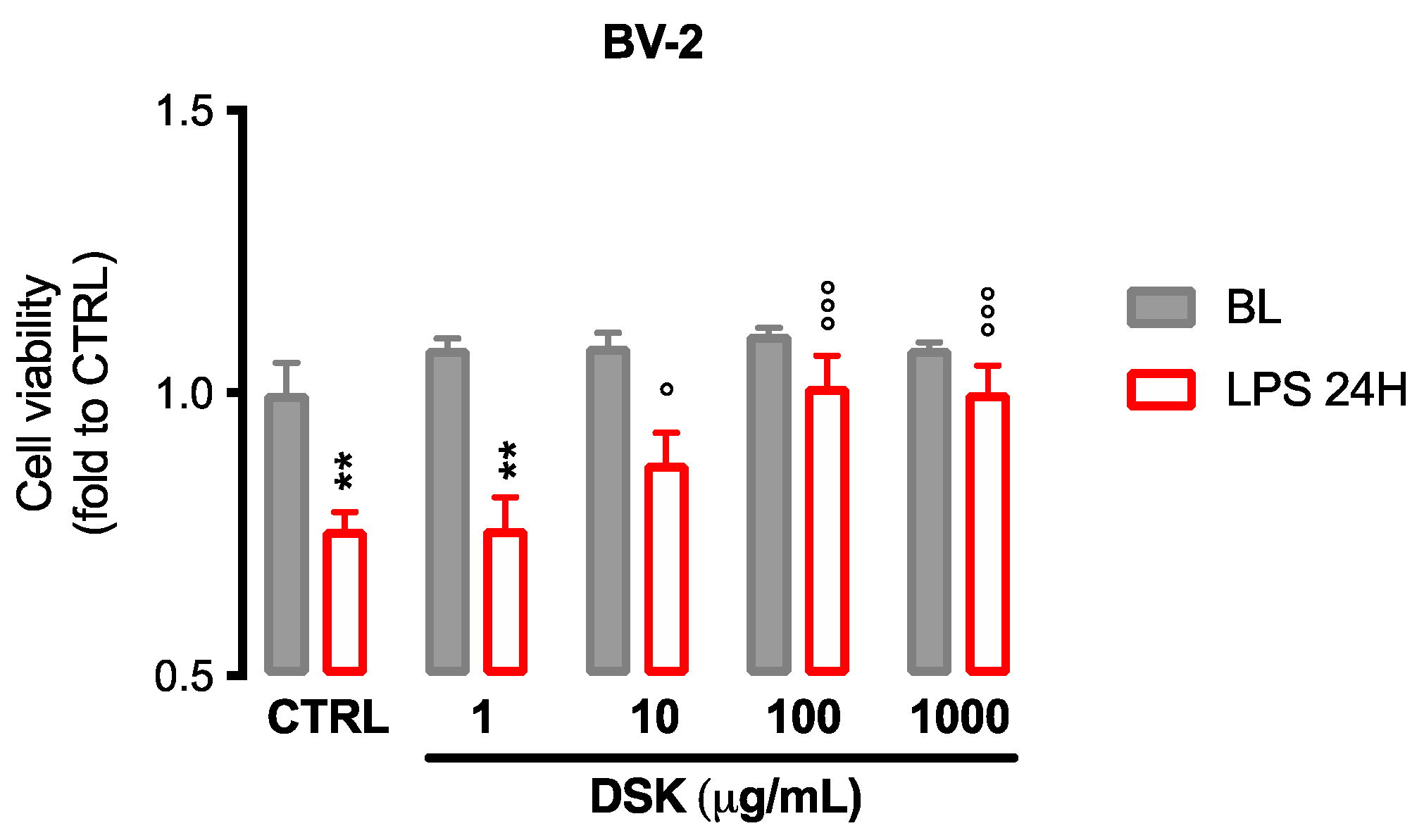
3.1. DSK Reduced Cytotoxicity Induced by LPS in Microglia Cells

To evaluate the anti-inflammatory activity of DSK, we stimulated BV-2 microglial cells with LPS 250 ng/mL for 24 h. After the inflammatory stimulus, microglial cells assume an inflammatory phenotype. Under basal conditions, none of the tested concentrations (1, 10, 100, and 1000 µg/mL) of DSK altered cell viability after 24 h of treatment. The inflammatory stimulus reduces cell viability by approximately 30% compared to the control group. The pre-treatment with DSK (10 to 1000 µg/mL) of LPS-stimulated cells was able to counteract the cytotoxic effect of the inflammatory stimulus (Figure 1).

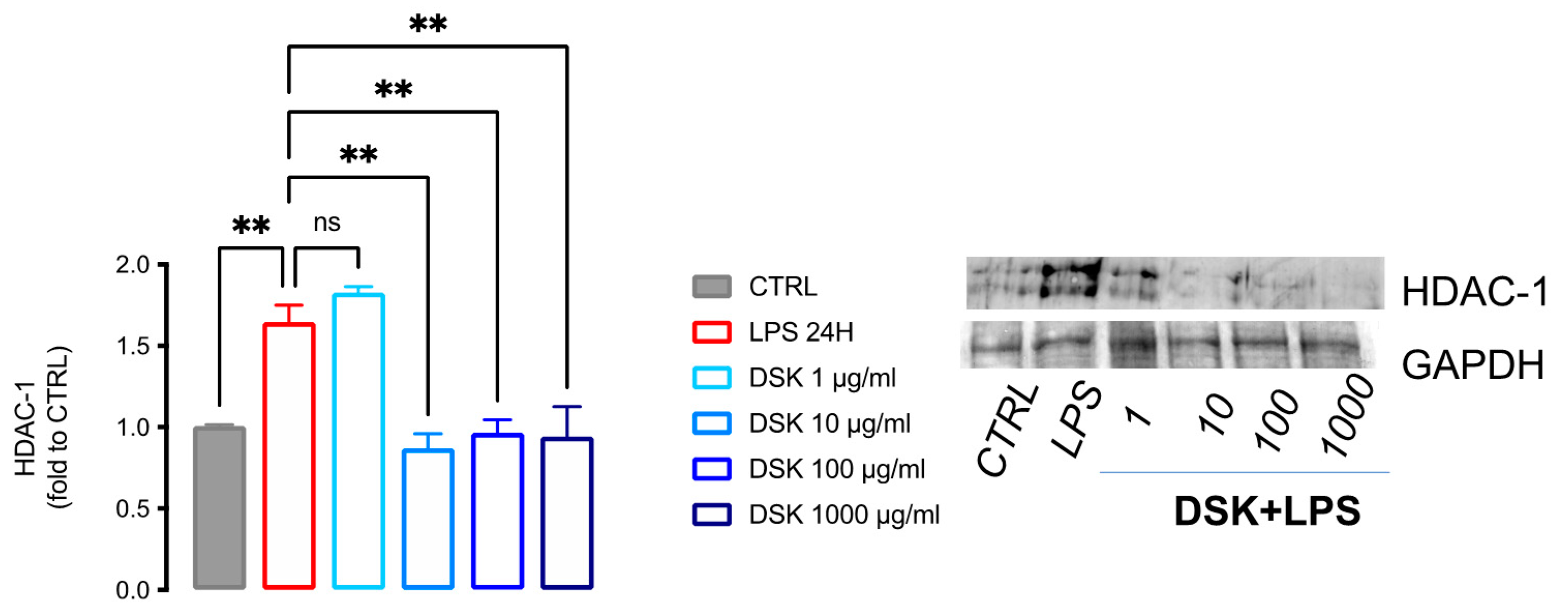
3.2. DSK Reduced HDAC-1 Protein Levels in LPS-Stimulated BV2

Experimental evidence suggests that HDAC inhibitors (HDACi) reduce the pro-inflammatory activity of LPS-stimulated isolated microglial cells [12]. As shown in Figure 2, LPS increased HDAC-1 expression (+65%), while pretreatment with DSK 10 (−13%), 100 (−4%), and 1000 (−6%) µg/mL was able to reduce HDAC1 expression to levels that are comparable to those of CTRL.

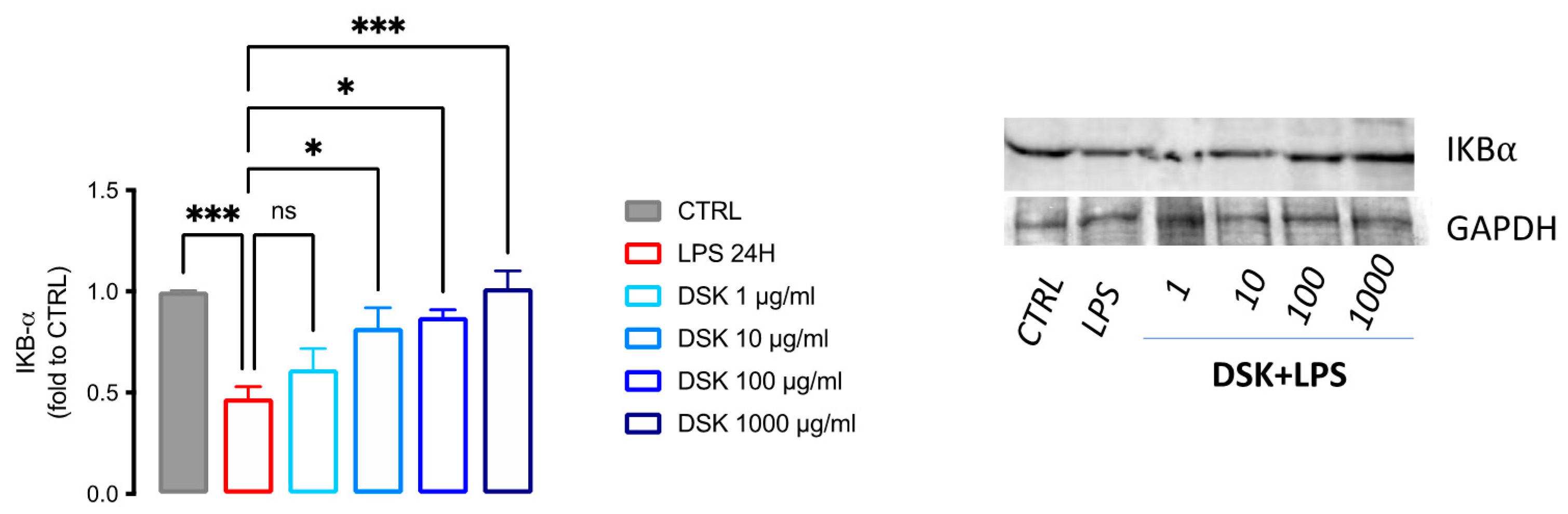
3.3. Activation of NF-κB Pathway Was Reduced by DSK Treatment in LPS-Stimulated BV2

It is well established that deacetylation by HDAC is not only directed to histone proteins, but also to enzymes and transcription factors. In fact, the deacetylation of specific lysine residues of transcription factors, such as NF-κB, can modulate their activity [14]. Activation of NF-κB p65 started with the degradation of IkB ⍺ proteins. With the degradation of IkB⍺, the p-NF-κBp65 complex is freed to move into the nucleus where it can trigger the expression of target genes resulting in an inflammatory response [15]. LPS increased the activity of this transcription factor by reducing (−53%) the levels of IKB⍺, the NF-κB inhibitory cytoplasmic protein. DSK 10 (−28%), 100 (−13%), and 1000 (+0.1%) µg/mL was able to counteract in a dose-dependent manner this effect reporting the value similar to the CTRL group, thus reducing the pro-inflammatory effect of NF-κB (Figure 3).

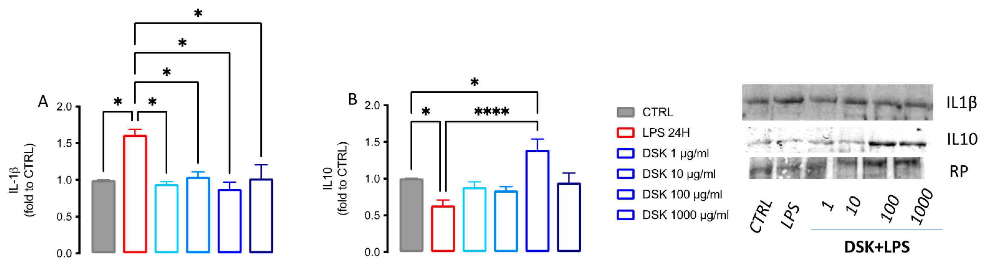
3.4. DSK Modulates Levels of Pro- and Anti-Inflammatory Cytokines Released from LPS-Stimulated BV2 Cells

NF-κB is a transcription factor that primarily regulates transcription of genes encoding for inflammatory factors, such as cytokines. Among these, the most peculiar for microglial activation is represented by IL-1β [16]. Further to the regulation of pro-inflammatory cytokines expression, NF-κB regulates transcription of cytokines with anti-inflammatory activity, reducing them in case of inflammation. Among these, one of the best known is IL-10 [17]. In the in vitro model of neuroinflammation, we observed that cells treated with LPS were characterized by a strong increase in the level of the pro-inflammatory cytokine IL-1β (+61%) and by a reduction of the anti-inflammatory cytokine IL-10 (−37%). DSK inhibited the release of IL-1β at each tested concentration (Figure 4A), counteracting a proinflammatory response. In addition, the concentration of 100 µg/mL significantly increased IL-10 release (+39%) (Figure 4B), promoting an anti-inflammatory response.

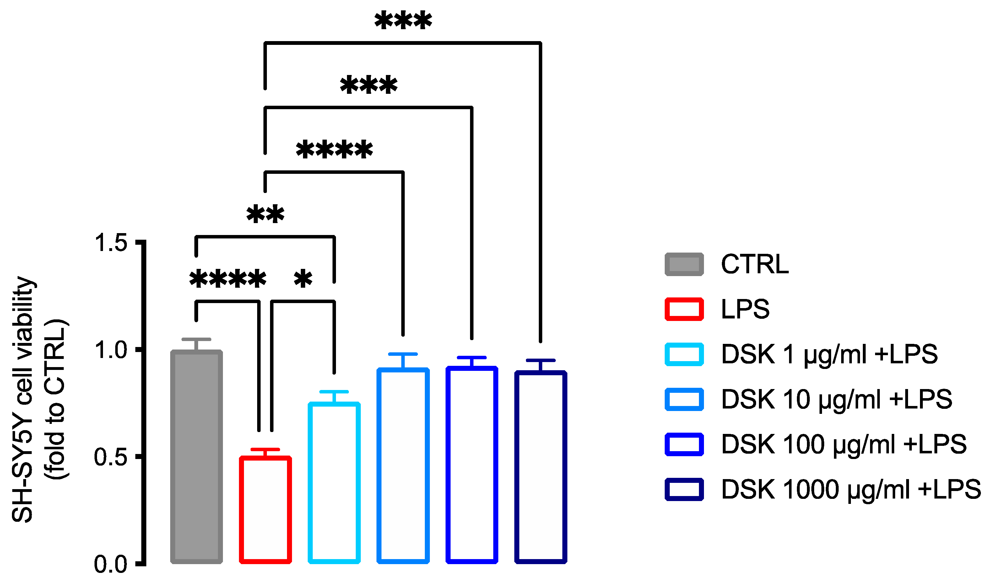
3.5. Neuroprotective Effect of DSK on Inflammation-Induced Neurotoxicity in SH-SY5Y

The anti-inflammatory effect observed in BV-2 cells was related to a neuroprotective effect in human neuronal cells SHSY5Y. LPS–stimulated BV-2 conditioned medium reduced neurons viability (−54.95%) compared to untreated neurons. The treatment with DSK increased the viability of the cells by +25%, +37.05%, +37.05%, and +36.05% at 1, 10, 100, and 1000 µg/mL, respectively, compared to the LPS group (Figure 5).

4. Discussion

Despite the degree of malnutrition existing on the planet, according to the World Health Organization (WHO), obesity represents one of the main public health problems in the world. It certainly represents an actual socio-economic problem [18]. The co-presence of systemic inflammation and obesity was recognized some time ago, with early reported evidence suggesting that obese people have elevated circulating pro-inflammatory cytokines compared to control groups [19]. Thus, a fat-rich diet leads to infiltration and activation of microglia with activation of inflammatory signaling [20]. Importantly, central inflammation may contribute to insulin resistance, promoting weight gain, and sustaining obesity [21]. Microglia are the main CNS immune cells and protect the homeostasis of the CNS [22]. Hence, if an unbalanced diet can induce an alteration in microglial activity, this can result in an alteration at the neuronal level, which can lead to a possible onset of degenerative diseases [23]. Thus, finding a product that can control both body weight and central inflammation could help prevent damage at the CNS level in overweight and obese patients. Complementary and alternative treatments for weight loss include medicinal plants, and their active components, that have been extensively used since ancient times [24].
The anti-obesity effect of polyphenols from commonly used herbal preparations has been documented [25]. Indeed, several clinical studies reported that the consumption of green tea [26] caffeine [27], cathechins [28], and epigallocatechin gallate (EGCG) [29] may improve weight maintenance by preventing or limiting weight regain. Moreover, several polyphenols could play a role in preventing the development of neuroinflammation [30]. For these reasons, our aim was to investigate the possible anti-inflammatory activity of DSK, a food supplement containing a combination of polyphenol-based herbal extracts, used to promote the metabolism of carbohydrates and lipids for the control of body weight and sense of hunger, in an in vitro model of neuroinflammation. Increasing evidence suggest that complex conditions with a multifactorial origin, such as obesity, benefits of a multitarget approach. In this contest, we want to emphasize and valorize the synergism of different plant extracts that can work together to reduce inflammation as a potential adjuvant in the control of obesity and related disorders.
LPS-stimulated BV2 cells have been widely used as a model of neuroinflammation. The stimulation with LPS increased the activation of several neuroinflammatory markers, such as HDAC-1, IKB⍺, and cytokines [12]. DSK was able to reduce the activation of all these pro-inflammatory markers. HDAC-1 is an epigenetic factor involved in inflammatory processes [31]. Recently, direct regulation of HDAC-1 has also been seen in modulating several transcription factors, including NF-κB [14,31]. Inhibition of HDAC-1 is, therefore, an important step towards reducing chronic inflammatory processes. Known inhibitors, such as Vorinostat, and others in the experimental phase, such as LG325, have been shown to reduce activation of the pro-inflammatory microglial phenotype in animal models in which microgliosis is certainly a key factor (e.g., neuropathic pain) [14,32,33,34,35]. In fact, HDAC-1 inhibition also leads to a reduction in the expression of genes involved in the transcription of the NF-κB factor and its entire pathway. NF-κB is undoubtedly an important marker of microglial activation both in the early stages of the inflammatory process and in the more advanced phases [36]. IKB is an inhibitory protein that, when present in large quantities, can block the nuclear translation of NF-κB. However, its degradation leads to the phosphorylation of the p65 subunit of NF-κB complex and its nuclear translation, thus, causing an increase in the expression of pro-inflammatory factors [15]. The receptors to which LPS, which is the stimulus we used to reproduce a model of chronic inflammation, binds mainly at the level of BV2 on TLR4 receptors, which are directly linked to the activation of the NF-κB pathway [30]. Thus, both through the epigenetic modulation of HDAC1 and TLR4, microglial cells lead to an increase in the degradation of IKB-alpha and simultaneously to an increase in the transcription of pro-inflammatory proteins. Among these, IL1ß is certainly one of the most characteristic of microglial cells. This cytokine is released by the inflammatory microglia and causes an alteration in normal activity in the CNS [12,37]. Cytotoxic factors from activated microglial cells cause damage to neurons and the use of polyphenols could potentially ameliorate this neurotoxic effect [38]. DSK was not only able to reduce pro-inflammatory markers, but it also increased the expression of IL-10, an anti-inflammatory cytokine able to restore the physiological function of microglial cells. An important role for IL-10 has been observed in obesity. Indeed, in high-fat diet-induced obesity in mice, IL-10 expression-inducing gut bacteria can suppress obesity [39]. Moreover, the reduction of IL-10 expression in serum was detected in obese children and in high-fat diet rats [40]. Thus, the increase of IL-10 expression could represent a protective approach for metabolic syndrome. Neuroinflammation plays a key role in the pathogenesis of neurological disorders in obese patients [41]. Currently, the interaction between glial cells and neurons is an increasingly interesting field for the development of new therapies [42,43,44]. By treating SH-SY5Y neuronal cells with the LPS-conditioned medium of DSK-pretreated BV-2 cells, we showed that the anti-inflammatory effect of DSK can prevent the induction of neurotoxicity caused by microglia hyperactivation.

5. Conclusions

In conclusion, in this work we wanted to investigate the possible anti-inflammatory activity of a DSK supplement, which is mainly used for weight control. The key role of microglia in producing inflammatory factors that may impair normal neuronal activity in overweight people has recently been highlighted. In an in vitro model of neuroinflammation, we have seen that DSK is able to reduce the main markers linked to microglial activation, and this may be a good starting point for further investigation of its activity in preclinical and clinical studies of neuroinflammatory-related pathologies.

Author Contributions

Conceptualization, N.G. and V.B.; methodology, formal analysis and data curation V.B. and N.G.; original draft preparation, N.G. and V.B.; review and editing, N.G., V.B., and L.C.; funding acquisition, N.G. All authors have read and agreed to the published version of the manuscript.

Funding

This work was supported by grant from the University of Florence.

Institutional Review Board Statement

Not applicable.

Informed Consent Statement

Not applicable.

Acknowledgments

Thanks to all the technical team of Gianluca Mech Srl for providing Dekosilhue.

Conflicts of Interest

The authors declare no conflict of interest.

References

  1. di Cesare, M.; Sorić, M.; Bovet, P.; Miranda, J.J.; Bhutta, Z.; Stevens, G.A.; Laxmaiah, A.; Kengne, A.-P.; Bentham, J. The Epidemiological Burden of Obesity in Childhood: A Worldwide Epidemic Requiring Urgent Action. BMC Med. 2019, 17, 212. [Google Scholar] [CrossRef] [PubMed] [Green Version]
  2. Loos, R.J.F.; Janssens, A.C.J.W. Predicting Polygenic Obesity Using Genetic Information. Cell Metabolism 2017, 25, 535–543. [Google Scholar] [CrossRef] [PubMed] [Green Version]
  3. Mayeux, R.; Stern, Y. Epidemiology of Alzheimer Disease. Cold Spring Harb. Perspect. Med. 2012, 2, a006239. [Google Scholar] [CrossRef] [PubMed] [Green Version]
  4. Marsland, A.L.; Walsh, C.; Lockwood, K.; John-Henderson, N.A. The Effects of Acute Psychological Stress on Circulating and Stimulated Inflammatory Markers: A Systematic Review and Meta-Analysis. Brain Behav. Immun. 2017, 64, 208–219. [Google Scholar] [CrossRef] [PubMed]
  5. Raz, L.; Knoefel, J.; Bhaskar, K. The Neuropathology and Cerebrovascular Mechanisms of Dementia. J. Cereb. Blood Flow Metab. 2015, 36, 172–186. [Google Scholar] [CrossRef] [Green Version]
  6. Bruce-Keller, A.J.; Keller, J.N.; Morrison, C.D. Obesity and Vulnerability of the CNS. Biochim. Et Biophys. Acta (BBA)-Mol. Basis Dis. 2009, 1792, 395–400. [Google Scholar] [CrossRef] [Green Version]
  7. Saijo, K.; Glass, C.K. Microglial Cell Origin and Phenotypes in Health and Disease. Nat. Rev. Immunol. 2011, 11, 775–787. [Google Scholar] [CrossRef]
  8. Hernández Bautista, R.J.; Mahmoud, A.M.; Königsberg, M.; López Díaz Guerrero, N.E. Obesity: Pathophysiology, Monosodium Glutamate-Induced Model and Anti-Obesity Medicinal Plants. Biomed. Pharmacother. 2019, 111, 503–516. [Google Scholar] [CrossRef]
  9. Vauzour, D. Dietary Polyphenols as Modulators of Brain Functions: Biological Actions and Molecular Mechanisms Underpinning Their Beneficial Effects. Oxidative Med. Cell. Longev. 2012, 2012, 914273. [Google Scholar] [CrossRef] [Green Version]
  10. Borgonetti, V.; Governa, P.; Biagi, M.; Dalia, P.; Corsi, L.; Rhodiola Rosea, L. Modulates Inflammatory Processes in a CRH-Activated BV2 Cell Model. Phytomedicine 2020, 68, 153143. [Google Scholar] [CrossRef]
  11. Wu, F.Y.-H.; Liao, W.-C.; Chang, H.-M. Comparison of Antitumor Activity of Vitamins K1, K2 and K3 on Human Tumor Cells by Two (MTT and SRB) Cell Viability Assays. Life Sci. 1993, 52, 1797–1804. [Google Scholar] [CrossRef]
  12. Borgonetti, V.; Governa, P.; Biagi, M.; Pellati, F.; Galeotti, N. Zingiber Officinale Roscoe Rhizome Extract Alleviates Neuropathic Pain by Inhibiting Neuroinflammation in Mice. Phytomedicine 2020, 78, 153307. [Google Scholar] [CrossRef] [PubMed]
  13. Isacchi, B.; Bergonzi, M.C.; Iacopi, R.; Ghelardini, C.; Galeotti, N.; Bilia, A.R. Liposomal Formulation to Increase Stability and Prolong Antineuropathic Activity of Verbascoside. Planta Med. 2017, 83, 412–419. [Google Scholar] [CrossRef] [PubMed]
  14. Romanelli, M.N.; Borgonetti, V.; Galeotti, N. Dual BET/HDAC Inhibition to Relieve Neuropathic Pain: Recent Advances, Perspectives, and Future Opportunities. Pharmacol. Res. 2021, 173, 105901. [Google Scholar] [CrossRef] [PubMed]
  15. Song, Q.; Jiang, Z.; Li, N.; Liu, P.; Liu, L.; Tang, M.; Cheng, G. Anti-Inflammatory Effects of Three-Dimensional Graphene Foams Cultured with Microglial Cells. Biomaterials 2014, 35, 6930–6940. [Google Scholar] [CrossRef]
  16. Mitchell, S.; Vargas, J.; Hoffmann, A. Signaling via the NFkB system. WIREs Syst. Biol. Med. 2016, 8, 227–241. [Google Scholar] [CrossRef] [Green Version]
  17. Sanjabi, S.; Zenewicz, L.A.; Kamanaka, M.; Flavell, R.A. Anti-Inflammatory and pro-Inflammatory Roles of TGF-β, IL-10, and IL-22 in Immunity and Autoimmunity. Curr. Opin. Pharmacol. 2009, 9, 447–453. [Google Scholar] [CrossRef] [Green Version]
  18. Kelly, T.; Yang, W.; Chen, C.-S.; Reynolds, K.; He, J. Global Burden of Obesity in 2005 and Projections to 2030. Int. J. Obes. 2008, 32, 1431–1437. [Google Scholar] [CrossRef] [Green Version]
  19. Wellen, K.E.; Hotamisligil, G.S. Obesity-Induced Inflammatory Changes in Adipose Tissue. J. Clin. Investig. 2003, 112, 1785–1788. [Google Scholar] [CrossRef]
  20. Miller, A.A.; Spencer, S.J. Obesity and Neuroinflammation: A Pathway to Cognitive Impairment. Brain Behav. Immun. 2014, 42, 10–21. [Google Scholar] [CrossRef]
  21. Thaler, J.P.; Yi, C.-X.; Schur, E.A.; Guyenet, S.J.; Hwang, B.H.; Dietrich, M.O.; Zhao, X.; Sarruf, D.A.; Izgur, V.; Maravilla, K.R.; et al. Obesity Is Associated with Hypothalamic Injury in Rodents and Humans. J. Clin. Investig. 2012, 122, 153–162. [Google Scholar] [CrossRef] [PubMed] [Green Version]
  22. Vainchtein, I.D.; Molofsky, A.V. Astrocytes and Microglia: In Sickness and in Health. Trends Neurosci. 2020, 43, 144–154. [Google Scholar] [CrossRef] [PubMed]
  23. Mazon, J.N.; de Mello, A.H.; Ferreira, G.K.; Rezin, G.T. The Impact of Obesity on Neurodegenerative Diseases. Life Sci. 2017, 182, 22–28. [Google Scholar] [CrossRef] [PubMed]
  24. Bahmani, M.; Eftekhari, Z.; Saki, K.; Fazeli-Moghadam, E.; Jelodari, M.; Rafieian-Kopaei, M. Obesity Phytotherapy: Review of Native Herbs Used in Traditional Medicine for Obesity. J. Evid. -Based Complementary Altern. Med. 2015, 21, 228–234. [Google Scholar] [CrossRef]
  25. Wang, S.; Moustaid-Moussa, N.; Chen, L.; Mo, H.; Shastri, A.; Su, R.; Bapat, P.; Kwun, I.; Shen, C.-L. Novel Insights of Dietary Polyphenols and Obesity. J. Nutr. Biochem. 2014, 25, 1–18. [Google Scholar] [CrossRef] [Green Version]
  26. Kovacs, E.M.R.; Lejeune, M.P.G.M.; Nijs, I.; Westerterp-Plantenga, M.S. Effects of Green Tea on Weight Maintenance after Body-Weight Loss. Br. J. Nutr. 2004, 91, 431–437. [Google Scholar] [CrossRef] [Green Version]
  27. Maki, K.C.; Reeves, M.S.; Farmer, M.; Yasunaga, K.; Matsuo, N.; Katsuragi, Y.; Komikado, M.; Tokimitsu, I.; Wilder, D.; Jones, F.; et al. Green Tea Catechin Consumption Enhances Exercise-Induced Abdominal Fat Loss in Overweight and Obese Adults. J. Nutr. 2009, 139, 264–270. [Google Scholar] [CrossRef]
  28. Nagao, T.; Hase, T.; Tokimitsu, I. A Green Tea Extract High in Catechins Reduces Body Fat and Cardiovascular Risks in Humans. Obesity 2007, 15, 1473–1483. [Google Scholar] [CrossRef]
  29. Suliburska, J.; Bogdanski, P.; Szulinska, M.; Stepien, M.; Pupek-Musialik, D.; Jablecka, A. Effects of Green Tea Supplementation on Elements, Total Antioxidants, Lipids, and Glucose Values in the Serum of Obese Patients. Biol. Trace Elem. Res. 2012, 149, 315–322. [Google Scholar] [CrossRef] [Green Version]
  30. Rahimifard, M.; Maqbool, F.; Moeini-Nodeh, S.; Niaz, K.; Abdollahi, M.; Braidy, N.; Nabavi, S.M.; Nabavi, S.F. Targeting the TLR4 Signaling Pathway by Polyphenols: A Novel Therapeutic Strategy for Neuroinflammation. Ageing Res. Rev. 2017, 36, 11–19. [Google Scholar] [CrossRef]
  31. Bandyopadhaya, A.; Tsurumi, A.; Rahme, L.G. NF-ΚBp50 and HDAC1 Interaction Is Implicated in the Host Tolerance to Infection Mediated by the Bacterial Quorum Sensing Signal 2-Aminoacetophenone. Front. Microbiol. 2017, 8, 1211. [Google Scholar] [CrossRef] [PubMed]
  32. Borgonetti, V.; Galeotti, N. Combined Inhibition of Histone Deacetylases and BET Family Proteins as Epigenetic Therapy for Nerve Injury-Induced Neuropathic Pain. Pharmacol. Res. 2021, 165, 105431. [Google Scholar] [CrossRef] [PubMed]
  33. Sanna, M.D.; Guandalini, L.; Romanelli, M.N.; Galeotti, N. The New HDAC1 Inhibitor LG325 Ameliorates Neuropathic Pain in a Mouse Model. Pharmacol. Biochem. Behav. 2017, 160, 70–75. [Google Scholar] [CrossRef] [PubMed]
  34. Chen, S.; Ye, J.; Chen, X.; Shi, J.; Wu, W.; Lin, W.; Lin, W.; Li, Y.; Fu, H.; Li, S. Valproic Acid Attenuates Traumatic Spinal Cord Injury-Induced Inflammation via STAT1 and NF-ΚB Pathway Dependent of HDAC3. J. Neuroinflammation 2018, 15, 150. [Google Scholar] [CrossRef] [PubMed]
  35. Denk, F.; Huang, W.; Sidders, B.; Bithell, A.; Crow, M.; Grist, J.; Sharma, S.; Ziemek, D.; Rice, A.S.C.; Buckley, N.J.; et al. HDAC Inhibitors Attenuate the Development of Hypersensitivity in Models of Neuropathic Pain. PAIN 2013, 154, 1668–1679. [Google Scholar] [CrossRef] [PubMed] [Green Version]
  36. Shih, R.H.; Wang, C.Y.; Yang, C.M. NF-KappaB Signaling Pathways in Neurological Inflammation: A Mini Review. Front. Mol. Neurosci. 2015, 8, 77. [Google Scholar] [CrossRef] [Green Version]
  37. Liu, X.; Quan, N. Microglia and CNS Interleukin-1: Beyond Immunological Concepts. Front. Neurol. 2018, 9, 8. [Google Scholar] [CrossRef] [Green Version]
  38. Chuang, D.Y.; Simonyi, A.; Cui, J.; Lubahn, D.B.; Gu, Z.; Sun, G.Y. Botanical Polyphenols Mitigate Microglial Activation and Microglia-Induced Neurotoxicity: Role of Cytosolic Phospholipase A2. NeuroMolecular Med. 2016, 18, 415–425. [Google Scholar] [CrossRef]
  39. Kim, H.I.; Yun, S.W.; Han, M.J.; Jang, S.E.; Kim, D.H. IL-10 Expression-Inducing Gut Bacteria Alleviate High-Fat Diet-Induced Obesity and Hyperlipidemia in Mice. J. Microbiol. Biotechnol. 2020, 30, 599–603. [Google Scholar] [CrossRef]
  40. Liu, Y.; Xu, D.; Yin, C.; Wang, S.; Wang, M.; Xiao, Y. IL-10/STAT3 Is Reduced in Childhood Obesity with Hypertriglyceridemia and Is Related to Triglyceride Level in Diet-Induced Obese Rats. BMC Endocr. Disord. 2018, 18, 39. [Google Scholar] [CrossRef] [Green Version]
  41. Lainez, N.M.; Coss, D. Obesity, Neuroinflammation, and Reproductive Function. Endocrinology 2019, 160, 2719–2736. [Google Scholar] [CrossRef] [PubMed]
  42. Sanna, M.D.; Quattrone, A.; Ghelardini, C.; Galeotti, N. PKC-Mediated HuD-GAP43 Pathway Activation in a Mouse Model of Antiretroviral Painful Neuropathy. Pharmacol. Res. 2014, 81, 44–53. [Google Scholar] [CrossRef] [PubMed]
  43. Gwak, Y.S.; Hulsebosch, C.E.; Leem, J.W. Neuronal-Glial Interactions Maintain Chronic Neuropathic Pain after Spinal Cord Injury. Neural Plast. 2017, 2017, 2480689. [Google Scholar] [CrossRef] [PubMed]
  44. Béchade, C.; Colasse, S.; Diana, M.A.; Rouault, M.; Bessis, A. NOS2 Expression Is Restricted to Neurons in the Healthy Brain but Is Triggered in Microglia upon Inflammation. Glia 2014, 62, 956–963. [Google Scholar] [CrossRef]
Figure 1. Cell viability of LPS-stimulated and unstimulated BV2 cells in presence of DSK 1, 10, 100, and 1000 µg/mL. BL= basal condition without LPS (2-way ANOVA ** p < 0.01 vs. CTRL BL; °°° p < 0.001 ° p < 0.05 vs. CTRL LPS; n = 6).
Nutraceuticals 02 00002 g001
Figure 2. HDAC-1 expression in BV2 cells stimulated with LPS 250 ng/mL and pretreated with DSK 1, 10, 100, and 1000 µg/mL. (1-way ANOVA ** p < 0.01; n = 3).
Nutraceuticals 02 00002 g002
Figure 3. IKB⍺ expression in BV2 cells stimulated with LPS 250 ng/mL and pretreated with DSK 1, 10, 100 and 1000 µg/mL. (1-way ANOVA, *** p < 0.01, * p < 0.05, n = 3).
Nutraceuticals 02 00002 g003
Figure 4. Quantification of the release of IL1ß (A) and IL-10 (B) from BV2 cells stimulated with LPS 250 ng/mL and pretreated with DSK 1, 10, 100, and 1000 µg/mL. (1-way ANOVA**** p < 0.0001 * p < 0.05; n = 3).
Nutraceuticals 02 00002 g004
Figure 5. Protective effect of DSK 1, 10, 100, and 1000 µg/mL on the neurotoxic effect induced by LPS-conditioned BV2 medium on SH-SY5Y. (1-way ANOVA, **** p < 0.0001, *** p < 0.001, ** p < 0.01, *p < 0.05, n = 6).
Nutraceuticals 02 00002 g005
Table 1. The list of aqueous extracts contained in the product DEKOSILHUE® (DSK). The content is expressed as grams of extracts in 1L of product.
Componentsg/L
Cinnamon: Cinnamomum zeylanicum Blume90
Orthosiphon: Orthosiphon stamineus Benth90
Green Tea: Camellia sinensis L. Kuntze90
Mate: Ilex paraguariensis A.St.Hill70
Gymnema: Gymnema sylvestre R. Br70
Bean: Phaseolus vulgaris L.60
Pineapple: Ananas comosus L. Merr.40
Migliarino: Lithospermum officinale L.40
Horsetail: Equisetum arvense L.40
Romice: Rumex crispus L.30
Asparagus: Asparagus officinalis L30
Fennel: Foeniculum vulgare Miller30
Birch: Betuta pendolo Roth.20
Publisher’s Note: MDPI stays neutral with regard to jurisdictional claims in published maps and institutional affiliations.

