Design of Chitosan and Its Water Soluble Derivatives-Based Drug Carriers with Polyelectrolyte Complexes

Abstract
:1. Introduction

| Polycationic Polyelectrolyte | Polyanionic Polyelectrolyte | Cross-Linking Agent | Preparation Method | Package Drugs | Reference |
|---|---|---|---|---|---|
| Chitosan | Sodium alginate | Calcium chloride | Coacervation | Rifampicin | [15] |
| Chitosan | Hyaluronic acid | TPP | Ionotropic gelation | Heparin | [17] |
| Chitosan | NaCS | — | Dipping-process | 5-ASA | [18] |
| Chitosan | Carrageenan | Glutaraldehyde | Complex coacervation | Pimenta-dioica oil | [19] |
| Chitosan | Carboxymethyl cellulose | Genipin | In situ synthesis | — | [20] |
| Chitosan | Pectin | — | Wet granulation | Theo-phylline | [21] |
| Chitosan | Xanthan gum | — | Hot-melt extrusion | CPM | [22] |
| Chitosan | — | Polyethylene glycol | Emulsification | 5-FU | [23] |
| WSCa | Poly-(l-aspartic acid) | — | Coagulation | BSA | [9] |
| WSCb | NaCS | PPS | Orifice-polymerization | Lactoferrin | [24] |
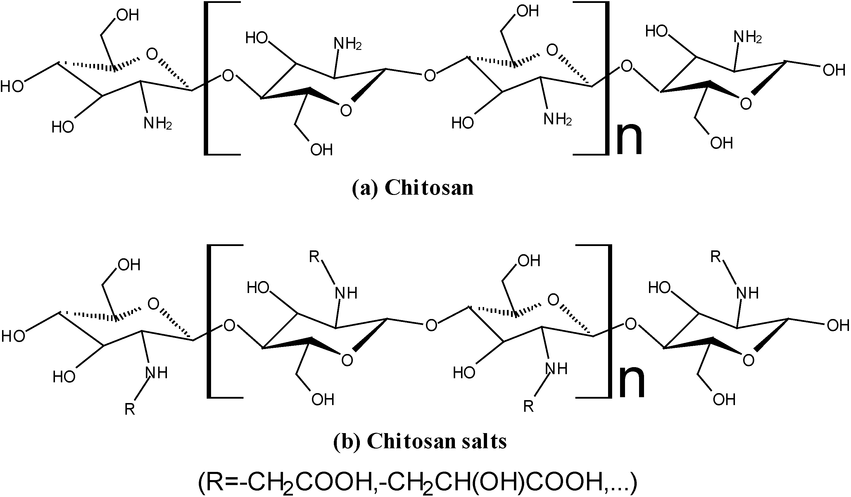
2. Properties of Chitosan and WSC
2.1. No-Toxicity
2.2. Solubility
2.3. Biocompatibility
2.4. Mucoadhesiveness
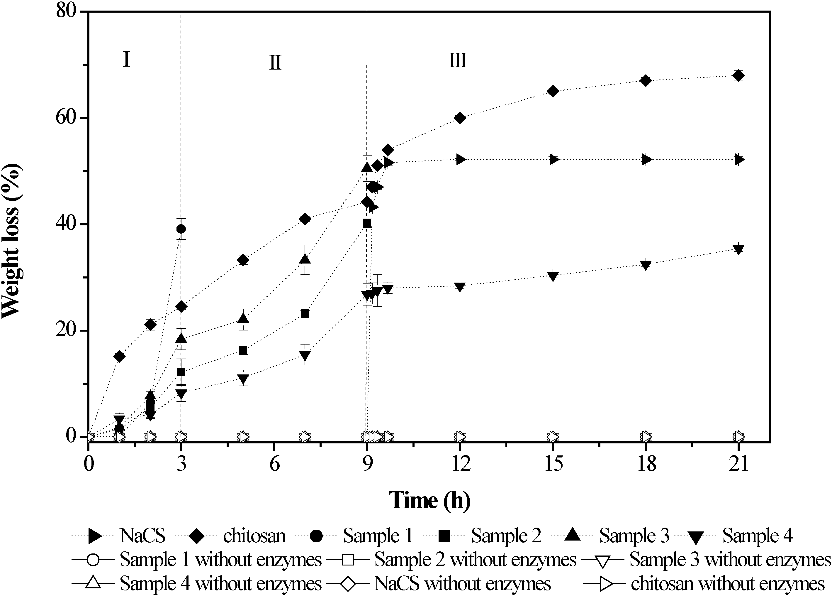
2.5. Biodegradability
3. Performances of PEC Based on Chitosan and WSC
4. Drug Carriers Designed with PECs Based on Chitosan and WSC
4.1. Films
| Polycationic Polyelectrolyte | Polyanionic Polyelectrolyte | Cross-Linking Agent | Preparation Method | Package Drugs | Reference |
|---|---|---|---|---|---|
| Chitosan | Polyacrylic acid | Glycerol/ | Cast | — | [63] |
| PEG200/ | |||||
| Hydrovance/ | |||||
| Trehalose | |||||
| Chitosan | NaCS | — | Cast | Paracetamol/5-ASA | [59,64] |
| Chitosan | Polyalkyleneoxide-maleic acid copolymer | — | Cast | Salicylic acid/Phenol | [65] |
| Chitosan | Hyaluronic acid | — | Self-assembly | — | [66] |

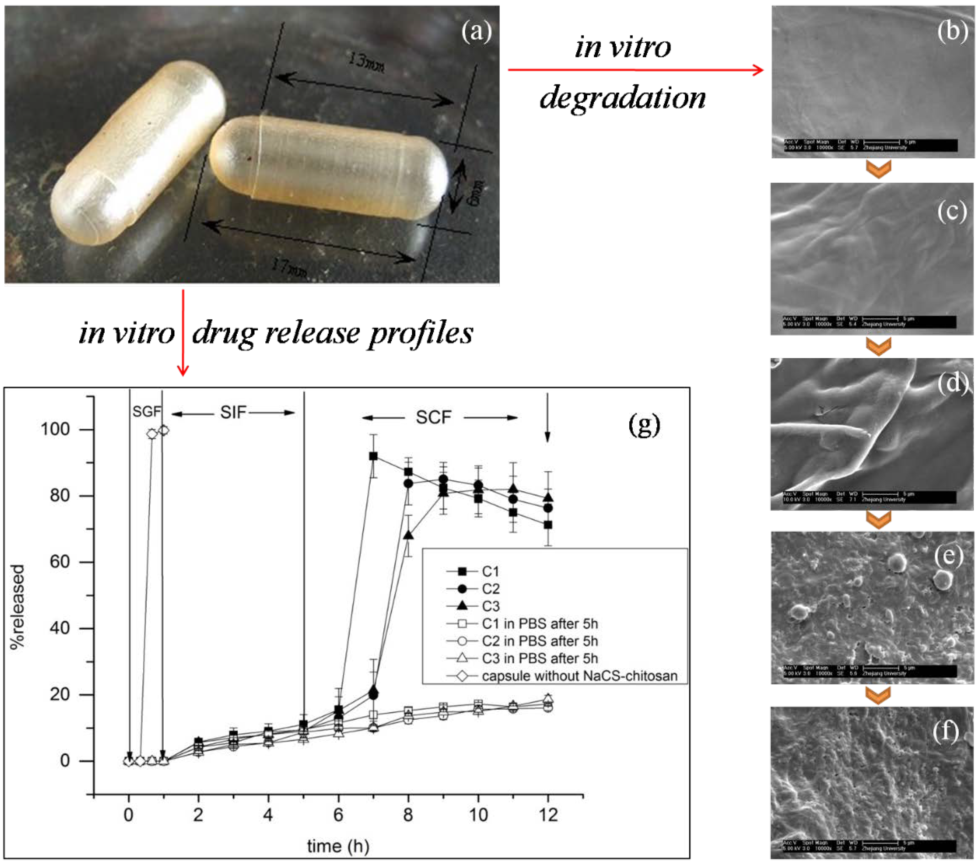
4.2. Hard Hollow Capsules

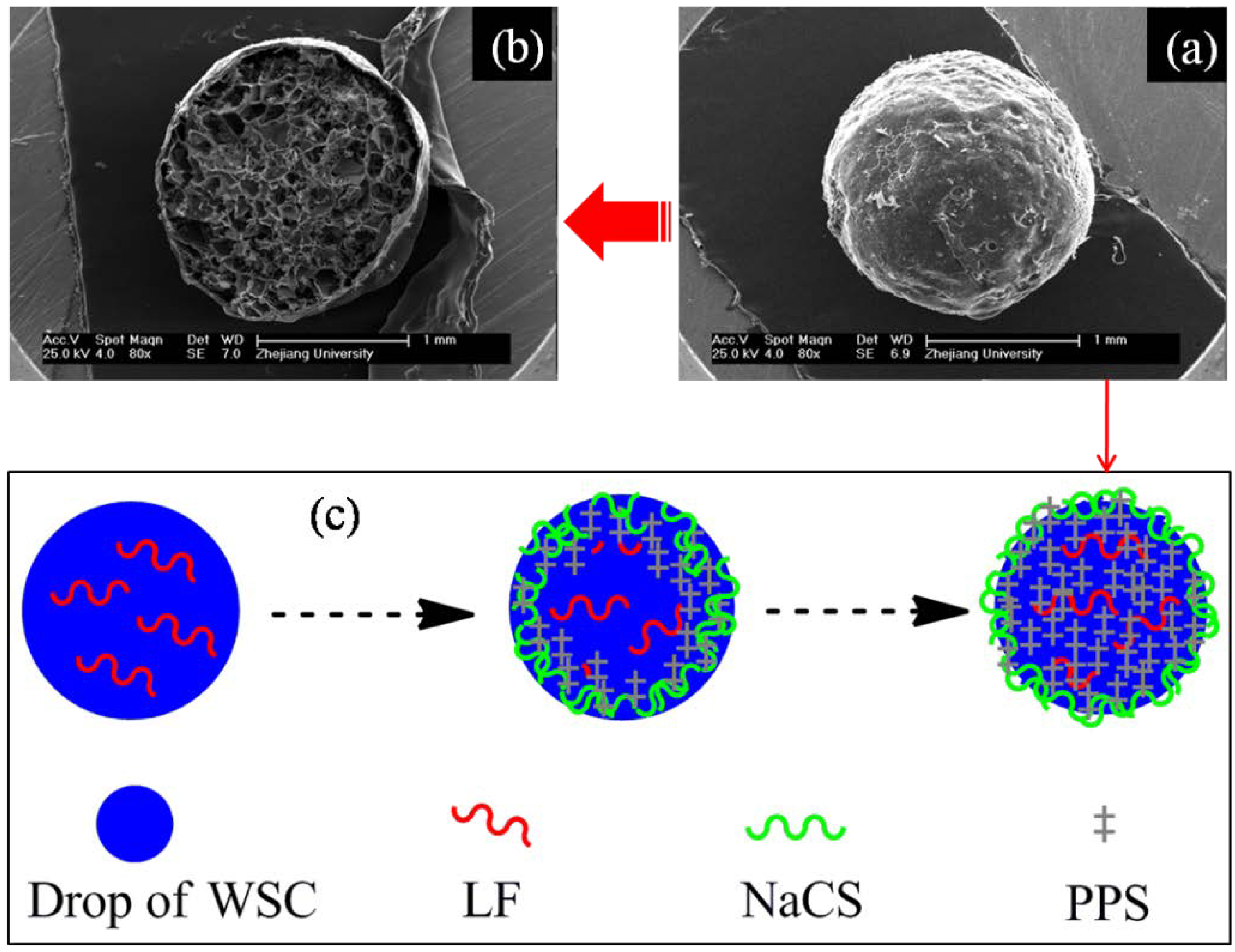
4.3. Microcapsules
| Polycationic Polyelectrolyte | Polyanionic Polyelectrolyte | Cross-Linking Agent | Preparation Method | Package Drugs | Reference |
|---|---|---|---|---|---|
| Chitosan | NaCS | Sodium polyphosphate | Orifice-polymerization | 5-ASA | [71] |
| Chitosan | Sodium alginate | — | Electrospray | Albumin | [72] |
| Chitosan | Sodium alginate | Calcium chloride | Modified orifice | Albendazole | [73] |
| Chitosan | NaCS | — | Self-assembly | — | [74] |
| Chitosan | kappa-carrageenan | Glutaraldehyde/Genipin/Tannic acid | Emulsion | Neem Seed Oil | [75] |
| WSCb | NaCS | Sodium polyphosphate | Orifice-polymerization | Lactoferrin | [76] |

| Polycationic Polyelectrolyte | Polyanionic Polyelectrolyte | Cross-Linking Agent | Preparation Method | Package Drugs | Reference |
|---|---|---|---|---|---|
| Chitosan | Dextran sulfate | — | Self-assembly | Insulin | [80] |
| WSCc | Sodium alginate | Calcium chloride | Coaxial air-flow | Naproxen | [10] |
| Chitosan | Pectin | Tripolyphosphate | Emulsion | Gliclazide | [81] |
| Chitosan | Hyaluronan sodium salt | — | Stirring (Non-solvent) | Chloramphenicol succinate sodium salt/Cefotaxime sodium salt | [82] |
| Chitosan | Polybetaine | — | Stirring | Chloramphenicol succinate sodium salt | [83] |
| Chitosan | Hyaluronic acid | — | Self-assembly | Paclitaxel | [84] |
| Chitosan | Carboxymethyl gum kondagogu | — | Coacervation | Ofloxacin | [85] |
| Chitosan | Sodium alginate | — | Ionic gelation | Amoxicillin | [86] |
4.4. Microparticles/Nanoparticles
5. Conclusions
Acknowledgments
Conflicts of Interest
References
- Onishi, H.; Machida, Y. Biodegradation and distribution of water-soluble chitosan in mice. Biomaterials 1999, 20, 175–182. [Google Scholar] [CrossRef] [PubMed]
- Xia, W.S.; Liu, P.; Liu, J. Advance in chitosan hydrolysis by non-specific cellulases. Bioresour. Technol. 2008, 99, 6751–6762. [Google Scholar] [CrossRef] [PubMed]
- Zhang, H.; Alsarra, I.A.; Neau, S.H. An in vitro evaluation of a chitosan-containing multiparticulate system for macromolecule delivery to the colon. Int. J. Pharm. 2002, 239, 197–205. [Google Scholar] [CrossRef] [PubMed]
- Zhang, Y.Q.; Niu, Y.G.; Luo, Y.C.; Ge, M.; Yang, T.; Yu, L.L.; Wang, Q. Fabrication, characterization and antimicrobial activities of thymol-loaded zein nanoparticles stabilized by sodium caseinate-chitosan hydrochloride double layers. Food Chem. 2014, 142, 269–275. [Google Scholar] [CrossRef] [PubMed]
- Dang, Q.F.; Yan, J.Q.; Li, Y.; Cheng, X.J.; Liu, C.S.; Chen, X.G. Chitosan acetate as an active coating material and its effects on the storing of Prunus avium L. J. Food Sci. 2010, 75, S125–S131. [Google Scholar] [CrossRef] [PubMed]
- Parodi, B.; Russo, E.; Caviglioli, G.; Baldassari, S.; Gaglianone, N.; Schito, A.M.; Cafaggi, S. A chitosan lactate/poloxamer 407-based matrix containing Eudragit RS microparticles for vaginal delivery of econazole: Design and in vitro evaluation. Drug Dev. Ind. Pharm. 2013, 39, 1911–1920. [Google Scholar] [CrossRef] [PubMed]
- Xu, P.S.; Bajaj, G.; Shugg, T.; van Alstine, W.G.; Yeo, Y. Zwitterionic chitosan derivatives for pH-sensitive stealth coating. Biomacromolecules 2010, 11, 2352–2358. [Google Scholar] [CrossRef] [PubMed]
- Bajaj, G.; van Alstine, W.G.; Yeo, Y. Zwitterionic chitosan derivative, a new biocompatible pharmaceutical excipient, prevents endotoxin-mediated cytokine release. PLoS One 2012, 7, 1–10. [Google Scholar]
- Shu, S.J.; Zhang, X.G.; Teng, D.Y.; Wang, Z.; Li, C.X. Polyelectrolyte nanoparticles based on water-soluble chitosan-poly(l-aspartic acid)-polyethylene glycol for controlled protein release. Carbohyd. Res. 2009, 344, 1197–1204. [Google Scholar] [CrossRef]
- Čalija, B.; Cekić, N.; Savić, S.; Daniels, R.; Marković, B.; Milić, J. pH-sensitive microparticles for oral drug delivery based on alginate/oligochitosan/Eudragit® L100–55 “sandwich” polyelectrolyte complex. Colloid. Surf. B 2013, 110, 395–402. [Google Scholar] [CrossRef]
- Franco, R.A.; Nguyen, T.H.; Lee, B.T. Preparation and characterization of electrospun PCL/PLGA membranes and chitosan/gelatin hydrogels for skin bioengineering applications. J. Mater. Sci. Mater. M. 2011, 22, 2207–2218. [Google Scholar] [CrossRef]
- Mahmoudzadeh, M.; Fassihi, A.; Emami, J.; Davies, N.M.; Dorkoosh, F. Physicochemical, pharmaceutical and biological approaches toward designing optimized and efficient hydrophobically modified chitosan-based polymeric micelles as a nanocarrier system for targeted delivery of anticancer drugs. J. Drug Target. 2013, 21, 693–709. [Google Scholar] [CrossRef] [PubMed]
- Supper, S.; Anton, N.; Seidel, N.; Riemenschnitter, M.; Curdy, C.; Vandamme, T. Thermosensitive chitosan/glycerophosphate-based hydrogel and its derivatives in pharmaceutical and biomedical applications. Expert Opin. Drug Deliv. 2014, 11, 249–267. [Google Scholar] [CrossRef] [PubMed]
- Zakhem, E.; Raghavan, S.; Bitar, K.N. Neo-innervation of a bioengineered intestinal smooth muscle construct around chitosan scaffold. Biomaterials 2014, 35, 1882–1889. [Google Scholar] [CrossRef] [PubMed]
- Lacerda, L.; Parize, A.L.; Fávere, V.; Laranjeira, M.C.M.; Stulzer, H.K. Development and evaluation of pH-sensitive sodium alginate/chitosan microparticles containing the antituberculosis drug rifampicin. Mat. Sci. Eng. C 2014, 39, 161–167. [Google Scholar] [CrossRef]
- Xiong, Y.; Yan, K.; Bentley, W.E.; Deng, H.B.; Du, Y.M.; Payne, G.F.; Shi, X.W. Compartmentalized Multilayer Hydrogel Formation Using a Stimulus-Responsive Self-Assembling Polysaccharide. ACS Appl. Mater. Inter. 2014, 6, 2948–2957. [Google Scholar] [CrossRef]
- Oyarzun-Ampuero, F.A.; Brea, J.; Loza, M.I.; Torres, D.; Alonso, M.J. Chitosan-hyaluronic acid nanoparticles loaded with heparin for the treatment of asthma. Int. J. Pharm. 2009, 381, 122–129. [Google Scholar] [CrossRef] [PubMed]
- Wang, M.J.; Xie, Y.L.; Zheng, Q.D.; Yao, S.J. A novel, potential microflora-activated carrier for a colon-specific drug delivery system and its characteristics. Ind. Eng. Chem. Res. 2009, 48, 5276–5284. [Google Scholar] [CrossRef]
- Dima, C.; Cotarlet, M.; Alexe, P.; Dima, S. Microencapsulation of essential oil of pimento [Pimenta dioica (L) Merr.] by chitosan/k-carrageenan complex coacervation method. Innov. Food Sci. Emerg. 2014, 22, 203–211. [Google Scholar] [CrossRef]
- Kaihara, S.; Suzuki, Y.; Fujimoto, K. In situ synthesis of polysaccharide nanoparticles via polyion complex of carboxymethyl cellulose and chitosan. Colloid. Surf. B. 2011, 85, 343–348. [Google Scholar] [CrossRef]
- Pandey, S.; Mishra, A.; Raval, P.; Patel, H.; Gupta, A.; Shah, D. Chitosan-pectin polyelectrolyte complex as a carrier for colon targeted drug delivery. J. Young Pharm. 2013, 5, 160–166. [Google Scholar] [CrossRef] [PubMed]
- Fukuda, M.; Peppas, N.A.; McGinity, J.W. Properties of sustained release hot-melt extruded tablets containing chitosan and xanthan gum. Int. J. Pharm. 2006, 310, 90–100. [Google Scholar] [CrossRef] [PubMed]
- Ganguly, K.; Aminabhavi, T.M.; Kulkarni, A.R. Colon targeting of 5-fluorouracil using polyethylene glycol cross-linked chitosan microspheres enteric coated with cellulose acetate phthalate. Ind. Eng. Chem. Res. 2011, 50, 11797–11807. [Google Scholar] [CrossRef]
- Wu, Q.X.; Zhang, Q.L.; Lin, D.Q.; Yao, S.J. Characterization of novel lactoferrin loaded capsules prepared with polyelectrolyte complexes. Int. J. Pharm. 2013, 455, 124–131. [Google Scholar] [CrossRef] [PubMed]
- Hamman, J.H. Chitosan based polyelectrolyte complexes as potential carrier materials in drug delivery systems. Mar. Drugs 2010, 8, 1305–1322. [Google Scholar] [CrossRef] [PubMed]
- Luo, Y.C.; Wang, Q. Recent development of chitosan-based polyelectrolyte complexes with natural polysaccharides for drug delivery. Int. J. Biol. Macromol. 2014, 64, 353–367. [Google Scholar] [CrossRef] [PubMed]
- Yan, S.F.; Zhang, K.X.; Liu, Z.W.; Zhang, X.; Gan, L.; Cao, B.; Chen, X.S.; Cui, L.; Yin, J.B. Fabrication of poly(l-glutamic acid)/chitosan polyelectrolyte complex porous scaffolds for tissue engineering. J. Mater. Chem. B 2013, 1, 1541–1551. [Google Scholar] [CrossRef]
- Ma, G.P.; Wang, Z.L.; Chen, J.; Yin, R.X.; Chen, B.L.; Nie, J. Freeze-dried chitosan-sodium hyaluronate polyelectrolyte complex fibers as tissue engineering scaffolds. New J. Chem. 2014, 38, 1211–1217. [Google Scholar] [CrossRef]
- Busilacchi, A.; Gigante, A.; Mattioli-Belmonte, M.; Manzotti, S.; Muzzarelli, R. Chitosan stabilizes platelet growth factors and modulates stem cell differentiation toward tissue regeneration. Carbohyd. Polym. 2013, 98, 665–676. [Google Scholar] [CrossRef]
- Arai, K.; Kinumaki, T.; Fujita, T. Toxicity of chitosan. Bull. Tokai Region. Fish. Res. Lab. 1968, 56, 88–94. [Google Scholar]
- Wedmore, I.; McManus, J.G.; Pusateri, A.E.; Holcomb, J.B. A special report on the chitosan-based hemostatic dressing: Experience in current combat operations. J. Trauma 2006, 60, 655–658. [Google Scholar] [CrossRef] [PubMed]
- Illum, L. Chitosan and its use as a pharmaceutical excipient. Pharm. Res. 1998, 15, 1326–1331. [Google Scholar] [CrossRef] [PubMed]
- Singh, V.; Tiwari, M. Hydrophobic modification of chitosan and its physicochemical evaluation as sustained release tablet formulation. Asian J. Chem. 2011, 23, 2141–2150. [Google Scholar]
- Costa, E.M.; Silva, S.; Costa, M.R.; Pereira, M.; Campos, D.A.; Odila, J.; Madureira, A.R.; Cardelle-Cobas, A.; Tavaria, F.K.; Rodrigues, A.S.; et al. Chitosan mouthwash: Toxicity and in vivo validation. Carbohyd. Polym. 2014, 111, 385–392. [Google Scholar] [CrossRef]
- Chen, C.Y.; Chung, Y.C. Antibacterial effect of water-soluble chitosan on representative dental pathogens Streptococcus Mutans and Lactobacilli Brevis. J. Appl. Oral Sci. 2012, 20, 620–627. [Google Scholar] [CrossRef] [PubMed]
- Kean, T.; Thanou, M. Biodegradation, biodistribution and toxicity of chitosan. Adv. Drug Deliver. Rev. 2010, 62, 3–11. [Google Scholar] [CrossRef]
- Hawary, D.L.; Motaleb, M.A.; Farag, H.; Guirguis, O.W.; Elsabee, M.Z. Water-soluble derivatives of chitosan as a target delivery system of Tc-99m to some organs in vivo for nuclear imaging and biodistribution. J. Radioanal. Nucl. Chem. 2011, 290, 557–567. [Google Scholar] [CrossRef]
- Hejazi, R.; Amiji, M. Chitosan-based gastrointestinal delivery systems. J. Control. Release 2003, 89, 151–165. [Google Scholar] [CrossRef] [PubMed]
- Nunthanid, J.; Huanbutta, K.; Luangtana-anan, M.; Sriamornsak, P.; Limmatvapirat, S.; Puttipipatkhachorn, S. Development of time-, pH-, and enzyme-controlled colonic drug delivery using spray-dried chitosan acetate and hydroxypropyl methylcellulose. Eur. J. Pharm. Biopharm. 2008, 68, 253–259. [Google Scholar] [CrossRef] [PubMed]
- Shu, X.Z.; Zhu, K.J. Controlled drug release properties of ionically cross-linked chitosan beads: The influence of anion structure. Int. J. Pharm. 2002, 233, 217–225. [Google Scholar] [CrossRef] [PubMed]
- Rai, G.; Jain, S.K.; Agrawal, S.; Bhadra, S.; Pancholi, S.S.; Agrawal, G.P. Chitosan hydrochloride based microspheres of albendazole for colonic drug delivery. Pharmazie 2005, 60, 131–134. [Google Scholar] [PubMed]
- Sobol, M.; Bartkowiak, A.; de Haan, B.; de Vos, P. Cytotoxicity study of novel water-soluble chitosan derivatives applied as membrane material of alginate microcapsules. J. Biomed. Mater. Res. A 2013, 101, 1907–1914. [Google Scholar] [CrossRef] [PubMed]
- Xiao, C.M.; Sun, F. Fabrication of distilled water-soluble chitosan/alginate functional multilayer composite microspheres. Carbohyd. Polym. 2013, 98, 1366–1370. [Google Scholar] [CrossRef]
- Sheng, Y.; He, H.J.; Zou, H. Poly(lactic acid) nanoparticles coated with combined WGA and water-soluble chitosan for mucosal delivery of beta-galactosidase. Drug Deliv. 2014, 21, 370–378. [Google Scholar] [CrossRef] [PubMed]
- Keong, L.C.; Halim, A.S. In vitro models in biocompatibility assessment for biomedical-grade chitosan derivatives in wound management. Int. J. Mol. Sci. 2009, 10, 1300–1313. [Google Scholar] [CrossRef] [PubMed]
- Muzzarelli, R.; Baldassarre, V.; Conti, F.; Ferrara, P.; Biagini, G.; Gazzanelli, G.; Vasi, V. Biological activity of chitosan: Ultrastructural study. Biomaterials 1988, 9, 247–252. [Google Scholar] [CrossRef] [PubMed]
- Henricus, M.M.; Fath, K.R.; Menzenski, M.Z.; Banerjee, I.A. Morphology controlled growth of chitosan-bound microtubes and a study of their biocompatibility and antibacterial activity. Macromol. Biosci. 2009, 9, 317–325. [Google Scholar] [CrossRef] [PubMed]
- Bravo-Osuna, I.; Vauthier, C.; Farabollini, A.; Palmieri, G.F.; Ponchel, G. Mucoadhesion mechanism of chitosan and thiolated chitosan-poly(isobutyl cyanoacrylate) core-shell nanoparticles. Biomaterials 2007, 28, 2233–2243. [Google Scholar] [CrossRef] [PubMed]
- Meng-Lund, E.; Muff-Westergaard, C.; Sander, C.; Madelung, P.; Jacobsen, J. A mechanistic based approach for enhancing buccal mucoadhesion of chitosan. Int. J. Pharm. 2014, 461, 280–285. [Google Scholar] [CrossRef] [PubMed]
- Gulbake, A.; Jain, S.K. Chitosan: A potential polymer for colon-specific drug delivery system. Expert Opin. Drug Deliv. 2012, 9, 713–729. [Google Scholar] [CrossRef] [PubMed]
- Wittaya-areekul, S.; Kruenate, J.; Prahsarn, C. Preparation and in vitro evaluation of mucoadhesive properties of alginate/chitosan microparticles containing prednisolone. Int. J. Pharm. 2006, 312, 113–118. [Google Scholar] [CrossRef] [PubMed]
- Fernandes, M.; Gonçalves, I.C.; Nardecchia, S.; Amaral, I.F.; Barbosa, M.A.; Martins, M.C.L. Modulation of stability and mucoadhesive properties of chitosan microspheres for therapeutic gastric application. Int. J. Pharm. 2013, 454, 116–124. [Google Scholar] [CrossRef] [PubMed]
- Muzzarelli, R. Human enzymatic activities related to the therapeutic administration of chitin derivatives. Cell. Mol. Life Sci. 1997, 53, 131–140. [Google Scholar] [CrossRef] [PubMed]
- Sinha, V.R.; Kumria, R. Polysaccharides in colon-specific drug delivery. Int. J. Pharm. 2001, 224, 19–38. [Google Scholar] [CrossRef] [PubMed]
- Tozaki, H.; Komoike, J.; Tada, C.; Maruyama, T.; Terabe, A.; Suzuki, T.; Yamamoto, A.; Muranishi, S. Chitosan capsules for colon-specific drug delivery: Improvement of insulin absorption from the rat colon. J. Pharm. Sci. 1997, 86, 1016–1021. [Google Scholar] [CrossRef] [PubMed]
- Orienti, I.; Cerchiara, T.; Luppi, B.; Bigucci, F.; Zuccari, G.; Zecchi, V. Influence of different chitosan salts on the release of sodium diclofenac in colon-specific delivery. Int. J. Pharm. 2002, 238, 51–59. [Google Scholar] [CrossRef] [PubMed]
- Yang, Y.M.; Hu, W.; Wang, X.D.; Gu, X.S. The controlling biodegradation of chitosan fibers by N-acetylation in vitro and in vivo. J. Mater. Sci.-Mater. M. 2007, 18, 2117–2121. [Google Scholar] [CrossRef]
- Xu, J.; McCarthy, S.P.; Gross, R.A.; Kaplan, D.L. Chitosan film acylation and effects on biodegradability. Macromolecules 1996, 29, 3436–3440. [Google Scholar] [CrossRef]
- Zhu, L.Y.; Lin, D.Q.; Yao, S.J. Biodegradation of polyelectrolyte complex films composed of chitosan and sodium cellulose sulfate as the controllable release carrier. Carbohyd. Polym. 2010, 82, 323–328. [Google Scholar] [CrossRef]
- Thunemann, A.F.; Muller, M.; Dautzenberg, H.; Joanny, J.; Lowen, H. Polyelectrolytes with defined molecular architecture II polyelectrolyte complexes. Adv. Polym. Sci. 2004, 166, 113–171. [Google Scholar]
- Ramaraj, B.; Radhakrishnan, G. Hydrogel capsules for sustained drug-release. J. Appl. Polym. Sci. 1994, 51, 979–988. [Google Scholar] [CrossRef]
- II’Ina, A.V.; Varlamov, V.P. Chitosan-based polyelectrolyte complexes: A review. Appl. Biochem. Microbiol. 2005, 41, 5–11. [Google Scholar] [CrossRef]
- Silva, C.L.; Pereira, J.C.; Ramalho, A.; Pais, A.A.C.C.; Sousa, J.J.S. Films based on chitosan polyelectrolyte complexes for skin drug delivery: Development and characterization. J. Membrane Sci. 2008, 320, 268–279. [Google Scholar] [CrossRef]
- Zhu, L.Y.; Yan, X.Q.; Zhang, H.M.; Lin, D.Q.; Yao, S.J.; Jiang, L. Determination of apparent drug permeability coefficients through chitosan-sodium cellulose sulfate polyelectrolyte complex films. Acta Phys.-Chim. Sin. 2014, 30, 365–370. [Google Scholar]
- Yoshizawa, T.; Shin-ya, Y.; Hong, K.; Kajiuchi, T. pH- and temperature-sensitive release behaviors from polyelectrolyte complex films composed of chitosan and PAOMA copolymer. Eur. J. Pharm. Biopharm. 2005, 59, 307–313. [Google Scholar] [CrossRef] [PubMed]
- Feng, Q.; Zeng, G.; Yang, P.; Wang, C.; Cai, J. Self-assembly and characterization of polyelectrolyte complex films of hyaluronic acid/chitosan. Colloid. Surf. A 2005, 257–258, 85–88. [Google Scholar] [CrossRef]
- Tozaki, H.; Odoriba, T.; Okada, N.; Fujita, T.; Terabe, A.; Suzuki, T.; Okabe, S.; Muranishi, S.; Yamamoto, A. Chitosan capsules for colon-specific drug delivery: enhanced localization of 5-aminosalicylic acid in the large intestine accelerates healing of TNBS-induced colitis in rats. J. Control. Release 2002, 82, 51–61. [Google Scholar] [CrossRef] [PubMed]
- Fetih, G.; Lindberg, S.; Itoh, K.; Okada, N.; Fujita, T.; Habib, F.; Artersson, P.; Attia, M.; Yamamoto, A. Improvement of absorption enhancing effects of n-dodecyl-β-d-maltopyranoside by its colon-specific delivery using chitosan capsules. Int. J. Pharm. 2005, 293, 127–135. [Google Scholar] [CrossRef] [PubMed]
- Huang, B.B.; Li, G.F.; Luo, J.H.; Duan, L.; Nobuaki, K.; Akira, Y. Permeabilities of rebamipide via rat intestinal membranes and its colon specific delivery using chitosan capsule as a carrier. World J. Gastroenterol. 2008, 14, 4928–4937. [Google Scholar] [CrossRef] [PubMed]
- Wang, M.J.; Xie, Y.L.; Chen, Z.J.; Yao, S.J. Optimizing preparation of NaCS-chitosan complex to form a potential material for the colon-specific drug delivery system. J. Appl. Polym. Sci. 2010, 117, 3001–3012. [Google Scholar]
- Wu, Q.X.; Yao, S.J. Novel NaCS-CS-PPS microcapsules as a potential enzyme-triggered release carrier for highly-loading 5-ASA. Colloid. Surf. B 2013, 109, 147–153. [Google Scholar] [CrossRef]
- Fukui, Y.; Maruyama, T.; Iwamatsu, Y.; Fujii, A.; Tanaka, T.; Ohmukai, Y.; Matsuyama, H. Preparation of monodispersed polyelectrolyte microcapsules with high encapsulation efficiency by an electrospray technique. Colloid. Surf. A 2010, 370, 28–34. [Google Scholar] [CrossRef]
- Simi, S.P.; Saraswathi, R.; Sankar, C.; Krishnan, P.N.; Dilip, C.; Ameena, K. Formulation and evaluation of Albendazole microcapsules for colon delivery using chitosan. Asian Pac. J. Trop. Med. 2010, 3, 374–378. [Google Scholar] [CrossRef]
- Xie, Y.L.; Wang, M.J.; Yao, S.J. Preparation and characterization of biocompatible microcapsules of sodium cellulose sulfate/chitosan by means of layer-by-layer self-assembly. Langmuir 2009, 25, 8999–9005. [Google Scholar] [CrossRef] [PubMed]
- Devi, N.; Maji, T.K. Effect of crosslinking agent on Neem (Azadirachta Indica A. Juss.) Seed Oil (NSO) encapsulated microcapsules kappa-carrageenan and chitosan polyelectrolyte complex. J. Macromol. Sci. A 2009, 46, 1114–1121. [Google Scholar] [CrossRef]
- Wu, Q.X.; Li, M.Z.; Yao, S.J. Performances of NaCS-WSC protein drug microcapsules with different degree of substitution of NaCS using sodium polyphosphate as cross-linking agent. Cellulose 2014, 21, 1897–1908. [Google Scholar] [CrossRef]
- Yu, W.T.; Song, H.Y.; Zheng, G.S.; Liu, X.D.; Zhang, Y.; Ma, X.J. Study on membrane characteristics of alginate-chitosan microcapsule with cell growth. J. Membr. Sci. 2011, 377, 214–220. [Google Scholar] [CrossRef]
- Cook, M.T.; Tzortzis, G.; Charalampopoulos, D.; Khutoryanskiy, V.V. Production and evaluation of dry alginate-chitosan microcapsules as an enteric delivery vehicle for probiotic bacteria. Biomacromolecules 2011, 12, 2834–2840. [Google Scholar] [CrossRef] [PubMed]
- Argin, S.; Kofinas, P.; Lo, Y.M. The cell release kinetics and the swelling behavior of physically crosslinked xanthan-chitosan hydrogels in simulated gastrointestinal conditions. Food Hydrocolloid. 2014, 40, 138–144. [Google Scholar] [CrossRef]
- Balabushevich, N.G.; Pechenkin, M.A.; Shibanova, E.D.; Volodkin, D.V.; Mikhalchik, E.V. Multifunctional polyelectrolyte microparticles for oral insulin delivery. Macromol. Biosci. 2013, 13, 1379–1388. [Google Scholar] [CrossRef] [PubMed]
- Barakat, N.S.; Almurshedi, A.S. Preparation and characterization of chitosan microparticles for oral sustained delivery of Gliclazide: In Vitro/In Vivo evaluation. Drug Dev. Res. 2011, 72, 235–246. [Google Scholar] [CrossRef]
- Vasiliu, S.; Popa, M.; Luca, C. Evaluation of retention and release processes of two antibiotics from the biocompatible core-shell microparticles. Eur. Polym. J. 2008, 44, 3894–3898. [Google Scholar] [CrossRef]
- Racovita, S.; Vasiliu, S.; Vlad, C.D. New drugs delivery systems based on polyelectrolyte complexes. Rev. Roum. Chim. 2010, 55, 659–666. [Google Scholar]
- Li, J.; Huang, P.; Chang, L.; Long, X.; Dong, A.; Liu, J.; Chu, L.; Hu, F.; Liu, J.; Deng, L. Tumor targeting and pH-responsive polyelectrolyte complex nanoparticles based on hyaluronic acid-paclitaxel conjugates and chitosan for oral delivery of paclitaxel. Macromol. Res. 2013, 21, 1331–1337. [Google Scholar] [CrossRef]
- Kumar, A.; Ahuja, M. Carboxymethyl gum kondagogu-chitosan polyelectrolyte complex nanoparticles: Preparation and characterization. Int. J. Biol. Macromol. 2013, 62, 80–84. [Google Scholar] [CrossRef] [PubMed]
- Arora, S.; Gupta, S.; Narang, R.K.; Budhiraja, R.D. Amoxicillin loaded chitosan-alginate polyelectrolyte complex nanoparticles as mucopenetrating delivery system for H. Pylori. Sci. Pharm. 2011, 79, 673–694. [Google Scholar] [CrossRef]
- Paliwal, R.; Paliwal, S.R.; Agrawal, G.P.; Vyas, S.P. Chitosan nanoconstructs for improved oral delivery of low molecular weight heparin: In vitro and in vivo evaluation. Int. J. Pharm. 2012, 422, 179–184. [Google Scholar] [CrossRef] [PubMed]
- Li, C.; Newman, R.A.; Wu, Q.P.; Ke, S.; Chen, W.; Hutto, T.; Kan, Z.; Brannan, M.D.; Charnsangavej, C.; Wallace, S. Biodistribution of paclitaxel and poly(l-glutamic acid)-paclitaxel conjugate in mice with ovarian OCa-1 tumor. Cancer Chemoth. Pharm. 2000, 46, 416–422. [Google Scholar] [CrossRef]
- Jain, A.K.; Swarnakar, N.K.; Godugu, C.; Singh, R.P.; Jain, S. The effect of the oral administration of polymeric nanoparticles on the efficacy and toxicity of tamoxifen. Biomaterials 2011, 32, 503–515. [Google Scholar] [CrossRef] [PubMed]
- Shelly; Ahuja, M.; Kumar, A. Gum ghatti-chitosan polyelectrolyte nanoparticles: Preparation and characterization. Int. J. Biol. Macromol. 2013, 61, 411–415. [Google Scholar] [CrossRef] [PubMed]
- Guo, R.; Chen, L.L.; Cai, S.S.; Liu, Z.H.; Zhu, Y.; Xue, W.; Zhang, Y.M. Novel alginate coated hydrophobically modified chitosan polyelectrolyte complex for the delivery of BSA. J. Mater. Sci.-Mater. Med. 2013, 24, 2093–2100. [Google Scholar] [CrossRef] [PubMed]
- Anitha, A.; Sreeranganathan, M.; Chennazhi, K.P.; Lakshmanan, V.; Jayakumar, R. In vitro combinatorial anticancer effects of 5-fluorouracil and curcumin loaded N,O-carboxymethyl chitosan nanoparticles toward colon cancer and in vivo pharmacokinetic studies. Eur. J. Pharm. Biopharm. 2014, 88, 238–251. [Google Scholar] [CrossRef] [PubMed]
© 2014 by the authors; licensee MDPI, Basel, Switzerland. This article is an open access article distributed under the terms and conditions of the Creative Commons Attribution license (http://creativecommons.org/licenses/by/4.0/).
Share and Cite
Wu, Q.-X.; Lin, D.-Q.; Yao, S.-J. Design of Chitosan and Its Water Soluble Derivatives-Based Drug Carriers with Polyelectrolyte Complexes. Mar. Drugs 2014, 12, 6236-6253. https://doi.org/10.3390/md12126236
Wu Q-X, Lin D-Q, Yao S-J. Design of Chitosan and Its Water Soluble Derivatives-Based Drug Carriers with Polyelectrolyte Complexes. Marine Drugs. 2014; 12(12):6236-6253. https://doi.org/10.3390/md12126236
Chicago/Turabian StyleWu, Qing-Xi, Dong-Qiang Lin, and Shan-Jing Yao. 2014. "Design of Chitosan and Its Water Soluble Derivatives-Based Drug Carriers with Polyelectrolyte Complexes" Marine Drugs 12, no. 12: 6236-6253. https://doi.org/10.3390/md12126236