Next Issue
Volume 43, December
Previous Issue
Volume 43, June
 
 
cimb-logo

Journal Browser

Journal Browser

Curr. Issues Mol. Biol., Volume 43, Issue 2 (September 2021) – 50 articles

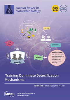
Cover Story (view full-size image): Some compounds we take (in foods, drinks, etc.) are potentially dangerous. However, evolution has endowed the human body with detoxification mechanisms whose training is achieved through a small amount of pro-oxidants; even food preservatives can be helpful in exercising antioxidant pathways. View this paper.
  • Issues are regarded as officially published after their release is announced to the table of contents alert mailing list.
  • You may sign up for e-mail alerts to receive table of contents of newly released issues.
  • PDF is the official format for papers published in both, html and pdf forms. To view the papers in pdf format, click on the "PDF Full-text" link, and use the free Adobe Reader to open them.
Order results
Result details
Section
Select all
Export citation of selected articles as:
15 pages, 3379 KiB  
Article
Oncological Properties of Intravenous Leiomyomatosis: Involvement of Mesenchymal Tumor Stem-Like Cells
by Saya Tamura, Takuma Hayashi, Hideki Tokunaga, Nobuo Yaegashi, Kaoru Abiko and Ikuo Konishi
Curr. Issues Mol. Biol. 2021, 43(2), 1188-1202; https://doi.org/10.3390/cimb43020084 - 19 Sep 2021
Cited by 7 | Viewed by 3234
Abstract
Uterine leiomyoma, also known as fibroids, is the most common benign neoplasm of the female genital tract. Leiomyoma is the most common uterine tumor. The leiomyoma subtypes account for approximately 10% of leiomyomas. Intravenous leiomyomatosis, a uterine leiomyoma subtype, is an intravascular growth [...] Read more.
Uterine leiomyoma, also known as fibroids, is the most common benign neoplasm of the female genital tract. Leiomyoma is the most common uterine tumor. The leiomyoma subtypes account for approximately 10% of leiomyomas. Intravenous leiomyomatosis, a uterine leiomyoma subtype, is an intravascular growth of benign smooth muscle cells, occasionally with pelvic or extrapelvic extension. Uterine leiomyosarcoma, a malignant tumor, tends to metastasize hematogenously, and distant metastasis to the lungs and liver is common. Therefore, the oncological properties of this intravenous leiomyomatosis resemble those of the malignant tumor uterine leiomyosarcoma. Cancer stem cells migrate to distant organs via intravascular infiltration, leading to micrometastases. We examined the oncological properties of intravenous leiomyomatosis using molecular pathological techniques on tissue excised from patients with uterine leiomyoma. CD44-positive mesenchymal tumor stem-like cells were detected in both patients with intravenous leiomyomatosis and uterine leiomyosarcoma. The oncological properties of intravenous leiomyomatosis were found to be similar to those of uterine leiomyosarcoma. However, in intravenous leiomyomatosis, cyclin E and Ki-67-positive cells were rare and no pathological findings suspecting malignancy were observed. It is expected that establishing a treatment method targeting cancer stem cells will lead to the treatment of malignant tumors with a low risk of recurrence and metastasis. Full article
(This article belongs to the Topic Cancer Biology and Therapy)
Show Figures

Figure 1

17 pages, 7191 KiB  
Article
Antioxidant and Anti-Melanogenic Activities of Heat-Treated Licorice (Wongam, Glycyrrhiza glabra × G. uralensis) Extract
by Min Hye Kang, Gwi Yeong Jang, Yun-Jeong Ji, Jeong Hoon Lee, Su Ji Choi, Tae Kyung Hyun and Hyung Don Kim
Curr. Issues Mol. Biol. 2021, 43(2), 1171-1187; https://doi.org/10.3390/cimb43020083 - 18 Sep 2021
Cited by 17 | Viewed by 5449
Abstract
Melanin is a brown or black pigment that protects skin from ultraviolet radiation and reactive oxygen species (ROS). However, overproduction of melanin is associated with lentigines, melasma, freckles and skin cancer. Licorice has shown antioxidant, anti-tumor, anti-platelet, anti-inflammatory and immunomodulatory activities and is [...] Read more.
Melanin is a brown or black pigment that protects skin from ultraviolet radiation and reactive oxygen species (ROS). However, overproduction of melanin is associated with lentigines, melasma, freckles and skin cancer. Licorice has shown antioxidant, anti-tumor, anti-platelet, anti-inflammatory and immunomodulatory activities and is used as a natural treatment for skin whitening. We aimed to confirm the potential of Wongam, a new cultivar of licorice developed by the Rural Development Administration (RDA), as a whitening agent in cosmetics. In addition, we verified the effect of heat treatment on the bioactivity of licorice by comparing antioxidant and anti-melanogenic activities of licorice extract before and after heating (130 °C). The heat-treated licorice extract (WH-130) showed higher radical-scavenging activities in the ABTS+ (2,2′-azino-bis-(3-ethylbenzothiazolin-6-sulfonic acid) diammonium salt) and DPPH (2,2-diphenyl-1-picrylhydrazyl) assays. In addition, WH-130 inhibited melanogenesis more effectively due to downregulation of tyrosinase in B16F10 melanoma cells than non-heated licorice extract. Moreover, heat treatment increased total phenolic content. In particular, isoliquiritigenin, an antioxidant and anti-melanogenic compound of licorice, was produced by heat treatment. In conclusion, WH-130, with increased levels of bioactive phenolics such as isoliquiritigenin, has potential for development into a novel skin whitening material with applications in cosmetics. Full article
Show Figures

Figure 1

15 pages, 2626 KiB  
Article
HPRep: Quantifying Reproducibility in HiChIP and PLAC-Seq Datasets
by Jonathan D. Rosen, Yuchen Yang, Armen Abnousi, Jiawen Chen, Michael Song, Ian R. Jones, Yin Shen, Ming Hu and Yun Li
Curr. Issues Mol. Biol. 2021, 43(2), 1156-1170; https://doi.org/10.3390/cimb43020082 - 17 Sep 2021
Cited by 5 | Viewed by 2795
Abstract
HiChIP and PLAC-Seq are emerging technologies for studying genome-wide long-range chromatin interactions mediated by the protein of interest, enabling more sensitive and cost-efficient interrogation of protein-centric chromatin conformation. However, due to the unbalanced read distribution introduced by protein immunoprecipitation, existing reproducibility measures developed [...] Read more.
HiChIP and PLAC-Seq are emerging technologies for studying genome-wide long-range chromatin interactions mediated by the protein of interest, enabling more sensitive and cost-efficient interrogation of protein-centric chromatin conformation. However, due to the unbalanced read distribution introduced by protein immunoprecipitation, existing reproducibility measures developed for Hi-C data are not appropriate for the analysis of HiChIP and PLAC-Seq data. Here, we present HPRep, a stratified and weighted correlation metric derived from normalized contact counts, to quantify reproducibility in HiChIP and PLAC-Seq data. We applied HPRep to multiple real datasets and demonstrate that HPRep outperforms existing reproducibility measures developed for Hi-C data. Specifically, we applied HPRep to H3K4me3 PLAC-Seq data from mouse embryonic stem cells and mouse brain tissues as well as H3K27ac HiChIP data from human lymphoblastoid cell line GM12878 and leukemia cell line K562, showing that HPRep can more clearly separate among pseudo-replicates, real replicates, and non-replicates. Furthermore, in an H3K4me3 PLAC-Seq dataset consisting of 11 samples from four human brain cell types, HPRep demonstrated the expected clustering of data that could not be achieved by existing methods developed for Hi-C data, highlighting the need for a reproducibility metric tailored to HiChIP and PLAC-Seq data. Full article
(This article belongs to the Section Bioinformatics and Systems Biology)
Show Figures

Figure 1

14 pages, 2308 KiB  
Article
Combined Transcriptome and Proteome Analysis of Maize (Zea mays L.) Reveals A Complementary Profile in Response to Phosphate Deficiency
by Zhi Nie, Bowen Luo, Xiao Zhang, Ling Wu, Dan Liu, Jialei Guo, Xuan He, Duojiang Gao, Shiqiang Gao and Shibin Gao
Curr. Issues Mol. Biol. 2021, 43(2), 1142-1155; https://doi.org/10.3390/cimb43020081 - 13 Sep 2021
Cited by 9 | Viewed by 2599
Abstract
A deficiency in the macronutrient phosphate (Pi) brings about various changes in plants at the morphological, physiological and molecular levels. However, the molecular mechanism for regulating Pi homeostasis in response to low-Pi remains poorly understood, particularly in maize (Zea mays L.), which [...] Read more.
A deficiency in the macronutrient phosphate (Pi) brings about various changes in plants at the morphological, physiological and molecular levels. However, the molecular mechanism for regulating Pi homeostasis in response to low-Pi remains poorly understood, particularly in maize (Zea mays L.), which is a staple crop and requires massive amounts of Pi. Therefore, in this study, we performed expression profiling of the shoots and roots of maize seedlings with Pi-tolerant genotype at both the transcriptomic and proteomic levels using RNA sequencing and isobaric tags for relative and absolute quantitation (iTRAQ). We identified 1944 differentially expressed transcripts and 340 differentially expressed proteins under low-Pi conditions. Most of the differentially expressed genes were clustered as regulators, such as transcription factors involved in the Pi signaling pathway at the transcript level. However, the more functional and metabolism-related genes showed expression changes at the protein level. Moreover, under low-Pi conditions, Pi transporters and phosphatases were specifically induced in the roots at both the transcript and protein levels, and increased amounts of mRNA and protein of two purple acid phosphatases (PAPs) and one UDP-sulfoquinovose synthase (SQD) were specifically detected in the roots. The new insights provided by this study will help to improve the P-utilization efficiency of maize. Full article
(This article belongs to the Section Bioinformatics and Systems Biology)
Show Figures

Figure 1

10 pages, 827 KiB  
Article
Chromosomal Location of Genes Differentially Expressed in Tumor Cells Surviving High-Dose X-ray Irradiation: A Preliminary Study on Radio-Fragile Sites
by Kaori Tsutsumi, Moe Masuda and Hiroyuki Date
Curr. Issues Mol. Biol. 2021, 43(2), 1133-1141; https://doi.org/10.3390/cimb43020080 - 8 Sep 2021
Viewed by 2565
Abstract
Altered gene expression is a common feature of tumor cells after irradiation. Our previous study showed that this phenomenon is not only an acute response to cytotoxic stress, instead, it was persistently detected in tumor cells that survived 10 Gy irradiation (IR cells). [...] Read more.
Altered gene expression is a common feature of tumor cells after irradiation. Our previous study showed that this phenomenon is not only an acute response to cytotoxic stress, instead, it was persistently detected in tumor cells that survived 10 Gy irradiation (IR cells). The current understanding is that DNA double-strand breaks (DSBs) are recognized by the phosphorylation of histone H2AX (H2AX) and triggers the ataxia-telangiectasia mutated (ATM) protein or the ATM- and Rad3-related (ATR) pathway, which activate or inactivate the DNA repair or apoptotic or senescence related molecules and causes the expression of genes in many instances. However, because changes in gene expression persist after passaging in IR cells, it may be due to the different pathways from these transient intracellular signaling pathways caused by DSBs. We performed microarray analysis of 30,000 genes in radiation-surviving cells (H1299-IR and MCF7-IR) and found an interesting relation between altered genes and their chromosomal loci. These loci formed a cluster on the chromosome, especially on 1q21 and 6p21-p22 in both irradiated cell lines. These chromosome sites might be regarded as “radio-fragile” sites. Full article
(This article belongs to the Special Issue Linking Genomic Changes with Cancer in the NGS Era)
Show Figures

Figure 1

20 pages, 7559 KiB  
Article
Saussurea lappa Exhibits Anti-Oncogenic Effect in Hepatocellular Carcinoma, HepG2 Cancer Cell Line by Bcl-2 Mediated Apoptotic Pathway and Mitochondrial Cytochrome C Release
by Amal A. Alotaibi, Asmatanzeem Bepari, Rasha Assad Assiri, Shaik Kalimulla Niazi, Sreenivasa Nayaka, Muthuraj Rudrappa, Shashiraj Kareyellapa Nagaraja and Meghashyama Prabhakara Bhat
Curr. Issues Mol. Biol. 2021, 43(2), 1114-1132; https://doi.org/10.3390/cimb43020079 - 8 Sep 2021
Cited by 25 | Viewed by 4015
Abstract
Background and Objectives: Saussurea lappa (S. lappa) is an important species of the Asteraceae family with several purposes in traditional medicine. This study intended to explore the cytotoxic effect of S. lappa on HepG2 cancer cell proliferation. Materials and Methods: The effects [...] Read more.
Background and Objectives: Saussurea lappa (S. lappa) is an important species of the Asteraceae family with several purposes in traditional medicine. This study intended to explore the cytotoxic effect of S. lappa on HepG2 cancer cell proliferation. Materials and Methods: The effects of an S. lappa n-butanol extract on the induction of apoptosis were investigated by flow cytometry and mitochondrial cytochrome C-releasing apoptosis assay. Additionally, real-time PCR was employed to confirm apoptosis initiation. Further, qualitative estimation of the active constituent of S. lappa was done by gas chromatography–mass spectroscopy (GC–MS). Results: The cell viability study revealed that the n-butanol extract of S. lappa demonstrated potent cytotoxicity against HepG2 cancer cells, with an IC50 value of 56.76 μg/mL. Cell morphology with dual staining of acridine orange (AO)-ethidium bromide (EB) showed an increase in orange/red nuclei due to cell death by S. lappa n-butanol extract compared to control cells. Apoptosis, as the mode of cell death, was also confirmed by the higher release of cytochrome C from mitochondria, the increased expression of caspase-3 and bax, along with down regulation of Bcl-2. Conclusion: These findings conclude that S. lappa is a cause of hepatic cancer cell death through apoptosis and a potential natural source suggesting furthermore investigation of its active compounds that are responsible for these observed activities. Full article
(This article belongs to the Topic Cancer Biology and Therapy)
Show Figures

Figure 1

24 pages, 927 KiB  
Article
Screening for Copy Number Variations of the 15q13.3 Hotspot in CHRNA7 Gene and Expression in Patients with Migraines
by Mehmet Fatih Özaltun, Sırma Geyik and Şenay Görücü Yılmaz
Curr. Issues Mol. Biol. 2021, 43(2), 1090-1113; https://doi.org/10.3390/cimb43020078 - 7 Sep 2021
Cited by 4 | Viewed by 2296
Abstract
Background: a migraine is a neurological disease. Copy number variation (CNV) is a phenomenon in which parts of the genome are repeated. We investigated the effects of the CNV and gene expression at the location 15q13.3 in the Cholinergic Receptor Nicotinic Alpha 7 [...] Read more.
Background: a migraine is a neurological disease. Copy number variation (CNV) is a phenomenon in which parts of the genome are repeated. We investigated the effects of the CNV and gene expression at the location 15q13.3 in the Cholinergic Receptor Nicotinic Alpha 7 Subunit (CHRNA7) gene, which we believe to be effective in the migraine clinic. Methods: we evaluated changes in CHRNA7 gene expression levels and CNV of 15q13.3 in patients with migraine (n = 102, with aura, n = 43; without aura, n = 59) according to healthy controls (n = 120) by q-PCR. The data obtained were analyzed against the reference telomerase reverse transcriptase (TERT) gene with the double copy number by standard curve analysis. Copy numbers were graded as a normal copy (2), gain (2>), and loss (<2). Results: we analyzed using the 2ΔΔCT calculation method. The CHRNA7 gene was significantly downregulated in patients (p < 0.05). The analysis of CNV in the CHRNA7 gene was statistically significant in the patient group, according to healthy controls (p < 0.05). A decreased copy number indicates a dosage loss. However, no significant difference was observed among gain, normal, and loss copy numbers and expression values in patients (p > 0.05). The change in CNV was not associated with the downregulation of the CHRNA7 gene. Conclusion: Downregulation of the CHRNA7 gene may contribute to the formation of migraine by inactivation of the alpha-7 nicotinic receptor (α7nAChR). The association of CNV gains and losses with migraines will lead to better understanding of the molecular mechanisms and pathogenesis, to better define the disease, to be used as a treatment target. Full article
(This article belongs to the Section Bioinformatics and Systems Biology)
Show Figures

Figure 1

10 pages, 2017 KiB  
Article
Hemopexin Modulates Expression of Complement Regulatory Proteins in Rat Glomeruli
by Maria G. Detsika and Elias A. Lianos
Curr. Issues Mol. Biol. 2021, 43(2), 1081-1089; https://doi.org/10.3390/cimb43020077 - 7 Sep 2021
Cited by 1 | Viewed by 1967
Abstract
In systemic hemolysis and in hematuric forms of kidney injury, the major heme scavenging protein, hemopexin (HPX), becomes depleted, and the glomerular microvasculature (glomeruli) is exposed to high concentrations of unbound heme, which, in addition to causing oxidative injury, can activate complement cascades; [...] Read more.
In systemic hemolysis and in hematuric forms of kidney injury, the major heme scavenging protein, hemopexin (HPX), becomes depleted, and the glomerular microvasculature (glomeruli) is exposed to high concentrations of unbound heme, which, in addition to causing oxidative injury, can activate complement cascades; thus, compounding extent of injury. It is unknown whether unbound heme can also activate specific complement regulatory proteins that could defend against complement-dependent injury. Isolated rat glomeruli were incubated in media supplemented with HPX-deficient (HPX) or HPX-containing (HPX+) sera as a means of achieving different degrees of heme partitioning between incubation media and glomerular cells. Expression of heme oxygenase (HO)-1 and of the complement activation inhibitors, decay-accelerating factor (DAF), CD59, and complement receptor-related gene Y (Crry), was assessed by western blot analysis. Expression of HO-1 and of the GPI-anchored DAF and CD59 proteins increased in isolated glomeruli incubated with HPX sera with no effect on Crry expression. Exogenous heme (hemin) did not further induce DAF but increased Crry expression. HPX modulates heme-mediated induction of complement activation controllers in glomeruli. This effect could be of translational relevance in glomerular injury associated with hematuria. Full article
(This article belongs to the Section Biochemistry, Molecular and Cellular Biology)
Show Figures

Figure 1

10 pages, 665 KiB  
Article
Regulation of Immunity in Clear Cell Renal Carcinoma: Role of PD-1, PD-L1, and PD-L2
by Liudmila Spirina, Zahar Yurmazov, Evgeny Usynin, Irina Kondakova, Ekaterine Ladutko and Evgeny Choynzonov
Curr. Issues Mol. Biol. 2021, 43(2), 1072-1080; https://doi.org/10.3390/cimb43020076 - 6 Sep 2021
Cited by 3 | Viewed by 2573
Abstract
Regulation of immunity is a unique oncogenic mechanism that differs in different cancers. VHL deficient clear cell renal cell carcinomas (ccRCC) trigger the immune response resulting in cancer progression. This study aimed to investigate PD-1, PD-L1, and PD-L2 expression in ccRCC primary cancers [...] Read more.
Regulation of immunity is a unique oncogenic mechanism that differs in different cancers. VHL deficient clear cell renal cell carcinomas (ccRCC) trigger the immune response resulting in cancer progression. This study aimed to investigate PD-1, PD-L1, and PD-L2 expression in ccRCC primary cancers and metastatic tissues associated with the p-VHL content, transcriptional, and growth factors expression. Methods: A total of 62 patients with RCC were enrolled in the study. Investigation of mRNA level was performed by PCR in real-time. Western blotting analysis was used for detecting the p-VHL protein content in tissues. Results: The PD-L2 prevalence in metastatic cancers is crucial in tumor progression. The VHL expression and p-VHL content determined the aggressive cancer behavior and elevated in disseminated tumors. The cancer dissemination was accompanied by an increase in both mRNA and VHL content. Conclusion: We present a new instrument targeting pathologies with p-VHL/HIF altered function that impact the PD-L2 expression through the change in transcriptional, growth factors, and AKT/mTOR modulation. Full article
(This article belongs to the Section Molecular Medicine)
Show Figures

Graphical abstract

16 pages, 2264 KiB  
Article
Resveratrol Ameliorates Aortic Calcification in Ovariectomized Rats via SIRT1 Signaling
by Sally K. Hammad, Rana G. Eissa, Mohamed A. Shaheen and Nahla N. Younis
Curr. Issues Mol. Biol. 2021, 43(2), 1057-1071; https://doi.org/10.3390/cimb43020075 - 5 Sep 2021
Cited by 15 | Viewed by 7388
Abstract
Postmenopausal women are at an increased risk of vascular calcification which is defined as the pathological deposition of minerals in the vasculature, and is strongly linked with increased cardiovascular disease risk. Since estrogen-replacement therapy is associated with increased cancer risk, there is a [...] Read more.
Postmenopausal women are at an increased risk of vascular calcification which is defined as the pathological deposition of minerals in the vasculature, and is strongly linked with increased cardiovascular disease risk. Since estrogen-replacement therapy is associated with increased cancer risk, there is a strong need for safer therapeutic approaches. In this study we aimed to investigate the protective and therapeutic effects of the phytoestrogen resveratrol against vascular calcification in ovariectomized rats, a preclinical model of postmenopause. Furthermore, we aimed to compare the effects of resveratrol to those of estrogen and to explore the mechanisms underpinning those effects. Treatment with resveratrol or estrogen ameliorated aortic calcification in ovariectomized rats, as shown by reduced calcium deposition in the arterial wall. Mechanistically, the effects of resveratrol and estrogen were mediated via the activation of SIRT1 signaling. SIRT1 protein expression was downregulated in the aortas of ovariectomized rats, and upregulated in rats treated with resveratrol or estrogen. Moreover, resveratrol and estrogen reduced the levels of the osteogenic markers: runt-related transcription factor 2 (RUNX2), osteocalcin and alkaline phosphatase (ALP) which have been shown to play a role during vascular calcification. Additionally, the senescence markers (p53, p16 and p21) which were also reported to play a role in the pathogenesis of vascular calcification, were reduced upon treatment with resveratrol and estrogen. In conclusion, the phytoestrogen resveratrol may be a safer alternative to estrogen, as a therapeutic approach against the progression of vascular calcification during postmenopause. Full article
Show Figures

Figure 1

14 pages, 1239 KiB  
Article
Proteomic Characterization of Colorectal Cancer Tissue from Patients Identifies Novel Putative Protein Biomarkers
by Maja Ludvigsen, Louise Thorlacius-Ussing, Henrik Vorum, Mogens Tornby Stender, Ole Thorlacius-Ussing and Bent Honoré
Curr. Issues Mol. Biol. 2021, 43(2), 1043-1056; https://doi.org/10.3390/cimb43020074 - 2 Sep 2021
Cited by 4 | Viewed by 3388
Abstract
Colorectal cancer (CRC) is one of the leading causes of cancer-related death over the world. There is a great need for biomarkers capable of early detection and as targets for treatment. Differential protein expression was investigated with two-dimensional gel electrophoresis (2D-PAGE) followed by [...] Read more.
Colorectal cancer (CRC) is one of the leading causes of cancer-related death over the world. There is a great need for biomarkers capable of early detection and as targets for treatment. Differential protein expression was investigated with two-dimensional gel electrophoresis (2D-PAGE) followed by identification with liquid chromatography–tandem mass spectrometry (LC-MS/MS) in CRC patient tissue from (i) the peripheral part of the tumor, (ii) the central part of the tumor as well as from (iii) a non-involved part of the colorectal tissue. The expression patterns of six identified proteins were further evaluated by one-dimensional Western blot (1D-WB) analysis of the CRC tissue. Proteins that were perturbed in expression level in the peripheral or in the central part of the tumor as compared with the non-involved part included S100A11, HNRNPF, HNRNPH1 or HNRNPH2, GSTP1, PKM and FABP1. These identified markers may have future diagnostic potential or may be novel treatment targets after further evaluation in larger patient cohorts. Full article
(This article belongs to the Section Molecular Medicine)
Show Figures

Figure 1

7 pages, 1048 KiB  
Case Report
Neonatal Diabetes in Patients Affected by Liang-Wang Syndrome Carrying KCNMA1 Variant p.(Gly375Arg) Suggest a Potential Role of Ca2+ and Voltage-Activated K+ Channel Activity in Human Insulin Secretion
by Chiara Mameli, Roberta Cazzola, Luigina Spaccini, Valeria Calcaterra, Maddalena Macedoni, Paola Azzurra La Verde, Enza D’Auria, Elvira Verduci, Gianluca Lista and Gian Vincenzo Zuccotti
Curr. Issues Mol. Biol. 2021, 43(2), 1036-1042; https://doi.org/10.3390/cimb43020073 - 31 Aug 2021
Cited by 10 | Viewed by 2866
Abstract
Liang-Wang syndrome (LIWAS) is a polymalformative syndrome first described in 2019 caused by heterozygous mutation of the KCNMA1 gene encoding the Ca2+ and voltage-activated K+ channel (BKC). The KCNMA1 variant p.(Gly356Arg) abolishes the function of BKC and blocks the generation of [...] Read more.
Liang-Wang syndrome (LIWAS) is a polymalformative syndrome first described in 2019 caused by heterozygous mutation of the KCNMA1 gene encoding the Ca2+ and voltage-activated K+ channel (BKC). The KCNMA1 variant p.(Gly356Arg) abolishes the function of BKC and blocks the generation of K+ current. The phenotype of this variant includes developmental delay, and visceral and connective tissue malformations. So far, only three cases of LWAS have been described, one of which also had neonatal diabetes (ND). We present the case of a newborn affected by LIWAS carrying the p.(Gly375Arg) variant who manifested diabetes in the first week of life. The description of our case strongly increases the frequency of ND in LIWAS patients and suggests a role of BK inactivation in human insulin secretion. The knowledge on the role of BKC in insulin secretion is very poor. Analyzing the possible mechanisms that could explain the association of LIWAS with ND, we speculate that BK inactivation might impair insulin secretion through the alteration of ion-dependent membrane activities and mitochondrial functions in β-cells, as well as the impaired intra-islet vessel reactivity. Full article
(This article belongs to the Section Molecular Medicine)
Show Figures

Figure 1

17 pages, 3116 KiB  
Systematic Review
Malondialdehyde, an Oxidative Stress Marker in Oral Squamous Cell Carcinoma—A Systematic Review and Meta-Analysis
by Khadijah Mohideen, Uma Sudhakar, Thayumanavan Balakrishnan, Mazen A. Almasri, Manea Musa Al-Ahmari, Hajar Saeed Al Dira, Malath Suhluli, Alok Dubey, Sheetal Mujoo, Zohaib Khurshid, A. Thirumal Raj and Shankargouda Patil
Curr. Issues Mol. Biol. 2021, 43(2), 1019-1035; https://doi.org/10.3390/cimb43020072 - 28 Aug 2021
Cited by 22 | Viewed by 5006
Abstract
Objective: To qualitative and quantitatively review published literature assessing the oxidative stress marker malondialdehyde (MDA) in oral squamous cell carcinoma (OSCC). Methodology: Pubmed (MeSH), Science Direct, Scopus, Web of Science, Willey Online Library, Cochrane, and Cross Reference were searched for studies assessing MDA [...] Read more.
Objective: To qualitative and quantitatively review published literature assessing the oxidative stress marker malondialdehyde (MDA) in oral squamous cell carcinoma (OSCC). Methodology: Pubmed (MeSH), Science Direct, Scopus, Web of Science, Willey Online Library, Cochrane, and Cross Reference were searched for studies assessing MDA levels in OSCC samples. Results: From the 1008 articles identified, 849 were excluded based on title and abstract screening due to duplication and irrelevance to the topic of interest. Full-text assessment of the remaining 159 articles led to the inclusion of only 46 articles that satisfied the selection criteria. Of these, only 26 studies had data compatible for quantitative analysis. The MDA levels in OSCC groups are significantly increased (p < 0.00001) in plasma, serum, and saliva samples in the majority of the studies evaluated. In contrast, MDA levels in OSCC tissue samples are significantly attenuated (p < 0.00001) compared to healthy controls, supported by fewer studies. Conclusions: The augmented MDA levels in plasma, serum, and saliva samples of the OSCC reflect the heightened oxidative stress level accurately. Further studies are required to understand the attenuated MDA levels in the tissue samples of OSCC. Correlation analysis between MDA levels with established clinicopathological prognostic markers could aid in formulating oxidative stress-based prognostication and treatment planning. Full article
Show Figures

Figure 1

23 pages, 2650 KiB  
Article
Active Ingredients from Euodia ruticarpa Steam Distilled Essential Oil Inhibit PC-3 Prostate Cancer Cell Growth via Direct Action and Indirect Immune Cells Conditioned Media In Vitro
by Tzu-He Yeh and Jin-Yuarn Lin
Curr. Issues Mol. Biol. 2021, 43(2), 996-1018; https://doi.org/10.3390/cimb43020071 - 28 Aug 2021
Cited by 7 | Viewed by 2739
Abstract
Active constituents isolated from Euodia ruticarpa (ER) steam distilled essential oil (SDEO) against PC-3 prostate cancer cell growth remain unclear. To clarify the puzzle, ER SDEO was extracted and further resolved into six isolated fractions ERF1–F6 with Sephadex LH-20 gel filtration chromatography to [...] Read more.
Active constituents isolated from Euodia ruticarpa (ER) steam distilled essential oil (SDEO) against PC-3 prostate cancer cell growth remain unclear. To clarify the puzzle, ER SDEO was extracted and further resolved into six isolated fractions ERF1–F6 with Sephadex LH-20 gel filtration chromatography to analyze their biological activities. Active ingredients in the isolated fractions were analyzed with GC-MS. Potential isolated fractions were selected to treat PC-3 cells with direct action and indirect treatment by mouse splenocyte- (SCM) and macrophage-conditioned media (MCM). The relationship between PC-3 cell viabilities and corresponding total polyphenols, flavonoid contents as well as Th1/Th2 cytokine profiles in SCM was analyzed using the Pearson product–moment correlation coefficient (r). As a result, ERF1–F3 was abundant in total polyphenols and flavonoids contents with diverse active ingredients. Treatments with ERF1–F3 at appropriate concentrations more or less inhibit PC-3 cell growth in a direct action manner. Only SCM, respectively, cultured with ER SDEO and ERF1–F3 markedly enhanced the effects to inhibit PC-3 cell growth, suggesting that secretions by splenocytes might involve anti-PC-3 effects. There are significantly negative correlations between PC-3 cell viabilities and IL-2, IL-10 as well as IL-10/IL-2 ratios in the corresponding SCM. Total polyphenol and flavonoid contents in the media cultured with ER SDEO isolated fractions positively correlated with IL-10 (Th2) and IL-10/IL-2 (Th2/Th1) cytokine secretion ratios by splenocytes, indicating that polyphenol and flavonoid components in ER SDEO isolated fractions promote Th2-polarized and anti-inflammatory characteristics. These new findings concluded that the inhibitory effects against PC-3 prostate cancer cell growth are attributed to active anti-inflammatory ingredients in ER SDEO and its active ERF1–F3 fractions through direct action and indirect treatment by modulating splenocytes’ cytokine secretion profiles. Full article
Show Figures

Figure 1

18 pages, 1923 KiB  
Article
Microbial Composition and Genes for Key Metabolic Attributes in the Gut Digesta of Sea Urchins Lytechinus variegatus and Strongylocentrotus purpuratus Using Shotgun Metagenomics
by Joseph A. Hakim, George B. H. Green, Stephen A. Watts, Michael R. Crowley, Casey D. Morrow and Asim K. Bej
Curr. Issues Mol. Biol. 2021, 43(2), 978-995; https://doi.org/10.3390/cimb43020070 - 26 Aug 2021
Cited by 4 | Viewed by 3040
Abstract
This paper describes the microbial community composition and genes for key metabolic genes, particularly the nitrogen fixation of the mucous-enveloped gut digesta of green (Lytechinus variegatus) and purple (Strongylocentrotus purpuratus) sea urchins by using the shotgun metagenomics approach. Both [...] Read more.
This paper describes the microbial community composition and genes for key metabolic genes, particularly the nitrogen fixation of the mucous-enveloped gut digesta of green (Lytechinus variegatus) and purple (Strongylocentrotus purpuratus) sea urchins by using the shotgun metagenomics approach. Both green and purple urchins showed high relative abundances of Gammaproteobacteria at 30% and 60%, respectively. However, Alphaproteobacteria in the green urchins had higher relative abundances (20%) than the purple urchins (2%). At the genus level, Vibrio was dominant in both green (~9%) and purple (~10%) urchins, whereas Psychromonas was prevalent only in purple urchins (~24%). An enrichment of Roseobacter and Ruegeria was found in the green urchins, whereas purple urchins revealed a higher abundance of Shewanella, Photobacterium, and Bacteroides (q-value < 0.01). Analysis of key metabolic genes at the KEGG-Level-2 categories revealed genes for amino acids (~20%), nucleotides (~5%), cofactors and vitamins (~6%), energy (~5%), carbohydrates (~13%) metabolisms, and an abundance of genes for assimilatory nitrogen reduction pathway in both urchins. Overall, the results from this study revealed the differences in the microbial community and genes designated for the metabolic processes in the nutrient-rich sea urchin gut digesta, suggesting their likely importance to the host and their environment. Full article
(This article belongs to the Section Bioinformatics and Systems Biology)
Show Figures

Figure 1

13 pages, 4605 KiB  
Article
Dynamic Diversity of NLR Genes in Triticum and Mining of Promising NLR Alleles for Disease Resistance
by Xiaolong Li and Shifeng Cheng
Curr. Issues Mol. Biol. 2021, 43(2), 965-977; https://doi.org/10.3390/cimb43020069 - 17 Aug 2021
Cited by 1 | Viewed by 2802
Abstract
Bread wheat is an essential crop with the second-highest global production after maize. Currently, wheat diseases are a serious threat to wheat production. Therefore, efficient breeding for disease resistance is extremely urgent in modern wheat. Here, we identified 2012 NLR genes from hexaploid [...] Read more.
Bread wheat is an essential crop with the second-highest global production after maize. Currently, wheat diseases are a serious threat to wheat production. Therefore, efficient breeding for disease resistance is extremely urgent in modern wheat. Here, we identified 2012 NLR genes from hexaploid wheat, and Ks values of paired syntenic NLRs showed a significant peak at 3.1–6.3 MYA, which exactly coincided with the first hybridization event between A and B genome lineages at ~5.5 MYA. We provided a landscape of dynamic diversity of NLRs from Triticum and Aegilops and found that NLR genes have higher diversity in wild progenitors and relatives. Further, most NLRs had opposite diversity patterns between genic and 2 Kb-promoter regions, which might respectively link sub/neofunctionalization and loss of duplicated NLR genes. Additionally, we identified an alien introgression of chromosome 4A in tetraploid emmer wheat, which was similar to that in hexaploid wheat. Transcriptome data from four experiments of wheat disease resistance helped to profile the expression pattern of NLR genes and identified promising NLRs involved in broad-spectrum disease resistance. Our study provided insights into the diversity evolution of NLR genes and identified beneficial NLRs to deploy into modern wheat in future wheat disease-resistance breeding. Full article
(This article belongs to the Section Bioinformatics and Systems Biology)
Show Figures

Figure 1

7 pages, 249 KiB  
Article
Assessment and Clinical Utility of a Non-Next-Generation Sequencing-Based Non-Invasive Prenatal Testing Technology
by Uzay Gormus, Alka Chaubey, Suresh Shenoy, Yong Wee Wong, Lee Yin Chan, Bao Ping Choo, Liza Oraha, Anna Gousseva, Fredrik Persson, Lawrence Prensky, Ephrem Chin and Madhuri Hegde
Curr. Issues Mol. Biol. 2021, 43(2), 958-964; https://doi.org/10.3390/cimb43020068 - 17 Aug 2021
Cited by 8 | Viewed by 4283
Abstract
Background: Rolling-circle replication (RCR) is a novel technology that has not been applied to cell-free DNA (cfDNA) testing until recently. Given the cost and simplicity advantages of this technology compared to other platforms currently used in cfDNA analysis, an assessment of RCR in [...] Read more.
Background: Rolling-circle replication (RCR) is a novel technology that has not been applied to cell-free DNA (cfDNA) testing until recently. Given the cost and simplicity advantages of this technology compared to other platforms currently used in cfDNA analysis, an assessment of RCR in clinical laboratories was performed. Here, we present the first validation study from clinical laboratories utilizing RCR technology. Methods: 831 samples from spontaneously pregnant women carrying a singleton fetus, and 25 synthetic samples, were analyzed for the fetal risk of trisomy 21 (T21), trisomy 18 (T18) and trisomy 13 (T13), by three laboratories on three continents. All the screen-positive pregnancies were provided post-test genetic counseling and confirmatory diagnostic invasive testing (e.g., amniocentesis). The screen-negative pregnancies were routinely evaluated at birth for fetal aneuploidies, using newborn examinations, and any suspected aneuploidies would have been offered diagnostic testing or confirmed with karyotyping. Results: The study found rolling-circle replication to be a highly viable technology for the clinical assessment of fetal aneuploidies, with 100% sensitivity for T21 (95% CI: 82.35–100.00%); 100.00% sensitivity for T18 (71.51–100.00%); and 100.00% sensitivity for T13 analyses (66.37–100.00%). The specificities were >99% for each trisomy (99.7% (99.01–99.97%) for T21; 99.5% (98.62–99.85%) for T18; 99.7% (99.03–99.97%) for T13), along with a first-pass no-call rate of 0.93%. Conclusions: The study showed that using a rolling-circle replication-based cfDNA system for the evaluation of the common aneuploidies would provide greater accuracy and clinical utility compared to conventional biochemical screening, and it would provide comparable results to other reported cfDNA methodologies. Full article
(This article belongs to the Section Bioinformatics and Systems Biology)
17 pages, 3936 KiB  
Article
Disease Progression in CNGA3 and CNGB3 Retinopathy; Characteristics of Slovenian Cohort and Proposed OCT Staging Based on Pooled Data from 126 Patients from 7 Studies
by Manca Tekavčič Pompe, Nika Vrabič, Marija Volk, Andrej Meglič, Martina Jarc-Vidmar, Borut Peterlin, Marko Hawlina and Ana Fakin
Curr. Issues Mol. Biol. 2021, 43(2), 941-957; https://doi.org/10.3390/cimb43020067 - 16 Aug 2021
Cited by 7 | Viewed by 2780
Abstract
Achromatopsia has been proposed to be a morphologically predominately stable retinopathy with rare reports of progression of structural changes in the macula. A five-grade system of optical coherence tomography (OCT) features has been used for the classification of structural macular changes. However, their [...] Read more.
Achromatopsia has been proposed to be a morphologically predominately stable retinopathy with rare reports of progression of structural changes in the macula. A five-grade system of optical coherence tomography (OCT) features has been used for the classification of structural macular changes. However, their association with age remains questionable. We characterized the Slovenian cohort of 12 patients with pathogenic variants in CNGA3 or CNGB3 who had been followed up with OCT for up to 9 years. Based on observed structural changes in association with age, the following four-stage classification of retinal morphological changes was proposed: (I) preserved inner segment ellipsoid band (Ise), (II) disrupted ISe, (III) ISe loss and (IV) ISe and RPE loss. Data from six previously published studies reporting OCT morphology in CNGA3 and CNGB3 patients were additionally collected, forming the largest CNGA3/CNGB3 cohort to date, comprising 126 patients aged 1–71 years. Multiple regression analysis showed a significant correlation of OCT stage with age (p < 0.001) and no correlation with gene (p > 0.05). The median ages of patients with stages I–IV were 12 years, 23 years, 27 years and 48 years, respectively, and no patient older than 50 years had continuous ISe. Our findings suggest that achromatopsia presents with slowly but steadily progressive structural changes of the macular outer retinal layers. However, whether morphological changes in time follow the proposed four-stage linear pattern needs to be confirmed in a long-term study. Full article
(This article belongs to the Section Molecular Medicine)
Show Figures

Figure 1

9 pages, 1086 KiB  
Article
Prostate Apoptotic Induction and NFκB Suppression by Dammarolic Acid: Mechanistic Insight into Onco-Therapeutic Action of an Aglycone Asiaticoside
by Ahmed Alafnan, Talib Hussain, Syed Mohd Danish Rizvi, Afrasim Moin and Abdulwahab Alamri
Curr. Issues Mol. Biol. 2021, 43(2), 932-940; https://doi.org/10.3390/cimb43020066 - 10 Aug 2021
Cited by 7 | Viewed by 2826
Abstract
Prostate cancer (PCa) is addressed as the second most common form of onco-threat worldwide and is usually considered as the major cause of mortality in men. Recent times have seen a surge in exploration of plant-derived components for alternative therapeutical interventions against different [...] Read more.
Prostate cancer (PCa) is addressed as the second most common form of onco-threat worldwide and is usually considered as the major cause of mortality in men. Recent times have seen a surge in exploration of plant-derived components for alternative therapeutical interventions against different oncological malignancies. Dammarolic acid or Asiatic acid (AsA) is an aglycone asiaticoside that has been reported for its efficacy in several ailments including cancer. The current study aimed to investigate the anti-proliferative potency of AsA against human prostate cancer PC-3 cells. Purified AsA was diluted and PC-3 cells were exposed to 20, 40, and 80 µM concentration and incubated for 24 h. Post-exposure, PC-3 cells showcased a substantial loss of their viability at 20 µM (p < 0.05), moreover, this reduction in cell viability escalated proportionally with an increase in AsA at concentrations of 40 and 80 µM (p < 0.01; p < 0.001) respectively. AsA-impelled loss of cellular viability was also evident from the acridine orange-stained photomicrographs, which was also used to quantify the viable and apoptotic cells using Image J software. Additionally, quantification of ROS within PC-3 cells also exhibited an increase in DCF-DA-mediated fluorescence intensity post-exposure to AsA in a dose-dependent manner. AsA-induced apoptosis in PC-3 cells was shown to be associated with augmented activity of caspase-3 proportionally to the AsA concentrations. Thus, initially, this exploratory study explicated that AsA treatment leads to anti-proliferative effects in PC-3 cells by enhancing oxidative stress and inciting apoptosis en route to onset of nuclear fragmentation. Full article
Show Figures

Figure 1

15 pages, 858 KiB  
Article
TET2 Mutation and High miR-22 Expression as Biomarkers to Predict Clinical Outcome in Myelodysplastic Syndrome Patients Treated with Hypomethylating Therapy
by Jina Yun, Young Sok Ji, Geum Ha Jang, Sung Hee Lim, Se Hyung Kim, Chan Kyu Kim, Sang Byung Bae, Jong Ho Won and Seong Kyu Park
Curr. Issues Mol. Biol. 2021, 43(2), 917-931; https://doi.org/10.3390/cimb43020065 - 5 Aug 2021
Cited by 6 | Viewed by 2883
Abstract
Tet methylcytosine dioxygenase 2 (TET2) is one of the most frequently mutated genes in myelodysplastic syndrome (MDS). TET2 is known to involve a demethylation process, and the loss of TET2 is thought to cause DNA hypermethylation. Loss of TET2 function is known to [...] Read more.
Tet methylcytosine dioxygenase 2 (TET2) is one of the most frequently mutated genes in myelodysplastic syndrome (MDS). TET2 is known to involve a demethylation process, and the loss of TET2 is thought to cause DNA hypermethylation. Loss of TET2 function is known to be caused by genetic mutations and miRNA, such as miR-22. We analyzed 41 MDS patients receiving hypomethylating therapy (HMT) to assess whether TET2 mutation status and miR-22 expression status were associated with their clinical characteristics and treatment outcomes. Responsiveness to HMT was not affected by both TET2 mutation (odds ratio (OR) 0.900, p = 0.909) and high miR-22 expression (OR 1.548, p = 0.631). There was a tendency for TET2 mutation to be associated with lower-risk disease based on IPSS (Gamma = −0.674, p = 0.073), lower leukemic transformation (OR 0.170, p = 0.040) and longer survival (Hazard ratio 0.354, p = 0.059). Although high miR-22 expression also showed a similar tendency, this tendency was weaker than that of TET2 mutation. In summary, the loss of TET2 function, including both TET2 mutation and high miR-22 expression, was not a good biomarker for predicting the response to HMT but may be associated with lower-risk disease based on IPSS, lower leukemic transformation and longer survival. Full article
(This article belongs to the Section Molecular Medicine)
Show Figures

Figure 1

17 pages, 614 KiB  
Review
Circulating miRNAs Related to Epithelial–Mesenchymal Transitions (EMT) as the New Molecular Markers in Endometriosis
by Anna Zubrzycka, Monika Migdalska-Sęk, Sławomir Jędrzejczyk and Ewa Brzeziańska-Lasota
Curr. Issues Mol. Biol. 2021, 43(2), 900-916; https://doi.org/10.3390/cimb43020064 - 5 Aug 2021
Cited by 11 | Viewed by 3421
Abstract
Endometriosis is a chronic gynecological disease defined by the presence of endometrial-like tissue found outside the uterus, most commonly in the peritoneal cavity. Endometriosis lesions are heterogenous but usually contain endometrial stromal cells and epithelial glands, immune cell infiltrates and are vascularized and [...] Read more.
Endometriosis is a chronic gynecological disease defined by the presence of endometrial-like tissue found outside the uterus, most commonly in the peritoneal cavity. Endometriosis lesions are heterogenous but usually contain endometrial stromal cells and epithelial glands, immune cell infiltrates and are vascularized and innervated by nerves. The complex etiopathogenesis and heterogenity of the clinical symptoms, as well as the lack of a specific non-invasive diagnostic biomarkers, underline the need for more advanced diagnostic tools. Unfortunately, the contribution of environmental, hormonal and immunological factors in the disease etiology is insufficient, and the contribution of genetic/epigenetic factors is still fragmentary. Therefore, there is a need for more focused study on the molecular mechanisms of endometriosis and non-invasive diagnostic monitoring systems. MicroRNAs (miRNAs) demonstrate high stability and tissue specificity and play a significant role in modulating a range of molecular pathways, and hence may be suitable diagnostic biomarkers for the origin and development of endometriosis. Of these, the most frequently studied are those related to endometriosis, including those involved in epithelial–mesenchymal transition (EMT), whose expression is altered in plasma or endometriotic lesion biopsies; however, the results are ambiguous. Specific miRNAs expressed in endometriosis may serve as diagnostics markers with prognostic value, and they have been proposed as molecular targets for treatment. The aim of this review is to present selected miRNAs associated with EMT known to have experimentally confirmed significance, and discuss their utility as biomarkers in endometriosis. Full article
(This article belongs to the Section Molecular Medicine)
Show Figures

Figure 1

13 pages, 1136 KiB  
Article
Protective Role of Genetic Variants in HSP90 Genes-Complex in COPD Secondary to Biomass-Burning Smoke Exposure and Non-Severe COPD Forms in Tobacco Smoking Subjects
by Enrique Ambrocio-Ortiz, Gloria Pérez-Rubio, Alejandra Ramírez-Venegas, Rafael de Jesús Hernández-Zenteno, Armando Paredes-López, Raúl H. Sansores, María Elena Ramírez-Díaz, Filiberto Cruz-Vicente, María de Lourdes Martínez-Gómez and Ramcés Falfán-Valencia
Curr. Issues Mol. Biol. 2021, 43(2), 887-899; https://doi.org/10.3390/cimb43020063 - 3 Aug 2021
Cited by 7 | Viewed by 3135
Abstract
Background: Chronic Obstructive Pulmonary Disease (COPD) is an inflammatory disease characterized by airflow obstruction, commonly present in smokers and subjects exposed to noxious particles product of biomass-burning smoke (BBS). Several association studies have identified single-nucleotide polymorphisms (SNP) in coding genes related to the [...] Read more.
Background: Chronic Obstructive Pulmonary Disease (COPD) is an inflammatory disease characterized by airflow obstruction, commonly present in smokers and subjects exposed to noxious particles product of biomass-burning smoke (BBS). Several association studies have identified single-nucleotide polymorphisms (SNP) in coding genes related to the heat shock proteins family-genes that codify the heat shock proteins (Hsp). Hsp accomplishes critical roles in regulating immune response, antigen-processing, eliminating protein aggregates and co-activating receptors. The presence of SNPs in these genes can lead to alterations in immune responses. We aimed to evaluate the association of SNPs in the HSP90 gene complex and COPD. Methods: We enrolled 1549 participants, divided into two comparison groups; 919 tobacco-smoking subjects (cases COPD-TS n = 294 and, controls SWOC n = 625) and 630 chronic exposed to BBS (cases COPD-BBS n = 186 and controls BBES n = 444). We genotyped 2 SNPs: the rs13296 in HSP90AB1 and rs2070908 in HSP90B1. Results: Through the dominant model (GC + CC), the rs2070908 is associated with decreased risk (p < 0.01, OR = 0.6) to suffer COPD among chronic exposed BBS subjects. We found an association between rs13296 GG genotype and lower risk (p = 0.01, OR = 0.22) to suffer severe COPD-TS forms in the severity analysis. Conclusions: single-nucleotide variants in the HSP90AB1 and HSP90B1 genes are associated with decreased COPD risk in subjects exposed to BBS and the most severe forms of COPD in tobacco-smoking subjects. Full article
(This article belongs to the Section Bioinformatics and Systems Biology)
Show Figures

Graphical abstract

19 pages, 1511 KiB  
Article
Paternal Finasteride Treatment Can Influence the Testicular Transcriptome Profile of Male Offspring—Preliminary Study
by Agnieszka Kolasa, Dorota Rogińska, Sylwia Rzeszotek, Bogusław Machaliński and Barbara Wiszniewska
Curr. Issues Mol. Biol. 2021, 43(2), 868-886; https://doi.org/10.3390/cimb43020062 - 31 Jul 2021
Cited by 2 | Viewed by 5701
Abstract
(1) Background: Hormone-dependent events that occur throughout spermatogenesis during postnatal testis maturation are significant for adult male fertility. Any disturbances in the T/DHT ratio in male progeny born from females fertilized by finasteride-treated male rats (F0:Fin) can result in the impairment of testicular [...] Read more.
(1) Background: Hormone-dependent events that occur throughout spermatogenesis during postnatal testis maturation are significant for adult male fertility. Any disturbances in the T/DHT ratio in male progeny born from females fertilized by finasteride-treated male rats (F0:Fin) can result in the impairment of testicular physiology. The goal of this work was to profile the testicular transcriptome in the male filial generation (F1:Fin) from paternal F0:Fin rats. (2) Methods: The subject material for the study were testis from immature and mature male rats born from females fertilized by finasteride-treated rats. Testicular tissues from the offspring were used in microarray analyses. (3) Results: The top 10 genes having the highest and lowest fold change values were mainly those that encoded odoriferous (Olfr: 31, 331, 365, 633, 774, 814, 890, 935, 1109, 1112, 1173, 1251, 1259, 1253, 1383) and vomeronasal (Vmn1r: 50, 103, 210, 211; Vmn2r: 3, 23, 99) receptors and RIKEN cDNA 5430402E10, also known as odorant-binding protein. (4) Conclusions: Finasteride treatment of male adult rats may cause changes in the testicular transcriptome of their male offspring, leading to a defective function of spermatozoa in response to odorant-like signals, which are recently more and more often noticed as significant players in male fertility. Full article
(This article belongs to the Section Bioinformatics and Systems Biology)
Show Figures

Figure 1

23 pages, 353 KiB  
Review
Next-Generation Sequencing (NGS) in COVID-19: A Tool for SARS-CoV-2 Diagnosis, Monitoring New Strains and Phylodynamic Modeling in Molecular Epidemiology
by Goldin John, Nikhil Shri Sahajpal, Ashis K. Mondal, Sudha Ananth, Colin Williams, Alka Chaubey, Amyn M. Rojiani and Ravindra Kolhe
Curr. Issues Mol. Biol. 2021, 43(2), 845-867; https://doi.org/10.3390/cimb43020061 - 30 Jul 2021
Cited by 67 | Viewed by 12943
Abstract
This review discusses the current testing methodologies for COVID-19 diagnosis and explores next-generation sequencing (NGS) technology for the detection of SARS-CoV-2 and monitoring phylogenetic evolution in the current COVID-19 pandemic. The review addresses the development, fundamentals, assay quality control and bioinformatics processing of [...] Read more.
This review discusses the current testing methodologies for COVID-19 diagnosis and explores next-generation sequencing (NGS) technology for the detection of SARS-CoV-2 and monitoring phylogenetic evolution in the current COVID-19 pandemic. The review addresses the development, fundamentals, assay quality control and bioinformatics processing of the NGS data. This article provides a comprehensive review of the obstacles and opportunities facing the application of NGS technologies for the diagnosis, surveillance, and study of SARS-CoV-2 and other infectious diseases. Further, we have contemplated the opportunities and challenges inherent in the adoption of NGS technology as a diagnostic test with real-world examples of its utility in the fight against COVID-19. Full article
(This article belongs to the Section Bioinformatics and Systems Biology)
14 pages, 532 KiB  
Article
Role of Periodontal Bacteria, Viruses, and Placental mir155 in Chronic Periodontitis and Preeclampsia—A Genetic Microbiological Study
by Jaideep Mahendra, Little Mahendra, Maryam H. Mugri, Mohammed E. Sayed, Shilpa Bhandi, Rahaf Turki Alshahrani, Thodur Madapusi Balaji, Saranya Varadarajan, Swetha Tanneeru, Abirami Nayaki Rao P., Sruthi Srinivasan, Rodolfo Reda, Luca Testarelli and Shankargouda Patil
Curr. Issues Mol. Biol. 2021, 43(2), 831-844; https://doi.org/10.3390/cimb43020060 - 29 Jul 2021
Cited by 25 | Viewed by 3953
Abstract
Previous studies assessed the involvement and impact of periodontal bacteria in preeclamptic women with chronic periodontitis. To explore further, the current study aimed to associate periodontal viruses and bacteria with mir155 levels in placental tissues of preeclamptic women with generalized chronic periodontitis. Four-hundred [...] Read more.
Previous studies assessed the involvement and impact of periodontal bacteria in preeclamptic women with chronic periodontitis. To explore further, the current study aimed to associate periodontal viruses and bacteria with mir155 levels in placental tissues of preeclamptic women with generalized chronic periodontitis. Four-hundred 45 pregnant women, 18–35 years of age, were selected and divided into four groups (controls, A, B, and C) where the Controls included 145 systemically and periodontally healthy pregnant women Group A-100 systemically healthy pregnant women with chronic periodontitis, Group B- 100 preeclamptic women with chronic periodontitis, Group C- 100 preeclamptic women without chronic periodontitis. Age, BMI, SES, and periodontal parameters such as PI, BOP, PPD, and CAL were noted. Periodontal pathogens such as Tf, Td, Pg, Pi, Fn, HSV, EBV, and HCMV were tested in subgingival plaque, placental tissues, and mir155. We observed that PI, BOP, PPD, CAL, Tf, and EBV were highly significant in Group B. We found a higher number of periodontal bacteria, viruses, and mir 155 in Group B showing a higher risk of preeclampsia. More genetic studies in this field are advised to ascertain the role of periodontopathogens and mir 155 in preeclampsia and periodontal inflammation. What is already known on this subject? Periodontal diseases pose an increased risk of developing preeclampsia and delivering preterm and/or low-birth-weight babies. What do the results of this study add? Periodontal variables such as PI, pocket depth, BOP, and clinical attachment levels, were found to be increased in the preeclamptic women with chronic periodontitis. The significant difference was seen in the relative fold expression of mir155 with higher gene expression of mir155 in groups B and A as compared to group C and controls. What are the implications of these findings for clinical practice and/or further research? In our study, mir155 correlation with the periodontal parameters and periodontal pathogens further strengthen the evidence of periodontal inflammation as a risk of preeclampsia in pregnant women especially when associated with chronic periodontitis. mir155 can be considered to be one of the genetic biomarkers and can be used as a diagnostic tool for the early detection of PE. Full article
(This article belongs to the Special Issue Advances in Research on Molecular Oral Microorganisms)
Show Figures

Figure 1

13 pages, 807 KiB  
Article
A High-Cholesterol Diet Increases Toll-like Receptors and Other Harmful Factors in the Rabbit Myocardium: The Beneficial Effect of Statins
by Alkistis Kapelouzou, Michalis Katsimpoulas, Christos Kontogiannis, Irene Lidoriki, Georgios Georgiopoulos, Christos Kourek, Christos Papageorgiou, Konstantinos S. Mylonas, Spyridon Dritsas, Alexandros Charalabopoulos and Dennis V. Cokkinos
Curr. Issues Mol. Biol. 2021, 43(2), 818-830; https://doi.org/10.3390/cimb43020059 - 26 Jul 2021
Cited by 4 | Viewed by 3164
Abstract
Background: A high-cholesterol diet (HCD) induces vascular atherosclerosis through vascular inflammatory and immunological processes via TLRs. The aim of this study is to investigate the mRNA expression of TLRs and other noxious biomarkers expressing inflammation, fibrosis, apoptosis, and cardiac dysfunction in the rabbit [...] Read more.
Background: A high-cholesterol diet (HCD) induces vascular atherosclerosis through vascular inflammatory and immunological processes via TLRs. The aim of this study is to investigate the mRNA expression of TLRs and other noxious biomarkers expressing inflammation, fibrosis, apoptosis, and cardiac dysfunction in the rabbit myocardium during (a) high-cholesterol diet (HCD), (b) normal diet resumption and (c) fluvastatin or rosuvastatin treatment. Methods: Forty-eight male rabbits were randomly divided into eight groups (n = 6/group). In the first experiment, three groups were fed with HCD for 1, 2 and 3 months. In the second experiment, three groups were fed with HCD for 3 months, followed by normal chow for 1 month and administration of fluvastatin or rosuvastatin for 1 month. Control groups were fed with normal chow for 90 and 120 days. The whole myocardium was removed; total RNA was isolated from acquired samples, and polymerase chain reaction, reverse transcription PCR and quantitative real-time PCR were performed. Results: mRNA of TLRs 2, 3, 4 and 8; interleukin-6; TNF-a; metalloproteinase-2; tissue inhibitor of metalloproteinase-1; tumor protein 53; cysteinyl aspartate specific proteinase-3; and brain natriuretic peptide (BNP) increased in HCD. Statins but not resumption of a normal diet decreased levels of these biomarkers and increased levels of antifibrotic factors. Conclusions: HCD increases the levels of TLRs; inflammatory, fibrotic and apoptotic factors; and BNP in the rabbit myocardium. Atherogenic diets adversely affect the myocardium at a molecular level and are reversed by statins. Full article
Show Figures

Figure 1

16 pages, 1348 KiB  
Article
The Immunohistochemical Expression of MCM-3, -5, and -7 Proteins in the Uterine Fibroids
by Piotr Rubisz, Lidia Hirnle and Christopher Kobierzycki
Curr. Issues Mol. Biol. 2021, 43(2), 802-817; https://doi.org/10.3390/cimb43020058 - 25 Jul 2021
Cited by 5 | Viewed by 4129
Abstract
Uterine fibroids are the most common mesenchymal uterine neoplasms; their prevalence is estimated in 40%–60% of women under 35 and in 70%–80% of women over 50 years of age. The current research aims to focus on the etiopathogenesis of uterine fibroids, the factors [...] Read more.
Uterine fibroids are the most common mesenchymal uterine neoplasms; their prevalence is estimated in 40%–60% of women under 35 and in 70%–80% of women over 50 years of age. The current research aims to focus on the etiopathogenesis of uterine fibroids, the factors that affect their growth, and markers with diagnostic and prognostic properties. The MCM (minichromosome maintenance) protein family consists of peptides whose primary function is participation in the molecular mechanism of creating replication forks while regulating DNA synthesis. The aim of this work was to determine the proliferative potential of uterine fibroid cells based on the expression of the Ki-67 antigen and the MCMs—i.e., MCM-3, MCM-5, and MCM-7. In addition, the expression of estrogen (ER) and progesterone (PgR) receptors was evaluated and correlated with the expression of the abovementioned observations. Ultimately, received results were analyzed in terms of clinical and pathological data. Materials and methods: In forty-four cases of uterine fibroids, immunohistochemical reactions were performed. A tissue microarray (TMA) technique was utilized and analyzed cases were assessed in triplicate. Immunohistochemistry was performed using antibodies against Ki-67 antigen, ER, PgR, MCM-3, MCM-5, and MCM-8 on an automated staining platform. Reactions were digitalized by a histologic scanner and quantified utilizing dedicated software for nuclear analysis. Assessment was based on quantification expression of the three histiospots, each representing one case in TMA. Results: In the study group (uterine fibroids), statistically significant stronger expression of all the investigated MCMs was observed, as compared to the control group. In addition, moderate and strong positive correlations were found between all tested proliferative markers. The expression of the MCM-7 protein also correlated positively with ER and PgR. With regard to clinical and pathological data, there was a negative correlation between the expression of MCMs and the number of both pregnancies and births. Significant reductions in MCM-5 and MCM-7 expression were observed in the group of women receiving oral hormonal contraceptives, while smoking women showed an increase in MCM-7, ER, and PgR. Conclusions: Uterine fibroid cells have greater proliferative potential, as evaluated by expression of the Ki-67 antigen and MCMs, than unaltered myometrial cells of the uterine corpus. The expression of MCM-7 was found to have strong or moderate correlations in all assessed relations. In the context of the clinical data, as well evident proliferative potential of MCMs, further studies are strongly recommended. Full article
(This article belongs to the Section Molecular Medicine)
Show Figures

Figure 1

20 pages, 2343 KiB  
Article
Application of Combined Long Amplicon Sequencing (CoLAS) for Genetic Analysis of Neurofibromatosis Type 1: A Pilot Study
by Sumihito Togi, Hiroki Ura and Yo Niida
Curr. Issues Mol. Biol. 2021, 43(2), 782-801; https://doi.org/10.3390/cimb43020057 - 23 Jul 2021
Cited by 10 | Viewed by 3150
Abstract
Elaborate analyses of the status of gene mutations in neurofibromatosis type 1 (NF1) are still difficult nowadays due to the large gene sizes, broad mutation spectrum, and the various effects of mutations on mRNA splicing. These problems cannot be solved simply by sequencing [...] Read more.
Elaborate analyses of the status of gene mutations in neurofibromatosis type 1 (NF1) are still difficult nowadays due to the large gene sizes, broad mutation spectrum, and the various effects of mutations on mRNA splicing. These problems cannot be solved simply by sequencing the entire coding region using next-generation sequencing (NGS). We recently developed a new strategy, named combined long amplicon sequencing (CoLAS), which is a method for simultaneously analysing the whole genomic DNA region and, also, the full-length cDNA of the disease-causative gene with long-range PCR-based NGS. In this study, CoLAS was specifically arranged for NF1 genetic analysis, then applied to 20 patients (five previously reported and 15 newly recruited patients, including suspicious cases) for optimising the method and to verify its efficacy and benefits. Among new cases, CoLAS detected not only 10 mutations, including three unreported mutations and one mosaic mutation, but also various splicing abnormalities and allelic expression ratios quantitatively. In addition, heterozygous mapping by polymorphisms, including introns, showed copy number monitoring of the entire NF1 gene region was possible in the majority of patients tested. Moreover, it was shown that, when a chromosomal level microdeletion was suspected from heterozygous mapping, it could be detected directly by breakpoint-specific long PCR. In conclusion, CoLAS not simply detect the causative mutation but accurately elucidated the entire structure of the NF1 gene, its mRNA expression, and also the splicing status, which reinforces its high usefulness in the gene analysis of NF1. Full article
(This article belongs to the Special Issue Linking Genomic Changes with Cancer in the NGS Era)
Show Figures

Figure 1

15 pages, 2777 KiB  
Article
Interactome Analysis of KIN (Kin17) Shows New Functions of This Protein
by Vanessa Pinatto Gaspar, Anelise Cardoso Ramos, Philippe Cloutier, José Renato Pattaro Junior, Francisco Ferreira Duarte Junior, Annie Bouchard, Flavio Augusto Vicente Seixas, Benoit Coulombe and Maria Aparecida Fernandez
Curr. Issues Mol. Biol. 2021, 43(2), 767-781; https://doi.org/10.3390/cimb43020056 - 22 Jul 2021
Cited by 5 | Viewed by 4021
Abstract
KIN (Kin17) protein is overexpressed in a number of cancerous cell lines, and is therefore considered a possible cancer biomarker. It is a well-conserved protein across eukaryotes and is ubiquitously expressed in all cell types studied, suggesting an important role in the maintenance [...] Read more.
KIN (Kin17) protein is overexpressed in a number of cancerous cell lines, and is therefore considered a possible cancer biomarker. It is a well-conserved protein across eukaryotes and is ubiquitously expressed in all cell types studied, suggesting an important role in the maintenance of basic cellular function which is yet to be well determined. Early studies on KIN suggested that this nuclear protein plays a role in cellular mechanisms such as DNA replication and/or repair; however, its association with chromatin depends on its methylation state. In order to provide a better understanding of the cellular role of this protein, we investigated its interactome by proximity-dependent biotin identification coupled to mass spectrometry (BioID-MS), used for identification of protein–protein interactions. Our analyses detected interaction with a novel set of proteins and reinforced previous observations linking KIN to factors involved in RNA processing, notably pre-mRNA splicing and ribosome biogenesis. However, little evidence supports that this protein is directly coupled to DNA replication and/or repair processes, as previously suggested. Furthermore, a novel interaction was observed with PRMT7 (protein arginine methyltransferase 7) and we demonstrated that KIN is modified by this enzyme. This interactome analysis indicates that KIN is associated with several cell metabolism functions, and shows for the first time an association with ribosome biogenesis, suggesting that KIN is likely a moonlight protein. Full article
(This article belongs to the Section Biochemistry, Molecular and Cellular Biology)
Show Figures

Figure 1

9 pages, 1263 KiB  
Article
Hepatic Gene Expression Changes in Rats Internally Exposed to Radioactive 56MnO2 Particles at Low Doses
by Bakhyt Ruslanova, Zhaslan Abishev, Nailya Chaizhunussova, Dariya Shabdarbayeva, Sholpan Tokesheva, Gaukhar Amantayeva, Ynkar Kairkhanova, Valeriy Stepanenko, Masaharu Hoshi and Nariaki Fujimoto
Curr. Issues Mol. Biol. 2021, 43(2), 758-766; https://doi.org/10.3390/cimb43020055 - 22 Jul 2021
Cited by 4 | Viewed by 2407
Abstract
We have studied the biological effects of the internal exposure to radioactive manganese-56 dioxide (56MnO2), the major radioisotope dust found in soil after atomic bomb explosions. Our previous study of blood chemistry indicated a possible adverse effect of 56 [...] Read more.
We have studied the biological effects of the internal exposure to radioactive manganese-56 dioxide (56MnO2), the major radioisotope dust found in soil after atomic bomb explosions. Our previous study of blood chemistry indicated a possible adverse effect of 56MnO2 on the liver. In the present study, we further examined the effects on the liver by determining changes in hepatic gene expressions. Male Wistar rats were exposed to 56MnO2 particles (three groups with the whole-body doses of 41, 90, and 100 mGy), stable MnO2 particles, or external 60Co γ-rays (2 Gy), and were examined together with the non-treated control group on postexposure day 3 and day 61. No histopathological changes were observed in the liver. The mRNA expression of a p53-related gene, the cyclin-dependent kinase inhibitor 1A, increased in 56MnO2 as well as in γ-ray irradiated groups on postexposure day 3 and day 61. The expression of a stress-responsive gene, nuclear factor κB, was also increased by 56MnO2 and γ-rays on postexposure day 3. However, the expression of cytokine genes (interleukin-6 or chemokine ligand 2) or fibrosis-related TGF-β/Smad genes (Tgfb1, Smad3, or Smad4) was not altered by the exposure. Our data demonstrated that the internal exposure to 56MnO2 particles at less than 0.1 Gy significantly affected the short-term gene expressions in the liver in a similar manner with 2 Gy of external γ-irradiation. These changes may be adaptive responses because no changes occurred in cytokine or TGF-β/Smad gene expressions. Full article
(This article belongs to the Section Bioinformatics and Systems Biology)
Show Figures

Figure 1

Previous Issue
Next Issue
Back to TopTop