
MicroRNAs in Esophageal Cancer: Implications for Diagnosis, Progression, Prognosis and Chemoresistance

 , and

Abstract
1. Introduction
2. Esophageal Squamous Cell Carcinoma (ESCC)
2.1. MicroRNAs as Noninvasive Diagnostic Biomarkers in ESCC
2.1.1. Multi-miRNA Signatures for Early Detection
2.1.2. Circulating OncomiRs
2.1.3. Tumor-Suppressive miRNAs
2.2. MicroRNAs Involved in the Development and Progression of ESCC
2.2.1. Tumor-Suppressive miRNAs in ESCC Progression
2.2.2. Oncogenic miRNAs in ESCC Progression
2.3. MicroRNAs as Prognostic Biomarkers in ESCC
2.4. MicroRNAs as Biomarkers for Cancer Resistance in ESCC
3. Esophageal Adenocarcinoma (EAC)
3.1. MicroRNAs as Noninvasive Diagnostic Biomarkers in EAC
3.2. MicroRNAs Involved in the Development and Progression of EAC
3.3. MicroRNAs as Prognostic Biomarkers in EAC
3.4. MicroRNAs as Biomarkers for Cancer Resistance in EAC
4. MicroRNA-Based Therapeutic Approaches in Esophageal Cancer
5. Conclusions
Author Contributions
Funding
Institutional Review Board Statement
Informed Consent Statement
Data Availability Statement
Acknowledgments
Conflicts of Interest
Abbreviations
| 5-FU | 5-Fluorouracil |
| AMOs | Anti-miRNA oligonucleotides |
| BE | Barrett’s Esophagus |
| BE-ND | Nondysplastic BE |
| CRT | Chemoradiotherapy |
| EC | Esophageal Cancer |
| EAC | Esophageal Adenocarcinoma |
| EMT | Epithelial–Mesenchymal Transition |
| ESCC | Esophageal Squamous Cell Carcinoma |
| GERD | Gastroesophageal reflux disease |
| HGD | High-grade dysplasia |
| LGD | Low-grade dysplasia |
| MET | Mesenchymal–Epithelial Transition |
| miRNA | MicroRNA |
| oncomiRs | Oncogenic microRNAs |
| T-ICs | Tumor-initiating Cells |
References
- Bray, F.; Laversanne, M.; Sung, H.; Ferlay, J.; Siegel, R.L.; Soerjomataram, I.; Jemal, A. Global Cancer Statistics 2022: GLOBOCAN Estimates of Incidence and Mortality Worldwide for 36 Cancers in 185 Countries. CA A Cancer J. Clin. 2024, 74, 229–263. [Google Scholar] [CrossRef] [Scilit] [PubMed]
- Ajani, J.A.; D’Amico, T.A.; Bentrem, D.J.; Cooke, D.; Corvera, C.; Das, P.; Enzinger, P.C.; Enzler, T.; Farjah, F.; Gerdes, H.; et al. Esophageal and Esophagogastric Junction Cancers, Version 2.2023, NCCN Clinical Practice Guidelines in Oncology. J. Natl. Compr. Cancer Netw. 2023, 21, 393–422. [Google Scholar] [CrossRef] [Scilit] [PubMed]
- Yang, H.; Wang, F.; Hallemeier, C.L.; Lerut, T.; Fu, J. Oesophageal Cancer. Lancet 2024, 404, 1991–2005. [Google Scholar] [CrossRef] [Scilit]
- Zhang, Y. Epidemiology of Esophageal Cancer. World J. Gastroenterol. 2013, 19, 5598–5606. [Google Scholar] [CrossRef] [Scilit]
- Ko, K.-P.; Huang, Y.; Zhang, S.; Zou, G.; Kim, B.; Zhang, J.; Jun, S.; Martin, C.; Dunbar, K.J.; Efe, G.; et al. Key Genetic Determinants Driving Esophageal Squamous Cell Carcinoma Initiation and Immune Evasion. Gastroenterology 2023, 165, 613–628.e20. [Google Scholar] [CrossRef] [Scilit]
- Sachdeva, U.M.; Shimonosono, M.; Flashner, S.; Cruz-Acuña, R.; Gabre, J.T.; Nakagawa, H. Understanding the Cellular Origin and Progression of Esophageal Cancer Using Esophageal Organoids. Cancer Lett. 2021, 509, 39–52. [Google Scholar] [CrossRef] [Scilit]
- Liu, X.; Zhang, M.; Ying, S.; Zhang, C.; Lin, R.; Zheng, J.; Zhang, G.; Tian, D.; Guo, Y.; Du, C.; et al. Genetic Alterations in Esophageal Tissues from Squamous Dysplasia to Carcinoma. Gastroenterology 2017, 153, 166–177. [Google Scholar] [CrossRef] [Scilit]
- Cancer Genome Atlas Research Network; Analysis Working Group: Asan University; BC Cancer Agency; Brigham and Women’s Hospital; Broad Institute; Brown University; Case Western Reserve University; Dana-Farber Cancer Institute; Duke University; Greater Poland Cancer Centre; et al. Integrated Genomic Characterization of Oesophageal Carcinoma. Nature 2017, 541, 169–175. [CrossRef] [Scilit]
- Engel, L.S.; Chow, W.; Vaughan, T.L.; Gammon, M.D.; Risch, H.A.; Stanford, J.L.; Schoenberg, J.B.; Mayne, S.T.; Dubrow, R.; Rotterdam, H.; et al. Population Attributable Risks of Esophageal and Gastric Cancers. J. Natl. Cancer Inst. 2003, 95, 1404–1413. [Google Scholar] [CrossRef] [Scilit]
- Castro, C.; Peleteiro, B.; Lunet, N. Modifiable Factors and Esophageal Cancer: A Systematic Review of Published Meta-Analyses. J. Gastroenterol. 2018, 53, 37–51. [Google Scholar] [CrossRef] [Scilit] [PubMed]
- Tarazi, M.; Chidambaram, S.; Markar, S.R. Risk Factors of Esophageal Squamous Cell Carcinoma beyond Alcohol and Smoking. Cancers 2021, 13, 1009. [Google Scholar] [CrossRef] [Scilit] [PubMed]
- Spechler, S.J. Barrett Esophagus and Risk of Esophageal Cancer: A Clinical Review. JAMA 2013, 310, 627–636. [Google Scholar] [CrossRef] [Scilit] [PubMed]
- Lander, S.; Lander, E.; Gibson, M.K. Esophageal Cancer: Overview, Risk Factors, and Reasons for the Rise. Curr. Gastroenterol. Rep. 2023, 25, 275–279. [Google Scholar] [CrossRef] [Scilit] [PubMed]
- Kota, L.; Tyberg, A. Risk of Barrett’s Esophagus Progression to Esophageal Adenocarcinoma. Clin. Endosc. 2024, 57, 763–764. [Google Scholar] [CrossRef] [Scilit]
- O’Brien, J.; Hayder, H.; Zayed, Y.; Peng, C. Overview of MicroRNA Biogenesis, Mechanisms of Actions, and Circulation. Front. Endocrinol. 2018, 9, 402. [Google Scholar] [CrossRef] [Scilit]
- Pandey, S.; Yadav, P. Exploring the Therapeutic Potential of microRNAs: Targeted Gene Regulation Strategies for Enhanced Cancer Therapy. J. Genet. Eng. Biotechnol. 2025, 23, 100556. [Google Scholar] [CrossRef] [Scilit]
- He, L.; Hannon, G.J. MicroRNAs: Small RNAs with a Big Role in Gene Regulation. Nat. Rev. Genet. 2004, 5, 522–531, Correction in Nat. Rev. Genet. 2004, 5, 631. [Google Scholar] [CrossRef] [Scilit]
- Ohtsuka, M.; Ling, H.; Doki, Y.; Mori, M.; Calin, G.A. MicroRNA Processing and Human Cancer. J. Clin. Med. 2015, 4, 1651–1667. [Google Scholar] [CrossRef] [Scilit]
- Calin, G.A.; Sevignani, C.; Dumitru, C.D.; Hyslop, T.; Noch, E.; Yendamuri, S.; Shimizu, M.; Rattan, S.; Bullrich, F.; Negrini, M.; et al. Human microRNA Genes Are Frequently Located at Fragile Sites and Genomic Regions Involved in Cancers. Proc. Natl. Acad. Sci. USA 2004, 101, 2999–3004. [Google Scholar] [CrossRef] [Scilit]
- Zarrilli, G.; Galuppini, F.; Angerilli, V.; Munari, G.; Sabbadin, M.; Lazzarin, V.; Nicolè, L.; Biancotti, R.; Fassan, M. miRNAs Involved in Esophageal Carcinogenesis and miRNA-Related Therapeutic Perspectives in Esophageal Carcinoma. Int. J. Mol. Sci. 2021, 22, 3640. [Google Scholar] [CrossRef] [Scilit]
- Yang, J.; Xu, J.; Wang, W.; Zhang, B.; Yu, X.; Shi, S. Epigenetic Regulation in the Tumor Microenvironment: Molecular Mechanisms and Therapeutic Targets. Signal Transduct. Target. Ther. 2023, 8, 210. [Google Scholar] [CrossRef] [Scilit] [PubMed]
- Metcalf, G.A.D. MicroRNAs: Circulating Biomarkers for the Early Detection of Imperceptible Cancers via Biosensor and Machine-Learning Advances. Oncogene 2024, 43, 2135–2142. [Google Scholar] [CrossRef] [Scilit] [PubMed]
- Zhang, C.; Wang, C.; Chen, X.; Yang, C.; Li, K.; Wang, J.; Dai, J.; Hu, Z.; Zhou, X.; Chen, L.; et al. Expression Profile of MicroRNAs in Serum: A Fingerprint for Esophageal Squamous Cell Carcinoma. Clin. Chem. 2010, 56, 1871–1879. [Google Scholar] [CrossRef] [Scilit] [PubMed]
- Li, S.; Li, Z.; Guo, F.; Qin, X.; Liu, B.; Lei, Z.; Song, Z.; Sun, L.; Zhang, H.-T.; You, J.; et al. miR-223 Regulates Migration and Invasion by Targeting Artemin in Human Esophageal Carcinoma. J. Biomed. Sci. 2011, 18, 24. [Google Scholar] [CrossRef] [Scilit]
- Yang, C.; Ning, S.; Li, Z.; Qin, X.; Xu, W. miR-22 Is down-Regulated in Esophageal Squamous Cell Carcinoma and Inhibits Cell Migration and Invasion. Cancer Cell Int. 2014, 14, 138. [Google Scholar] [CrossRef] [Scilit]
- Li, S.; Qin, X.; Li, Y.; Zhang, X.; Niu, R.; Zhang, H.; Cui, A.; An, W.; Wang, X. MiR-133a Suppresses the Migration and Invasion of Esophageal Cancer Cells by Targeting the EMT Regulator SOX4. Am. J. Transl. Res. 2015, 7, 1390–1403. [Google Scholar]
- Zhang, H.; Ye, Q.; Du, Z.; Huang, M.; Zhang, M.; Tan, H. MiR-148b-3p Inhibits Renal Carcinoma Cell Growth and pro-Angiogenic Phenotype of Endothelial Cell Potentially by Modulating FGF2. Biomed. Pharmacother. 2018, 107, 359–367. [Google Scholar] [CrossRef] [Scilit]
- Ji, L.; Zhu, Z.-N.; He, C.-J.; Shen, X. MiR-127-3p Targets KIF3B to Inhibit the Development of Oral Squamous Cell Carcinoma. Eur. Rev. Med. Pharmacol. Sci. 2019, 23, 630–640. [Google Scholar]
- Borelli Bovo, T.J.; de Camargo, J.A.; Pimenta, R.; Ribeiro Guimarães, V.; Candido, P.; Leite, K.R.M.; Camargo Passerotti, C.; Nahas, W.C.; Reis, S.T. MiR-10a as a Potential Biomarker and Therapeutic Target in Localized and Metastatic Prostate Cancer. Curr. Issues Mol. Biol. 2025, 47, 913. [Google Scholar] [CrossRef] [Scilit]
- Han, M.T.T.; Oo, T.Z.M.; Chewaskulyong, B.; Pornprasert, S.; Choocheep, K.; Punturee, K.; Kumsaiyai, W.; Wuttiin, Y.; Chiampanichayakul, S.; Cressey, R. MiR-125b-5p and miR-100-5p as Biomarkers and Therapeutic Targets for the Prevention of Particulate Matter-Induced Non-Smoker Lung Cancer. PLoS ONE 2025, 20, e0337805. [Google Scholar] [CrossRef] [Scilit]
- Huang, Z.; Zhang, L.; Zhu, D.; Shan, X.; Zhou, X.; Qi, L.; Wu, L.; Zhu, J.; Cheng, W.; Zhang, H.; et al. A Novel Serum microRNA Signature to Screen Esophageal Squamous Cell Carcinoma. Cancer Med. 2016, 6, 109–119. [Google Scholar] [CrossRef] [Scilit] [PubMed]
- Zhu, J.; Chen, L.; Zou, L.; Yang, P.; Wu, R.; Mao, Y.; Zhou, H.; Li, R.; Wang, K.; Wang, W.; et al. MiR-20b, -21, and -130b Inhibit PTEN Expression Resulting in B7-H1 over-Expression in Advanced Colorectal Cancer. Hum. Immunol. 2014, 75, 348–353. [Google Scholar] [CrossRef] [Scilit] [PubMed]
- Yu, J.; Chen, S.; Niu, Y.; Liu, M.; Zhang, J.; Yang, Z.; Gao, P.; Wang, W.; Han, X.; Sun, G. Functional Significance and Therapeutic Potential of miRNA-20b-5p in Esophageal Squamous Cell Carcinoma. Mol. Ther. Nucleic Acids 2020, 21, 315–331. [Google Scholar] [CrossRef] [Scilit] [PubMed]
- Liu, S.-G.; Qin, X.-G.; Zhao, B.-S.; Qi, B.; Yao, W.-J.; Wang, T.-Y.; Li, H.-C.; Wu, X.-N. Differential Expression of miRNAs in Esophageal Cancer Tissue. Oncol. Lett. 2013, 5, 1639–1642. [Google Scholar] [CrossRef] [Scilit]
- Ji, H.; Liu, S.; Yang, L.; Wu, Y.; Zhang, H.; Liu, X.; Li, L.; Li, L. miR-28–3p Suppresses Gastric Cancer Growth and EMT-Driven Metastasis by Targeting the ARF6/Hedgehog Axis. Mol. Cell. Probes 2025, 79, 102010. [Google Scholar] [CrossRef] [Scilit]
- Li, S.; Li, F.; Niu, R.; Zhang, H.; Cui, A.; An, W.; Wang, X. Mir-192 Suppresses Apoptosis and Promotes Proliferation in Esophageal Aquamous Cell Caicinoma by Targeting Bim. Int. J. Clin. Exp. Pathol. 2015, 8, 8048–8056. [Google Scholar]
- Kurashige, J.; Watanabe, M.; Iwatsuki, M.; Kinoshita, K.; Saito, S.; Hiyoshi, Y.; Kamohara, H.; Baba, Y.; Mimori, K.; Baba, H. Overexpression of microRNA-223 Regulates the Ubiquitin Ligase FBXW7 in Oesophageal Squamous Cell Carcinoma. Br. J. Cancer 2012, 106, 182–188. [Google Scholar] [CrossRef] [Scilit]
- Tang, H.; Zhong, Y.; Wang, J.; Meng, S.; Yu, D.; Fan, B.; Dong, X.; Wang, M.; Fu, M.; Qian, Y.; et al. N2 Neutrophils Induce Cell Stemness Reprogramming to Promote Gastric Cancer Progression via Exosomal miRNAs. Cell. Signal 2025, 136, 112085. [Google Scholar] [CrossRef] [Scilit]
- Würdinger, T.; Tannous, B.A.; Saydam, O.; Skog, J.; Grau, S.; Soutschek, J.; Weissleder, R.; Breakefield, X.O.; Krichevsky, A.M. miR-296 Regulates Growth Factor Receptor Overexpression in Angiogenic Endothelial Cells. Cancer Cell 2008, 14, 382–393. [Google Scholar] [CrossRef] [Scilit]
- Wang, Z.-Z.; Luo, Y.-R.; Du, J.; Yu, Y.; Yang, X.-Z.; Cui, Y.-J.; Jin, X.-F. MiR-296-5p Inhibits Cell Invasion and Migration of Esophageal Squamous Cell Carcinoma by Downregulating STAT3 Signaling. Eur. Rev. Med. Pharmacol. Sci. 2019, 23, 5206–5214. [Google Scholar] [CrossRef] [Scilit]
- He, Z.; Yu, L.; Luo, S.; Li, M.; Li, J.; Li, Q.; Sun, Y.; Wang, C. miR-296 Inhibits the Metastasis and Epithelial-Mesenchymal Transition of Colorectal Cancer by Targeting S100A4. BMC Cancer 2017, 17, 140. [Google Scholar] [CrossRef] [Scilit] [PubMed]
- Zhou, X.; Wen, W.; Zhu, J.; Huang, Z.; Zhang, L.; Zhang, H.; Qi, L.-W.; Shan, X.; Wang, T.; Cheng, W.; et al. A Six-microRNA Signature in Plasma Was Identified as a Potential Biomarker in Diagnosis of Esophageal Squamous Cell Carcinoma. Oncotarget 2017, 8, 34468–34480. [Google Scholar] [CrossRef] [Scilit] [PubMed]
- Sudo, K.; Kato, K.; Matsuzaki, J.; Boku, N.; Abe, S.; Saito, Y.; Daiko, H.; Takizawa, S.; Aoki, Y.; Sakamoto, H.; et al. Development and Validation of an Esophageal Squamous Cell Carcinoma Detection Model by Large-Scale MicroRNA Profiling. JAMA Netw. Open 2019, 2, e194573, Correction in JAMA Netw. Open 2019, 2, e197401. [Google Scholar] [CrossRef] [Scilit] [PubMed]
- Kim, H.G.; Cho, Y.; Lee, J.-S.; Oh, E.-T.; Park, H.J. Identification of miR-6794-3p as a Suppressor in Pancreatic Cancer Metastasis. Int. J. Biol. Sci. 2024, 20, 5272–5292. [Google Scholar] [CrossRef] [Scilit]
- Xie, L.; Li, W.; Li, Y. Mir-744-5p Inhibits Cell Growth and Angiogenesis in Osteosarcoma by Targeting NFIX. J. Orthop. Surg. Res. 2024, 19, 485. [Google Scholar] [CrossRef] [Scilit]
- Skóra, K.; Strojny, D.; Sobański, D.; Staszkiewicz, R.; Bryś, K.; Gogol, P.; Bereza, K.; Masternak, M.; Grabarek, B.O. Multi-Omics Profiling of TGF-β Isoforms and Regulatory miRNAs in Astrocytic Tumors Reveals TGF-β-3 as a Prognostic Biomarker. Front. Oncol. 2025, 15, 1592685. [Google Scholar] [CrossRef] [Scilit]
- Mizoguchi, A.; Takayama, A.; Arai, T.; Kawauchi, J.; Sudo, H. MicroRNA-8073: Tumor Suppressor and Potential Therapeutic Treatment. PLoS ONE 2018, 13, e0209750. [Google Scholar] [CrossRef] [Scilit]
- Komatsu, S.; Ichikawa, D.; Takeshita, H.; Tsujiura, M.; Morimura, R.; Nagata, H.; Kosuga, T.; Iitaka, D.; Konishi, H.; Shiozaki, A.; et al. Circulating microRNAs in Plasma of Patients with Oesophageal Squamous Cell Carcinoma. Br. J. Cancer 2011, 105, 104–111. [Google Scholar] [CrossRef] [Scilit]
- Kurashige, J.; Kamohara, H.; Watanabe, M.; Tanaka, Y.; Kinoshita, K.; Saito, S.; Hiyoshi, Y.; Iwatsuki, M.; Baba, Y.; Baba, H. Serum microRNA-21 Is a Novel Biomarker in Patients with Esophageal Squamous Cell Carcinoma. J. Surg. Oncol. 2012, 106, 188–192. [Google Scholar] [CrossRef] [Scilit]
- Li, B.-X.; Yu, Q.; Shi, Z.-L.; Li, P.; Fu, S. Circulating microRNAs in Esophageal Squamous Cell Carcinoma: Association with Locoregional Staging and Survival. Int. J. Clin. Exp. Med. 2015, 8, 7241–7250. [Google Scholar]
- Luo, D.; Huang, Z.; Lv, H.; Wang, Y.; Sun, W.; Sun, X. Up-Regulation of MicroRNA-21 Indicates Poor Prognosis and Promotes Cell Proliferation in Esophageal Squamous Cell Carcinoma via Upregulation of lncRNA SNHG1. Cancer Manag. Res. 2020, 12, 1–14. [Google Scholar] [CrossRef] [Scilit]
- Kahng, D.H.; Kim, G.H.; Park, S.J.; Kim, S.; Lee, M.W.; Lee, B.E.; I, H. MicroRNA Expression in Plasma of Esophageal Squamous Cell Carcinoma Patients. J. Korean Med. Sci. 2022, 37, e197. [Google Scholar] [CrossRef] [Scilit] [PubMed]
- Sun, L.-H.; Tian, D.; Yang, Z.-C.; Li, J.-L. Exosomal miR-21 Promotes Proliferation, Invasion and Therapy Resistance of Colon Adenocarcinoma Cells through Its Target PDCD4. Sci. Rep. 2020, 10, 8271. [Google Scholar] [CrossRef] [Scilit] [PubMed]
- Hirajima, S.; Komatsu, S.; Ichikawa, D.; Takeshita, H.; Konishi, H.; Shiozaki, A.; Morimura, R.; Tsujiura, M.; Nagata, H.; Kawaguchi, T.; et al. Clinical Impact of Circulating miR-18a in Plasma of Patients with Oesophageal Squamous Cell Carcinoma. Br. J. Cancer 2013, 108, 1822–1829. [Google Scholar] [CrossRef] [Scilit] [PubMed]
- Zhang, W.; Lei, C.; Fan, J.; Wang, J. miR-18a Promotes Cell Proliferation of Esophageal Squamous Cell Carcinoma Cells by Increasing Cylin D1 via Regulating PTEN-PI3K-AKT-mTOR Signaling Axis. Biochem. Biophys. Res. Commun. 2016, 477, 144–149. [Google Scholar] [CrossRef] [Scilit]
- Kim, S.; Kim, G.H.; Park, S.J.; Kwon, C.H.; I, H.; Lee, M.W.; Lee, B.E. Exosomal MicroRNA Analyses in Esophageal Squamous Cell Carcinoma Cell Lines. J. Clin. Med. 2022, 11, 4426. [Google Scholar] [CrossRef] [Scilit]
- Gandellini, P.; Folini, M.; Longoni, N.; Pennati, M.; Binda, M.; Colecchia, M.; Salvioni, R.; Supino, R.; Moretti, R.; Limonta, P.; et al. miR-205 Exerts Tumor-Suppressive Functions in Human Prostate through down-Regulation of Protein Kinase Cepsilon. Cancer Res. 2009, 69, 2287–2295. [Google Scholar] [CrossRef] [Scilit]
- Jiang, M.; Zhang, P.; Hu, G.; Xiao, Z.; Xu, F.; Zhong, T.; Huang, F.; Kuang, H.; Zhang, W. Relative Expressions of miR-205-5p, miR-205-3p, and miR-21 in Tissues and Serum of Non-Small Cell Lung Cancer Patients. Mol. Cell. Biochem. 2013, 383, 67–75. [Google Scholar] [CrossRef] [Scilit]
- Li, J.; Hu, K.; Gong, G.; Zhu, D.; Wang, Y.; Liu, H.; Wu, X. Upregulation of MiR-205 Transcriptionally Suppresses SMAD4 and PTEN and Contributes to Human Ovarian Cancer Progression. Sci. Rep. 2017, 7, 41330. [Google Scholar] [CrossRef] [Scilit]
- Nagai, H.; Hasegawa, S.; Uchida, F.; Terabe, T.; Ishibashi Kanno, N.; Kato, K.; Yamagata, K.; Sakai, S.; Kawashiri, S.; Sato, H.; et al. MicroRNA-205-5p Suppresses the Invasiveness of Oral Squamous Cell Carcinoma by Inhibiting TIMP-2 Expression. Int. J. Oncol. 2018, 52, 841–850. [Google Scholar] [CrossRef] [Scilit]
- Komatsu, S.; Ichikawa, D.; Hirajima, S.; Kawaguchi, T.; Miyamae, M.; Okajima, W.; Ohashi, T.; Arita, T.; Konishi, H.; Shiozaki, A.; et al. Plasma microRNA Profiles: Identification of miR-25 as a Novel Diagnostic and Monitoring Biomarker in Oesophageal Squamous Cell Carcinoma. Br. J. Cancer 2014, 111, 1614–1624. [Google Scholar] [CrossRef] [Scilit] [PubMed]
- Karunakara, S.H.; Moorthy, M.; Ramaswamy, G.; Puttamallapa, V.S.; Prakash, M.; Mehtani, R.; Kabekkodu, S.P.; Vishwanath, P.M.; Santhekadur, P.K. Identification of Potential Oncogenic miRNA Clusters with a Special Focus on miR-106b/25 Cluster-Regulated Networks and Their Clinical Utility in Hepatocellular Carcinoma. Discov. Oncol. 2025, 16, 2047. [Google Scholar] [CrossRef] [Scilit] [PubMed]
- Zhang, T.; Zhao, D.; Wang, Q.; Yu, X.; Cui, Y.; Guo, L.; Lu, S.H. MicroRNA-1322 Regulates ECRG2 Allele Specifically and Acts as a Potential Biomarker in Patients with Esophageal Squamous Cell Carcinoma. Mol. Carcinog. 2013, 52, 581–590. [Google Scholar] [CrossRef] [Scilit] [PubMed]
- Zheng, D.; Ding, Y.; Ma, Q.; Zhao, L.; Guo, X.; Shen, Y.; He, Y.; Wei, W.; Liu, F. Identification of Serum MicroRNAs as Novel Biomarkers in Esophageal Squamous Cell Carcinoma Using Feature Selection Algorithms. Front. Oncol. 2019, 8, 674. [Google Scholar] [CrossRef] [Scilit]
- Lv, Y.; Ma, X.; Liu, Q.; Long, Z.; Li, S.; Tan, Z.; Wang, D.; Xing, X.; Chen, L.; Chen, W.; et al. C-Jun Targets miR-451a to Regulate HQ-Induced Inhibition of Erythroid Differentiation via the BATF/SETD5/ARHGEF3 Axis. Toxicology 2024, 505, 153843. [Google Scholar] [CrossRef] [Scilit]
- Huang, W.; Zhao, Y.; Xu, Z.; Wu, X.; Qiao, M.; Zhu, Z.; Zhao, Z. The Regulatory Mechanism of miR-574-5p Expression in Cancer. Biomolecules 2022, 13, 40. [Google Scholar] [CrossRef] [Scilit]
- Osan, C.; Chira, S.; Nutu, A.M.; Braicu, C.; Baciut, M.; Korban, S.S.; Berindan-Neagoe, I. The Connection between MicroRNAs and Oral Cancer Pathogenesis: Emerging Biomarkers in Oral Cancer Management. Genes 2021, 12, 1989. [Google Scholar] [CrossRef] [Scilit]
- Barroso, L.; Veiga, P.; Melo, J.B.; Carreira, I.M.; Ribeiro, I.P. Molecular and Genetic Pathogenesis of Oral Cancer: A Basis for Customized Diagnosis and Treatment. Biology 2025, 14, 842. [Google Scholar] [CrossRef] [Scilit]
- Dong, S.; Yin, H.; Dong, C.; Sun, K.; Lv, P.; Meng, W.; Ming, L.; He, F. Predictive Value of Plasma MicroRNA-216a/b in the Diagnosis of Esophageal Squamous Cell Carcinoma. Dis. Markers 2016, 2016, 1857067, Erratum in Dis. Markers 2017, 2017, 3437679. [Google Scholar] [CrossRef] [Scilit]
- Wang, R.-T.; Xu, M.; Xu, C.-X.; Song, Z.-G.; Jin, H. Decreased Expression of miR216a Contributes to Non-Small-Cell Lung Cancer Progression. Clin. Cancer Res. 2014, 20, 4705–4716. [Google Scholar] [CrossRef] [Scilit]
- Liu, F.; Zhou, S.; Deng, Y.; Zhang, Z.; Zhang, E.; Wu, Z.; Huang, Z.; Chen, X. MiR-216b Is Involved in Pathogenesis and Progression of Hepatocellular Carcinoma through HBx-miR-216b-IGF2BP2 Signaling Pathway. Cell Death Dis. 2015, 6, e1670. [Google Scholar] [CrossRef] [Scilit] [PubMed]
- Deng, M.; Tang, H.; Zhou, Y.; Zhou, M.; Xiong, W.; Zheng, Y.; Ye, Q.; Zeng, X.; Liao, Q.; Guo, X.; et al. miR-216b Suppresses Tumor Growth and Invasion by Targeting KRAS in Nasopharyngeal Carcinoma. J. Cell Sci. 2011, 124, 2997–3005. [Google Scholar] [CrossRef] [Scilit]
- Chen, P.-J.; Yeh, S.-H.; Liu, W.-H.; Lin, C.-C.; Huang, H.-C.; Chen, C.-L.; Chen, D.-S.; Chen, P.-J. Androgen Pathway Stimulates microRNA-216a Transcription to Suppress the Tumor Suppressor in Lung Cancer-1 Gene in Early Hepatocarcinogenesis. Hepatology 2012, 56, 632–643. [Google Scholar] [CrossRef] [Scilit] [PubMed]
- Liu, H.; Chen, K.; Wang, L.; Zeng, X.; Huang, Z.; Li, M.; Dong, P.; Chen, X. miR-613 Inhibits Warburg Effect in Gastric Cancer by Targeting PFKFB2. Biochem. Biophys. Res. Commun. 2019, 515, 37–43. [Google Scholar] [CrossRef] [Scilit] [PubMed]
- Huang, Y.; Zhang, H.; Wang, L.; Liu, C.; Guo, M.; Tan, H.; Liu, Z. MiR-613 Inhibits the Proliferation, Migration, and Invasion of Papillary Thyroid Carcinoma Cells by Directly Targeting TAGLN2. Cancer Cell Int. 2021, 21, 494. [Google Scholar] [CrossRef] [Scilit]
- Guan, S.; Wang, C.; Chen, X.; Liu, B.; Tan, B.; Liu, F.; Wang, D.; Han, L.; Wang, L.; Huang, X.; et al. MiR-613: A Novel Diagnostic and Prognostic Biomarker for Patients with Esophageal Squamous Cell Carcinoma. Tumour Biol. 2016, 37, 4383–4391. [Google Scholar] [CrossRef] [Scilit]
- Yang, Z.; Yuan, Z.; Fan, Y.; Deng, X.; Zheng, Q. Integrated analyses of microRNA and mRNA expression profiles in aggressive papillary thyroid carcinoma. Mol. Med. Rep. 2013, 8, 1353–1358. [Google Scholar] [CrossRef] [Scilit]
- Wan, H.; Yuan, B.; Jiang, K.; Wei, J.; Feng, X.; Sun, B.; Wang, F. CircRNA CircRIMS Is Overexpressed in Esophageal Squamous Cell Carcinoma and Downregulate miR-613 Through Methylation to Increase Cell Proliferation. Cancer Manag. Res. 2021, 13, 4587–4595. [Google Scholar] [CrossRef] [Scilit]
- Wang, C.; Li, Q.; Liu, F.; Chen, X.; Nesa, E.U.; Guan, S.; Liu, B.; Han, L.; Tan, B.; Wang, D.; et al. Serum miR-1297: A Promising Diagnostic Biomarker in Esophageal Squamous Cell Carcinoma. Biomarkers 2016, 21, 517–522. [Google Scholar] [CrossRef] [Scilit]
- Yan, S.; Wang, M. HCG11 Inhibits Salivary Adenoid Cystic Carcinoma by Upregulating EphA2 via Binding to miR-1297. Oral. Surg. Oral. Med. Oral. Pathol. Oral. Radiol. 2023, 135, 257–267. [Google Scholar] [CrossRef] [Scilit]
- Scafetta, G.; Rampioni Vinciguerra, G.L.; Giglio, S.; Faruq, O.; Cirombella, R.; Segatto, I.; Citron, F.; Mattevi, M.C.; Di Renzi, E.; Cascione, L.; et al. miR-1297 Is Frequently Downmodulated in Flat Epithelial Atypia of the Breast and Promotes Mammary Neoplastic Transformation via EphrinA2 Regulation. J. Exp. Clin. Cancer Res. 2025, 44, 96. [Google Scholar] [CrossRef] [Scilit]
- Pubu, S.; Zhang, J.-W.; Yang, J. Early Diagnosis of Esophageal Cancer: How to Put “Early Detection” into Effect? World J. Gastrointest. Oncol. 2024, 16, 3386–3392. [Google Scholar] [CrossRef] [Scilit]
- Chen, X.; Ba, Y.; Ma, L.; Cai, X.; Yin, Y.; Wang, K.; Guo, J.; Zhang, Y.; Chen, J.; Guo, X.; et al. Characterization of microRNAs in Serum: A Novel Class of Biomarkers for Diagnosis of Cancer and Other Diseases. Cell Res. 2008, 18, 997–1006. [Google Scholar] [CrossRef] [Scilit]
- Mitchell, P.S.; Parkin, R.K.; Kroh, E.M.; Fritz, B.R.; Wyman, S.K.; Pogosova-Agadjanyan, E.L.; Peterson, A.; Noteboom, J.; O’Briant, K.C.; Allen, A.; et al. Circulating microRNAs as Stable Blood-Based Markers for Cancer Detection. Proc. Natl. Acad. Sci. USA 2008, 105, 10513–10518. [Google Scholar] [CrossRef] [Scilit]
- Qi, Y.; Li, X.; Zhao, S. miR-29b Inhibits the Progression of Esophageal Squamous Cell Carcinoma by Targeting MMP-2. Neoplasma 2015, 62, 384–390. [Google Scholar] [CrossRef] [Scilit] [PubMed]
- Dai, F.; Zhang, Y.; Chen, Y. Involvement of miR-29b Signaling in the Sensitivity to Chemotherapy in Patients with Ovarian Carcinoma. Hum. Pathol. 2014, 45, 1285–1293. [Google Scholar] [CrossRef] [Scilit] [PubMed]
- Qiang, W.; Liu, Z.; Serna, V.A.; Druschitz, S.A.; Liu, Y.; Espona-Fiedler, M.; Wei, J.-J.; Kurita, T. Down-Regulation of miR-29b Is Essential for Pathogenesis of Uterine Leiomyoma. Endocrinology 2014, 155, 663–669. [Google Scholar] [CrossRef] [Scilit] [PubMed]
- Hou, S.-M.; Cheng, C.-Y.; Chen, W.-L.; Chang, E.-M.; Lin, C.-Y. NGF-TrkA Axis Enhances PDGF-C-Mediated Angiogenesis in Osteosarcoma via miR-29b-3p Suppression: A Potential Therapeutic Strategy Using Larotrectinib. Life 2025, 15, 99. [Google Scholar] [CrossRef] [Scilit]
- Ma, T.; Zhao, Y.; Lu, Q.; Lu, Y.; Liu, Z.; Xue, T.; Shao, Y. MicroRNA-30c Functions as a Tumor Suppressor via Targeting SNAI1 in Esophageal Squamous Cell Carcinoma. Biomed. Pharmacother. 2018, 98, 680–686, Erratum in Biomed. Pharmacother. 2024, 179, 117376. [Google Scholar] [CrossRef] [Scilit]
- Shi, H.; Pan, B.; Liang, J.; Cai, B.; Wu, G.; Bian, Y.; Shan, G.; Ren, S.; Huang, Y.; Guo, W. miR-30c-5p Inhibits Esophageal Squamous Cell Carcinoma Progression by Repressing the PI3K/AKT Signaling Pathway. Thorac. Cancer 2024, 15, 2206–2216. [Google Scholar] [CrossRef] [Scilit]
- Bockhorn, J.; Yee, K.; Chang, Y.-F.; Prat, A.; Huo, D.; Nwachukwu, C.; Dalton, R.; Huang, S.; Swanson, K.E.; Perou, C.M.; et al. MicroRNA-30c Targets Cytoskeleton Genes Involved in Breast Cancer Cell Invasion. Breast Cancer Res. Treat. 2013, 137, 373–382. [Google Scholar] [CrossRef] [Scilit]
- Zhong, Z.; Xia, Y.; Wang, P.; Liu, B.; Chen, Y. Low Expression of microRNA-30c Promotes Invasion by Inducing Epithelial Mesenchymal Transition in Non-Small Cell Lung Cancer. Mol. Med. Rep. 2014, 10, 2575–2579. [Google Scholar] [CrossRef] [Scilit] [PubMed]
- Tian, X.-Y.; Mu, Y.-P. Serum miR-30c Serves as Potential Biomarkers for the Diagnosis and Prognosis of Gastric Cancer. J. Toxicol. Environ. Health A 2025, 88, 701–709. [Google Scholar] [CrossRef] [Scilit] [PubMed]
- Yokobori, T.; Suzuki, S.; Tanaka, N.; Inose, T.; Sohda, M.; Sano, A.; Sakai, M.; Nakajima, M.; Miyazaki, T.; Kato, H.; et al. MiR-150 Is Associated with Poor Prognosis in Esophageal Squamous Cell Carcinoma via Targeting the EMT Inducer ZEB1. Cancer Sci. 2013, 104, 48–54. [Google Scholar] [CrossRef] [Scilit]
- Xiao, C.; Calado, D.P.; Galler, G.; Thai, T.-H.; Patterson, H.C.; Wang, J.; Rajewsky, N.; Bender, T.P.; Rajewsky, K. MiR-150 Controls B Cell Differentiation by Targeting the Transcription Factor c-Myb. Cell 2007, 131, 146–159, Erratum in Cell 2016, 165, 1027. [Google Scholar] [CrossRef] [Scilit] [PubMed]
- Ghisi, M.; Corradin, A.; Basso, K.; Frasson, C.; Serafin, V.; Mukherjee, S.; Mussolin, L.; Ruggero, K.; Bonanno, L.; Guffanti, A.; et al. Modulation of microRNA Expression in Human T-Cell Development: Targeting of NOTCH3 by miR-150. Blood 2011, 117, 7053–7062. [Google Scholar] [CrossRef] [Scilit]
- Tano, N.; Kim, H.W.; Ashraf, M. microRNA-150 Regulates Mobilization and Migration of Bone Marrow-Derived Mononuclear Cells by Targeting Cxcr4. PLoS ONE 2011, 6, e23114. [Google Scholar] [CrossRef] [Scilit]
- Avissar, M.; Christensen, B.C.; Kelsey, K.T.; Marsit, C.J. MicroRNA Expression Ratio Is Predictive of Head and Neck Squamous Cell Carcinoma. Clin. Cancer Res. 2009, 15, 2850–2855. [Google Scholar] [CrossRef] [Scilit]
- Li, L.; Xiao, C.; He, K.; Xiang, G. Circ_0072088 Promotes Progression of Hepatocellular Carcinoma by Activating JAK2/STAT3 Signaling Pathway via miR-375. IUBMB Life 2021, 73, 1153–1165. [Google Scholar] [CrossRef] [Scilit]
- Ding, L.; Xu, Y.; Zhang, W.; Deng, Y.; Si, M.; Du, Y.; Yao, H.; Liu, X.; Ke, Y.; Si, J.; et al. MiR-375 Frequently Downregulated in Gastric Cancer Inhibits Cell Proliferation by Targeting JAK2. Cell Res. 2010, 20, 784–793. [Google Scholar] [CrossRef] [Scilit]
- Yan, J.-W.; Lin, J.-S.; He, X.-X. The Emerging Role of miR-375 in Cancer. Int. J. Cancer 2014, 135, 1011–1018. [Google Scholar] [CrossRef] [Scilit]
- Wu, K.; Liu, F.; Zhang, T.; Zhou, Z.; Yu, S.; Quan, Y.; Zhu, S. miR-375 Suppresses the Growth and Metastasis of Esophageal Squamous Cell Carcinoma by Targeting PRDX1. J. Gastrointest. Oncol. 2022, 13, 2154–2168. [Google Scholar] [CrossRef] [Scilit] [PubMed]
- Kong, K.L.; Kwong, D.L.W.; Chan, T.H.-M.; Law, S.Y.-K.; Chen, L.; Li, Y.; Qin, Y.-R.; Guan, X.-Y. MicroRNA-375 Inhibits Tumour Growth and Metastasis in Oesophageal Squamous Cell Carcinoma through Repressing Insulin-like Growth Factor 1 Receptor. Gut 2012, 61, 33–42, Correction in Gut 2016, 65, 531. Erratum in Gut 2016, 65, 1388. [Google Scholar] [CrossRef] [Scilit] [PubMed]
- Wu, W.; Zhang, Y.; Li, X.; Wang, X.; Yuan, Y. miR-375 Inhibits the Proliferation, Migration and Invasion of Esophageal Squamous Cell Carcinoma by Targeting XPR1. Curr. Gene Ther. 2021, 21, 290–298. [Google Scholar] [CrossRef] [Scilit] [PubMed]
- Su, Y.; Mei, L.; Wu, Y.; Li, C.; Jiang, T.; Zhao, Y.; Feng, X.; Sun, T.; Li, Y.; Wang, Z.; et al. Xenotropic and Polytropic Retrovirus Receptor 1 (XPR1) Promotes Progression of Papillary Thyroid Carcinoma via the BRAF-ERK1/2-P53 Signaling Pathway. J. Endocrinol. Investig. 2025, 48, 633–652. [Google Scholar] [CrossRef] [Scilit]
- Yi, Y.; Lu, X.; Chen, J.; Jiao, C.; Zhong, J.; Song, Z.; Yu, X.; Lin, B. Downregulated miR-486-5p Acts as a Tumor Suppressor in Esophageal Squamous Cell Carcinoma. Exp. Ther. Med. 2016, 12, 3411–3416. [Google Scholar] [CrossRef] [Scilit]
- Lang, B.; Zhao, S. miR-486 Functions as a Tumor Suppressor in Esophageal Cancer by Targeting CDK4/BCAS2. Oncol. Rep. 2018, 39, 71–80. [Google Scholar] [CrossRef] [Scilit]
- Jin, L.; Yi, J.; Gao, Y.; Han, S.; He, Z.; Chen, L.; Song, H. MiR-630 Inhibits Invasion and Metastasis in Esophageal Squamous Cell Carcinoma. Acta Biochim. Biophys. Sin. 2016, 48, 810–819. [Google Scholar] [CrossRef] [Scilit]
- Liu, X.; Wu, W.; Zhang, S.; Tan, W.; Qiu, Y.; Liao, K.; Yang, K. Effect of miR-630 Expression on Esophageal Cancer Cell Invasion and Migration. J. Clin. Lab. Anal. 2021, 35, e23815. [Google Scholar] [CrossRef] [Scilit]
- Sarkar, S.; Bristow, C.A.; Dey, P.; Rai, K.; Perets, R.; Ramirez-Cardenas, A.; Malasi, S.; Huang-Hobbs, E.; Haemmerle, M.; Wu, S.Y.; et al. PRKCI Promotes Immune Suppression in Ovarian Cancer. Genes. Dev. 2017, 31, 1109–1121. [Google Scholar] [CrossRef] [Scilit]
- Sakurai, M.A.; Ozaki, Y.; Okuzaki, D.; Naito, Y.; Sasakura, T.; Okamoto, A.; Tabara, H.; Inoue, T.; Hagiyama, M.; Ito, A.; et al. Gefitinib and Luteolin Cause Growth Arrest of Human Prostate Cancer PC-3 Cells via Inhibition of Cyclin G-Associated Kinase and Induction of miR-630. PLoS ONE 2014, 9, e100124. [Google Scholar] [CrossRef] [Scilit] [PubMed]
- Wang, B.; Li, J.; Li, Y.; Liang, T.; Chu, X. MiR-630 Suppresses Non-Small Cell Lung Cancer by Targeting Vimentin. J. Clin. Lab. Anal. 2022, 36, e24536. [Google Scholar] [CrossRef] [Scilit] [PubMed]
- Zou, J.; Yang, S.; He, C.; Deng, L.; Xu, B.; Chen, S. miR-630 as a Therapeutic Target in Pancreatic Cancer Stem Cells: Modulation of the PRKCI-Hedgehog Signaling Axis. Biol. Direct 2024, 19, 109. [Google Scholar] [CrossRef] [Scilit] [PubMed]
- Sun, L.; Dong, S.; Dong, C.; Sun, K.; Meng, W.; Lv, P.; Yin, H.; Ming, L.; He, F. Predictive Value of Plasma miRNA-718 for Esophageal Squamous Cell Carcinoma. Cancer Biomark. 2016, 16, 265–273. [Google Scholar] [CrossRef] [Scilit]
- Leng, R.; Zha, L.; Tang, L. MiR-718 Represses VEGF and Inhibits Ovarian Cancer Cell Progression. FEBS Lett. 2014, 588, 2078–2086. [Google Scholar] [CrossRef] [Scilit]
- Song, Y.; Li, J.; Zhu, Y.; Dai, Y.; Zeng, T.; Liu, L.; Li, J.; Wang, H.; Qin, Y.; Zeng, M.; et al. MicroRNA-9 Promotes Tumor Metastasis via Repressing E-Cadherin in Esophageal Squamous Cell Carcinoma. Oncotarget 2014, 5, 11669–11680. [Google Scholar] [CrossRef] [Scilit]
- Cui, Y.; Xue, Y.; Dong, S.; Zhang, P. Plasma microRNA-9 as a Diagnostic and Prognostic Biomarker in Patients with Esophageal Squamous Cell Carcinoma. J. Int. Med. Res. 2017, 45, 1310–1317. [Google Scholar] [CrossRef] [Scilit]
- Polyak, K.; Weinberg, R.A. Transitions between Epithelial and Mesenchymal States: Acquisition of Malignant and Stem Cell Traits. Nat. Rev. Cancer 2009, 9, 265–273. [Google Scholar] [CrossRef] [Scilit]
- Ma, L.; Young, J.; Prabhala, H.; Pan, E.; Mestdagh, P.; Muth, D.; Teruya-Feldstein, J.; Reinhardt, F.; Onder, T.T.; Valastyan, S.; et al. miR-9, a MYC/MYCN-Activated microRNA, Regulates E-Cadherin and Cancer Metastasis. Nat. Cell Biol. 2010, 12, 247–256. [Google Scholar] [CrossRef] [Scilit]
- Zheng, Y.-J.; Liang, T.-S.; Wang, J.; Zhao, J.-Y.; Zhai, S.-N.; Yang, D.-K.; Wang, L.-D. MicroRNA-155 Acts as a Diagnostic and Prognostic Biomarker for Oesophageal Squamous Cell Carcinoma. Artif. Cells Nanomed. Biotechnol. 2020, 48, 977–982. [Google Scholar] [CrossRef] [Scilit]
- Xu, W.; Song, C.; Wang, X.; Li, Y.; Bai, X.; Liang, X.; Wu, J.; Liu, J. Downregulation of miR-155-5p Enhances the Anti-Tumor Effect of Cetuximab on Triple-Negative Breast Cancer Cells via Inducing Cell Apoptosis and Pyroptosis. Aging 2021, 13, 228–240. [Google Scholar] [CrossRef] [Scilit] [PubMed]
- Xu, R.; Guo, C.-X.; Qian, J.; Yin, F.-X.; Zhang, M.-L. Exosomal miR-155-5p Modulates Breast Cancer Proliferation and Metastasis Via NF-κB Activation. J. Biochem. Mol. Toxicol. 2025, 39, e70542. [Google Scholar] [CrossRef] [Scilit]
- Zhang, J.; Cheng, C.; Yuan, X.; He, J.-T.; Pan, Q.-H.; Sun, F.-Y. microRNA-155 Acts as an Oncogene by Targeting the Tumor Protein 53-Induced Nuclear Protein 1 in Esophageal Squamous Cell Carcinoma. Int. J. Clin. Exp. Pathol. 2014, 7, 602–610. [Google Scholar] [PubMed]
- Ren, L.-H.; Chen, W.-X.; Li, S.; He, X.-Y.; Zhang, Z.-M.; Li, M.; Cao, R.-S.; Hao, B.; Zhang, H.-J.; Qiu, H.-Q.; et al. MicroRNA-183 Promotes Proliferation and Invasion in Oesophageal Squamous Cell Carcinoma by Targeting Programmed Cell Death 4. Br. J. Cancer 2014, 111, 2003–2013. [Google Scholar] [CrossRef] [Scilit] [PubMed]
- Yang, M.; Liu, R.; Li, X.; Liao, J.; Pu, Y.; Pan, E.; Yin, L.; Wang, Y. miRNA-183 Suppresses Apoptosis and Promotes Proliferation in Esophageal Cancer by Targeting PDCD4. Mol. Cells 2014, 37, 873–880. [Google Scholar] [CrossRef] [Scilit]
- Xu, X.; Zheng, L.; Hang, N.; Zhu, G.; Mao, W.; Fan, Y.; Tao, K. The Transcription Factor C-Jun Regulates Smad4 Expression by Upregulating Pre-miR-183 Expression to Promote Invasion and Metastasis of Esophageal Squamous Cell Carcinomas. Vitr. Cell. Dev. Biol. Anim. 2020, 56, 550–558. [Google Scholar] [CrossRef] [Scilit]
- Sun, J.; Song, K.; Feng, X.; Gao, S. MicroRNA-367 Is a Potential Diagnostic Biomarker for Patients with Esophageal Squamous Cell Carcinoma. Biochem. Biophys. Res. Commun. 2016, 473, 363–369. [Google Scholar] [CrossRef] [Scilit]
- Wei, X.; Jiang, Y.; Yang, G.; Chang, T.; Sun, G.; Chen, S.; Wu, S.; Liu, R. MicroRNA-367-3p Directly Targets RAB23 and Inhibits Proliferation, Migration and Invasion of Bladder Cancer Cells and Increases Cisplatin Sensitivity. J. Cancer Res. Clin. Oncol. 2023, 149, 17807–17821. [Google Scholar] [CrossRef] [Scilit]
- Tanaka, H.; Maruyama, S.; Shoda, K.; Kawaguchi, Y.; Higuchi, Y.; Ozawa, T.; Nakayama, T.; Saito, R.; Izumo, W.; Takiguchi, K.; et al. miR-100-5p and miR-203a-3p Suppress Esophageal Squamous Cell Carcinoma Progression by Targeting FKBP5. Oncol. Rep. 2025, 54, 170. [Google Scholar] [CrossRef] [Scilit]
- Chen, C.; Yang, C.; Tian, X.; Liang, Y.; Wang, S.; Wang, X.; Shou, Y.; Li, H.; Xiao, Q.; Shu, J.; et al. Downregulation of miR-100-5p in Cancer-associated Fibroblast-derived Exosomes Facilitates Lymphangiogenesis in Esophageal Squamous Cell Carcinoma. Cancer Med. 2023, 12, 14468–14483. [Google Scholar] [CrossRef] [Scilit]
- Zhou, S.-M.; Zhang, F.; Chen, X.-B.; Jun, C.-M.; Jing, X.; Wei, D.-X.; Xia, Y.; Zhou, Y.-B.; Xiao, X.-Q.; Jia, R.-Q.; et al. miR-100 Suppresses the Proliferation and Tumor Growth of Esophageal Squamous Cancer Cells via Targeting CXCR7. Oncol. Rep. 2016, 35, 3453–3459. [Google Scholar] [CrossRef] [Scilit] [PubMed]
- Wang, L.; Zhang, Z.; Yu, X.; Li, Q.; Wang, Q.; Chang, A.; Huang, X.; Han, X.; Song, Y.; Hu, J.; et al. SOX9/miR-203a Axis Drives PI3K/AKT Signaling to Promote Esophageal Cancer Progression. Cancer Lett. 2020, 468, 14–26. [Google Scholar] [CrossRef] [Scilit] [PubMed]
- Cugliari, G. FKBP5, a Modulator of Stress Responses Involved in Malignant Mesothelioma: The Link between Stress and Cancer. Int. J. Mol. Sci. 2023, 24, 8183. [Google Scholar] [CrossRef] [Scilit] [PubMed]
- Li, P.; Mao, W.-M.; Zheng, Z.-G.; Dong, Z.-M.; Ling, Z.-Q. Down-Regulation of PTEN Expression Modulated by Dysregulated miR-21 Contributes to the Progression of Esophageal Cancer. Dig. Dis. Sci. 2013, 58, 3483–3493. [Google Scholar] [CrossRef] [Scilit]
- Zhang, J.; Ma, L.; Shi, D.; Zhang, Z.; Yao, C.; Zhao, X.; Xu, Q.; Wen, P.; He, L. Prognostic Significance of miR-21 and PDCD4 in Patients with Stage II Esophageal Carcinoma after Surgical Resection. J. Cell. Biochem. 2018, 119, 4783–4791. [Google Scholar] [CrossRef] [Scilit]
- Zhao, M.-Y.; Wang, L.-M.; Liu, J.; Huang, X.; Liu, J.; Zhang, Y.-F. MiR-21 Suppresses Anoikis through Targeting PDCD4 and PTEN in Human Esophageal Adenocarcinoma. Curr. Med. Sci. 2018, 38, 245–251. [Google Scholar] [CrossRef] [Scilit]
- Gao, S.; Liu, J.; Zhang, H.; Zhao, N.; Zhang, J. Low miR-133a Expression Is a Predictor of Outcome in Patients with Esophageal Squamous Cell Cancer. Eur. Rev. Med. Pharmacol. Sci. 2016, 20, 3788–3792. [Google Scholar]
- Akanuma, N.; Hoshino, I.; Akutsu, Y.; Murakami, K.; Isozaki, Y.; Maruyama, T.; Yusup, G.; Qin, W.; Toyozumi, T.; Takahashi, M.; et al. MicroRNA-133a Regulates the mRNAs of Two Invadopodia-Related Proteins, FSCN1 and MMP14, in Esophageal Cancer. Br. J. Cancer 2014, 110, 189–198. [Google Scholar] [CrossRef] [Scilit]
- Zhang, H.; Tang, K. Clinical Value of Integrated-signature miRNAs in Esophageal Cancer. Cancer Med. 2017, 6, 1893–1903. [Google Scholar] [CrossRef] [Scilit]
- Huang, C.-G.; Liu, Q.; Zheng, S.-T.; Liu, T.; Tan, Y.-Y.; Peng, T.-Y.; Chen, J.; Lu, X.-M. miR-133b Promotes Esophageal Squamous Cell Carcinoma Metastasis. Clin. Med. Insights Oncol. 2023, 17, 11795549231219502. [Google Scholar] [CrossRef] [Scilit]
- Lin, C.; Zhang, S.; Wang, Y.; Wang, Y.; Nice, E.; Guo, C.; Zhang, E.; Yu, L.; Li, M.; Liu, C.; et al. Functional Role of a Novel Long Noncoding RNA TTN-AS1 in Esophageal Squamous Cell Carcinoma Progression and Metastasis. Clin. Cancer Res. 2018, 24, 486–498. [Google Scholar] [CrossRef] [Scilit] [PubMed]
- Gong, H.; Song, L.; Lin, C.; Liu, A.; Lin, X.; Wu, J.; Li, M.; Li, J. Downregulation of miR-138 Sustains NF-κB Activation and Promotes Lipid Raft Formation in Esophageal Squamous Cell Carcinoma. Clin. Cancer Res. 2013, 19, 1083–1093. [Google Scholar] [CrossRef] [Scilit]
- Zheng, S.; Zhang, X.; Wang, X.; Li, J. Downregulation of miR-138 Predicts Poor Prognosis in Patients with Esophageal Squamous Cell Carcinoma. Cancer Biomark. 2017, 20, 49–54. [Google Scholar] [CrossRef] [Scilit] [PubMed]
- Huang, B.; Zhang, Y.; Sun, P.; Yuan, Y.; Wang, C. MiR-138-5p Inhibits Thyroid Cancer Cell Growth and Stemness by Targeting TRPC5/Wnt/β-Catenin Pathway. Mol. Biotechnol. 2024, 66, 544–553. [Google Scholar] [CrossRef] [Scilit] [PubMed]
- Ye, Z.; Fang, B.; Pan, J.; Zhang, N.; Huang, J.; Xie, C.; Lou, T.; Cao, Z. miR-138 Suppresses the Proliferation, Metastasis and Autophagy of Non-Small Cell Lung Cancer by Targeting Sirt1. Oncol. Rep. 2017, 37, 3244–3252. [Google Scholar] [CrossRef] [Scilit]
- Jiang, B.; Mu, W.; Wang, J.; Lu, J.; Jiang, S.; Li, L.; Xu, H.; Tian, H. MicroRNA-138 Functions as a Tumor Suppressor in Osteosarcoma by Targeting Differentiated Embryonic Chondrocyte Gene 2. J. Exp. Clin. Cancer Res. 2016, 35, 69. [Google Scholar] [CrossRef] [Scilit]
- Isozaki, Y.; Hoshino, I.; Akutsu, Y.; Hanari, N.; Mori, M.; Nishimori, T.; Murakami, K.; Akanuma, N.; Takeshita, N.; Maruyama, T.; et al. Usefulness of microRNA-375 as a Prognostic and Therapeutic Tool in Esophageal Squamous Cell Carcinoma. Int. J. Oncol. 2015, 46, 1059–1066. [Google Scholar] [CrossRef] [Scilit]
- Luo, H.-S.; Wu, D.-H. Identification of miR-375 as a Potential Prognostic Biomarker for Esophageal Squamous Cell Cancer: A Bioinformatics Analysis Based on TCGA and Meta-Analysis. Pathol. Res. Pract. 2019, 215, 512–518. [Google Scholar] [CrossRef] [Scilit]
- Li, X.; Lin, R.; Li, J. Epigenetic Silencing of MicroRNA-375 Regulates PDK1 Expression in Esophageal Cancer. Dig. Dis. Sci. 2011, 56, 2849–2856. [Google Scholar] [CrossRef] [Scilit]
- Takeshita, N.; Hoshino, I.; Mori, M.; Akutsu, Y.; Hanari, N.; Yoneyama, Y.; Ikeda, N.; Isozaki, Y.; Maruyama, T.; Akanuma, N.; et al. Serum microRNA Expression Profile: miR-1246 as a Novel Diagnostic and Prognostic Biomarker for Oesophageal Squamous Cell Carcinoma. Br. J. Cancer 2013, 108, 644–652, Correction in Br. J. Cancer 2024, 131, 1106. [Google Scholar] [CrossRef] [Scilit]
- Chang, P.; Wang, X.; Zhou, Y.; Hou, Y. Analysis of the Correlation between the Expression of miR-655 and Esophageal Cancer Prognosis. Oncol. Lett. 2017, 13, 4691–4694. [Google Scholar] [CrossRef] [Scilit] [PubMed]
- Harazono, Y.; Muramatsu, T.; Endo, H.; Uzawa, N.; Kawano, T.; Harada, K.; Inazawa, J.; Kozaki, K. miR-655 Is an EMT-Suppressive MicroRNA Targeting ZEB1 and TGFBR2. PLoS ONE 2013, 8, e62757. [Google Scholar] [CrossRef] [Scilit] [PubMed]
- Kiuchi, J.; Komatsu, S.; Imamura, T.; Nishibeppu, K.; Shoda, K.; Arita, T.; Kosuga, T.; Konishi, H.; Shiozaki, A.; Okamoto, K.; et al. Low Levels of Tumour Suppressor miR-655 in Plasma Contribute to Lymphatic Progression and Poor Outcomes in Oesophageal Squamous Cell Carcinoma. Mol. Cancer 2019, 18, 2. [Google Scholar] [CrossRef] [Scilit] [PubMed]
- Okumura, T.; Shimada, Y.; Moriyama, M.; Takei, Y.; Omura, T.; Sekine, S.; Nagata, T.; Shimizu, K.; Tsukada, K. MicroRNA-203 inhibits the progression of esophageal squamous cell carcinoma with restored epithelial tissue architecture in vivo. Int. J. Oncol. 2014, 44, 1923–1932. [Google Scholar] [CrossRef] [Scilit]
- Ogawa, R.; Ishiguro, H.; Kuwabara, Y.; Kimura, M.; Mitsui, A.; Katada, T.; Harata, K.; Tanaka, T.; Fujii, Y. Expression Profiling of Micro-RNAs in Human Esophageal Squamous Cell Carcinoma Using RT-PCR. Med. Mol. Morphol. 2009, 42, 102–109. [Google Scholar] [CrossRef] [Scilit]
- Hong, L.; Han, Y.; Zhang, H.; Li, M.; Gong, T.; Sun, L.; Wu, K.; Zhao, Q.; Fan, D. The Prognostic and Chemotherapeutic Value of miR-296 in Esophageal Squamous Cell Carcinoma. Ann. Surg. 2010, 251, 1056–1063. [Google Scholar] [CrossRef] [Scilit]
- Lin, R.-J.; Xiao, D.-W.; Liao, L.-D.; Chen, T.; Xie, Z.-F.; Huang, W.-Z.; Wang, W.-S.; Jiang, T.-F.; Wu, B.-L.; Li, E.-M.; et al. MiR-142-3p as a Potential Prognostic Biomarker for Esophageal Squamous Cell Carcinoma. J. Surg. Oncol. 2012, 105, 175–182. [Google Scholar] [CrossRef] [Scilit]
- Zhao, Y.-X.; Zhao, H.-P.; Zhao, M.-Y.; Yu, Y.; Qi, X.; Wang, J.-H.; Lv, J. Latest Insights into the Global Epidemiological Features, Screening, Early Diagnosis and Prognosis Prediction of Esophageal Squamous Cell Carcinoma. World J. Gastroenterol. 2024, 30, 2638–2656. [Google Scholar] [CrossRef] [Scilit]
- Takahashi, K.; Osaka, Y.; Ota, Y.; Watanabe, T.; Iwasaki, K.; Tachibana, S.; Nagakawa, Y.; Katsumata, K.; Tsuchida, A. Phase II Study of Docetaxel, Cisplatin, and 5-Fluorouracil Chemoradiotherapy for Unresectable Esophageal Cancer. Anticancer. Res. 2020, 40, 2827–2832. [Google Scholar] [CrossRef] [Scilit]
- Zhang, H.; Li, M.; Han, Y.; Hong, L.; Gong, T.; Sun, L.; Zheng, X. Down-Regulation of miR-27a Might Reverse Multidrug Resistance of Esophageal Squamous Cell Carcinoma. Dig. Dis. Sci. 2010, 55, 2545–2551. [Google Scholar] [CrossRef] [Scilit]
- Hummel, R.; Watson, D.I.; Smith, C.; Kist, J.; Michael, M.Z.; Haier, J.; Hussey, D.J. Mir-148a Improves Response to Chemotherapy in Sensitive and Resistant Oesophageal Adenocarcinoma and Squamous Cell Carcinoma Cells. J. Gastrointest. Surg. 2011, 15, 429–438. [Google Scholar] [CrossRef] [Scilit]
- Imanaka, Y.; Tsuchiya, S.; Sato, F.; Shimada, Y.; Shimizu, K.; Tsujimoto, G. MicroRNA-141 Confers Resistance to Cisplatin-Induced Apoptosis by Targeting YAP1 in Human Esophageal Squamous Cell Carcinoma. J. Hum. Genet. 2011, 56, 270–276. [Google Scholar] [CrossRef] [Scilit] [PubMed]
- Komatsu, S.; Ichikawa, D.; Kawaguchi, T.; Takeshita, H.; Miyamae, M.; Ohashi, T.; Okajima, W.; Imamura, T.; Kiuchi, J.; Arita, T.; et al. Plasma microRNA Profiles: Identification of miR-23a as a Novel Biomarker for Chemoresistance in Esophageal Squamous Cell Carcinoma. Oncotarget 2016, 7, 62034–62048. [Google Scholar] [CrossRef] [Scilit] [PubMed]
- Jingjing, L.; Wangyue, W.; Qiaoqiao, X.; Jietong, Y. MiR-218 Increases Sensitivity to Cisplatin in Esophageal Cancer Cells via Targeting Survivin Expression. Open Med. 2016, 11, 31–35. [Google Scholar] [CrossRef] [Scilit] [PubMed]
- Tang, K.H.; Dai, Y.D.; Tong, M.; Chan, Y.P.; Kwan, P.S.; Fu, L.; Qin, Y.R.; Tsao, S.W.; Lung, H.L.; Lung, M.L.; et al. A CD90+ Tumor-Initiating Cell Population with an Aggressive Signature and Metastatic Capacity in Esophageal Cancer. Cancer Res. 2013, 73, 2322–2332. [Google Scholar] [CrossRef] [Scilit]
- Zhao, J.-S.; Li, W.-J.; Ge, D.; Zhang, P.-J.; Li, J.-J.; Lu, C.-L.; Ji, X.-D.; Guan, D.-X.; Gao, H.; Xu, L.-Y.; et al. Tumor Initiating Cells in Esophageal Squamous Cell Carcinomas Express High Levels of CD44. PLoS ONE 2011, 6, e21419. [Google Scholar] [CrossRef] [Scilit]
- Liu, A.; Zhu, J.; Wu, G.; Cao, L.; Tan, Z.; Zhang, S.; Jiang, L.; Wu, J.; Li, M.; Song, L.; et al. Antagonizing miR-455-3p Inhibits Chemoresistance and Aggressiveness in Esophageal Squamous Cell Carcinoma. Mol. Cancer 2017, 16, 106, Correction in Mol. Cancer 2021, 20, 152. [Google Scholar] [CrossRef] [Scilit]
- Tanaka, Y.; Kamohara, H.; Kinoshita, K.; Kurashige, J.; Ishimoto, T.; Iwatsuki, M.; Watanabe, M.; Baba, H. Clinical Impact of Serum Exosomal microRNA-21 as a Clinical Biomarker in Human Esophageal Squamous Cell Carcinoma. Cancer 2013, 119, 1159–1167. [Google Scholar] [CrossRef] [Scilit]
- Komatsu, S.; Ichikawa, D.; Kawaguchi, T.; Miyamae, M.; Okajima, W.; Ohashi, T.; Imamura, T.; Kiuchi, J.; Konishi, H.; Shiozaki, A.; et al. Circulating miR-21 as an Independent Predictive Biomarker for Chemoresistance in Esophageal Squamous Cell Carcinoma. Am. J. Cancer Res. 2016, 6, 1511–1523. [Google Scholar]
- Wang, W.-T.; Guo, C.-Q.; Cui, G.-H.; Zhao, S. Correlation of Plasma miR-21 and miR-93 with Radiotherapy and Chemotherapy Efficacy and Prognosis in Patients with Esophageal Squamous Cell Carcinoma. World J. Gastroenterol. 2019, 25, 5604–5618. [Google Scholar] [CrossRef] [Scilit]
- Li, B.; Hong, P.; Zheng, C.-C.; Dai, W.; Chen, W.-Y.; Yang, Q.-S.; Han, L.; Tsao, S.W.; Chan, K.T.; Lee, N.P.Y.; et al. Identification of miR-29c and Its Target FBXO31 as a Key Regulatory Mechanism in Esophageal Cancer Chemoresistance: Functional Validation and Clinical Significance. Theranostics 2019, 9, 1599–1613. [Google Scholar] [CrossRef] [Scilit] [PubMed]
- Tan, X.; Ren, S.; Fu, M.Z.; Ren, S.; Yang, C.; Wu, X.; Chen, T.; Latham, P.S.; Meltzer, S.J.; Fu, S.W. microRNA-196b Promotes Esophageal Squamous Cell Carcinogenesis and Chemoradioresistance by Inhibiting EPHA7, Thereby Restoring EPHA2 Activity. Am. J. Cancer Res. 2021, 11, 3594–3610. [Google Scholar] [PubMed]
- Odenthal, M.; Bollschweiler, E.; Grimminger, P.P.; Schröder, W.; Brabender, J.; Drebber, U.; Hölscher, A.H.; Metzger, R.; Vallböhmer, D. MicroRNA Profiling in Locally Advanced Esophageal Cancer Indicates a High Potential of miR-192 in Prediction of Multimodality Therapy Response. Int. J. Cancer 2013, 133, 2454–2463. [Google Scholar] [CrossRef] [Scilit] [PubMed]
- Furuke, H.; Konishi, H.; Arita, T.; Kataoka, S.; Shibamoto, J.; Takabatake, K.; Takaki, W.; Shimizu, H.; Yamamoto, Y.; Komatsu, S.; et al. Plasma microRNA-192-5p Can Predict the Response to Neoadjuvant Chemotherapy and Prognosis in Esophageal Cancer. Cancer Sci. 2023, 114, 1686–1696. [Google Scholar] [CrossRef] [Scilit]
- Dulai, G.S.; Guha, S.; Kahn, K.L.; Gornbein, J.; Weinstein, W.M. Preoperative Prevalence of Barrett’s Esophagus in Esophageal Adenocarcinoma: A Systematic Review. Gastroenterology 2002, 122, 26–33. [Google Scholar] [CrossRef] [Scilit]
- Shaheen, N.J.; Falk, G.W.; Iyer, P.G.; Gerson, L.B. ACG Clinical Guideline: Diagnosis and Management of Barrett’s Esophagus. Am. J. Gastroenterol. 2016, 111, 30–51, Erratum in Am. J. Gastroenterol. 2016, 111, 1077. [Google Scholar] [CrossRef] [Scilit]
- Kadri, S.; Lao-Sirieix, P.; Fitzgerald, R.C. Developing a Nonendoscopic Screening Test for Barrett’s Esophagus. Biomark. Med. 2011, 5, 397–404. [Google Scholar] [CrossRef] [Scilit]
- Bao, C.; Li, Y.; Huan, L.; Zhang, Y.; Zhao, F.; Wang, Q.; Liang, L.; Ding, J.; Liu, L.; Chen, T.; et al. NF-κB Signaling Relieves Negative Regulation by miR-194 in Hepatocellular Carcinoma by Suppressing the Transcription Factor HNF-1α. Sci. Signal 2015, 8, ra75. [Google Scholar] [CrossRef] [Scilit]
- Krützfeldt, J.; Rösch, N.; Hausser, J.; Manoharan, M.; Zavolan, M.; Stoffel, M. MicroRNA-194 Is a Target of Transcription Factor 1 (Tcf1, HNF1α) in Adult Liver and Controls Expression of Frizzled-6. Hepatology 2012, 55, 98–107. [Google Scholar] [CrossRef] [Scilit]
- Wijnhoven, B.P.L.; Hussey, D.J.; Watson, D.I.; Tsykin, A.; Smith, C.M.; Michael, M.Z. MicroRNA Profiling of Barrett’s Oesophagus and Oesophageal Adenocarcinoma. Br. J. Surg. 2010, 97, 853–861. [Google Scholar] [CrossRef] [Scilit]
- Cabibi, D.; Caruso, S.; Bazan, V.; Castiglia, M.; Bronte, G.; Ingrao, S.; Fanale, D.; Cangemi, A.; Calò, V.; Listì, A.; et al. Analysis of Tissue and Circulating microRNA Expression during Metaplastic Transformation of the Esophagus. Oncotarget 2016, 7, 47821–47830. [Google Scholar] [CrossRef] [Scilit] [PubMed]
- Bus, P.; Kestens, C.; Ten Kate, F.J.W.; Peters, W.; Drenth, J.P.H.; Roodhart, J.M.L.; Siersema, P.D.; van Baal, J.W.P.M. Profiling of Circulating microRNAs in Patients with Barrett’s Esophagus and Esophageal Adenocarcinoma. J. Gastroenterol. 2016, 51, 560–570. [Google Scholar] [CrossRef] [Scilit] [PubMed]
- Slaby, O.; Srovnal, J.; Radova, L.; Gregar, J.; Juracek, J.; Luzna, P.; Svoboda, M.; Hajduch, M.; Ehrmann, J. Dynamic Changes in microRNA Expression Profiles Reflect Progression of Barrett’s Esophagus to Esophageal Adenocarcinoma. Carcinogenesis 2015, 36, 521–527. [Google Scholar] [CrossRef] [Scilit] [PubMed]
- Drahos, J.; Schwameis, K.; Orzolek, L.D.; Hao, H.; Birner, P.; Taylor, P.R.; Pfeiffer, R.M.; Schoppmann, S.F.; Cook, M.B. MicroRNA Profiles of Barrett’s Esophagus and Esophageal Adenocarcinoma: Differences in Glandular Non-Native Epithelium. Cancer Epidemiol. Biomark. Prev. 2016, 25, 429–437. [Google Scholar] [CrossRef] [Scilit]
- Zhang, K.; Wu, X.; Wang, J.; Lopez, J.; Zhou, W.; Yang, L.; Wang, S.E.; Raz, D.J.; Kim, J.Y. Circulating miRNA Profile in Esophageal Adenocarcinoma. Am. J. Cancer Res. 2016, 6, 2713–2721. [Google Scholar]
- Miyoshi, J.; Mannucci, A.; Scarpa, M.; Gao, F.; Toden, S.; Whitsett, T.; Inge, L.J.; Bremner, R.M.; Takayama, T.; Cheng, Y.; et al. Liquid Biopsy to Identify Barrett’s Oesophagus, Dysplasia and Oesophageal Adenocarcinoma: The EMERALD Multicentre Study. Gut 2025, 74, 169–181. [Google Scholar] [CrossRef] [Scilit]
- Maru, D.M.; Singh, R.R.; Hannah, C.; Albarracin, C.T.; Li, Y.X.; Abraham, R.; Romans, A.M.; Yao, H.; Luthra, M.G.; Anandasabapathy, S.; et al. MicroRNA-196a Is a Potential Marker of Progression during Barrett’s Metaplasia-Dysplasia-Invasive Adenocarcinoma Sequence in Esophagus. Am. J. Pathol. 2009, 174, 1940–1948. [Google Scholar] [CrossRef] [Scilit]
- García-Castillo, J.; Martínez-Cáceres, C.M.; Bernabé-García, M.; Munitiz, V.; Ruiz de Angulo, D.; Parrilla, P.; Ortiz, Á.; Martínez de Haro, L.F.; Cayuela, M.L. MicroRNA 196a Contributes to the Aggressiveness of Esophageal Adenocarcinoma through the MYC/TERT/NFκB Axis. Mol. Oncol. 2025, 19, 3305–3324. [Google Scholar] [CrossRef] [Scilit]
- Leidner, R.S.; Ravi, L.; Leahy, P.; Chen, Y.; Bednarchik, B.; Streppel, M.; Canto, M.; Wang, J.S.; Maitra, A.; Willis, J.; et al. The MicroRNAs, MiR-31 and MiR-375, as Candidate Markers in Barrett’s Esophageal Carcinogenesis. Genes. Chromosomes Cancer 2012, 51, 473–479. [Google Scholar] [CrossRef] [Scilit]
- Fassan, M.; Volinia, S.; Palatini, J.; Pizzi, M.; Baffa, R.; De Bernard, M.; Battaglia, G.; Parente, P.; Croce, C.M.; Zaninotto, G.; et al. MicroRNA Expression Profiling in Human Barrett’s Carcinogenesis. Int. J. Cancer 2011, 129, 1661–1670. [Google Scholar] [CrossRef] [Scilit]
- Winther, M.; Alsner, J.; Tramm, T.; Baeksgaard, L.; Holtved, E.; Nordsmark, M. Evaluation of miR-21 and miR-375 as Prognostic Biomarkers in Esophageal Cancer. Acta Oncol. 2015, 54, 1582–1591. [Google Scholar] [CrossRef] [Scilit] [PubMed]
- Fassan, M.; Realdon, S.; Cascione, L.; Hahne, J.C.; Munari, G.; Guzzardo, V.; Arcidiacono, D.; Lampis, A.; Brignola, S.; Dal Santo, L.; et al. Circulating microRNA Expression Profiling Revealed miR-92a-3p as a Novel Biomarker of Barrett’s Carcinogenesis. Pathol. Res. Pract. 2020, 216, 152907. [Google Scholar] [CrossRef] [Scilit] [PubMed]
- Isidori, F.; Bozzarelli, I.; Mastracci, L.; Malvi, D.; Lugaresi, M.; Molinari, C.; Söderström, H.; Räsänen, J.; D’Errico, A.; Fiocca, R.; et al. Targeted Sequencing of Sorted Esophageal Adenocarcinoma Cells Unveils Known and Novel Mutations in the Separated Subpopulations. Clin. Transl. Gastroenterol. 2020, 11, e00202. [Google Scholar] [CrossRef] [Scilit] [PubMed]
- Fiocca, R.; Mastracci, L.; Lugaresi, M.; Grillo, F.; D’Errico, A.; Malvi, D.; Spaggiari, P.; Tomezzoli, A.; Albarello, L.; Ristimäki, A.; et al. The Prognostic Impact of Histology in Esophageal and Esophago-Gastric Junction Adenocarcinoma. Cancers 2021, 13, 5211. [Google Scholar] [CrossRef] [Scilit]
- Bozzarelli, I.; Orsini, A.; Isidori, F.; Mastracci, L.; Malvi, D.; Lugaresi, M.; Fittipaldi, S.; Gozzellino, L.; Astolfi, A.; Räsänen, J.; et al. miRNA-221 and miRNA-483-3p Dysregulation in Esophageal Adenocarcinoma. Cancers 2024, 16, 591. [Google Scholar] [CrossRef] [Scilit]
- Fornari, F.; Gramantieri, L.; Ferracin, M.; Veronese, A.; Sabbioni, S.; Calin, G.A.; Grazi, G.L.; Giovannini, C.; Croce, C.M.; Bolondi, L.; et al. MiR-221 Controls CDKN1C/P57 and CDKN1B/P27 Expression in Human Hepatocellular Carcinoma. Oncogene 2008, 27, 5651–5661. [Google Scholar] [CrossRef] [Scilit]
- Zhu, J.; Liu, F.; Wu, Q.; Liu, X. MiR-221 Increases Osteosarcoma Cell Proliferation, Invasion and Migration Partly through the Downregulation of PTEN. Int. J. Mol. Med. 2015, 36, 1377–1383. [Google Scholar] [CrossRef] [Scilit]
- Yang, Z.-G.; Ma, X.-D.; He, Z.-H.; Guo, Y.-X. miR-483-5p Promotes Prostate Cancer Cell Proliferation and Invasion by Targeting RBM5. Int. Braz. J. Urol. 2017, 43, 1060–1067. [Google Scholar] [CrossRef] [Scilit]
- Wu, K.; Wang, J.; He, J.; Chen, Q.; Yang, L. miR-483-3p Promotes Proliferation and Migration of Neuroblastoma Cells by Targeting PUMA. Int. J. Clin. Exp. Pathol. 2018, 11, 490–501. [Google Scholar]
- Chang, W.; Chang, Q.; Lu, H.; Li, Y.; Chen, C. MiR-221-3p Facilitates Thyroid Cancer Cell Proliferation and Inhibit Apoptosis by Targeting FOXP2 Through Hedgehog Pathway. Mol. Biotechnol. 2022, 64, 919–927. [Google Scholar] [CrossRef] [Scilit]
- Matsui, D.; Zaidi, A.H.; Martin, S.A.; Omstead, A.N.; Kosovec, J.E.; Huleihel, L.; Saldin, L.T.; DiCarlo, C.; Silverman, J.F.; Hoppo, T.; et al. Primary Tumor microRNA Signature Predicts Recurrence and Survival in Patients with Locally Advanced Esophageal Adenocarcinoma. Oncotarget 2016, 7, 81281–81291. [Google Scholar] [CrossRef] [Scilit] [PubMed]
- Wang, P.; Gong, S.; Liao, B.; Liu, J.; Zhao, L.; Yao, H.; Wu, N. miR-652-5p Promoter Hypermethylation Enhances Glioblastoma Malignancy through the SDC1/TGFβ2/pERBB4 Complex via HIF2α-Mediated Regulation under Hypoxia. Cancer Cell Int. 2025. [Google Scholar] [CrossRef] [Scilit]
- Lai, T.; Qiu, H.; Si, L.; Zhen, Y.; Chu, D.; Guo, R. Long Noncoding RNA BMPR1B-AS1 Facilitates Endometrial Cancer Cell Proliferation and Metastasis by Sponging miR-7-2-3p to Modulate the DCLK1/Akt/NF-κB Pathway. Cell Cycle 2022, 21, 1599–1618. [Google Scholar] [CrossRef] [Scilit] [PubMed]
- Gu, J.; Zhang, J.; Zheng, L.; Ajani, J.A.; Wu, X.; Ye, Y. Serum miR-331-3p Predicts Tumor Recurrence in Esophageal Adenocarcinoma. Sci. Rep. 2018, 8, 14006. [Google Scholar] [CrossRef] [Scilit] [PubMed]
- Guo, X.; Guo, L.; Ji, J.; Zhang, J.; Zhang, J.; Chen, X.; Cai, Q.; Li, J.; Gu, Q.; Liu, B.; et al. miRNA-331-3p Directly Targets E2F1 and Induces Growth Arrest in Human Gastric Cancer. Biochem. Biophys. Res. Commun. 2010, 398, 1–6. [Google Scholar] [CrossRef] [Scilit]
- Epis, M.R.; Giles, K.M.; Barker, A.; Kendrick, T.S.; Leedman, P.J. miR-331-3p Regulates ERBB-2 Expression and Androgen Receptor Signaling in Prostate Cancer. J. Biol. Chem. 2009, 284, 24696–24704. [Google Scholar] [CrossRef] [Scilit]
- Orlandella, F.M.; Imperlini, E.; Pane, K.; Luciano, N.; Braile, M.; De Stefano, A.E.; Iervolino, P.L.C.; Ruocco, A.; Orrù, S.; Franzese, M.; et al. miR-331-5p Affects Motility of Thyroid Cancer Cell Lines and Regulates BID Expression. Biomedicines 2024, 12, 658. [Google Scholar] [CrossRef] [Scilit]
- Jiang, F.; Zhang, L.; Liu, Y.; Zhou, Y.; Wang, H. Overexpression of miR-331 Indicates Poor Prognosis and Promotes Progression of Breast Cancer. Oncol. Res. Treat. 2020, 43, 441–448. [Google Scholar] [CrossRef] [Scilit]
- Petrick, J.L.; Pfeiffer, R.M.; Liao, L.M.; Abnet, C.C.; Wu, X.; Gammon, M.D.; Vaughan, T.L.; Cook, M.B. Circulating MicroRNAs in Relation to Esophageal Adenocarcinoma Diagnosis and Survival. Dig. Dis. Sci. 2021, 66, 3831–3841. [Google Scholar] [CrossRef] [Scilit]
- Vos, S.; Vesuna, F.; Raman, V.; van Diest, P.J.; van der Groep, P. miRNA Expression Patterns in Normal Breast Tissue and Invasive Breast Cancers of BRCA1 and BRCA2 Germ-Line Mutation Carriers. Oncotarget 2015, 6, 32115–32137. [Google Scholar] [CrossRef] [Scilit]
- Luo, Y.; Wen, X.; Wang, L.; Gao, J.; Wang, Z.; Zhang, C.; Zhang, P.; Lu, C.; Duan, L.; Tian, Y. Identification of MicroRNAs Involved in Growth Arrest and Apoptosis in Hydrogen Peroxide-Treated Human Hepatocellular Carcinoma Cell Line HepG2. Oxid. Med. Cell Longev. 2016, 2016, 7530853. [Google Scholar] [CrossRef] [Scilit]
- Qi, H.; Sun, Y.; Jiang, Y.; Li, X. Upregulation of Circular RNA Circ_0000502 Predicts Unfavorable Prognosis in Osteosarcoma and Facilitates Cell Progression via Sponging miR-1238. J. Cell. Biochem. 2019, 120, 8475–8482. [Google Scholar] [CrossRef] [Scilit]
- Yin, J.; Zeng, A.; Zhang, Z.; Shi, Z.; Yan, W.; You, Y. Exosomal Transfer of miR-1238 Contributes to Temozolomide-Resistance in Glioblastoma. eBioMedicine 2019, 42, 238–251. [Google Scholar] [CrossRef] [Scilit]
- Odenthal, M.; Hee, J.; Gockel, I.; Sisic, L.; Schmitz, J.; Stoecklein, N.H.; Driemel, C.; Möhlendick, B.; Schmidt, T.; Knoefel, W.T.; et al. Serum microRNA Profiles as Prognostic/Predictive Markers in the Multimodality Therapy of Locally Advanced Adenocarcinomas of the Gastroesophageal Junction. Int. J. Cancer 2015, 137, 230–237. [Google Scholar] [CrossRef] [Scilit] [PubMed]
- Ye, J.; Boileau, R.M.; Parchem, R.J.; Judson-Torres, R.L.; Blelloch, R. The miR-290 and miR-302 Clusters Are Essential for Reprogramming of Fibroblasts to Induced Pluripotent Stem Cells. Stem Cells 2025, 43, sxae080. [Google Scholar] [CrossRef] [Scilit] [PubMed]
- Lipchina, I.; Elkabetz, Y.; Hafner, M.; Sheridan, R.; Mihailovic, A.; Tuschl, T.; Sander, C.; Studer, L.; Betel, D. Genome-Wide Identification of microRNA Targets in Human ES Cells Reveals a Role for miR-302 in Modulating BMP Response. Genes. Dev. 2011, 25, 2173–2186. [Google Scholar] [CrossRef] [Scilit]
- Liu, X.; Yu, J.; Jiang, L.; Wang, A.; Shi, F.; Ye, H.; Zhou, X. MicroRNA-222 Regulates Cell Invasion by Targeting Matrix Metalloproteinase 1 (MMP1) and Manganese Superoxide Dismutase 2 (SOD2) in Tongue Squamous Cell Carcinoma Cell Lines. Cancer Genom. Proteom. 2009, 6, 131–139. [Google Scholar]
- Mathé, E.A.; Nguyen, G.H.; Bowman, E.D.; Zhao, Y.; Budhu, A.; Schetter, A.J.; Braun, R.; Reimers, M.; Kumamoto, K.; Hughes, D.; et al. MicroRNA Expression in Squamous Cell Carcinoma and Adenocarcinoma of the Esophagus: Associations with Survival. Clin. Cancer Res. 2009, 15, 6192–6200, Correction in Clin. Cancer Res. 2010, 16, 1692. [Google Scholar] [CrossRef] [Scilit]
- Boult, J.K.R.; Tanière, P.; Hallissey, M.T.; Campbell, M.J.; Tselepis, C. Oesophageal Adenocarcinoma Is Associated with a Deregulation in the MYC/MAX/MAD Network. Br. J. Cancer 2008, 98, 1985–1992. [Google Scholar] [CrossRef] [Scilit]
- Ogunwobi, O.O.; Beales, I.L.P. Glycine-Extended Gastrin Stimulates Proliferation via JAK2- and Akt-Dependent NF-kappaB Activation in Barrett’s Oesophageal Adenocarcinoma Cells. Mol. Cell Endocrinol. 2008, 296, 94–102. [Google Scholar] [CrossRef] [Scilit]
- Feber, A.; Xi, L.; Pennathur, A.; Gooding, W.E.; Bandla, S.; Wu, M.; Luketich, J.D.; Godfrey, T.E.; Litle, V.R. MicroRNA Prognostic Signature for Nodal Metastases and Survival in Esophageal Adenocarcinoma. Ann. Thorac. Surg. 2011, 91, 1523–1530. [Google Scholar] [CrossRef] [Scilit]
- Tanaka, N.; Minemura, C.; Asai, S.; Kikkawa, N.; Kinoshita, T.; Oshima, S.; Koma, A.; Kasamatsu, A.; Hanazawa, T.; Uzawa, K.; et al. Identification of miR-199-5p and miR-199-3p Target Genes: Paxillin Facilities Cancer Cell Aggressiveness in Head and Neck Squamous Cell Carcinoma. Genes 2021, 12, 1910. [Google Scholar] [CrossRef] [Scilit]
- Garzon, R.; Volinia, S.; Liu, C.-G.; Fernandez-Cymering, C.; Palumbo, T.; Pichiorri, F.; Fabbri, M.; Coombes, K.; Alder, H.; Nakamura, T.; et al. MicroRNA Signatures Associated with Cytogenetics and Prognosis in Acute Myeloid Leukemia. Blood 2008, 111, 3183–3189. [Google Scholar] [CrossRef] [Scilit] [PubMed]
- Hu, B.; El Hajj, N.; Sittler, S.; Lammert, N.; Barnes, R.; Meloni-Ehrig, A. Gastric Cancer: Classification, Histology and Application of Molecular Pathology. J. Gastrointest. Oncol. 2012, 3, 251–261. [Google Scholar] [CrossRef] [Scilit] [PubMed]
- Liang, Z.D.; Lippman, S.M.; Wu, T.-T.; Lotan, R.; Xu, X.-C. RRIG1 Mediates Effects of Retinoic Acid Receptor Β2 on Tumor Cell Growth and Gene Expression through Binding to and Inhibition of RhoA. Cancer Res. 2006, 66, 7111–7118. [Google Scholar] [CrossRef] [Scilit] [PubMed]
- Yang, Y.; Fan, X.; Ren, Y.; Wu, K.; Tian, X.; Wen, F.; Liu, D.; Fan, Y.; Zhao, S. SOX2-Upregulated microRNA-30e Promotes the Progression of Esophageal Cancer via Regulation of the USP4/SMAD4/CK2 Axis. Mol. Ther. Nucleic Acids 2020, 23, 200–214. [Google Scholar] [CrossRef] [Scilit]
- Lynam-Lennon, N.; Reynolds, J.V.; Marignol, L.; Sheils, O.M.; Pidgeon, G.P.; Maher, S.G. MicroRNA-31 Modulates Tumour Sensitivity to Radiation in Oesophageal Adenocarcinoma. J. Mol. Med. 2012, 90, 1449–1458. [Google Scholar] [CrossRef] [Scilit]
- Streppel, M.M.; Pai, S.; Campbell, N.R.; Hu, C.; Yabuuchi, S.; Canto, M.I.; Wang, J.S.; Montgomery, E.A.; Maitra, A. microRNA 223 Is Upregulated in the Multistep Progression of Barrett’s Esophagus and Modulates Sensitivity to Chemotherapy by Targeting PARP1. Clin. Cancer Res. 2013, 19, 4067–4078. [Google Scholar] [CrossRef] [Scilit]
- Lynam-Lennon, N.; Bibby, B.A.S.; Mongan, A.M.; Marignol, L.; Paxton, C.N.; Geiersbach, K.; Bronner, M.P.; O’Sullivan, J.; Reynolds, J.V.; Maher, S.G. Low MiR-187 Expression Promotes Resistance to Chemoradiation Therapy In Vitro and Correlates with Treatment Failure in Patients with Esophageal Adenocarcinoma. Mol. Med. 2016, 22, 388–397. [Google Scholar] [CrossRef] [Scilit]
- Lynam-Lennon, N.; Heavey, S.; Sommerville, G.; Bibby, B.A.S.; Ffrench, B.; Quinn, J.; Gasch, C.; O’Leary, J.J.; Gallagher, M.F.; Reynolds, J.V.; et al. MicroRNA-17 Is Downregulated in Esophageal Adenocarcinoma Cancer Stem-like Cells and Promotes a Radioresistant Phenotype. Oncotarget 2017, 8, 11400–11413. [Google Scholar] [CrossRef] [Scilit]
- Matuszcak, C.; Lindner, K.; Eichelmann, A.-K.; Hussey, D.J.; Haier, J.; Hummel, R. microRNAs: Key Regulators of Chemotherapy Response and Metastatic Potential via Complex Control of Target Pathways in Esophageal Adenocarcinoma. Surg. Oncol. 2018, 27, 392–401. [Google Scholar] [CrossRef] [Scilit]
- Huang, J.; Zhang, S.-Y.; Gao, Y.-M.; Liu, Y.-F.; Liu, Y.-B.; Zhao, Z.-G.; Yang, K. MicroRNAs as Oncogenes or Tumour Suppressors in Oesophageal Cancer: Potential Biomarkers and Therapeutic Targets. Cell Prolif. 2014, 47, 277–286. [Google Scholar] [CrossRef] [Scilit]
- Lennox, K.A.; Behlke, M.A. Chemical Modification and Design of Anti-miRNA Oligonucleotides. Gene Ther. 2011, 18, 1111–1120. [Google Scholar] [CrossRef] [Scilit] [PubMed]
- Nana-Sinkam, S.P.; Croce, C.M. Clinical Applications for microRNAs in Cancer. Clin. Pharmacol. Ther. 2013, 93, 98–104. [Google Scholar] [CrossRef] [Scilit] [PubMed]
- Lu, T.X.; Rothenberg, M.E. MicroRNA. J. Allergy Clin. Immunol. 2018, 141, 1202–1207. [Google Scholar] [CrossRef] [Scilit] [PubMed]
- Weng, W.; Wu, Q.; Yu, Y.; Mei, W.; Wang, X. A Novel Chemotherapeutic Arene Ruthenium(II) Drug Rawq01 Altered the Effect of microRNA-21 on PTEN/AKT Signaling Pathway in Esophageal Cancer Cells. Anticancer Res. 2013, 33, 5407–5414. [Google Scholar]
- Doghish, A.S.; El-Husseiny, A.A.; Abdelmaksoud, N.M.; El-Mahdy, H.A.; Elsakka, E.G.E.; Abdel Mageed, S.S.; Mahmoud, A.M.A.; Raouf, A.A.; Elballal, M.S.; El-Dakroury, W.A.; et al. The Interplay of Signaling Pathways and miRNAs in the Pathogenesis and Targeted Therapy of Esophageal Cancer. Pathol. Res. Pract. 2023, 246, 154529. [Google Scholar] [CrossRef] [Scilit]
- Hong, D.S.; Kang, Y.-K.; Borad, M.; Sachdev, J.; Ejadi, S.; Lim, H.Y.; Brenner, A.J.; Park, K.; Lee, J.-L.; Kim, T.-Y.; et al. Phase 1 Study of MRX34, a Liposomal miR-34a Mimic, in Patients with Advanced Solid Tumours. Br. J. Cancer 2020, 122, 1630–1637. [Google Scholar] [CrossRef] [Scilit]
- Janssen, H.L.A.; Reesink, H.W.; Lawitz, E.J.; Zeuzem, S.; Rodriguez-Torres, M.; Patel, K.; van der Meer, A.J.; Patick, A.K.; Chen, A.; Zhou, Y.; et al. Treatment of HCV Infection by Targeting microRNA. N. Engl. J. Med. 2013, 368, 1685–1694. [Google Scholar] [CrossRef] [Scilit]
- van Zandwijk, N.; Pavlakis, N.; Kao, S.C.; Linton, A.; Boyer, M.J.; Clarke, S.; Huynh, Y.; Chrzanowska, A.; Fulham, M.J.; Bailey, D.L.; et al. Safety and Activity of microRNA-Loaded Minicells in Patients with Recurrent Malignant Pleural Mesothelioma: A First-in-Man, Phase 1, Open-Label, Dose-Escalation Study. Lancet Oncol. 2017, 18, 1386–1396. [Google Scholar] [CrossRef] [Scilit]
- Asakiya, C.; Zhu, L.; Yuhan, J.; Zhu, L.; Huang, K.; Xu, W. Current Progress of miRNA-Derivative Nucleotide Drugs: Modifications, Delivery Systems, Applications. Expert Opin. Drug Deliv. 2022, 19, 435–450. [Google Scholar] [CrossRef] [Scilit]
- Kuran, S.A.; Adıyıl, R.; Zıkşahna, K.; Özkan, M.; Ihlamur, M. Nanotechnology-Enabled miRNA Delivery Systems next-Generation Molecular Strategies in Cancer Therapy. Biochem. Biophys. Res. Commun. 2025, 792, 152923. [Google Scholar] [CrossRef] [Scilit]
- Mayne, G.C.; Hussey, D.J.; Watson, D.I. microRNAs and Esophageal Cancer–Implications for Pathogenesis and Therapy. Curr. Pharm. Des. 2013, 19, 1211–1226. [Google Scholar] [CrossRef] [Scilit]



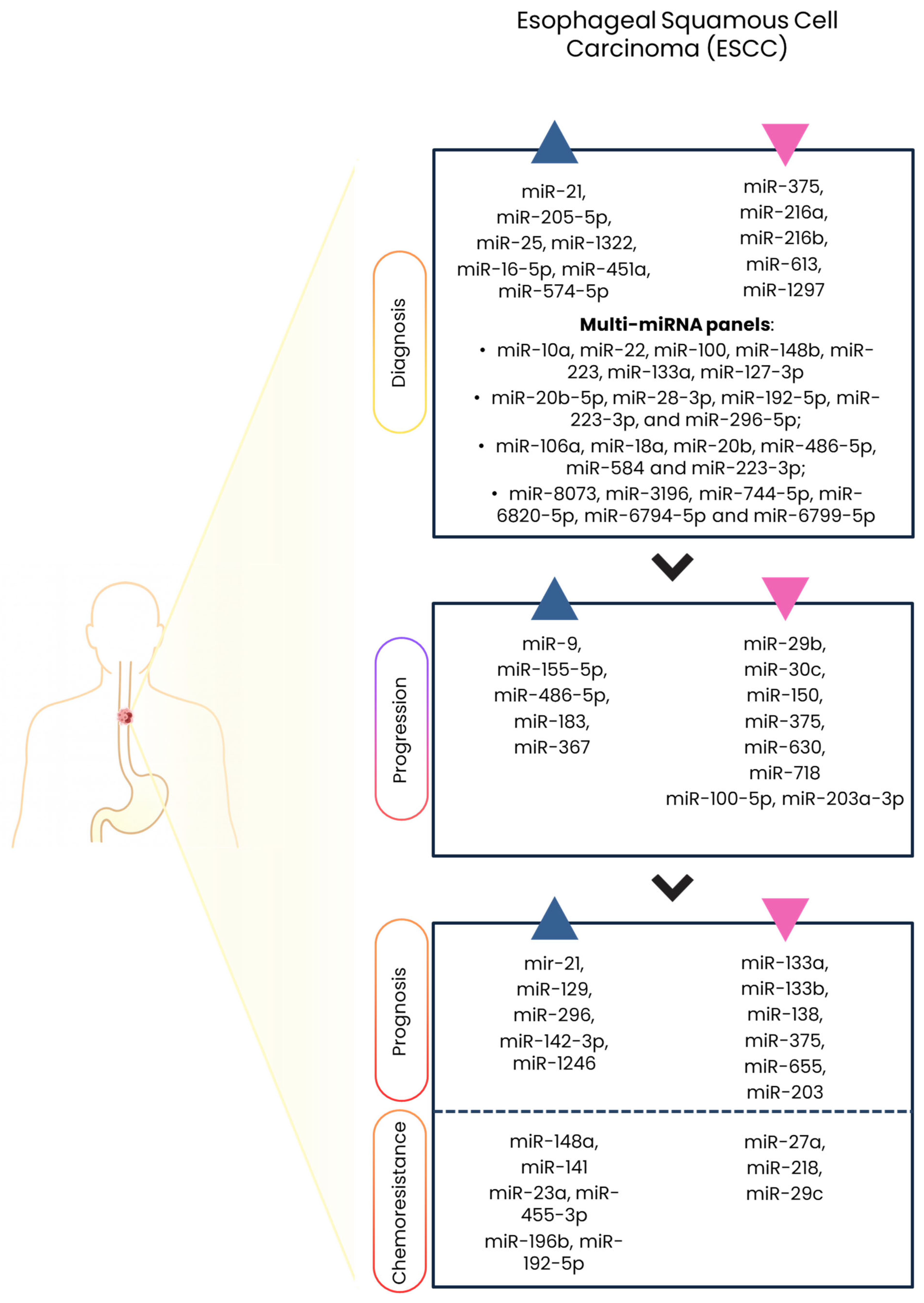
| miRNA | Expression in ESCC | Sample | Main Targets/Pathways | Functional Role | Clinical Relevance | References |
|---|---|---|---|---|---|---|
| miR-10a, miR-22, miR-100, miR-148b, miR-223, miR-133a, miR-127-3p | All upregulated (miR-223 downregulated) | Serum | SOX4, ARTN, VEGF-A, FGF2-FGFR2, mTOR, Wnt/β-catenin, CXCR7, PI3K/AKT, FBXW7, IGF1R, HIF-1α | Angiogenesis, motility, EMT suppression, migration, proliferation | Diagnostic | [23,24,25,26,27,28,29,30] |
| miR-20b-5p, miR-28-3p, miR-192-5p, miR-296-5p | All upregulated | Serum | PTEN, FBXW7 (PI3K/AKT pathway), BIM, ARF6 (Hedgehog signaling) | Proliferation, migration, invasion | Diagnostic | [31,32,33,34,35,36,37,38,39,40,41] |
| miR-106a, miR-18a, miR-20b, miR-486-5p, and miR-584, miR-223-3p | All upregulated (miR-223-3p downregulated) | Serum | PTEN (PI3K/AKT pathway), CDK4, BCAS2 | Proliferation, cell-cycle progression, apoptosis | Diagnostic | [32,42,51,54,55,103,107] |
| miR-8073, miR-3196, miR-744-5p, miR-6820-5p, miR-6794-5p, miR-6799-5p | miR-8073, miR-3196, miR-744-5p upregulated; miR-6820-5p, miR-6794-5p, miR-6799-5p downregulated | Serum | TGF-β signaling, PI3K/AKT, chromatin remodeling | Cell cycle regulation, angiogenesis, EMT, apoptosis, metastasis | Diagnostic | [43,44,45,46,47] |
| miR-21 | Upregulated | Serum/tissue | PTEN, PDCD4, TPM1 | Proliferation, invasion, resistance | Diagnostic, prognostic, predictive | [53,134,135,136,168,169,170] |
| miR-205-5p | Upregulated | Plasma | PTEN, SMAD4, PI3K/AKT pathway, TGF-β pathway | Proliferation, EMT | Diagnostic | [56,57,58,59,60] |
| miR-25 | Upregulated | Serum | TGF-β, VEGF, EGFR, Wnt/β-catenin | Angiogenesis, metastasis | Diagnostic | [61,62] |
| miR-1322 | Upregulated | Serum | ECRG2 | Proliferation | Diagnostic | [63,64] |
| miR-16-5p, miR-451a, miR-574-5p | Upregulated | Serum | RECK, SOX6 | Proliferation | Diagnostic | [50,65,66] |
| miR-375 | Downregulated | Serum/tissue | IGF1R, PDK1 (PI3K/AKT pathway), XPR1 (BRAF-ERK1/2-p53), PRDX1 | Proliferation, survival, metastasis | Diagnostic, progression, prognostic | [48,52,56,67,68,98,99,100,101,102,103,104,105,147,148,149] |
| miR-216a/b | Downregulated | Serum | KRAS, eIF4B, ZEB1, IGF2BP2 | EMT, proliferation | Diagnostic | [70,71,72,73] |
| miR-613 | Downregulated | Serum/tissue | FN1 | Migration, invasion | Diagnostic | [74,75,76,77,78] |
| miR-1297 | Downregulated | Serum | EphA2 | Metastasis | Diagnostic | [79,80,81] |
| miR-29b | Downregulated | Tissue | MMP2 | Invasion, metastasis | Progression | [85,86,87,88] |
| miR-30c | Downregulated | Serum/tissue | SNAI1, PI3K/AKT pathway | EMT suppression | Progression | [89,90,91,92,93] |
| miR-150 | Downregulated | Tissue | ZEB1, c-MYB, NOTCH3, CXCR4 | EMT/MET | Progression | [94,95,96,97] |
| miR-486-5p | Upregulated | Serum | CDK4, BCAS2 | Cell-cycle progression, apoptosis | Progression | [34,106,107] |
| miR-630 | Downregulated | Tissue | SLUG, GAK, PRKCI, JAK2/STAT3 pathway | EMT, invasion | Progression | [108,109,110,111,112,113] |
| miR-718 | Downregulated | Plasma | VEGF, HOXB8 | Proliferation, invasion, angiogenesis, apoptosis | Progression | [114,115] |
| miR-9 | Upregulated | Plasma/tissue | α-catenin, β-catenin | EMT, metastasis | Progression | [118,119] |
| miR-155-5p | Upregulated | Tissue | TP53INP1 | Proliferation | Progression | [120,121,122,123] |
| miR-183 | Upregulated | Tissue | PDCD4, SMAD4 | EMT, survival | Progression | [124,125,126] |
| miR-367 | Upregulated | Plasma | RAB23 | Proliferation | Progression | [127,128] |
| miR-100-5p, miR-203a-3p | Downregulated | Tissue | CXCR7, FKBP5, PI3K/AKT pathway | Invasion suppression | Progression | [129,130,131,132,133] |
| miR-133a | Downregulated | Tissue | SOX4, IGF14 | Proliferation, migration, invasion | Prognostic | [137,138] |
| miR-133b | Downregulated | Tissue | JAK2/STAT3 pathway, SNAIL1 | Survival, progression | Prognostic | [139,140,141] |
| miR-138 | Downregulated | Serum/tissue | NF-κB | Proliferation, invasion | Prognostic | [142,143,144,145,146] |
| miR-1246 | Upregulated | Serum | p53 network, DYRK1A | Stemness, invasion | Prognostic | [150] |
| miR-655 | Downregulated | Tissue | ZEB1, TGFBR2 | Migration, invasion | Prognostic | [151,152,153] |
| miR-203 | Downregulated | Tissue | TP63 | Survival, differentiation | Prognostic | [154] |
| miR-129 | Upregulated | Tissue | APC, RAB11 | Progression | Prognostic | [155] |
| miR-296 | Upregulated | Tissue | cyclin D1, p27, MDR1 (P-gp) | Proliferation | Prognostic | [156] |
| miR-142-3p | Upregulated | Tissue | APC, KLF4, BCL2L1 | Proliferation | Prognostic | [157] |
| miR-27a | Downregulated | Cell lines | P-gp, BCL2 | Drug resistance | Predictive | [160] |
| miR-148a | Upregulated | Cell lines | DNMT1, DNMT3B, PXR | Chemosensitivity | Predictive | [161] |
| miR-141 | Upregulated | Cell lines | YAP1 | Cisplatin resistance | Predictive | [162] |
| miR-23a | Upregulated | Plasma/tissue | PTEN, CDH1, IRF1, APAF1, TOP2B | Chemoresistance | Predictive | [163] |
| miR-218 | Downregulated | Cell lines | Survivin | Chemosensitivity | Predictive | [164] |
| miR-455-3p | Upregulated | Tissue | Wnt/β-catenin pathway, TGF-β/SMAD pathway | Chemoresistance | Predictive | [165,166,167] |
| miR-29c | Downregulated | Serum/tissue | FBXO31 | Chemoresistance | Predictive | [171] |
| miR-196b | Upregulated | Cell lines | EPHA7/EPHA2 | EMT, resistance | Predictive | [172] |
| miR-192-5p | Upregulated | Plasma | ERCC3, ERCC4 | Cisplatin resistance | Predictive | [173,174] |
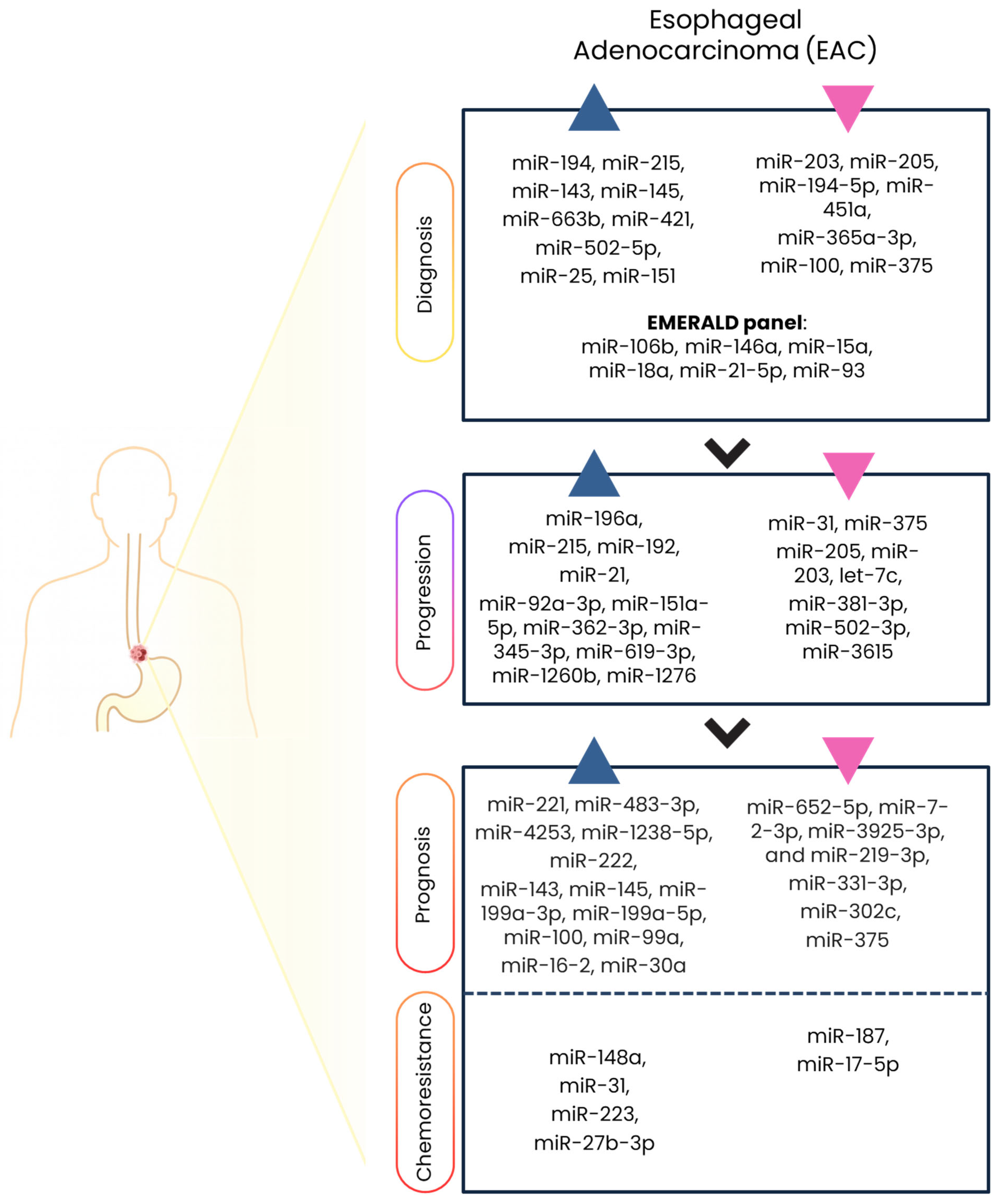
| miRNA | Expression in EAC | Sample | Main Targets/Pathways | Functional Role | Clinical Relevance | References |
|---|---|---|---|---|---|---|
| miR-194, miR-215, miR-143, miR-145 | All upregulated | Serum/Tissue | HNF1α, TP53 | Metaplasia acquisition | Diagnostic | [178,179,180,181,221] |
| miR-203, miR-205 | Downregulated | Tissue | TP63 | EMT | Diagnostic/progression | [180,190] |
| miR-375 | Downregulated | Tissue, serum | PDK1, IGF1R, MXI1, JAK2 | Proliferation | Diagnostic/progression/prognostic | [183,188,218,219,220] |
| miR-663b, miR-421, miR-502-5p | Upregulated | Tissue | SMAD4, PTEN, c-MYC, BCL2 | Tumorigenesis | Diagnostic | [184] |
| miR-25, miR-151, miR-100, miR-375 | miR-25, miR-151 upregulated; miR-100, miR-375 downregulated | Serum | CDH1, p21, Bim, PDK1, IGF1R, MXI1, JAK2 | Proliferation, invasion | Diagnostic | [185] |
| miR-106b, miR-146a, miR-15a, miR-18a, miR-93, miR-21-5p | Dysregulated | Plasma | Cell-cycle regulation | Proliferation | Diagnostic | [186] |
| miR-196a | Upregulated | Tissue | SPRR2C, S100A9, KRT5, NF-κB, c-MYC | EMT, proliferation | Progression | [183,187,188] |
| miR-31 | Downregulated | Tissue | E2F2/CDKN2A | Proliferation | Progression/predictive | [189,227] |
| miR-215, miR-192, miR-205, let-7c, miR-203 | miR-215, miR-192 upregulated; miR-205, let-7c, miR-203 downregulated. | Tissue | TP53, TP63, HMGA2 | Progression, EMT | Progression | [180,190] |
| miR-21 | Upregulated | Tissue | PDCD4, PTEN (PI3K/AKT pathway) | Proliferation | Progression | [136,191] |
| miR-92a-3p | Upregulated | Serum/tissue | PTEN, BIM, TGF-β/SMAD pathway | Early progression | Progression | [192] |
| miR-221, miR-483-3p | Upregulated | Tissue | MALAT1 | Invasion, recurrence | Prognostic | [194,195,196,197,198,199,200] |
| miR-652-5p, miR-7-2-3p | Downregulated | Tissue | SDC1/TGFβ2/pERBB4, DCLK1 | EMT, proliferation | Prognostic | [201,202,203] |
| miR-331-3p | Downregulated | Serum | HER2, PI3K/AKT pathway | Proliferation, migration | Prognostic | [204,205,206,207,208] |
| miR-4253, miR-1238-5p | Dysregulated | Serum | LHX2 | Proliferation, invasion, apoptosis | Prognostic | [209,210,211,212,213] |
| miR-222, miR-302c | Dysregulated | Serum | TOB2, DAZAP2, SLAIN1, MMP1 | Invasion | Prognostic | [214,215,216,217] |
| miR-199a-3p, miR-199a-5p | Upregulated | Tissue | SOX4, FXR1, PXN | Proliferation | Prognostic | [221,222,223] |
| miR-16-2 | Upregulated | Tissue | RAR-β2 | Tumor progression | Prognostic | [224,225] |
| miR-30e | Upregulated | Tissue | USP4 | EMT, invasion | Prognostic | [226] |
| miR-148a | Upregulated | Cell lines | MSK1, PXR | Chemosensitivity | Predictive | [161] |
| miR-223 | Upregulated | Cell lines | PARP1 | Chemosensitivity | Predictive | [228] |
| miR-187 | Downregulated | Tissue | Complement cascade (C3) | CRT resistance | Predictive | [229] |
| miR-17-5p | Downregulated | Cell lines | PRKACB C6orf120 | CRT resistance | Predictive | [230] |
| miR-27b-3p | Upregulated | Cell lines | SP1, PPARγ | Chemosensitivity | Predictive | [231] |
Disclaimer/Publisher’s Note: The statements, opinions and data contained in all publications are solely those of the individual author(s) and contributor(s) and not of MDPI and/or the editor(s). MDPI and/or the editor(s) disclaim responsibility for any injury to people or property resulting from any ideas, methods, instructions or products referred to in the content. |
© 2026 by the authors. Licensee MDPI, Basel, Switzerland. This article is an open access article distributed under the terms and conditions of the Creative Commons Attribution (CC BY) license.
Share and Cite
Cataldi-Stagetti, E.; Governatori, G.; Orsini, A.; De Nicolo, B.; Zagari, R.M.; Bonora, E. MicroRNAs in Esophageal Cancer: Implications for Diagnosis, Progression, Prognosis and Chemoresistance. Int. J. Mol. Sci. 2026, 27, 878. https://doi.org/10.3390/ijms27020878
Cataldi-Stagetti E, Governatori G, Orsini A, De Nicolo B, Zagari RM, Bonora E. MicroRNAs in Esophageal Cancer: Implications for Diagnosis, Progression, Prognosis and Chemoresistance. International Journal of Molecular Sciences. 2026; 27(2):878. https://doi.org/10.3390/ijms27020878
Chicago/Turabian StyleCataldi-Stagetti, Erica, Giulia Governatori, Arianna Orsini, Bianca De Nicolo, Rocco Maurizio Zagari, and Elena Bonora. 2026. "MicroRNAs in Esophageal Cancer: Implications for Diagnosis, Progression, Prognosis and Chemoresistance" International Journal of Molecular Sciences 27, no. 2: 878. https://doi.org/10.3390/ijms27020878
APA StyleCataldi-Stagetti, E., Governatori, G., Orsini, A., De Nicolo, B., Zagari, R. M., & Bonora, E. (2026). MicroRNAs in Esophageal Cancer: Implications for Diagnosis, Progression, Prognosis and Chemoresistance. International Journal of Molecular Sciences, 27(2), 878. https://doi.org/10.3390/ijms27020878