


Abstract
In this study, thiazole derivatives 13–23 were synthesized as novel cholinesterase inhibitors. Their structures were elucidated using 1H-NMR, 13C-NMR, and elemental analysis techniques. The binding positions and efficiencies of the designed compounds on the enzyme were investigated through molecular docking studies, and the properties of ADMET were examined using computational methods. Cytotoxicity analyses of the synthesized compounds were performed to screen their inhibitory effects on acetylcholinesterase (AChE) and butyrylcholinesterase (BChE) in vitro. It has been determined that these compounds do not exhibit a significant cytotoxic effect on healthy endothelial cells. Although the compounds showed lower efficacy in AChE inhibition compared to the reference compound tacrine (IC50: 0.136 μM; KI: 0.46 ± 0.01 nM), they exhibited moderate inhibitory effects. Among these, compounds 16 and 23 were found to be the most potent AChE inhibitors, with IC50 values of 36.47 µM and 34.65 µM, and KI values of 22.20 ± 1.23 and 21.10 ± 1.58 µM, respectively, while compounds 15 and 20 were found to be the most potent BChE inhibitors, with IC50 values of 17.76 µM and 16.90 µM, and KI values of 14.10 ± 0.58 and 13.40 ± 0.48 µM, respectively, on other cholinesterases. These tested compounds can be considered potential candidates for further research in the treatment of Alzheimer’s disease (AD).
1. Introduction
The global burden of neurodegenerative disorders has increased considerably, largely as a consequence of the progressive aging of the population. Among these disorders, Alzheimer’s disease (AD) is the predominant cause of dementia and represents a progressive neurological condition characterized by a gradual deterioration of cognitive abilities [1,2]. The clinical manifestations of AD commonly include memory deficits and impairments in language, behavior, visuospatial processing, and, particularly in advanced stages, motor functions [3]. Dysfunction of the cholinergic system in the cerebral cortex is also considered an important feature associated with cognitive decline in AD [2]. The increasing life expectancy observed worldwide has contributed substantially to the growing prevalence of AD, making this disorder an increasingly important public health concern and highlighting the need for effective therapeutic strategies [4]. Current epidemiological estimates indicate that approximately 50 million individuals worldwide are living with severe dementia, with AD accounting for approximately 60–70% of these cases [2]. Moreover, the global number of individuals affected by AD is expected to increase markedly in the coming decades, potentially reaching approximately 100 million by 2050 [1]. Because the disease predominantly affects older adults and progressively compromises cognitive and functional independence, AD constitutes a major challenge for healthcare systems and places a considerable physical, psychological, and social burden on both patients and their caregivers [5,6].
Although the etiology of AD is unknown, it is thought that several factors play a significant role in its onset and progression. AD is characterized by several interconnected pathological processes, including extracellular amyloid-β (A) accumulation and plaque formation, oxidative imbalance, impaired cholinergic signaling associated with reduced acetylcholine (ACh) levels, and tau hyperphosphorylation leading to neurofibrillary tangle formation [1,7]. Consequently, several pathological hypotheses have been put forward in connection with the development of AD. Among these hypotheses, the cholinergic hypothesis stands out [8]. According to the cholinergic hypothesis, one of the fundamental causes of AD is a deficiency in cholinergic neurotransmission [1]. This involves the disruption of neurotransmitters in the synaptic cleft, known as acetylcholine. The enzymes that exacerbate this disruption and trigger AD are acetylcholinesterase (AChE) and butyrylcholinesterase (BChE). These enzymes also contribute to the accumulation of amyloid-β (A) on protein structures in the brain [9]. Furthermore, the accumulation of tau, a microtubule-associated protein, may also contribute to cholinergic neuron degeneration in cases of tauopathy [10].
Unfortunately, there is no cure for AD, and the disease ultimately leads to death. Neuroscientists worldwide are actively conducting research and development into potential drugs to treat AD [6]. When evaluating treatments for neurodegenerative diseases, the use of cholinesterase inhibitors, which block the enzymes that catalyze the breakdown of butyrylcholine (BCh) or ACh, is often considered [10]. Researchers have suggested that AD’s resistance to AChE inhibitors is likely due to BChE activity, which interferes with AChE’s ability to perform or sustain its enzymatic function. Consequently, dual inhibition of AChE and BChE would enhance therapeutic efficacy in the treatment of AD. To date, “rivastigmine” is the only approved dual AChE/BChE inhibitor; the others are single-target molecules [8]. Inhibition of both enzymes is the strategy used to increase ACh levels, and the cholinesterase inhibitor drugs used in current treatment have been developed on this basis [4]. Unfortunately, however, the effects of Food and Drug Administration (FDA) approved drugs are limited, and none of them can halt the progression of the disease. In this context, a growing body of evidence suggests that selective AChE and BChE dual enzyme inhibitors may be more effective in the advanced stages of the disease compared to the drugs used in current treatments. Consequently, cholinesterase inhibitors are regarded as one of the primary targets in the design of multifunctional anti-AD compounds, owing to the beneficial role they play in the early and middle stages of the disease [4,11].
AChE is an enzyme related to BChE, and both cholinesterases exhibit a similar structure, featuring five binding sites within a 20 Å deep binding pocket. This pocket is primarily bounded by the catalytic aromatic site (CAS), which is located near the base, and the peripheral aromatic site (PAS), situated at the entrance to the pocket. The PAS is the first binding site for the substrate; in the CAS, the charged substrate interacts with the aromatic residues. Whilst the hydrolysis reaction takes place in the esteratic region, the oxyanion hole ensures the stabilization of the substrate’s transition state. The final section, the acyl pocket, ensures substrate selectivity. Although AChE and BChE share approximately 65% amino acid sequence identity, structural differences within their active-site regions arise largely from the substitution of aromatic residues by aliphatic amino acids in BChE. These differences are most pronounced in the PAS. PAS (AChE: Tyr341, Asp74, Tyr124, Tyr72, Trp286; BChE: Tyr332, Asp70, Gln119, Ala277, Asn68) and the CAS regions (AChE: Phe338, Tyr337, Tyr133, Trp86; BChE: Phe329, Trp82, Ala328, Tyr128) result in an increase in throat volume of 200 Å, allowing larger ligands to bind to BChE. Although there are fewer aromatic residues in the PAS region of BChE, the Asp70 residue facilitates the transfer of the substrate to the CAS [3,5].
Thiazole-based compounds have attracted considerable interest in medicinal chemistry owing to the broad spectrum of biological activities associated with this heterocyclic scaffold, making it an important structural framework in drug discovery [12]. The thiazole moiety is also a key component of many active pharmaceutical ingredients. Heterocyclic rings are frequently used as the main structural unit in the design of biological compounds. Various studies have demonstrated that compounds containing a thiazole ring are effective against neurodegenerative disorders caused by low levels of cholinergic neurotransmitters [13,14,15,16]. In a study on 1,3-thiazole derivatives by Modrić et al. in 2022, compound 7 was observed to inhibit AChE with an IC50 value of 91 µM [17]. In BChE inhibition, compound 6 stood out with a value of 195 µM [17]. In a study by Hafez et al. (2023), novel benzothiazole derivatives were synthesized and their AChE inhibition was measured [18]. Compound 1e showed the highest efficacy with an IC50 value of 2.92 ± 0.37 µM. Donepezil was used as a positive control (IC50 value 36.45 ± 8.31 nM) [18]. In another study by Mirjalili et al. in 2024, hydroxycoumarin-based 4-(benzo[d]thiazole-2-yl)phenols were designed and synthesized [19]. AChE inhibition profiles were investigated and Compound 3i showed the highest activity against AChE with an IC50 value of 2.7 µM [19]. In another study revealing the multiple target potential of the thiazole core, Hussain et al. synthesized novel thiazolidinedione–thiazole hybrids in 2025. Among these, compound 49 was the most active compound, showing IC50 values of 0.072 ± 0.002 µM for AChE, 9.81 ± 0.07 µM for BChE and 0.024 ± 0.001 µM for MAO-B. Donepezil (0.055 ± 0.003 µM) was used as the reference drug for AChE inhibition, while safinamide (8.50 ± 0.19 µM for MAO-A, 0.034 ± 0.002 µM for MAO-B) was used as the reference drug for MAO-A and MAO-B [20]. In a study conducted in 2026, Doğan et al. synthesized novel thiazole derivatives and investigated their double cholinesterase inhibition and antioxidant properties. Among these, compound 3 (IC50 1.440 ± 0.017 µM for AChE and IC50 3.620 ± 0.017 µM for BChE) was found to be the most active compound. In this study, donepezil HCl was used as a positive control (IC50 values for AChE and BChE were 33.490 ± 0.031 µM and 36.710 ± 0.020 µM, respectively). Compound 3 (80.145 ± 0.021 µM) showed a value close to the reference drug BHT (65.410 ± 0.021 µM) in terms of antioxidant properties according to the DPPH method [21]. These examples shown in the current literature highlight that the thiazole core has an important place in obtaining new target agents that can be the subject of Alzheimer’s treatment and also have antioxidant properties.
This study reports a novel approach to the design and synthesis of new thiazole derivatives 13–23 as cholinesterase inhibitors that may demonstrate efficacy as therapeutic agents in the treatment of AD. In this study, 11 new thiazole compounds were synthesized through the design of these new thiazole derivatives. Subsequently, the inhibitory activities of the synthesized compounds against AChE and BChE were determined. Furthermore, to provide a molecular-level perspective on the study, in silico studies were conducted, including the prediction of physicochemical properties and molecular docking analyses to investigate the interactions of these compounds with protein domains. Consequently, these compounds demonstrated significant inhibitory activity against both AChE and BChE, acting as anti-Alzheimer’s agents by inhibiting these enzymes. The present study will contribute to future research aimed at developing dual-enzyme inhibitor drugs for the treatment of neurodegenerative diseases caused by AD, utilizing thiazole ring compounds.
2. Results and Discussion
2.1. Chemistry
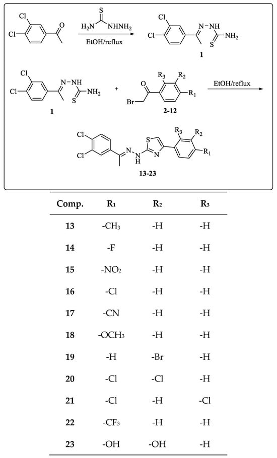
The synthesis of the targeted thiazole derivative compounds (13–23) was carried out in two steps as shown in Scheme 1. In the first step of the synthesis, thiosemicarbazide was refluxed with 1-(3,4-dichlorophenyl)-ethane-1-one as a ketone derivative for 4 h. 2-(1-(3,4-dichlorophenyl)ethylidene)hydrazine-1-carbothiamide (Compound 1), which will be used in the other step, was successfully obtained. The resulting end product was precipitated in an ice bath and filtered. In the second step, the target compounds (13–23) were obtained by reacting compound 1 with appropriate acetophenone derivatives (2–12) by refluxing in ethanol for 4–8 h. The progress of all synthesis steps and the depletion of starting materials were monitored by TLC. The resulting products were precipitated in an ice bath, filtered, and then crystallized from ethanol. The structures of the compounds were elucidated by 1H NMR, 13C NMR, and elemental analysis (Figures S1–S25). The COSY, HSQC, and HMBC spectra of compound 18 are shown in Figures S13–S15. The COSY spectrum showed the expected correlations between the vicinal aromatic protons of the 4-methoxyphenyl group and between the adjacent protons of the dichlorophenyl ring, supporting the substitution patterns of both aromatic moieties. In the HSQC spectrum, the methoxy protons at δ_H 3.79 correlated directly with the carbon signal at δ_C 55.59, whereas the methyl protons at δ_H 2.32 correlated with the carbon signal at δ_C 14.20. The proton of the thiazole ring showed a direct HSQC correlation with the carbon resonance at δ_C 102.74. The remaining HSQC cross-peaks in the aromatic region connected the proton signals between δ_H 6.97 and 7.95 with their corresponding protonated aromatic carbon atoms. In the HMBC spectrum, the methoxy protons exhibited a diagnostic long-range correlation with the oxygen-substituted aromatic carbon at δ_C 159.27, confirming the 4-methoxyphenyl moiety. In addition, long-range correlations involving the thiazole proton, the hydrazone NH proton, and the methyl group supported the connectivity of the thiazole, hydrazone, and dichlorophenyl fragments. Collectively, the COSY, HSQC, and HMBC results were consistent with and further supported the proposed structure of compound 18.
Scheme 1.
General procedure for synthesis of the final compounds 13–23.
2.2. Cytotoxicity Assay
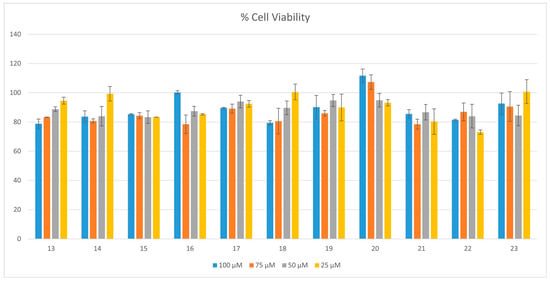
The in vitro cytotoxic effects of the synthesized thiazole (13–23) compounds on the human umbilical vein endothelial cell (HUVEC) line, representing a healthy endothelial model, were evaluated utilizing the MTT assay. Cells were exposed to four distinct concentrations ranging from 25 µM to 100 µM, and relative cell viability percentages were calculated in comparison to the control group. Overall findings revealed that none of the tested compounds reduced HUVEC viability below 70%, even at the highest applied concentration of 100 µM (Figure 1). This indicates that the investigated compounds do not exert a marked cytotoxic effect on healthy endothelial cells and demonstrate a reliable safety profile at the cellular level.
Figure 1.
Cell viability of HUVECs for 24 h.
2.3. Cholinesterase Inhibition Activity
This study investigated the inhibitory properties of novel thiazole derivatives (13–23) against cholinesterase enzymes. The main objectives of this study were to determine the inhibitory activities of the synthesized derivatives against cholinesterase enzymes associated with AD and to provide preliminary structure–activity relationship data for the future optimization of this compound series. Table 1 presents the IC50 and KI values obtained for the thiazole derivatives and reference inhibitors against the tested cholinesterase enzymes.
Table 1.
IC50 and KI values of compounds 13–23 against AChE and BChE.
Thiazole derivatives (13–23) were evaluated for their inhibitory effects on the AD-associated cholinesterase enzymes, AChE and BChE. The inhibitory activities of these compounds against cholinesterase enzymes were determined as follows: IC50 values of 34.65–77.08 µM and KI values of 21.10 ± 1.58–46.84 ± 2.63 µM for AChE, and IC50 values of 16.90–69.30 µM and KI values of 13.40 ± 0.48–54.80 ± 2.38 µM for BChE. Although all compounds exhibited lower inhibitory activity against both cholinesterase enzymes than the reference inhibitor tacrine, several derivatives showed measurable inhibition and provided preliminary structure–activity relationship data for further optimization. Tacrine exhibited IC50 values of 0.136 and 0.207 µM and KI values of 0.46 ± 0.01 and 0.33 ± 0.00 µM against AChE and BChE, respectively. These findings indicate that the investigated compounds possess appreciable inhibitory activity within the micromolar concentration range. Furthermore, the determined KI values provide valuable insights into the strength of inhibitor–enzyme interactions, thereby contributing to the assessment of inhibitor affinity and selectivity toward the target enzyme. As shown in Table 1, all synthesized derivatives exhibited measurable AChE inhibitory activity in the micromolar range. According to these results, compounds 16 and 23 were found to be the strongest AChE inhibitors with IC50 values of 34.65 and 36.47, and KI values of 21.10 ± 1.58 and 22.20 ± 1.23 µM, respectively, while compounds 15 and 20 were the strongest BChE inhibitors with IC50 values of 16.90 and 17.76, and KI values of 14.10 ± 0.58 and 13.40 ± 0.48 µM, respectively, on other cholinesterases. In contrast, compounds 13 and 14 have a weaker AChE inhibitory effect compared to the other derivatives, with an IC50 value of approximately 77 and a KI value of 47 µM. Specifically, as can be seen from the selectivity index values presented in Table 1, compound 16 exhibited a higher degree of selectivity for AChE compared to all others. Many studies have been conducted in the literature on the enzyme inhibitory potential of thiazole derivatives. The inhibitory potential of the thiazole derivatives synthesized in this study, especially against AChE and BChE cholinesterase enzymes, once again demonstrates that this structural skeleton is an important pharmacophore based on enzyme inhibition.
In one study, based on the cholinergic hypothesis, novel thiazolylhydrazone derivatives were synthesized and their inhibitory potential against cholinesterase enzymes was investigated. These derivatives showed significant inhibitory activity with IC50 values in the range of 0.031 ± 0.001–>100 µM for AChE, and with IC50 values of >100 µM for BChE compared to the reference donepezil (IC50: 0.021 ± 0.001 µM) [22]. Similarly, in another study, a series of benzothiazole-based fused 1,3,4-triazole/1,3,4-thiadiazole hybrid derivatives exhibited IC50 values of 2.10 ± 0.20–14.10 ± 0.10 µM for AChE, and IC50 values of 3.10 ± 0.50–15.30 ± 0.10 µM for BChE [23]. In another study, thiazole-sulfonamide derivatives were synthesized, and the inhibitory effect of these derivatives against AChE and BChE was investigated. The compounds exhibited IC50 values of 0.10–11.40 µM and 0.20–14.30 µM [24]. In the study conducted by Karakaya et al., 5-hydroxymethylfuran-incorporated thiazole-based furan derivatives showed a significant inhibitory effect with IC50 values between 21.105 and 86.625 µM for AChE, and 6.932 and 34.652 µM for BChE [25].
2.4. Molecular Docking
The molecular docking results of the selected compounds showed favorable binding within the catalytic gorge of both AChE and BChE, consistent with their enzyme inhibitory activities (Table 2 and Figure 2). Overall, the predicted binding affinities were higher toward AChE than BChE, as reflected by the more negative Glide GScores and docking scores. Among the investigated derivatives, compound 23 exhibited the strongest predicted affinity for AChE, with a Glide GScore of −8.729 kcal/mol, followed closely by compound 16 (−8.636 kcal/mol), whereas compound 20 showed the most favorable binding toward BChE (Glide GScore = −6.406 kcal/mol), followed by compound 15 (−6.303 kcal/mol). Although the co-ligands displayed the lowest docking scores in both enzymes, the synthesized compounds demonstrated comparable interaction patterns within the active sites, supporting their potential as effective cholinesterase inhibitors.
Table 2.
Molecular docking results of the most potent compounds for AChE and BChE (15, 16, 20 and 23). The remaining synthesized compounds are summarized as ranges (kcal/mol).
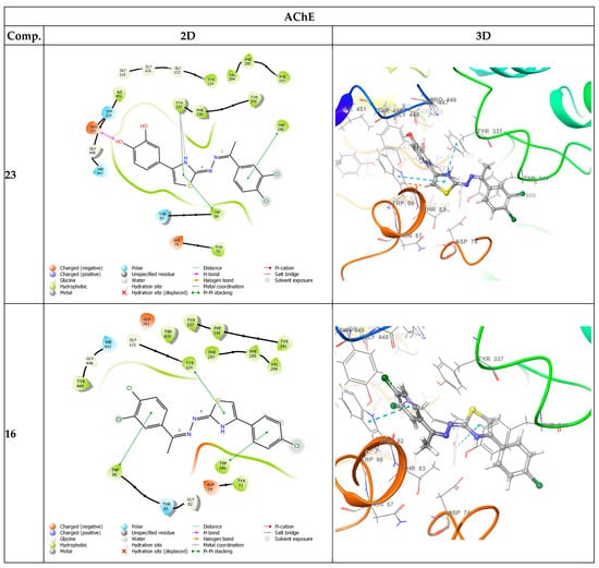
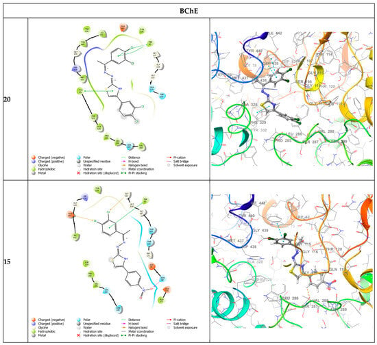
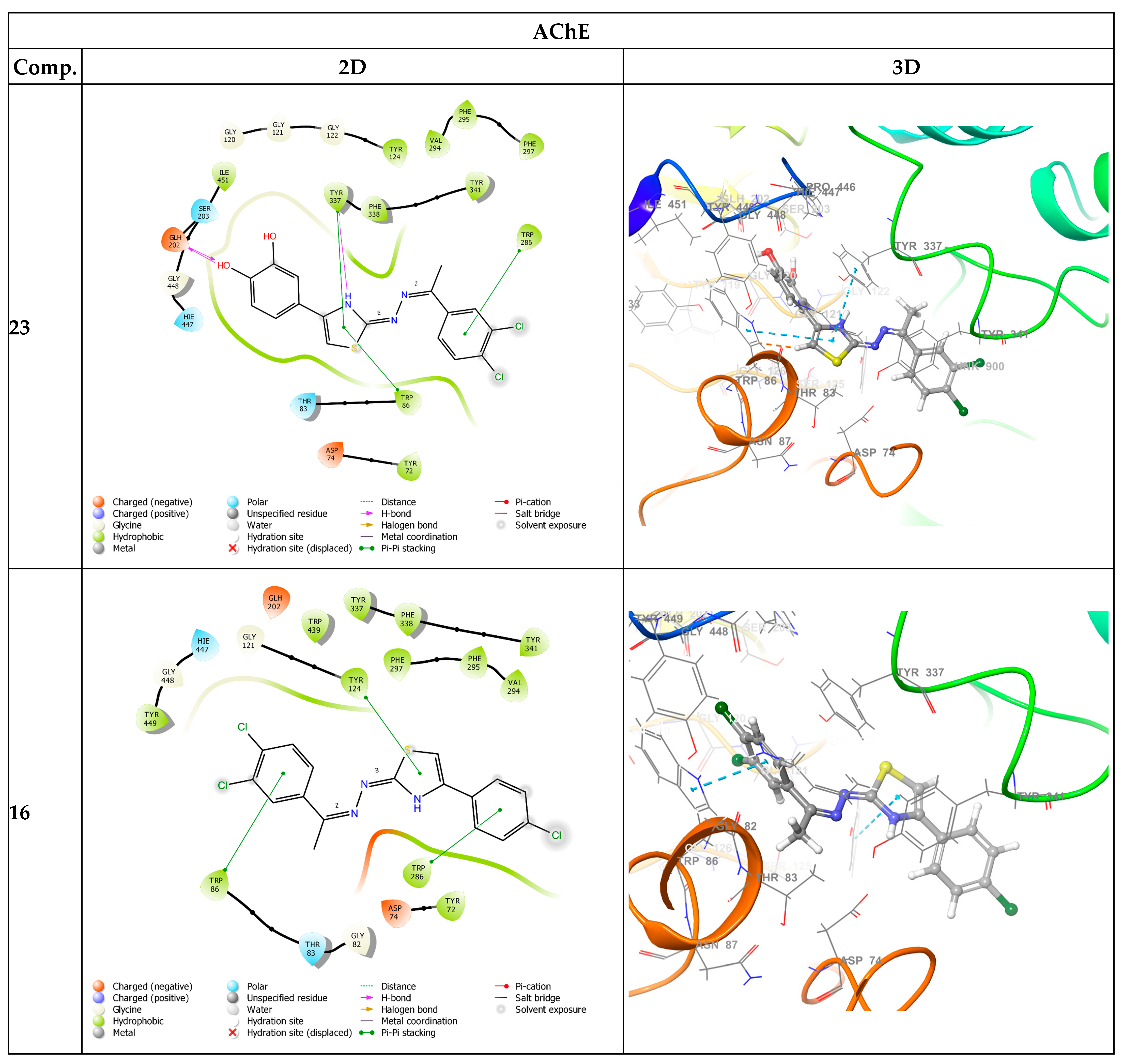
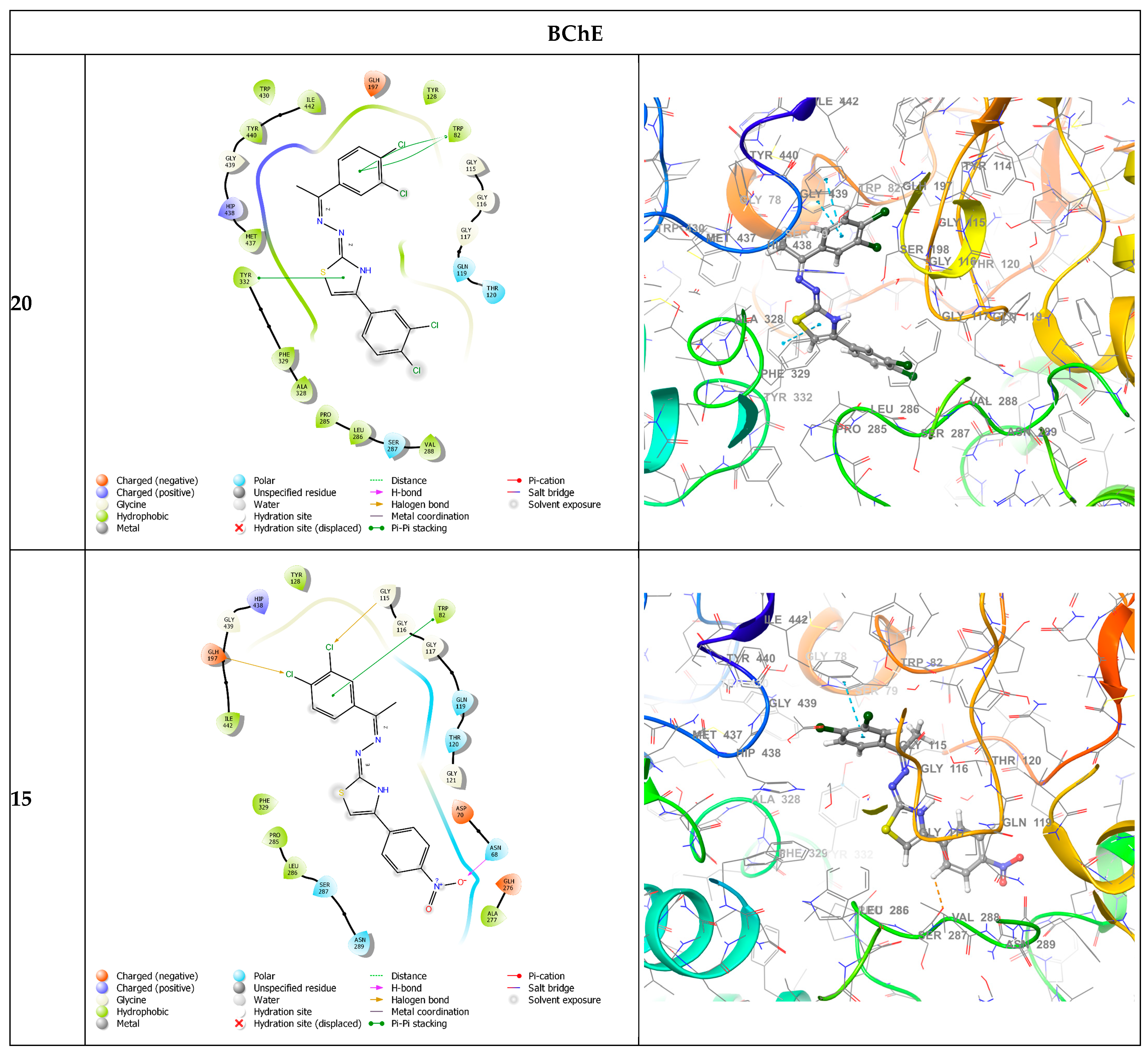
Figure 2.
2D and 3D protein–ligand interaction profiles of selected compounds (15, 16, 20 and 23) within the active sites of AChE (PDB ID: 7XN1) and BChE (PDB ID: 7Q1M). In AChE, compound 23 forms three π–π stacking interactions with Trp86, Trp286 and Tyr337, along with hydrogen bonding with Tyr337 and Glh202 (Glide GScore: −8.729 kcal/mol), indicating simultaneous engagement of the catalytic and peripheral sites. Compound 16 interacts primarily with Trp86, Trp 286 and Tyr 124Asp74 (−8.636 kcal/mol), suggesting a dual-binding mode. In BChE, compound 20 shows strong aromatic stabilization through π–π stacking with Trp82, Tyr332 (−6.38 kcal/mol). Similarly, compound 15 interacts with key aromatic residues within the hydrophobic gorge, with Trp86 additionally forming a hydrogen bond with Asn68, as well as two halogen bonds with Gly115 and Glh197 (−6.123 kcal/mol). Overall, ligand binding is predominantly governed by aromatic and hydrophobic interactions, supporting stable accommodation within the enzyme active-site gorge.
Within the AChE binding pocket, compound 23 established an extensive interaction network that included three π–π stacking interactions with Trp86, Trp286, and Tyr337, together with hydrogen bonds involving Tyr337 and Glu202. Such simultaneous engagement of residues belonging to both the catalytic active site (CAS) and the peripheral anionic site (PAS) is considered advantageous because it enhances ligand stabilization throughout the active-site gorge and may contribute to stronger enzyme inhibition. This binding mode is further supported by its favorable MM-GBSA binding free energy (−50.18 kcal/mol), suggesting the formation of a thermodynamically stable enzyme–ligand complex. Similarly, compound 16 exhibited a dual-binding orientation by interacting with Trp86, Trp286, Tyr124, and Asp74, yielding a Glide GScore of −8.636 kcal/mol and an MM-GBSA binding energy of −46.65 kcal/mol. The involvement of both catalytic and peripheral aromatic residues indicates efficient occupation of the enzyme gorge and provides a structural rationale for its potent experimental AChE inhibitory activity.
For BChE, compound 20 displayed the best docking performance among the synthesized derivatives, with a Glide GScore of −6.406 kcal/mol and the most favorable MM-GBSA binding energy (−45.44 kcal/mol). Its binding was predominantly stabilized through π–π stacking interactions with the aromatic residues Trp82 and Tyr332, which are essential components of the hydrophobic binding gorge. These interactions likely promote efficient ligand accommodation through aromatic and hydrophobic complementarity. In comparison, compound 15 showed a similar binding orientation within the BChE active site (Glide GScore = −6.303 kcal/mol), where aromatic interactions were reinforced by a hydrogen bond with Asn68 and two halogen bonds involving Gly115 and Glu197. The additional hydrogen- and halogen-bonding interactions may enhance binding specificity and compensate for its slightly less favorable docking score relative to compound 20.
Comparison of the docking metrics revealed that the selected compounds fall within or exceed the binding affinity range observed for the remaining synthesized derivatives, confirming their selection for further interaction analysis. In particular, compound 23 exhibited the most favorable AChE binding profile, whereas compound 20 showed the strongest predicted interaction with BChE. Despite small differences in Glide GScores, variations in MM-GBSA binding energies indicate that non-covalent interactions beyond docking pose geometry, including hydrogen bonding, π–π stacking, and halogen bonding, contribute substantially to overall complex stability. Collectively, these findings suggest that aromatic stacking interactions with conserved tryptophan and tyrosine residues represent the principal driving force for ligand recognition, while complementary hydrogen- and halogen-bonding interactions further stabilize the complexes. The docking results therefore provide a structural explanation for the experimentally observed cholinesterase inhibitory activities of compounds 23, 16, 20, and 15, highlighting their ability to occupy the active-site gorge through favorable hydrophobic and polar interactions.
2.5. ADMET
The ADMET properties of the most active compounds (15, 16, 20, and 23) were predicted using the QikProp module (Table 3). Overall, the selected derivatives exhibited favorable pharmacokinetic profiles, although some parameters deviated slightly from the recommended ranges. The predicted logP values (4.507–6.981) indicated moderate to high lipophilicity, with compound 23 showing the most balanced profile, while compounds 16 and 20 slightly exceeded the recommended limit. As expected, all compounds displayed relatively low aqueous solubility (logS = −6.411 to −8.635), suggesting that their high lipophilicity contributes to limited dissolution.
Table 3.
Predicted ADMET properties of the synthesized compounds (QikProp analysis). For the most active compounds (15, 16, 20 and 23).
Despite their low solubility, all compounds demonstrated excellent membrane permeability, with Caco-2 (683–6331 nm/s) and MDCK (3149–10,000 nm/s) values well above the threshold for highly permeable molecules. Accordingly, all derivatives were predicted to have 100% human oral absorption. Furthermore, the calculated logBB values (−0.582 to 0.893) suggest adequate blood–brain barrier penetration, an important characteristic for cholinesterase inhibitors intended for Alzheimer’s disease.
The selected compounds also showed acceptable human serum albumin binding and moderate numbers of predicted metabolic reactions, indicating suitable distribution and metabolic stability. However, all derivatives exhibited predicted hERG inhibition values below the recommended safety threshold, suggesting a potential cardiotoxicity risk that should be confirmed experimentally. Among the evaluated compounds, 23 displayed the most balanced overall ADMET profile owing to its favorable lipophilicity, relatively improved solubility, excellent permeability, and predicted CNS penetration. Overall, the ADMET analysis supports the drug-like potential of these lead compounds while indicating that improving aqueous solubility and reducing hERG liability should be priorities for future optimization.
2.6. Structure–Activity Relationship (SAR)
The SAR analysis revealed that the nature and position of substituents on the phenyl ring markedly influenced the AChE inhibitory activity (Table 1). Among the synthesized derivatives, compound 23 bearing 2,4-difluoro substituents exhibited the highest AChE inhibitory activity (IC50 = 34.65 μM), followed by the 4-chloro derivative 16 (36.47 μM) and the 4-nitro derivative 15 (38.50 μM). These findings suggest that electron-withdrawing substituents, particularly halogens, enhance AChE inhibition, probably by strengthening hydrophobic and aromatic interactions within the enzyme gorge. In contrast, electron-donating (–CH3, –OCH3) or strongly polar (–CN) substituents resulted in lower AChE activity.
A different trend was observed for BChE inhibition. The 3,4-dichloro derivative 20 displayed the highest potency (IC50 = 16.90 μM), closely followed by the 4-nitro derivative 15 (17.76 μM). The enhanced activity of compound 20 indicates that increasing hydrophobicity through multiple chlorine substituents favors binding within the larger and more hydrophobic BChE active-site gorge. In comparison, the mono-chloro analog 16 showed significantly weaker BChE inhibition, highlighting the importance of both the number and position of halogen substituents.
Overall, halogenated derivatives emerged as the most promising compounds in this series. While compound 23 demonstrated preferential inhibition toward AChE, compound 20 exhibited the highest selectivity for BChE (SI = 2.417). The docking studies further support these observations by showing that the lead compounds establish favorable π–π stacking and hydrophobic interactions with key aromatic residues in the catalytic gorge. Collectively, these findings indicate that appropriate incorporation of electron-withdrawing halogen substituents represents an effective strategy for improving cholinesterase inhibitory activity in this benzimidazole-based scaffold.
3. Materials and Methods
3.1. Chemistry
All the chemicals employed in the synthetic procedure were purchased from Sigma-Aldrich Chemicals (Sigma-Aldrich Corp., St. Louis, MO, USA) or Merck Chemicals (Merck KGaA, Darmstadt, Germany). Melting points of the obtained compounds were determined by the MP90 digital melting point apparatus (Mettler Toledo, Columbus, OH, USA) and were uncorrected. 1H-NMR and 13C-NMR spectra of the synthesized compounds were recorded on a Bruker 400 MHz and 100 MHz digital FT-NMR spectrometer (Bruker Bioscience, Billerica, MA, USA) in DMSO-d6, respectively. Splitting patterns were designated as follows: s: singlet; d: doublet; t: triplet; m: multiplet in the NMR spectra. Coupling constants (J) were reported in Hertz. All reactions were monitored by thin-layer chromatography (TLC) using Silica Gel 60 F254 TLC plates (Merck KGaA, Darmstadt, Germany).
Synthesis of 2-(1-(3,4-dichlorophenyl)ethylidene)hydrazine-1-carbothioamide derivative (1):
1-(3,4-dichlorophenyl)-ethane-1-one (1 meq) and thiosemicarbazide (1 meq) were dissolved in ethanol and refluxed for 4 h. The end of the experiment was checked by TLC analysis. After the reaction was complete, the solution was cooled in an ice bath to obtain the compound 2-(1-(3,4-dichlorophenyl)ethylidene)hydrazine-1-carbothioamide. The resulting product was filtered.
Synthesis of target compounds (13–23):
The resulting 2-(1-(3,4-dichlorophenyl)ethylidene)hydrazine-1-carbothioamide derivative (compound 1) was mixed with appropriate bromoacetophenone derivatives (2–12) in ethanol under reflux for 4–8 h to obtain the target compounds. The reaction’s completion was monitored by TLC. The product formed at the end of the reaction was cooled in an ice bath. The precipitate was filtered and crystallized from ethanol
2-(2-(1-(3,4-Dichlorophenyl)ethylidene)hydrazineyl)-4-(p-tolyl)thiazole (13): M.p.:149.8 °C. Yield: 81%.
1H-NMR (400 MHz, DMSO-d6): δ: 2.33 (6H, s, CH3), 7.22 (2H, d, J = 5.84 Hz, Aromatic CH), 7.27 (1H, br.s, Aromatic CH), 6.98 (1H, d, J = 6.92 Hz, Aromatic CH), 7.76–7.78 (2H, m, Aromatic CH), 7.95 (1H, s, Aromatic CH), 11.43 (1H, s, NH).
13C-NMR (100 MHz, DMSO-d6): δ = 14.22 (CH3), 21.27 (CH3), 125.95, 126.13, 127.27, 127.70, 128.72, 129.66, 131.08, 131.46, 131.80, 137.29, 139.07, 145.78, 166.49, 171.32.
Anal. calcd. For C18H15N3SCl2, C, 57.45; H, 4.02; N, 11.17. Found: C, 57.38; H, 4.01; N, 11.20.
2-(2-(1-(3,4-Dichlorophenyl)ethylidene)hydrazineyl)-4-(4-fluorophenyl)thiazole (14): M.p.:176.2 °C. Yield: 77%.
1H-NMR (400 MHz, DMSO-d6): δ: 2.32 (3H, s, CH3), 7.25 (2H, t, J = 7.92 Hz, Aromatic CH), 7.34 (1H, s, Aromatic CH), 7.68 (1H, d, J = 8.40 Hz, Aromatic CH), 7.75 (1H, d, J = 8.04 Hz, Aromatic CH), 7.92–7.95 (3H, m, Aromatic CH), 11.45 (1H, s, NH).
13C-NMR (100 MHz, DMSO-d6): δ = 14.25 (CH3), 104.63, 115.92 (d, J = 21.31 Hz), 126.13, 127.71, 127.97 (d, J = 7.98 Hz), 131.07, 131.51, 131.80, 139.00, 144.39, 150.11, 162.07 (d, J = 242.45 Hz), 168.02, 170.03.
Anal. calcd. For C17H12N3SCl2F, C, 53.69; H, 3.18; N, 11.05. Found: C, 53.84; H, 3.19; N, 11.07.
2-(2-(1-(3,4-Dichlorophenyl)ethylidene)hydrazineyl)-4-(4-nitrophenyl)thiazole (15): M.p.:162.3 °C. Yield: 79%.
1H-NMR (400 MHz, DMSO-d6): δ: 2.34 (3H, s, CH3), 7.70 (1H, d, J = 7.84 Hz, Aromatic CH), 7.78 (2H, s, Aromatic CH), 7.97 (1H, s, Aromatic CH), 8.14 (2H, d, J = 7.68 Hz, 1,4-disubstituted benzene CH), 8.30 (2H, d, J = 8.00 Hz, 1,4-disubstituted benzene CH), (1H, s, Aromatic CH).
13C-NMR (100 MHz, DMSO-d6): δ = 14.26 (CH3), 105.01, 120.80, 125.99, 126.15, 127.72, 128.03, 129.10, 131.09, 131.49, 131.81, 135.15, 139.05, 169.94, 173.90.
Anal. calcd. For C17H12N4O2SCl2, C, 50.13; H, 2.97; N, 13.76. Found: C, 50.26; H, 2.98; N, 13.80.
2-(2-(1-(3,4-Dichlorophenyl)ethylidene)hydrazineyl)-4-(4-chlorophenyl)thiazole (16): M.p.:152.4 °C. Yield: 80%.
1H-NMR (400 MHz, DMSO-d6): δ: 2.32 (3H, s, CH3), 7.46 (1H, s, Aromatic CH), 7.52 (1H, d, J = 8.64 Hz, Aromatic CH), 7.70–7.71 (3H, m, Aromatic CH), 7.76 (1H, d, J = 8.44 Hz, Aromatic CH), 7.93–7.96 (2H, m, Aromatic CH), 11.51 (1H, s, NH).
13C-NMR (100 MHz, DMSO-d6): δ = 14.30 (CH3), 106.21, 126.18, 127.76, 127.98, 130.25, 131.12, 131.82, 132.69, 135.70, 138.98, 146.84, 151.92, 166.88, 171.96.
Anal. calcd. For C17H12N3SCl3, C, 51.47; H, 3.05; N, 10.59. Found: C, 51.63; H, 3.04; N, 10.57.
2-(2-(1-(3,4-Dichlorophenyl)ethylidene)hydrazineyl)-4-(4-cyanophenyl)thiazole (17): M.p.:243.5 °C. Yield: 79%.
1H-NMR (400 MHz, DMSO-d6): δ: 2.38 (3H, s, CH3), 7.74–7.75 (2H, m, Aromatic CH), 7.81 (1H, d, J = 8.36 Hz, Aromatic CH), 7.93 (2H, d, J = 7.52 Hz, 1,4-disubstituted benzene CH), 8.00 (1H, s, Aromatic CH), 8.11 (2H, d, J = 7.44 Hz, 1,4-disubstituted benzene CH), 11.59 (1H, s, NH).
13C-NMR (100 MHz, DMSO-d6): δ = 14.33 (CH3), 108.79, 110.07, 119.44, 126.18, 126.59, 127.77, 131.09, 131.62, 131.82, 133.18, 138.90, 139.29, 144.79, 149.47, 170.25.
Anal. calcd. For C18H12N4SCl2, C, 55.82; H, 3.12; N, 14.47. Found: C, 55.67; H, 3.13; N, 14.41.
2-(2-(1-(3,4-Dichlorophenyl)ethylidene)hydrazineyl)-4-(4-methoxyphenyl)thiazole (18): M.p.:176.2 °C. Yield: 76%.
1H-NMR (400 MHz, DMSO-d6): δ: 2.32 (3H, s, CH3), 3.79 (3H, s, OCH3), 6.97 (2H, d, J = 7.08 Hz, 1,4-disubstituted benzene CH), 7.17 (1H, br.s, Aromatic CH), 7.68 (1H, d, J = 7.48 Hz, Aromatic CH), 7.75 (1H, d, J = 8.32 Hz, Aromatic CH), 7.81 (2H, d, J = 7.04 Hz, 1,4-disubstituted benzene CH), 7.95 (1H, s, Aromatic CH), 11.41 (1H, s, NH).
13C-NMR (100 MHz, DMSO-d6): δ = 14.20 (CH3), 55.59, 102.74, 114.45, 126.10, 127.32, 127.68, 127.88, 128.08, 131.05, 131.44, 131.80, 139.07, 144.09, 159.27, 169.83.
Anal. calcd. For C18H15N3SOCl2, C, 55.11; H, 3.85; N, 10.71. Found: C, 55.29; H, 3.84; N, 10.68.
2-(2-(1-(3,4-Dichlorophenyl)ethylidene)hydrazineyl)-4-(3-bromophenyl)thiazole (19): M.p.:148.7 °C. Yield: 78%.
1H-NMR (400 MHz, DMSO-d6): δ: 2.33 (3H, s, CH3), 7.61 (1H, s, Aromatic CH), 7.69 (1H, d, J = 8.96 Hz, Aromatic CH), 7.77 (3H, br.s, Aromatic CH), 7.96 (1H, s, Aromatic CH), 8.09 (2H, d, J = 7.20 Hz, Aromatic CH), 11.53 (1H, s, NH).
13C-NMR (100 MHz, DMSO-d6): δ = 14.27 (CH3), 106.52, 122.57, 124.82, 126.17, 127.74, 128.66, 130.58, 131.08, 131.30, 131.57, 131.82, 137.46, 138.95, 144.62, 149.54, 170.01.
Anal. calcd. For C17H12N3SCl2Br, C, 46.28; H, 2.74; N, 9.52. Found: C, 46.14; H, 2.75; N, 9.55.
2-(2-(1-(3,4-Dichlorophenyl)ethylidene)hydrazineyl)-4-(3,4-dichlorophenyl)thiazole (20): M.p.:180.5 °C. Yield: 82%.
1H-NMR (400 MHz, DMSO-d6): δ: 2.32 (3H, s, CH3), 7.59 (1H, s, Aromatic CH), 7.68–7.70 (2H, m, Aromatic CH), 7.75 (1H, d, J = 8.44 Hz, Aromatic CH), 7.86 (1H, d, J = 8.36 Hz, Aromatic CH), 7.95 (1H, s, Aromatic CH), 8.12 (1H, s, Aromatic CH), 11.49 (1H, s, NH).
13C-NMR (100 MHz, DMSO-d6): δ = 14.30 (CH3), 107.22, 126.01, 126.19, 127.68, 127.76, 130.16, 131.10, 131.36, 131.61, 131.82, 131.92, 138.91, 144.78, 149.98, 166.06, 170.13.
Anal. calcd. For C17H11N3SCl4, C, 47.36; H, 2.57; N, 9.75. Found: C, 47.48; H, 2.58; N, 9.72.
2-(2-(1-(3,4-Dichlorophenyl)ethylidene)hydrazineyl)-4-(2,4-dichlorophenyl)thiazole (21): M.p.:191.2 °C. Yield: 82%.
1H-NMR (400 MHz, DMSO-d6): δ: 2.29 (3H, s, CH3), 7.31–7.32 (1H, m, Aromatic CH), 7.50 (1H, d, J = 8.60 Hz, Aromatic CH), 7.64–7.69 (2H, m, Aromatic CH), 7.72–7.74 (1H, m, Aromatic CH), 7.93 (1H, s, Aromatic CH), 7.96–7.98 (1H, m, Aromatic CH).
13C-NMR (100 MHz, DMSO-d6): δ = 14.33 (CH3), 105.84, 127.97, 128.46, 130.16, 130.26, 131.76, 131.93, 132.82, 132.92, 133.02, 133.48, 136.48, 140.91, 150.99, 165.53, 173.31.
Anal. calcd. For C17H11N3SCl4, C, 47.36; H, 2.57; N, 9.75. Found: C, 47.51; H, 2.58; N, 9.71.
2-(2-(1-(3,4-Dichlorophenyl)ethylidene)hydrazineyl)-4-(4-trifluorophenyl)thiazole (22): M.p.:163.2 °C. Yield: 76%.
1H-NMR (400 MHz, DMSO-d6): δ: 2.32 (3H, s, CH3), 7.38 (1H, t, J = 7.76 Hz, Aromatic CH), 7.49–7.52 (2H, m, Aromatic CH), 7.68 (1H, d, J = 8.36 Hz, Aromatic CH), 7.75 (1H, d, J = 7.96 Hz, Aromatic CH), 7.88 (1H, d, J = 7.20 Hz, Aromatic CH), 7.95 (1H, s, Aromatic CH), 8.09 (1H, s, Aromatic CH), 11.46 (1H, s, NH).
13C-NMR (100 MHz, DMSO-d6): δ = 14.29 (CH3), 107.68, 125.32, 126.07, 126.19, 126.53, 127.11, 127.76, 128.19, 131.10, 131.59, 131.82, 138.95, 153.22, 165.31, 170.23.
Anal. calcd. For C18H12N3SCl2F3, C, 50.25; H, 2.81; N, 9.77. Found: C, 50.34; H, 2.80; N, 9.74.
2-(2-(1-(3,4-Dichlorophenyl)ethylidene)hydrazineyl)-4-(3,4-dihydroxyphenyl)thiazole (23): M.p.:224.2 °C. Yield: 74%.
1H-NMR (400 MHz, DMSO-d6): δ: 2.33 (3H, s, CH3), 7.19 (1H, t, J = 8.36 Hz, Aromatic CH), 7.25 (1H, s, Aromatic CH), 7.33–7.38 (1H, m, Aromatic CH), 7.69–7.70 (1H, m, Aromatic CH), 7.75 (1H, d, J = 7.72 Hz, Aromatic CH), 7.95 (1H, s, Aromatic CH), 8.04–8.10 (1H, m, Aromatic CH), 11.48 (1H, s, NH).
13C-NMR (100 MHz, DMSO-d6): δ = 14.27 (CH3), 105.03, 112.17, 119.64, 126.16, 127.73, 129.61, 130.75, 131.08, 131.56, 131.82, 135.71, 138.96, 144.10, 150.92, 161.19, 169.42.
Anal. calcd. For C17H13N3SO2Cl2, C, 51.79; H, 3.32; N, 10.66. Found: C, 51.88; H, 3.33; N, 10.67.
3.2. Cytotoxicity Assay
Cell Culture
Human umbilical vein endothelial cells (HUVECs) were cultured using Dulbecco’s Modified Eagle Medium (DMEM; Gibco, Paisley, UK), supplemented with 10% fetal bovine serum and 1% penicillin–streptomycin. The cell cultures were maintained in a standard humidified incubator at 37 °C with an atmosphere containing 5% CO2. Routine passaging of the cells was performed at a subcultivation ratio of 1:3.
Cytotoxicity Assay
The in vitro cell viability was determined utilizing the 3-[4,5-dimethylthiazol-2-yl]-2,5-diphenyltetrazolium bromide (MTT) assay, based on a slightly modified version of the protocol originally established by Mosmann (1983) [26]. Briefly, HUVECs were seeded into 96-well flat-bottom microtiter plates at a seeding density of 1 × 10⁴ cells per well, suspended in 200 μL of complete culture medium. The plates were incubated for 24 h under standard physiological conditions (37 °C, 5% CO2, humidified environment) to allow for proper cell adhesion.
Following attachment, the cells were exposed to the synthesized thiazole compounds at four distinct concentrations (25, 50, 75, and 100 µM) and incubated for an additional 24 h. Upon completion of the treatment phase, 20 μL of sterile-filtered MTT solution (5 mg/mL dissolved in PBS) was introduced to each well, and the microplates were incubated in the dark at 37 °C for 3 to 4 h. The medium containing unreacted MTT was then carefully discarded, and 100 μL of dimethyl sulfoxide (DMSO) was added to every well to dissolve the synthesized purple formazan crystals. To ensure homogeneous solubilization, the plates were subjected to gentle agitation on a shaker for 10–15 min at room temperature. The optical density (OD) of the wells was subsequently measured at a wavelength of 540 nm utilizing a microplate reader (BioTek Instruments, Winooski, VT, USA).
3.3. Cholinesterase Activity
Cholinesterase (ChE) activity was determined based on a modified protocol originally described by Ellman et al. (1961) [27]. The enzymatic reactions were performed at 37 °C using acetylthiocholine iodide (ATChI) as the substrate for AChE isolated from electric eel and butyrylcholine iodide (BChI) as the substrate for BChE obtained from horse serum. For each assay, the reaction mixture was prepared in a cuvette containing 100 µL of Tris–HCl buffer (1 M, pH 8.0), 100 µL of DTNB solution (0.01 M), and 50 µL of the corresponding substrate solution (ATChI or BChI; 0.050 M). The assay was initiated by the addition of 50 µL of enzyme solution, corresponding to 5.32 × 10⁻³ U. After a 5 min incubation period, the absorbance was measured spectrophotometrically at 412 nm. Each experiment was conducted in triplicate to ensure the reproducibility of the measurements [28,29].
3.4. Molecular Docking Study
Molecular docking studies were performed to investigate the binding modes of the synthesized compounds within the active sites of acetylcholinesterase (AChE) and butyrylcholinesterase (BChE). Docking calculations were carried out using the crystal structures of AChE (PDB ID: 7XN1, 2.85 Å) and BChE (PDB ID: 7Q1M, 2.79 Å), retrieved from the RCSB Protein Data Bank. These structures were selected because they contain co-crystallized tacrine, allowing reliable definition of the active-site binding pocket and validation of the docking protocol. All calculations were performed using Maestro v12.8.117 (Schrödinger Suite 2021-2) under physiological conditions (pH 7.4 ± 1.0), following previously validated protocols [30,31,32,33,34,35,36].
The synthesized compounds were constructed in Maestro and prepared using the LigPrep module. Ionization states were generated with Epik at pH 7.4 ± 1.0, while relevant tautomers, stereoisomers, and low-energy conformers were generated prior to docking.
Protein structures were prepared using the Protein Preparation Wizard. Missing hydrogen atoms were added, bond orders were assigned, and hydrogen-bonding networks were optimized. Protein protonation states were generated using Epik and refined with PROPKA at pH 7.4 ± 1.0. The co-crystallized tacrine was retained only for active-site definition and docking validation, whereas all other ligands were removed. Protein structures were then energy-minimized using the OPLS4 force field with a heavy-atom RMSD convergence criterion of 0.3 Å. Crystallographic water molecules were removed from AChE, whereas structurally relevant water molecules were retained in BChE when appropriate.
Docking grids were generated by centering the receptor box on the co-crystallized tacrine within the catalytic gorge. A grid size of 10 Å in each direction from the ligand centroid was used, with default Glide parameters (van der Waals scaling factor = 1.0; partial charge cutoff = 0.25). Molecular docking was performed using the Glide Standard Precision (SP) protocol, and the optimal binding pose for each compound was selected according to the lowest Glide GScore. Binding interactions with residues located in both the catalytic anionic site (CAS) and peripheral anionic site (PAS) were subsequently analyzed to rationalize the observed cholinesterase inhibitory activities and structure–activity relationships.
3.5. ADMET
ADMET (absorption, distribution, metabolism, excretion, and toxicity) properties were predicted using the QikProp module [37] implemented in Schrödinger Suite 2021-2. This tool was used to estimate the physicochemical and pharmacokinetic properties of the synthesized compounds.
4. Conclusions
In this study, the newly synthesized thiazole derivatives (13–23) were designed and evaluated for their inhibitory activities against both AChE and BChE. In vitro tests of the compounds showed moderate-to-high inhibition against both cholinesterase enzymes (AChE and BChE). In addition to their anti-Alzheimer’s effect, the cytotoxic properties of the compounds were also investigated on HUVEC lines. The cytotoxic test results also showed that the compounds exhibited a safe cytotoxicity profile. Among the compounds, compound 23, containing the 3,4,-dihydroxy substituents, showed the highest activity in AChE inhibition (IC50 = 34.65 µM). Compound 16 followed, also showing high activity (IC50 = 36.47 µM). Compound 20, containing a 3,4-dichloro substitute, showed the highest BChE inhibition (IC50 = 16.90 µM), followed by compound 15, which also showed high activity (IC50 = 17.76 µM). Tacrine was used as the reference drug in the cholinesterase inhibition test (IC50 = 0.136 µM for AChE, IC50 = 0.207 µM for BChE). These results indicate moderate activity compared to findings from a recent literature review. Some studies have shown that thiazole-based anti-Alzheimer’s agents exhibit inhibition at low micromolar levels. For example, in the study by Hussain et al., it was shown that the AChE IC50 values of benzimidazole-thiazole derivative compounds ranged from 0.052 ± 0.001 to 9.03 ± 0.36 µM and the BChE IC50 values ranged from 0.11 ± 0.01 to 17.4 ± 1.08 µM. It was determined that the activity increased when the thiazole ring was enhanced with benzimidazole. In the study by Modrić et al. [17], it was shown that the AChE and BChE inhibitions of 1,3-thiazole derivatives ranged from 91 to 765 µM and 195 to 760 µM, respectively. It is understood that the obtained derivatives (13–23) show higher activity compared to simple 1,3-thiazole derivatives. In the study by Hafez et al. [18], the study proceeded with the benzothiazole form of thiazole. In the results obtained, most of the compounds showed low activity (>10 µM), while compound 1e (2.92 ± 0.37 µM) showed the highest activity. It was observed that the activity profile remained moderate, depending on the substituent, by diversifying the thiazole core in this way. In their study, Mirjalili et al. synthesized a series of new benzothiazole rings and investigated their activity [19]. According to their results, the compounds showed AChE inhibition in the range of 2.7 ± 0.10 to 35.8 ± 0.30 µM. Similarly, it is understood that the benzothiazole form of the thiazole ring has a broad and moderate activity profile depending on the substituents. In the study by Doğan et al., new thiazole derivatives were synthesized and their dual cholinesterase inhibition and antioxidant properties were investigated. According to the results obtained, AChE inhibition was observed in the range of 1.440 ± 0.017 to 39.160 ± 0.028 µM, and BChE inhibition in the range of 3.620 ± 0.017 to 40.850 ± 0.023 µM [17,18,19,20,21]. A comparison of recent thiazole-focused anti-Alzheimer studies in the literature highlights the thiazole ring as a focal point in studies targeting Alzheimer’s disease. Molecular docking studies supported the results obtained from in vitro tests. The strong π-π interactions and complementary halogen interactions of the precursor compounds highlighted their binding ability to the enzyme’s active site. ADMET predictions also emphasized that the compounds exhibited good critical pharmacokinetic properties such as blood–brain barrier penetration and membrane permeability. Taken together, although these thiazole derivatives bearing a dihalogenated phenyl ring exhibited lower cholinesterase inhibitory activity than tacrine, the findings provide preliminary structure–activity relationship data that may guide the further optimization of this chemical scaffold.
Supplementary Materials
The following supporting information can be downloaded at https://www.mdpi.com/article/10.3390/molecules31193568/s1: Figures S1–12, S16–25: 1H and 13C NMR spectra of the synthesized compounds; Figures S13–S15: COSY, HMBC, and HSQC spectra of compound 18.
Author Contributions
Conceptualization, U.A.Ç., M.I. and G.H.S.; methodology, U.A.Ç., A.K., M.F., G.H.S., Y.Ö.K., N.E.-H.D. and M.I.; software, N.E.-H.D.; investigation, U.A.Ç., M.F., A.K., G.H.S., Y.Ö.K., N.E.-H.D. and M.I.; writing—original draft preparation, all authors; writing—review and editing, all authors; visualization, Z.A.K. and Ş.B.; supervision, Y.Ö., Z.A.K. and Ş.B.; project administration, Ş.B.; funding acquisition, Ş.B. All authors have read and agreed to the published version of the manuscript.
Funding
This research was funded by Anadolu University, grant number THD-2026-3611, ID: 3611.
Institutional Review Board Statement
The current study does not involve human samples.
Informed Consent Statement
The current study does not involve human samples.
Data Availability Statement
The data supporting the findings of this study are provided in the manuscript and the Supplementary Information files.
Acknowledgments
The authors have reviewed and edited the output and take full responsibility for the content of this publication.
Conflicts of Interest
The authors declare no conflicts of interest.
Abbreviations
The following abbreviations are used in this manuscript:
| Aβ | Amyloid-beta |
| ACh | Acetylcholine |
| AChE | Acetylcholinesterase |
| AD | Alzheimer’s Disease |
| ADMET | Absorption, distribution, metabolism, excretion, and toxicity |
| ATChI | Acetylthiocholine Iodide |
| BCh | Butyrylcholine |
| BChE | Butyrylcholinesterase |
| BChI | Butyrylcholine Iodide |
| ChE | Cholinesterase |
| CAS | Catalytic aromatic site |
| DMEM | Dulbecco’s Modified Eagle Medium |
| DMSO | Dimethyl Sulfoxide |
| FDA | Food and Drug Administration |
| HUVEC | Human Umbilical Vein Endothelial Cells |
| J | Coupling constants |
| MTT | 3-[4,5-dimethylthiazol-2-yl]-2,5-diphenyltetrazolium bromide |
| OD | Optical Density |
| PAS | Peripheral Aromatic Site |
| SAR | Structure–Activity Relationship |
| SI | Selectivity index |
References
- Dadhich, I.A.; Panchal, N.B.; Dadhich, K.A.; Dadhich, C.S. Exploring Benzimidazole Chemistry: Synthesis, Biological activity, and Molecular Docking Studies for Alzheimer’s Treatment. Orient. J. Chem. 2024, 40, 1045–1055. [Google Scholar] [CrossRef] [Scilit]
- Khan, S.; Iqbal, T.; Rehman, M.U.; Khan, M.B.; Islam, M.S.; Dahlous, K.A. Integrated insight and in silico investigation of hybrid bis-thiazolidinone derivatives along with anti-Alzheimer activity. 3 Biotech 2025, 15, 176. [Google Scholar] [CrossRef] [Scilit] [PubMed]
- Conger, N.; Osmaniye, D.; Sağlık, B.N.; Levent, S.; Ozkay, Y.; Kaplancıklı, Z.A. Design, synthesis, biological activities, and evaluation of molecular docking-dynamics studies of new thiosemicarbazones that may be effective against Alzheimer’ s disease. J. Mol. Recognit. 2023, 36, e3059. [Google Scholar] [CrossRef] [Scilit] [PubMed]
- Shakila, S.; Kilic, B.; Bardakkaya, M.; Aksakal, F.; Sagkan, R.I.; Dogruer, D.S. Multifunctional anti-Alzheimer’ s agents: Synthesis, biological evaluation, and molecular docking study of new 2-phenoxyacetamide/3-phenoxypropanamide/4-oxobutanamide derivatives. J. Mol. Struct. 2024, 1317, 139106. [Google Scholar] [CrossRef] [Scilit]
- da Conceição, R.A.; von Ranke, N.; Azevedo, L.; Franco, D.; Nadur, N.F.; Kummerle, A.E.; Barbosa, M.L.d.C.; Souza, A.M.T. Structure—Based design of new N-benzy-piperidine derivatives as multitarget directed AChE/BuChE inhibitors for Alzheimer’ s disease. J. Cell Biochem. 2023, 124, 1734–1748. [Google Scholar] [CrossRef] [Scilit] [PubMed]
- Mohammadi, Y.; Emadi, R.; Maddahi, A.; Shirdel, S.; Morowvat, M.H. Identifying potential Alzheimer’ s disease therapeutics through GSK-3 β inhibition: A molecular docking and dynamics approach. Comput. Biol. Chem. 2024, 111, 108095. [Google Scholar] [CrossRef] [Scilit] [PubMed]
- Xu, Y.; Zhang, J.; Wang, H.; Mao, F.; Bao, K.; Liu, W.; Zhu, J.; Li, X.; Zhang, H.; Li, J. Rational design of novel selective dual-target inhibitors of acetylcholinesterase and monoamine oxidase B as potential anti-Alzheimer’s disease agents. ACS Chem. Neurosci. 2019, 10, 482–496. [Google Scholar] [CrossRef] [Scilit] [PubMed]
- Sağlık, B.N.; Levent, S.; Osmaniye, D.; Acar Çevik, U.; Kaya Çavuşoğlu, B.; Özkay, Y.; Koparal, A.S.; Kaplancıklı, Z.A. Design, synthesis, and biological activity evaluation of new donepezil-like compounds bearing thiazole ring for the treatment of Alzheimer’s disease. Crystals 2020, 10, 637. [Google Scholar] [CrossRef] [Scilit]
- Khan, S.; Iqbal, T.; Hussain, R.; Fiaz, Z.; Abdellattif, M.H.; Kashtoh, H. Novel thiazole—Thiadiazole as a potential anti- Alzheimer agent: Synthesis and molecular interactions via an in silico study. Future Med. Chem. 2025, 17, 1349–1362. [Google Scholar] [CrossRef] [Scilit] [PubMed]
- Tj, S.; James, J.P.; Fathima, C.Z.; Dixit, S.; Kolachi, K.Y. MD Simulations, DFT and Molecular Docking Studies of Schiff Base—Derived Heterocycles as Potent BuChE Inhibitors for Alzheimer’ s Disease. ChemistrySelect 2026, 11, e03914. [Google Scholar] [CrossRef] [Scilit]
- Durgun, M.; Akocak, S.; Lolak, N.; Topal, F.; Koçyiğit, Ü.M.; Türkeş, C.; Işık, M.; Beydemir, Ş. Design and synthesis of pyrazole carboxamide derivatives as selective cholinesterase and carbonic anhydrase inhibitors: Molecular docking and biological evaluation. Chem. Biodiver. 2024, 21, e202301824. [Google Scholar] [CrossRef] [Scilit] [PubMed]
- Drozdowska, D.; Ratkiewicz, A.; Wróbel-Tałałaj, A.; Parzych, C.; El-Dershaby, H.A.; Abd El-Razik, H.A.; Soliman, F.S.G.; El-Wakil, M.H. Repurposed arylidene-hydrazinyl-1,3-thiazoles as multitarget anti-Alzheimer’s agents via combined computational and experimental studies. ACS Omega 2025, 10, 59328–59344. [Google Scholar] [CrossRef] [Scilit] [PubMed]
- Liu, C.; Zou, M.; Zuo, J.; Xie, H.; Lyu, W.; Xu, J.; Feng, F.; Sun, H.; Liu, W.; Jiang, X. Discovery of thiazole salt AChE inhibitors and development of thiamine disulfide prodrugs targeting the central nervous system. Bioorganic Chem. 2023, 139, 106702. [Google Scholar] [CrossRef] [Scilit] [PubMed]
- Zerroug, E.; Belaidi, S.; Chtita, S.; Tuffaha, G.; AbulQais, F.; Kciuk, M.; Dubey, A.; Taha, M.O. Structure-Based Approaches for the Prediction of Alzheimer’ s Disease Inhibitors: Comparative Interactions Analysis, Pharmacophore Modeling and Molecular Dynamics Simulations. ChemistrySelect 2024, 9, e202303307. [Google Scholar] [CrossRef] [Scilit]
- Asfour, A.A.R.; Evren, A.E.; Özkan, B.N.S.; Yurttaş, L. Investigating the potential of novel thiazole derivatives in treating Alzheimer’ s and Parkinson’ s diseases. J. Biomol. Struct. Dyn. 2026, 44, 2939–2955. [Google Scholar] [CrossRef] [Scilit] [PubMed]
- Kassem, A.F.; Althomali, R.H.; Anwar, M.M.; El-Sofany, W.I. Thiazole moiety: A promising scaffold for anticancer drug discovery. J. Mol. Struct. 2024, 1303, 137510. [Google Scholar] [CrossRef] [Scilit]
- Modrić, M.; Božičević, M.; Odak, I.; Talić, S.; Barić, D.; Mlakić, M.; Raspudić, A.; Škorić, I. The structure–activity relationship and computational studies of 1, 3-thiazole derivatives as cholinesterase inhibitors with anti-inflammatory activity. Comptes Rendus. Chim. 2022, 25, 267–279. [Google Scholar] [CrossRef] [Scilit]
- Hafez, D.E.; Dubiel, M.; La Spada, G.; Catto, M.; Reiner-Link, D.; Syu, Y.T.; Abadi, A.H. Novel benzothiazole derivatives as multitargeted-directed ligands for the treatment of Alzheimer’s disease. J. Enzym. Inhib. Med. Chem. 2023, 38, 2175821. [Google Scholar] [CrossRef] [Scilit] [PubMed]
- Mirjalili, B.B.F.; Fazeli Attar, S.A.; Shiri, F. Synthesis, biological evaluation and in silico study of 4-(benzo[d]thiazole-2-yl) phenols based on 4-hydroxy coumarin as acetylcholinesterase inhibitors. Sci. Rep. 2024, 14, 25055. [Google Scholar] [CrossRef] [Scilit] [PubMed]
- Hussain, F.; Tahir, A.; Rehman, H.M.; Wu, Y.; Shah, M.; Rashid, U. Promising thiazolidinedione-thiazole based multi-target and neuroprotective hybrids for Alzheimer’s disease: Design, synthesis, in-vitro, in-vivo and in-silico studies. Eur. J. Med. Chem. 2025, 287, 117327. [Google Scholar] [CrossRef] [Scilit] [PubMed]
- Doğan, M.O.; Polat, Y.; Göksu, S.; Baltaş, N.; Kemaloğlu, F.; Baday, S.; Sungur, F.A.; Tok, F. Synthesis, Molecular Docking, Molecular Dynamics and DFT Studies of New Thiazole Derivatives as Dual Cholinesterase Inhibitors Against Alzheimer’s Disease. Polycycl. Aromat. Compd. 2026, 46, 981–1005. [Google Scholar] [CrossRef] [Scilit]
- Sağlık, B.N.; Osmaniye, D.; Acar Çevik, U.; Levent, S.; Kaya Çavuşoğlu, B.; Özkay, Y.; Kaplancıklı, Z.A. Design, synthesis, and structure–activity relationships of thiazole analogs as anticholinesterase agents for alzheimer’s disease. Molecules 2020, 25, 4312. [Google Scholar] [CrossRef] [Scilit] [PubMed]
- Khan, S.; Hussain, R.; Khan, Y.; Iqbal, T.; Chinnam, S.; Akif, M.; Dera, A.A. Hybrid benzothiazole derived fused triazole/thiazole derivatives as versatile anti-Alzheimer agents: Synthesis, characterization, biological evaluation and molecular docking studies. J. Mol. Struct. 2024, 1318, 139200. [Google Scholar] [CrossRef] [Scilit]
- Khan, S.; Ullah, H.; Taha, M.; Rahim, F.; Sarfraz, M.; Iqbal, R.; Iqbal, N.; Hussain, R.; Shah, S.A.A.; Ayub, K.; et al. Synthesis, DFT studies, molecular docking and biological activity evaluation of thiazole-sulfonamide derivatives as potent Alzheimer’s inhibitors. Molecules 2023, 28, 559. [Google Scholar] [CrossRef] [Scilit] [PubMed]
- Karakaya, A.; Çevik, U.A.; Kaya, B.; Çiftçi, B.; Necip, A.; Işık, M.; Beydemir, Ş.; Özkay, Y.; Kaplancıklı, Z.A. Design, Synthesis, In Silico Absorption, Distribution, Metabolism, and Elimination and Molecular Docking Studies of Thiazole-Based Furan Derivatives, and Their Biological Evaluation for Alzheimer Disease Therapy. Chem. Open 2025, 14, e202500305. [Google Scholar] [CrossRef] [Scilit] [PubMed]
- Mosmann, T. Rapid colorimetric assay for cellular growth and survival: Application to proliferation and cytotoxicity assays. J. Immunol. Methods 1983, 65, 55–63. [Google Scholar] [CrossRef] [Scilit] [PubMed]
- Ellman, G.L.; Courtney, K.D.; Andres, V., Jr.; Featherstone, R.M. A new and rapid colorimetric determination of acetylcholinesterase activity. Biochem. Pharmacol. 1961, 7, 88–95. [Google Scholar] [CrossRef] [Scilit] [PubMed]
- Gülçin, İ.; Scozzafava, A.; Supuran, C.T.; Akıncıoğlu, H.; Koksal, Z.; Turkan, F.; Alwasel, S. The effect of caffeic acid phenethyl ester (CAPE) on metabolic enzymes including acetylcholinesterase, butyrylcholinesterase, glutathione S-transferase, lactoperoxidase, and carbonic anhydrase isoenzymes I, II, IX, and XII. J. Enzym. Inhib. Med. Chem. 2016, 31, 1095–1101. [Google Scholar] [CrossRef] [Scilit] [PubMed]
- Lolak, N.; Akocak, S.; Durgun, M.; Duran, H.E.; Necip, A.; Türkeş, C.; Işık, M.; Beydemir, Ş. Novel bis-ureido-substituted sulfaguanidines and sulfisoxazoles as carbonic anhydrase and acetylcholinesterase inhibitors. Mol. Divers 2023, 27, 1735–1749. [Google Scholar] [CrossRef] [Scilit] [PubMed]
- Bank, R.P.D. RCSB PDB—7XN1: Crystal Structure of Human Acetylcholinesterase in Complex with Tacrine, Resolution: 2.85 Å. 2023. Available online: https://www.rcsb.org/structure/7XN1 (accessed on 1 September 2026).
- Bank, R.P.D. RCSB PDB—7Q1M: Crystal Structure of Human Butyrylcholinesterase in Complex with N-[(2S)-3-[(cyclohexylmethyl)amino]-2-hydroxypropyl]-2,2-diphenylacetamide, Resolution: 2.79 Å. 2021. Available online: https://www.wwpdb.org/pdb?id=pdb_00007q1m (accessed on 1 September 2026). [CrossRef] [Scilit]
- Schrödinger, L. Schrödinger Release 2021-2: Maestro v12.8.117; Schrödinger Inc.: New York, NY, USA, 2021. [Google Scholar]
- Lolak, N.; Akocak, S.; Topal, M.; Koçyiğit, Ü.M.; Işık, M.; Türkeş, C.; Topal, F.; Durgun, M.; Beydemir, Ş. Sulfonamide-Bearing Pyrazolone Derivatives as Multitarget Therapeutic Agents: Design, Synthesis, Characterization, Biological Evaluation, In Silico ADME/T Profiling and Molecular Docking Study. Pharmacol. Res. Perspect. 2025, 13, e70088. [Google Scholar] [CrossRef] [Scilit] [PubMed]
- Albadrani, H.M.; Alsaweed, M.; Jamal, Q.M.S.; Alasiry, S.M.; Jahan, S.; Hamed, M.; Kamal, M.; Rehman, T.; Iqbal, D. In-Vitro enzyme inhibition, kinetics, molecular docking and dynamics simulation approaches to decoding the mechanism of Ficus virens in cholinesterase inhibition. J. Taibah Univ. Sci. 2024, 18, 2403813. [Google Scholar] [CrossRef] [Scilit]
- de Almeida, R.B.; Barbosa, D.B.; do Bomfim, M.R.; Amparo, J.A.; Andrade, B.S.; Costa, S.L.; Botura, M.B. Identification of a novel dual inhibitor of acetylcholinesterase and butyrylcholinesterase: In vitro and in silico studies. Pharmaceuticals 2023, 16, 95. [Google Scholar] [CrossRef] [Scilit] [PubMed]
- He, Z.-C.; Zhang, T.; Peng, W.; Ding, F. Protonation State Insights into the Influence of Biocatalytic Function for Acetylcholinesterase Mediated by Neonicotinoids. Biochemistry 2025, 64, 1996–2009. [Google Scholar] [CrossRef] [Scilit] [PubMed]
- Schrödinger, L. Schrödinger Release 2021-2: QikProp; Schrödinger Inc.: New York, NY, USA, 2021. [Google Scholar]

Disclaimer/Publisher’s Note: The statements, opinions and data contained in all publications are solely those of the individual author(s) and contributor(s) and not of MDPI and/or the editor(s). MDPI and/or the editor(s) disclaim responsibility for any injury to people or property resulting from any ideas, methods, instructions or products referred to in the content. |
© 2026 by the authors. Licensee MDPI, Basel, Switzerland. This article is an open access article distributed under the terms and conditions of the Creative Commons Attribution (CC BY) license.