Article

Fusaproliferin, a Fungal Mycotoxin, Shows Cytotoxicity against Pancreatic Cancer Cell Lines

by
Nazia Hoque
1,2,3,
Choudhury Mahmood Hasan
4,
Md. Sohel Rana
2,
Amrit Varsha
5,
Md. Hossain Sohrab
3,* and
Khondaker Miraz Rahman
5,*
1
Department of Pharmacy, East West University, Dhaka 1212, Bangladesh
2
Department of Pharmacy, Jahangirnagar University, Savar, Dhaka 1342, Bangladesh
3
Pharmaceutical Sciences Research Division (PSRD), BCSIR Laboratories, Dhaka 1205, Bangladesh
4
Department of Pharmaceutical Chemistry, University of Dhaka, Dhaka-1000, Bangladesh
5
School of Cancer and Pharmaceutical Science, King’s College London, 150 Stamford Street, London SE1 9NH, UK
*
Authors to whom correspondence should be addressed.
Molecules 2018, 23(12), 3288; https://doi.org/10.3390/molecules23123288
Submission received: 17 October 2018 / Revised: 24 November 2018 / Accepted: 4 December 2018 / Published: 11 December 2018

Abstract

:
As a part of our ongoing research on endophytic fungi, we have isolated a sesterterpene mycotoxin, fusaproliferin (FUS), from a Fusarium solani strain, which is associated with the plant Aglaonema hookerianum Schott. FUS showed rapid and sub-micromolar IC50 against pancreatic cancer cell lines. Time-dependent survival analysis and microscopy imaging showed rapid morphological changes in cancer cell lines 4 h after incubation with FUS. This provides a new chemical scaffold that can be further developed to obtain more potent synthetic agents against pancreatic cancer.

Graphical Abstract

1. Introduction

Pancreatic adenocarcinoma is a leading cause of adult cancer mortality. It is presently untreatable, with a 5-year survival rate of ~5% [1]. As early detection is difficult, most patients present with locally advanced or metastatic disease [2]. Therapeutic options are limited, and metastatic disease frequently develops after surgery [3]. Pancreatic cancer is the seventh leading cause of cancer-related deaths worldwide, and annually more than 200,000 deaths are attributed to pancreatic cancer every year [4,5]. Natural sources, particularly plants, represent an important source of new anticancer chemical scaffolds, and there is an increasing interest in searching for natural products with drug-like properties as potential leads for drug discovery projects [6,7].
Endophytic fungi are symbiotically associated with plants, capable of synthesizing bioactive compounds without causing any damage to the host [8]. Some of these compounds have proven useful for novel drug discoveries, and provide a defense against harmful pathogens for the plants [9,10]. As a part of our ongoing research on endophytic fungi [11,12,13,14], a Fusarium solani strain, isolated from the plant Aglaonema hookerianum Schott. (Family: Araceae), was investigated. F. solani has been proved as a potent source of structurally-diverse natural compounds with cytotoxic activity, such as karuquinone A and karuquinone B [15], 9-desmethylherbarine, 7-desmethylscorpinone and 7-desmethyl-6-methylbostrycoidin [13], camptothecin and 10-hydroxycamptothecin [16], as well as paclitaxel [17]. F. solani is considered a plant pathogen, and it accounts for more than 50% infections caused by Fusarium spp. Infections by other Fusarium spp. strains are relatively uncommon [18] Chemical investigation of the ethyl acetate extract of the F. solani led to the isolation of fusaproliferin (FUS), a mycotoxin which was first isolated from the Italian F. proliferatum strains, named “proliferin” [19] and later “fusaproliferin” [20]. The absolute stereochemistry of the compound was confirmed by Santini et al. in 1996 [21]. FUS is also produced by Fusarium subglutinans and fifteen other ex-type strains of Fusarium species [22,23]. FUS produced a toxic effect on Artemia salina, insect cells, and human B lymphocytes [24]. It was also reported to produce a teratogenic effect on chicken embryos [25]. In this study, we examined the anticancer activity of FUS against two pancreatic and two breast cancer cell lines, and compared the activity of the compound with that of gemcitabine and doxorubicin, the current drugs of choice for pancreatic and breast cancer, respectively.

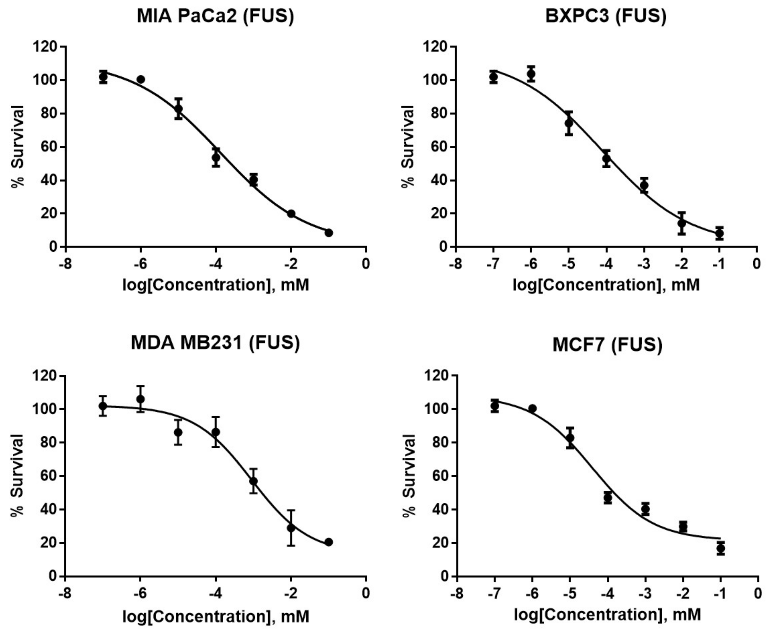
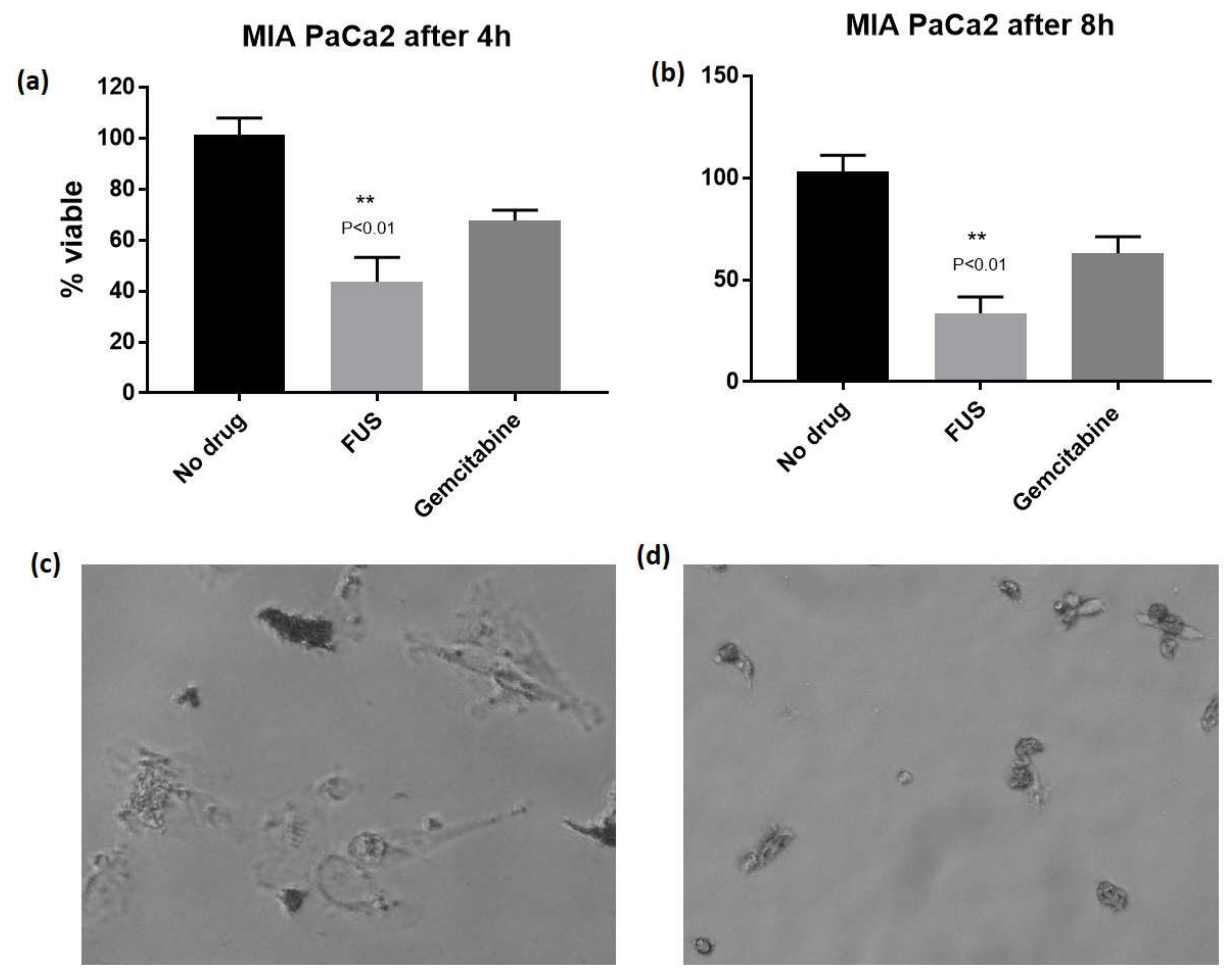
2. Results and Discussion

FUS was obtained as a white gum. The structure of FUS (Figure 1) was confirmed by spectroscopic analysis (1H, 13C-NMR, DEPT-135, 2D-NMR and HR-ESIMS) and by comparison with the published spectral values [19]. Accurate mass measurement of FUS obtained by FT-ESI-MS yielded a parent mass at m/z 467.2778 in positive ionization mode, corresponding to the sodium adduct [M + Na]+ with a molecular formula of C27H40O5 (calcd. mass 467.2773, [C27H40O5 + Na]+), accounting for 8 degrees of unsaturation. The resonances at δ 170.9 and 207.9 ppm in the 13C-NMR spectrum were characteristic of the presence of two carbonyl carbons of an ester and a ketone, respectively. The 1H and 13C-NMR data, in conjunction with the DEPT-135 spectrum (Figure S13), proved the presence of 27 carbon atom signals corresponding to six methyls (20-, 21-, 22-, 23-, 25-, and 27-), seven sp3 methylenes (1-, 4-, 5-, 8-, 9-, 13- and 24-), three sp3 methines (10-, 14- and 19-), three sp2 methines (2-,6- and 12-), one sp3 quaternary carbon (15-), five sp2 quaternary carbons (3-, 7-, 11-, 17- and 18-), two carbonyl carbons of an ester (26-OCOCH3) and a ketone (16-CO-). The presence of three sp2 methines and five sp2 quaternary carbons, along with one each of an ester and carbonyl moiety, proved the presence of six double bonds in this compound, and thus indicated that it was a bicyclic compound. After deducting the acetyl moiety ‘OCOCH3’ (δH = 2.06, δC = 20.9 and 170.9 ppm), the compound consisted of 25 carbons, which indicated it as a sesterterpene.
The cytotoxicity of FUS was determined against two pancreatic cancer cell lines, BxPc3 and MIA PaCa2, the ER-positive breast cancer cell line MCF7, and the triple negative breast cancer cell line MDA MB 231; gemcitabine was used as the positive control for the pancreatic cancer cell lines and doxorubicin for the breast cancer cell lines. FUS was active against all four cell lines tested (Figure 2 and Table 1) with sub to low micromolar IC50, but the activity against the pancreatic cancer cell lines were notably better than the breast cancer cell lines. FUS was between 3 to 58 times more potent than gemcitabine in pancreatic cancer cell lines, but doxorubicin was superior against both breast cancer cell lines compared to FUS. The therapeutic utility of FUS was further investigated against WI38, a non-tumor lung fibroblast cell line. FUS was found to be cytotoxic against WI38 with a high micromolar IC50 (Table 1), but was between 23 to 138 times more selective for the pancreatic cancer cell lines, and between 4.6 to 9.4 more selective for the breast cancer cell lines. This suggests a good therapeutic index against the pancreatic cancer cell lines which can be exploited if FUS is considered as a starting point for a medicinal chemistry program to develop a more potent analogue.
The relatively rapid cytotoxicity observed for FUS during the cell culture experiments led us to carry out a time-dependent cytotoxicity assay by monitoring percentage survival after 4- and 8-h post-incubation. FUS showed greater toxicity at both 4 and 8 h at 4 × IC50concentration in MIA PaCa2 cell line compared to gemcitabine. The differences were statistically significant (p < 0.01) (Figure 3a). Similarly, rapid toxicity was observed against MDA MB 231 cell line at 4 h (p < 0.03) and at 8 h (p < 0.01) compared to doxorubicin (Figures S5 and S6), although doxorubicin was notably more potent than FUS after 24 h incubation.
The morphological changes in the MIAPaCa2 cell line after incubating with FUS were monitored using a Nikon TS100 inverted microscope fitted with a camera. The cells appeared to show both apoptotic and necrotic damages within 4 h post-incubation, and the damages were fully evident at the 8-h time point (Figure 3c,d). These images, along with the survival analysis, point to the ability of the compound to induce severe stress resulting in rapid toxicity against the cell lines. This rapid cytotoxicity is intriguing and potentially a useful characteristic for an anticancer scaffold that can be developed against pancreatic cancer. Further studies are required to ascertain the mechanism of action of this compound, and will be reported in due course.
In summary, FUS is a known sesterterpene mycotoxin isolated from the endophytic fungus F. solani. This compound showed potent and rapid cytotoxicity against both pancreatic and breast cancer cell lines tested in this study. The complex structure and intriguing biological activity of FUS make it a good target for chemical synthesis and a lead structure for a medicinal chemistry project to develop a new anticancer drug against pancreatic cancer.

3. Experimental Section

3.1. Collection and Identification of the Plant Material

The aerial part of A. hookerianum was collected from Pablakhali, Rangamati, Chittagong Hill tracts, Bangladesh on 10 August 2014 and identified by the taxonomist of Bangladesh National Herbarium, Mirpur, Dhaka. A voucher specimen of the plant has been deposited (Accession no.: DACB 40633) in the herbarium for further reference (Figure S1).

3.2. General Experimental Procedures

The NMR spectra were recorded on a Bruker 400 MHz NMR spectrometer using CDCl3. The HRMS spectrum was recorded on an Exactive Orbitrap by a Thermo Scientific mass spectrometer at King’s College London, (London, UK), and the data were processed by Thermo XCalibur 2.2. Column chromatography was carried out on silica gel (70–230 mesh and 230–400 mesh, Merck, Darmstadt, Germany). Organic solvents, potato dextrose agar (PDA) medium, and TLC plates were purchased from Merck, Germany.

3.3. Isolation of Fungal Material

About 300 g of fresh and healthy parts of the plant (leaves, roots, and petioles) was cut with a sterile scalpel and stored at 4 °C in a sterile polyethene bag prior to use. Endophytic fungi were isolated from the fresh plant parts following the procedure, established at Pharmaceutical Sciences Research Division, BCSIR Laboratories, Dhaka, Bangladesh [11,12,13,14]. Total four endophytic fungi were isolated from different parts of A. hookerianum bearing the internal strain no. AHPE-3, AHPE-4 (Figure S2), AHLE-1 and AHLE-4. All the endophytic fungi were taxonomically identified up to genus level on the basis of macroscopic and microscopic morphological characters as Fusarium sp. (Figure S3). (AHPE-3), Fusarium sp. (AHPE-4), Colletotrichum sp. (AHLE-1) and Colletotrichum sp. (AHLE-4). The fungus AHPE-4 was selected for further investigation, based on the brine shrimp lethality bioassay data (Figure S4), and was cultured at a large scale to isolate bioactive secondary metabolites.

3.4. Molecular Identification of the Endophytic Fungus AHPE-4

For identification and differentiation, the Internal Transcript Spacer regions (ITS4 and ITS5) and the intervening 5.8S rRNA region was amplified and sequenced using electrophoretic sequencing on an ABI 3730 × l DNA analyzer (Applied Biosystems, Waltham, MA, USA) using Big Dye Terminator v 3.1 cycle sequencing kit (Thermo Fisher Scientific, Waltham, MA, USA). The ITS regions of the fungus were amplified using PCR (Hot Start Green Master Mix, Promega, Madison, WI, USA) and the universal ITS primers, ITS4 (5′-TCC GTA GGT GAA CCT GCG G-3′) and ITS5 (5′-GGA AGT AAA AGT CGT AAC AAG G-3′). The PCR products were purified and desalted using the Hot Start Green Master Mix (Cat: M7432, Promega, USA.) and sequenced on an ABI 3730 × l DNA analyzer (Applied Biosystems, USA). The sequences were aligned and prepared with the software Chromas (V 2.6.2, Technelysium, Brisbane, Australia) and matched against the nucleotide-nucleotide database (BLASTn) of the U.S. National Center for Biotechnology Information (NCBI) for final identification of the endophytic isolate. Finally, the sequence data (SI) were deposited in the Gen Bank database (accession number MG75792), which revealed 99% similarity other related fungal isolates of F. solani bearing accession numbers KX 497027, KJ863503, AB 190389, AY433805 etc. deposited in NCBI.

3.5. Extraction of the Fungal Material and Isolation of FUS
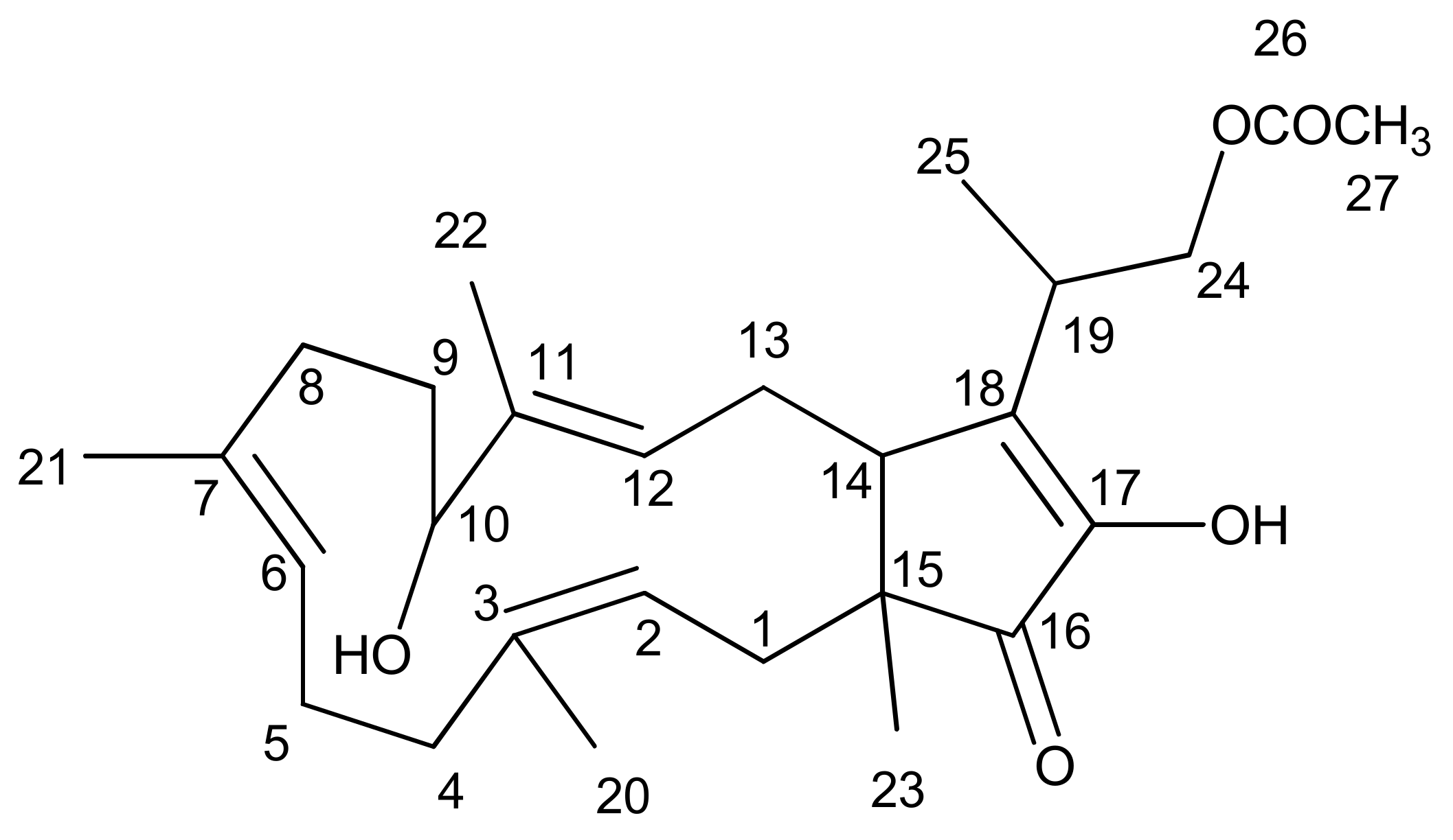
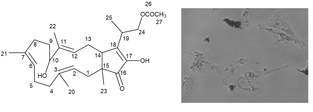
The fungus F. solani (AHPE-4), isolated from the petiole of the plant A. hookerianum, was cultivated at 28 ± 2 °C for 28 days on potato dextrose agar (PDA). The culture media were extracted with ethyl acetate for seven days in an air-tight, flat-bottom container with occasional shaking and stirring. This procedure was repeated three times to obtain the crude extract. The extract of endophytic fungi was then filtered using sterilized cotton filter followed by Whatman no. 1 filter papers. The solvent was evaporated with a rotary evaporator at low temperature (40 °C–50 °C) and reduced pressure.
The crude fungal extract (8 gm) was subjected to column chromatography for fractionation on silica gel (70–230 mesh) using gradients of petroleum ether/ethyl acetate, then ethyl acetate, followed by a gradient of ethyl acetate/methanol, and finally methanol, to afford a total of 15 fractions. These fractions were screened by TLC on silica gel under UV light and by spraying with vanillin-H2SO4 spray reagents. The column fraction of petroleum ether/15% ethyl acetate was subjected to preparative TLC on silica gel (toluene/20% ethyl acetate, 3 developments) to obtain FUS.

Fusaproliferin

18 mg, white, amorphous sticky mass; (1H-NMR, CDCl3): δ 2.40 (1H, dd, J = 10.8, 13.6 Hz, H-1´), 1.74 (1H, m, H-1″), 5.27 (1H, dd, J = 5.0, 10.2 Hz, H-2), 2.30 (1H, m, H-4´), 2.06 (1H, m, H-4″), 2.30 (1H, m, H-5´), 2.11 (1H, m, H-5″), 5.15 (1H, bs, H-6), 2.11 (1H, m, H-8´), 1.82 (1H, d, J = 9.2 Hz, H-8″), 1.82 (1H, d, J = 9.2 Hz, H-9´), 1.65 (1H, m, H-9″), 4.08 (1H, dd, J = 3.4, 9.8 Hz, H-10), 5.40 (1H, bt, H-12), 2.40 (1H, dd, J = 10.8, 13.6 Hz, H-13´), 1.95 (1H, m, H-13″), 2.69 (1H, dd, J = 2.0, 11.2 Hz, H-14), 2.80 (1H, sextet, H-19), 1.66 (3H, s, H-20), 1.66 (3H, s, H-21), 1.59 (3H, s, H-22), 1.02 (3H, s, H-23), 4.31 (1H, dd, J = 8.0, 10.4 Hz, H-24´), 4.27 (1H, dd, J = 7.2, 10.4 Hz, H-24″), 1.33 (3H, d, J = 6.8 Hz, H-25), 2.06 (3H, s, H-27), 5.56 ( 1H, s, 17- OH). 13C-NMR: δC 39.1 (C-1), 121.4 (C-2), 138.2 (C-3), 40.3 (C-4), 23.8 (C-5), 124.3 (C-6), 132.9 (C-7), 34.9 (C-8), 29.7 (C-9), 76.5 (C-10), 136.5 (C-11), 128.9 (C-12), 28.7 (C-13), 49.6 (C-14), 49.0 (C-15), 207.9 (C-16), 147.3 (C-17), 146.7 (C-18), 33.7 (C-19), 15.5 (C-20), 15.3 (C-21), 10.4 (C-22), 16.2 (C-23), 66.4 (C-24), 14.5 (C-25), 170.9 (C-26), 20.9 (C-27). HRESIMS m/z 467.2778 [M + Na]+ (calcd mass 467.2773, [C27H40O5 + Na]+).

3.6. Bioassays

3.6.1. Cell Culture

The MIA PaCa2 (pancreatic adenocarcinoma), BXPC3 (pancreatic adenocarcinoma), MDA-MB-231 (triple-negative breast cancer), MCF-7 (estrogen receptor positive breast cancer) cell lines were obtained from the American Type Culture Collection. The MIA PaCa2 cell line was maintained in Dulbecco’s modified Eagle’s medium (DMEM; Invitrogen, Carlsbad, CA, USA), supplemented with fetal bovine serum (10% v/v; Invitrogen), horse serum (2.5% v/v; Invitrogen) and penicillin-streptomycin (1% v/v, Invitrogen). The BXPC3 cell line was maintained in RPMI-1640 medium (DMEM; Invitrogen), supplemented with fetal bovine serum (10% v/v; Invitrogen), and penicillin-streptomycin (1% v/v, Invitrogen). The MDA MB 231 cell line was maintained in Dulbecco’s modified Eagle’s medium (DMEM; Invitrogen), supplemented with fetal bovine serum (10% v/v; Invitrogen), l-glutamine (2 mM; Invitrogen), non-essential amino acids (1×; Invitrogen) and penicillin-streptomycin (1% v/v, Invitrogen). The MCF7 cell line was maintained in Eagle’s Minimum Essential medium supplemented with fetal bovine serum (10% v/v; Invitrogen), 0.01 mg/mL human recombinant insulin and penicillin-streptomycin (1% v/v, Invitrogen). During seeding, cells were counted using a Neubauer hemocytometer (Assistant, Hanover, Germany) by microscopy (Nikon, Melville, NY, USA) on a non-adherent suspension of cells that were washed in PBS, trypsinized, centrifuged at 8 °C at 8000 rpm for 5 min, and re-suspended in fresh medium.

3.6.2. MTT Assay

The cells were grown in normal cell culture conditions at 37 °C under a 5% CO2 humidified atmosphere using an appropriate medium. The cell count was adjusted to 105 cells/mL and 2500 cells (MDA-MB-231) or 5000 cells (A4 and WI-38) were added per well. The cells were incubated for 24 h, and 1 μL of the appropriate inhibitor concentrations was added to the wells in triplicate. After 96 h of continuous exposure to each compound, the cytotoxicity was determined using the 3-(4,5-dimethylthiazol-2-yl)-2,5-diphenyltetrazolium bromide (MTT) (Lancaster Synthesis Ltd., Morecambe, Lancashire, UK) colorimetric assay. Absorbance was quantified by spectrophotometry at λ = 570 nm (Envision Plate Reader, PerkinElmer, Waltham, MA, USA). IC50 values were calculated by a dose-response analysis using the Prism GraphPad Prism® software.

Supplementary Materials

The experimental procedures and 1H, 13C-NMR, DEPT-135 and HRMS spectra of FUS (PDF) are available online.

Author Contributions

Conceptualization, C.M.H. and M.H.S.; Formal analysis, C.M.H. and M.H.S.; Investigation, N.H., A.V., M.H.S. and K.M.R.; Methodology, N.H. and A.V.; Project administration, M.H.S.; Resources, K.M.R.; Supervision, C.M.H., M.S.R., M.H.S. and K.M.R.; Writing-original draft, N.H. and M.H.S.; Writing-review & editing, C.M.H., M.S.R. and K.M.R.

Funding

This research received no external funding. The APC was funded by King’s College London ACAC11460.

Acknowledgments

The authors are thankful to the authority of BCSIR for supporting the work in this paper through providing the all lab facilities related to the work on endophytic fungi.

Conflicts of Interest

The authors declare no competing financial interest.

References

  1. Kleeff, J.; Korc, M.; Apte, M.; La Vecchia, C.; Johnson, C.D.; Biankin, A.V.; Neale, R.E.; Tempero, M.; Tuveson, D.A.; Hruban, R.H.; et al. Pancreatic cancer. Nat. Rev. Dis. Prim. 2016, 2, 16022. [Google Scholar] [CrossRef] [PubMed]
  2. Van Cutsem, E.; Vervenne, W.L.; Bennouna, J.; Humblet, Y.; Gill, S.; Van Laethem, J.-L.; Verslype, C.; Scheithauer, W.; Shang, A.; Cosaert, J. Phase III trial of bevacizumab in combination with gemcitabine and erlotinib in patients with metastatic pancreatic cancer. J. Clin. Oncol. 2009, 27, 2231–2237. [Google Scholar] [CrossRef]
  3. Siriwardena, A.K.; Siriwardena, A.M. Pancreatic cancer. BMJ Br. Med. J. 2014, 349. [Google Scholar] [CrossRef] [PubMed]
  4. Wong, M.C.S.; Jiang, J.Y.; Liang, M.; Fang, Y.; Yeung, M.S.; Sung, J.J.Y. Global temporal patterns of pancreatic cancer and association with socioeconomic development. Sci. Rep. 2017, 7, 3165. [Google Scholar] [CrossRef] [Green Version]
  5. Vincent, A.; Herman, J.; Schulick, R.; Hruban, R.H.; Goggins, M. Pancreatic cancer. Lancet 2011, 378, 607–620. [Google Scholar] [CrossRef]
  6. Ouyang, L.; Luo, Y.; Tian, M.; Zhang, S.Y.; Lu, R.; Wang, J.H.; Kasimu, R.; Li, X. Plant natural products: From traditional compounds to new emerging drugs in cancer therapy. Cell Prolif. 2014, 47, 506–515. [Google Scholar] [CrossRef] [PubMed]
  7. Harvey, A.L.; Edrada-Ebel, R.; Quinn, R.J. The re-emergence of natural products for drug discovery in the genomics era. Nat. Rev. Drug Disc. 2015, 14, 111–129. [Google Scholar] [CrossRef] [Green Version]
  8. Kumar, A.; Patil, D.; Rajamohanan, P.R.; Ahmad, A. Isolation, purification and characterization of vinblastine and vincristine from endophytic fungus Fusarium oxysporum isolated from Catharanthus roseus. PLoS ONE 2013, 8, e71805. [Google Scholar] [CrossRef]
  9. Nascimento, A.M.d.; Conti, R.; Turatti, I.C.; Cavalcanti, B.C.; Costa-Lotufo, L.V.; Pessoa, C.; de Moraes, M.O.; Manfrim, V.; Toledo, J.S.; Cruz, A.K. Bioactive extracts and chemical constituents of two endophytic strains of Fusarium oxysporum. Rev. Bras. Farmacogn. 2012, 22, 1276–1281. [Google Scholar] [CrossRef]
  10. Cui, Y.; Yi, D.; Bai, X.; Sun, B.; Zhao, Y.; Zhang, Y. Ginkgolide B produced endophytic fungus (Fusarium oxysporum) isolated from Ginkgo biloba. Fitoterapia 2012, 83, 913–920. [Google Scholar] [CrossRef]
  11. Khan, M.I.H.; Sohrab, M.H.; Rony, S.R.; Tareq, F.S.; Hasan, C.M.; Mazid, M.A. Cytotoxic and antibacterial naphthoquinones from an endophytic fungus, Cladosporium sp. Toxicol. Rep. 2016, 3, 861–865. [Google Scholar] [CrossRef] [PubMed]
  12. Chowdhury, N.S.; Sohrab, M.H.; Rony, S.R.; Sharmin, S.; Begum, M.N.; Rana, M.S.; Hasan, C.M. Identification and bioactive potential of endophytic fungi from Monochoria hastata (L.) Solms. Bangladesh J. Bot. 2016, 45, 187–193. [Google Scholar]
  13. Chowdhury, N.S.; Sohrab, M.H.; Rana, M.S.; Hasan, C.M.; Jamshidi, S.; Rahman, K.M. Cytotoxic naphthoquinone and azaanthraquinone derivatives from an endophytic Fusarium solani. J. Nat. Prod. 2017, 80, 1173–1177. [Google Scholar] [CrossRef] [PubMed]
  14. Khan, N.; Afroz, F.; Begum, M.N.; Rony, S.R.; Sharmin, S.; Moni, F.; Hasan, C.M.; Shaha, K.; Sohrab, M.H. Endophytic Fusarium solani: A rich source of cytotoxic and antimicrobial napthoquinone and aza-anthraquinone derivatives. Toxicol. Rep. 2018, 5, 970–976. [Google Scholar] [CrossRef] [PubMed]
  15. Takemoto, K.; Kamisuki, S.; Chia, P.T.; Kuriyama, I.; Mizushina, Y.; Sugawara, F. Bioactive dihydronaphthoquinone derivatives from Fusarium solani. J. Nat. Prod. 2014, 77, 1992–1996. [Google Scholar] [CrossRef] [PubMed]
  16. Shweta, S.; Zuehlke, S.; Ramesha, B.T.; Priti, V.; Kumar, M.P.; Ravikanth, G.; Spiteller, M.; Vasudeva, R.; Shaanker, U.R. Endophytic fungal strains of Fusarium solani, from Apodytes dimidiata E. Mey. ex Arn (Icacinaceae) produce camptothecin, 10-hydroxycamptothecin and 9-methoxycamptothecin. Phytochemistry 2010, 71, 117–122. [Google Scholar] [CrossRef] [PubMed]
  17. Chakravarthi, B.; Das, P.; Surendranath, K.; Karande, A.A.; Jayabaskaran, C. Production of paclitaxel by Fusarium solani isolated from Taxus celebica. J. Biosci. 2008, 33, 259–267. [Google Scholar] [CrossRef]
  18. Kosmidis, C.; Denning, D.W. Opportunistic and Systemic Fungi. In Infectious Diseases, 4th ed.; Elsevier: Amsterdam, The Netherlands, 2017; pp. 1681–1709.e3. [Google Scholar]
  19. Randazzo, G.; Foglianoa, V.; Ritieni, A.; Rossin, L.M.E.; Scarallo, A.; Segred, A.L. Proliferin, a new sesterterpene from Fusarium proliferatum. Tetrahedron 1993, 49, 10883–10896. [Google Scholar] [CrossRef]
  20. Ritieni, A.; Fogliano, V.; Randazzo, G.; Scarallo, A.; Logrieco, A.; Moretti, A.; Manndina, L.; Bottalico, A. Isolation and characterization of fusaproliferin, a new toxic metabolite from Fusarium proliferatum. Nat. Toxins 1995, 3, 17–20. [Google Scholar] [CrossRef]
  21. Santini, A.; Ritieni, A.; Fogliano, V.; Randazzo, G.; Mannina, L.; Logrieco, A.; Benedetti, E. Structure and absolute stereochemistry of fusaproliferin, a toxic metabolite from Fusarium proliferatum. J. Nat. Prod. 1996, 59, 109–112. [Google Scholar] [CrossRef]
  22. Fotso, J.; Leslie, J.F.; Smith, S.J. Production of beauvericin, moniliformin, fusaproliferin, and fumonisins B1, B2, and B3 by fifteen ex-type strains of Fusarium Species. Appl. Environ. Microbiol. 2002, 68, 5195–5197. [Google Scholar] [CrossRef] [PubMed]
  23. Ritieni, A.; Monti, S.M.; Moretti, A.; Logrieco, A.; Gallo, A.; Ferracane, R.; Fogliano, V. Stability of fusaproliferin, a mycotoxin from Fusarium spp. J. Sci. Food Agric. 1999, 79, 1676–1680. [Google Scholar] [CrossRef]
  24. Logrieco, A.; Moretti, A.; Fornelli, F.; Fogliano, V.; Ritieni, A.; Caiaffa, M.F.; Randazzo, G.; Bottalico, A.; Macchia, L. Fusaproliferin production by Fusarium subglutinans and its toxicity to Artemia salina, SF-9 insect cells, and IARC/LCL 171 human B lymphocytes. Appl. Environ. Microbiol. 1996, 62, 3378–3384. [Google Scholar] [PubMed]
  25. Ritieni, A.; Monti, S.M.; Randazzo, G.; Logrieco, A.; Moretti, A.; Peluso, G.; Ferracane, R.; Fogliano, V. Teratogenic effects of fusaproliferin on chicken embryos. J. Agric. Food Chem. 1997, 45, 3039–3043. [Google Scholar] [CrossRef]
Sample Availability: Samples of the compounds are not available from the authors. However, the compounds can be extracted again from the natural source.
Figure 1. Structure of Fusaproliferin.
Molecules 23 03288 g001
Figure 2. MTT cell-viability assay profile in pancreatic (MIA PaCa2 and BXPC3) and breast (MDA MB 231 and MCF7) cancer cell lines treated with FUS for 24 h.
Molecules 23 03288 g002
Figure 3. Effect of FUS on pancreatic cancer cell line. (a,b) FUS showed statistically significant rapid toxicity (p < 0.01) against MIA PaCa2 cell line after 4 h and 8 h incubation; (c,d) morphological changes observed in MIA PaCa2 cell lines after 4 h and 8 h incubation, respectively.
Molecules 23 03288 g003
Table 1. Cytotoxicity assay results of FUS against human tumor cells (IC50 in μM).
Compound/StandardMIA PaCa 2 (Pancreatic)BXPC3
(Pancreatic)
MDA MB 231
(Breast)
MCF7
(Breast)
WI 38 (Lung Fibroblast)
FUS0.13 ± 0.090.76 ± 0.241.9 ± 0.323.9 ± 0.7518 ± 0.66
Gemcitabine7.6 ± 0.662.2 ± 0.43NTNTNT
DoxorubicinNTNT0.06 ± 0.030.02 ± 0.018NT

