Article

Chemical Composition and in-Vitro Evaluation of the Antimicrobial and Antioxidant Activities of Essential Oils Extracted from Seven Eucalyptus Species

1
Department of Applied Chemistry and Biochemistry, Government College University, Faisalabad 38000, Pakistan
2
Department of Zoology and Fisheries, Government College University, Faisalabad 38000, Pakistan
3
Department of Chemistry, Government College for Women University, Faisalabad 38000, Pakistan
4
Department of Biochemistry/US-Pakistan Center for Advanced Studies in Agriculture and Food Security (USPCAS/AFS), University of Agriculture, Faisalabad 38000, Pakistan
5
Department of Chemistry, Government College University, Faisalabad 38000, Pakistan
*
Authors to whom correspondence should be addressed.
Molecules 2015, 20(11), 20487-20498; https://doi.org/10.3390/molecules201119706
Submission received: 27 July 2015 / Revised: 26 October 2015 / Accepted: 29 October 2015 / Published: 18 November 2015
(This article belongs to the Section Medicinal Chemistry)

Abstract

:
Eucalyptus is well reputed for its use as medicinal plant around the globe. The present study was planned to evaluate chemical composition, antimicrobial and antioxidant activity of the essential oils (EOs) extracted from seven Eucalyptus species frequently found in South East Asia (Pakistan). EOs from Eucalyptus citriodora, Eucalyptus melanophloia, Eucalyptus crebra, Eucalyptus tereticornis, Eucalyptus globulus, Eucalyptus camaldulensis and Eucalyptus microtheca were extracted from leaves through hydrodistillation. The chemical composition of the EOs was determined through GC-MS-FID analysis. The study revealed presence of 31 compounds in E. citriodora and E. melanophloia, 27 compounds in E. crebra, 24 compounds in E. tereticornis, 10 compounds in E. globulus, 13 compounds in E. camaldulensis and 12 compounds in E. microtheca. 1,8-Cineole (56.5%), α-pinene (31.4%), citrinyl acetate (13.3%), eugenol (11.8%) and terpenene-4-ol (10.2%) were the highest principal components in these EOs. E. citriodora exhibited the highest antimicrobial activity against the five microbial species tested (Staphylococcus aureus, Bacillus subtilis, Escherichia coli, Aspergillus niger and Rhizopus solani). Gram positive bacteria were found more sensitive than Gram negative bacteria to all EOs. The diphenyl-1-picrylhydazyl (DPPH) radical scavenging activity and percentage inhibition of linoleic acid oxidation were highest in E. citriodora (82.1% and 83.8%, respectively) followed by E. camaldulensis (81.9% and 83.3%, respectively). The great variation in chemical composition of EOs from Eucalyptus, highlight its potential for medicinal and nutraceutical applications.

1. Introduction

The interest in use of natural medicines from plants has been increasing in the past few years in industrialized societies, particularly against microbial agents because of the ever growing problem of antibiotic resistance [1]. The applications of essential, volatile and hydrophobic oils from natural resources are rapidly increasing in medicinal, food and cosmetic industries to replace harmful synthetic additives [2,3]. The EOs are aromatic essences usually derived from the aerial parts of the plants containing oxygenated compounds; phenols, alcohols, esters, ethers, ketones, aldehydes and oxides, hydrocarbons; terpenes, organic sulphur and nitrogenous compounds and benzene derivatives [4,5]. EOs used as food flavorings and preservatives are generally recommended as safe (GRAS) and have broad spectrum of activity against different bacterial and fungal strains due to the presence of high percentages of monoterpenes, eugenol, cinnamic aldehydes, thymol and polyphenols [6,7].
Eucalyptus, with almost 900 species, is found worldwide, with some species being native to South East Asia. Due to its rapid growth Eucalyptus is an important hardwood forest crop used in the pulp and paper industry. Its timber is used for construction and fuel [8], while the gum is used for the treatment of diarrhoea and as an astringent in dentistry [9]. More than 300 species of this genus contain volatile oils in their leaves [10]. Worldwide production of Eucalyptus EOs is 3000 tons and major producers of EOs of Eucalyptus are China, Spain, Portugal, South Africa and Chile [11]). Eucalyptus EOs are used as fragrance elements in household products and cosmetics such as soaps, detergents, lotions and perfumes. They are also used as flavor elements in foods and beverages such as baked goods, confectionaries, meat products, ice creams and soft drinks. In addition their use to help prevent and treat human diseases has undergone a relatively recent revival and expansion [12].
Eucalyptus EO is officially listed in the Indian Pharmacopeia as a counterirritant and mild expectorant and also in the Chinese Pharmacopeia as a skin irritant. EOs of Eucalyptus have been extensively used as expectorant in cough and cold compounds in various oral dosage forms, including lozenges and syrups, and as an inhalant in vapour baths. It is also used externally for percutaneus absorption in dosage forms including the EOs, liniments and ointments [13]. EO of Eucalyptus is ingested orally to treat catarrh, used as inhalant and applied tropically as a rubefacient [14].
Volatile oils of Eucalyptus are used as antioxidants and anti-inflammatory agents for skin diseases, rheumatism and as expectorant in cases of bronchitis [15,16]. Many researchers have reported the chemical composition, antioxidant and antimicrobial activities of Eucalyptus species [17]. However, geographical distribution and species variation greatly affect these properties which require extensive studies to explore the potential of this plant. The chemical composition of eight Eucalyptus species’ essential oils from Tunisia reported the presence of 1,8-cineole as the major ingredient, followed by cryptone, α-pinene, p-cymene, α-terpineol, phellandral, cuminal, globulol, limonene, aromadendrene, sapthulenol and terpinene-4-ol [18]. 1,8-Cineole and α-pinene have been reported as major components of EOs from seven Eucalyptus species [19].
Eucalyptus was introduced in Pakistan long ago, but interest in the tree has increased considerably in the last two decades and lots of renewed efforts towards its propagation have been made. The large scale plantation of Eucalyptus in Pakistan has been recommended [20]. The growth of Eucalyptus in Pakistan is popular with public sector institutions, roadside plantations are widely distributed and it is being introduced to farmers in irrigated, saline and waterlogged soil areas as a purported cash crop. Its wood has been widely used for houses and furniture. However, its leaves are not yet used commercially. Currently Pakistan is importing more than 10 tons of Eucalyptus EOs from different countries as compared to 8.9 and 8.16 tons of Eucalyptus EOs in 1999–2000 and 2000–2001.
Presently, the volatile constituents and antioxidant activity of EOs from Pakistani Eucalyptus have not been rationally investigated and no systematic information about its chemical composition is available. To address this gap in our knowledge, the present study reports the chemical composition, antimicrobial and antioxidant activity of the EOs from seven Eucalyptus species for their potential use in food and medicine. The objectives of the study were: (i) the extraction of EOs from leaves of Eucalyptus and quantification of its yield; (ii) the identification and quantification of chemical compounds in each oil and (iii) determination of antioxidant and antimicrobial activities against some selected strains of bacteria and fungi.

2. Results and Discussion

2.1. Percentage Yield of EOs by Steam Distillation

The average yield of EOs from the different species of Eucalyptus was 1.84% (w/w) after 7 h of distillation. E. globulus had highest oil content (1.91%), while E. melanophloia contained least amount (1.73%). These findings are similar to results reporting a 2.4% yield of oil from E. gillii cultivated in Iran and 2.3% in Tunisia [21,22]. The small differences may be due to different climatic, geographic and ecological parameters.

2.2. Physiochemical Analysis of Essential Oils

The physiochemical properties of seven different Eucalyptus EOs revealed that initially Eucalyptus EOs were colourless, but after about 2 weeks they showed yellow colours (Table 1). All the EOs were soluble in 2 mL of 80% ethanol, but insoluble in water. The specific gravity of the EOs extracted from the seven Eucalyptus species ranged from 0.84 to 0.94. The boiling point of E. melanophloia EO was the highest at 178 °C, followed by E. citriodora (177 °C) and the lowest was for E. globulus (161 °C).
Table 1. Physiochemical properties of Eucalyptus essential oils.
Physiochemical PropertyE. citriodoraE. camaldulensisE. crebraE. tereticornisE. globulesE. melanophloiaE. microtheca
Percentage yield1.821.901.841.831.891.731.84
Color Pale yellowSlightly yellowishLight yellowYellow to orangeColorless to pale yellowYellow to brownYellow reddish,
OdorCitronellal odor1,8 Cineole odorCamphor odorCineole-pinene odorHerbal odorPinene odorCymene odor
SolubilityInsoluble in water, soluble in alcoholInsoluble in water, soluble in alcoholInsoluble in water, soluble in alcoholInsoluble in water, soluble in alcoholInsoluble in water, soluble in alcoholInsoluble in water, soluble in alcoholInsoluble in water, soluble in alcohol
Boiling point (°C)177 °C178 °C165 °C173 °C161 °C174 °C166 °C
Specific gravity0.850.920.900.840.890.940.86
Refractive index1.491.451.421.441.381.411.47

2.3. Chemical Nature of Essential Oils

The chemical composition of the seven selected Eucalyptus EOs revealed great variation in the concentration and types of constituents (Table 2). Identification of the components of EOs was based on their GC retention times and mass spectra, which were compared to the published data and spectra of respective standards to those constituents. The GC-MS analysis thus revealed the presence of 31 components in E. citriodora and E. melanophloia, 27 components in E. crebra, 24 components in E. tereticornis, 14 components in E. globulus, 13 components in E. camaldulensis and 12 components in E. microtheca. The results are comparable to the 25 major compounds at an average concentration greater than 0.9% ± 0.2% reported earlier in eight Eucalyptus species EOs. E. gillii EO contained 34 compounds as reported by another study [23].
Previous studies on four species of Eucalyptus grown in Tunisia reported the chemical compositions of E. salubris (27 compounds; 99.2%), E. salmonophloia (31 compounds; 99.2%), E. oleosa (32 compounds; 97.6%), and E. gracilis (18 compounds; 97.7%) [23].
E. citriodora and E. melanophloia EOs consisted of the highest number (31) of chemical compounds. The major components in EO of E. citriodora were citronellal (22.3%), citronellol (20%), patchoulene (9.4%), germacrene-D (7.5%), α-terpinol (6.3%), eugenol (3.9%), α-pinene (3.6%), and β-citronellal (3.2%). Previous studies on E. citriodora EO showed total 17 components out of which citronellal (21.2%) citronellol (12.2%) and isopulegol (11.9%) were listed as the main components [9]. Analysis of E. melanophloia EO revealed 31 components including α-pinene (16.0%), β-phellandrene (14.3%), farnesol (10.0%) and verbenol (9.2%). These results show a very efficient production of such compounds as compared to monoterpenoid oils, with α-pinene (1.6%), 1,8-cineole (0.2%–60%) and p-cymene (0.2%–21%) as the major constituents [24]. The chemical composition of E. camaldulensis EO showed 13 components, with major components such as linalool (17.0%), 1,8-cineole (16.1%), p-cymene (12.2%), β-farnesol (11.1%), γ-terpinene (10.7%), phenythyl acetate (7.1%), geranial (6.6%), terpinene-4-ol (5.3%), paraldehyde (5.3%) and p-ment-1-en-3, 8-diol (5.1%). EO from E. oleosa stem, immature flowers and the fruit showed 1,8-cineole as principal compound, representing 31.5%, 47.0% and 29.1%, respectively [25]. The results show small variation from a previous study describing a total of 11 components out of which p-cymene (17.4%–28.6%), β-phellandrene (12.3%–14.4%) and β-pinene (0.9%–11.4%) were the main ingredients [26] and great variation from the 1,8-cineole (21.7%), β-pinene (20.5%) and methyl eugenol (6.1%) levels in E. camaldulensis [27]. The study is also comparable to a E. camaldulensis EO analysis reporting 1,8-cineole (45.7%) and p-cymene (17.1%) as major compounds obtained by hydrodistillation and 8,14-cedrane oxide (43.8%) and elemol (6.3%) by supercritical fluid extraction [28]. The findings of present study show a greater number of oxygenated compounds. The differences may be attributed to geographical, environmental and climatic variations affecting chemical composition of Eucalyptus species.
E. crebra oil was composed of 27 components. Limonene (14.3%) was the major constituent, followed by terpinene-4-ol (10.2%) and citrinyl acetate (9.1%). E. tereticornis EO showed 24 components with 1,8-cineole (15.2%), α-pinene (12.1%), myrtenal (8.1%), linalool, (7.4%) and paraldehyde nitrile (7.1%) as the most abundant. Another study reported that E. tereticornis EO contained 19 components out of which α-pinene (21.4%) was the major constituent but 1,8 cineole was absent [29]. In this study 1,8-cineole was the principal component (56.5%) found in E. globulus along with limonene (28.0%), α-pinene (4.2%), α-terpinol (4.0%) and globulol (2.4%). The present study shows higher contents of these compounds as compared to earlier reports for a total of 10 components in this plant, out of which 1,8 cineole (17.5%), α-pinene (1.7%) and α-phellandrene (1.1%) were the major constituents [30]. E. microtheca EO showed total 12 components, with α-pinene (31.4%), citrinyl acetate (13.2%), cymene (12.4%), eugenol (11.8%) and eucalyptol (8.1%) as the major ones. The results of the present study showed higher amounts than the cymene (10.3%) and α-pinene (10.0%) as the major constituents reported previously [31]. The chemical composition of EOs was statistically significantly different in all Eucalyptus species. Some components were found in abundance in some species and absent in some species.
Table 2. Relative percentage chemical composition of essential oils of the EOs extracted from seven Eucalyptus species.
Serial No.ComponentsE. citriodoraE. camaldulensisE. crebraE. tereticornisE. globulesE. melanophloiaE. microtheca
1.1,8-Cineole 16.14.915.256.53.12.0
2.cis-β-Ocimene0.1 2.1
3.4-Methylene-1-(1-methylethyl)-3-Cyclo-hexene-1-ol 5.7
4.3-Carene 1.7
5.α-Cubebene 2.22.20.2
6.α-Elemene 1.3
7.α-Humelene 2.4
8.α-Terpinol6.3 4
9.α-Phellandrene 8.1
10.α-Pinene3.60.92.512.14.21631.4
11.β-Caryophyllene 1.2
12.β-Citronellal3.2
13.β-Farnesol 11.1 2.8 10
14.β-Phellandrene0.9 14.3
15.β-Pinene 2.6 0.21.52.5
16.Amorphane 0.3
17.Benzaldehyde 0.8
18.Camphene 1.10.10.9
19.Camphor 1.3
20.Citrinyl acetate2.8 9.1 2.813.2
21.Citral 0.1
22.Citronellal22.3
23.Citronellol20
24.Citronellal oxime1.4 1.03.6 1.4
25.Cymene-8-ol0.3 1.6
26.Cyclopentanone
27.Eugenol3.90.60.71.8 1.711.8
28.Eucalyptol 0.1 3.30.21.18.1
29.Digitoxigenine 1.81.7 0.4
30.Geranial0.1 4.1 2.7
31.Germacrene-D7.5 2.5
32.Geranial oxime1.9 4.2
33.Geranial0.16.63.6 0.11.4
34.Geranial nitrile0.7 1.1 3.6
35.Geranyl acetate 2.7
36.Globulol 2.40.6
37.Isopulegol0.1 2.3 0.10.6
38.Isosativene 3.7 4.5
39.Limonene0.1 14.30.7281.21.5
40.Linalool0.5171.17.40.30.6
41.2-Methylprop-1-enyl-cyclohexa-1,5-diene 0.9
42.Myrtenal 0.6 8.1 9.2
43.Neriine 2.30.40.8
44.Neral 1.75.4 2.71.7
45.Neral oxime 1,4
46.Paraldehyde 5.3 6.0 1.4
47.Paraldehyde nitrile1.9 5.97.1 0.7
48.Patchoulene9.4 3.0
49.p-Cymene 12.2 0.212.4
50.Phenythyl acetate 7.1 0.21.81.3
51.Pinocarveol
52.p-Ment-1-en-3,8-diol 5.1
53.Sabinene4.2 1.4
54.Spathulenol 0.4
55.Solanone 0.3
56.Terpinene-4-ol0.35.310.2 1.2
57.Verbenol 9.2
58.γ -Terpinene0.710.7
59.γ-Terpinene1 1.8 0.4
60.Trance-pinocarveol1.1 6.8
61.γ-phellandrene3.2
62.Ylangene 0.9

2.4. Antimicrobial Activity

The EOs extracted from the seven Eucalyptus species showed great potential for their application as antimicrobial agents. The activities were determined by using three strains of bacteria and two strains of fungi.

2.4.1. Antibacterial Activity

The EOs extracted from all seven Eucalyptus spp. showed antibacterial activity against S. aureus, B. subtilis and E. coli (Table 3). Maximum inhibition zones were observed for the E. citriodora EO against Gram positive bacteria (31 mm against S. aureus and 28 mm against B. subtilis). E. melanophloia and E. citriodora showed significant activity against the Gram negative bacterium E. coli.
Table 3. Comparative antimicrobial activities of essential oils of Eucalyptus species.
Microbial SpeciesZones of Growth Inhibition ( in mm) by Eucalyptus Species
E. citriodoraE. camaldulensisE. crebraE. tereticornisE. globulesE. melanophloiaE. microthecaAmoxilNizoral
Bacterial speciesS. aureus31 ± 0.83 Aa21 ± 0.851 Ade23 ± 0.836 Acd22 ± 0.853 Ade28 ± 0.835 Acd26 ± 0.836 Abc16 ± 0.831 Ae22 ± 0.833 Aab-
B. subtilis28 ± 0.833 Aa24 ± 0.835 Ade21 ± 0.848 Acd18 ± 0.835 Ade17 ± 0.833 Acd22 ± 0.836 Abc20 ± 0.838 Ae28 ± 0.833 Aab-
E. coli15 ± 0.835 Ba10 ± 0.835 Bde12 ± 0.835 Bcd14 ± 0.836 Bde13 ± 0.83 Bcd16 ± 0.833 Bbc11 ± 0.835 Be20 ± 0.831 Bab-
Fungal speciesA. niger29 ± 0.831 Aa28 ± 0.835 Aab25 ± 0.836 Abc26 ± 0.833 Ab24 ± 0.835 Abc27 ± 0.835 Ac21 ± 0.835 Ac-17 ± 0.835 Ac
R. solani26 ± 0.836 Bb22 ± 0.829 Bab19 ± 0.835 Bbc21 ± 0.836 Bb20 ± 0.835 Bbc12 ± 0.835 Bc17 ± 0.838 Bc-20 ± 0.835 Bc
Each values is means of three. The capital letters represent significant difference in microbial species, while small letters represent significant difference in EOs.
However, all the species showed higher activity for the Gram positive bacteria as compared to the Gram negative bacteria. Gram positive bacteria were found to be more sensitive to E. gillii EO and extracts than Gram negative ones [22]. E. oleosa EO exhibited an interesting antibacterial activity against all microorganisms tested (L. monocytogenes, S. aureus, E. coli, K. pneumoniae, S. cerevisiae, C. albicans, M. ramamnianus and A. ochraceus). The activity in this study was better against Gram-positive bacteria except for S. aureus and E. coli [23]. Results have proved that E. citriodora EO is more potent in antibacterial activity than the other six species. The EOs of Eucalyptus have previously shown antimicrobial and antiplasmid activities [32]. Previous studies also report that EOs against food spoilage organisms and food-borne pathogens are slightly more active against Gram-positive than Gram-negative bacteria [33].

2.4.2. Antifungal Activity

E. citriodora EO was found most effective against A. niger (29 mm) and R. solani (26 mm). Significant antimicrobial activity was shown by EO of E. microtheca against A. niger (21 mm) while EO of E. melanophloia was found least effective (12 mm) against R. solani (Table 3). The results showed that all the tested Eucalyptus EOs presented significant antifungal activity against A. niger and R. solani and had equal or more antifungal effect than amoxil and nizoral. These properties could be correlated to the chemical composition of the oils with good phenolic, alcoholic or aldehydic contents that may be correlated to the geographical distribution and environmental effect on production of phytochemicals in plants.

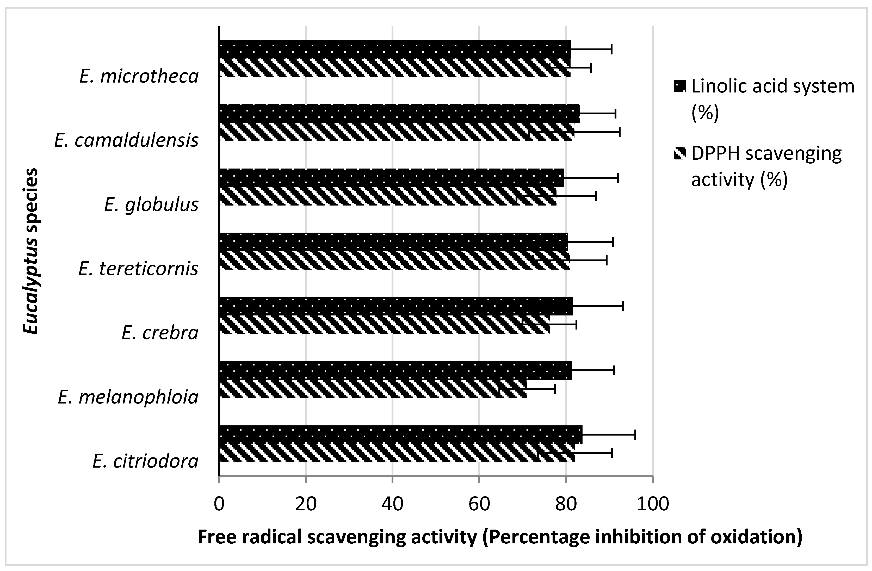
2.5. Antioxidant Activity

The DPPH scavenging activity was highest in E. citriodora (82.1%), followed by E. camaldulensis (81.9%) and E. microtheca (81.8%) as compared to positive control BHT. The results are compared to the DPPH assay results for E. oleosa EO activity in the range of 12.0–52.8 mg/mL, whereas in the 2,2′-azinobis-3-ethylbenzothiazoline-6-sulfonate assay (176.5 ± 3.1 mg/L) it showed the best inhibition result [18]. All the EOs strongly suppressed the peroxide formation in linoleic acid system during incubation. The inhibition of oxidation of linoleic acid system was higher for the EOs of E. citriodora (83.8%) and E. camaldulensis (83.2%) than the other five Eucalyptus species (Figure 1). These findings are also supported by another study reporting 86.07% inhibition of linoleic acid for non-polar methanol extract of E. sargentii [34].
Figure 1. Antioxidant activities of Eucalyptus EOs through linoleic acid and diphenyl-1-picrylhydazyl radical scavenging assays. (Each value is mean of three).
Molecules 20 19706 g001

3. Experimental Section

3.1. Chemicals and Microbial Strains

All the chemicals used in this study were of analytical grade and purchased from Sigma Aldrich (Taufkirchen, Germany). S. aureus, B. subtilis, E. coli, A. niger and R. solani were obtained from and identified by the Department of Microbiology, University of Agriculture (Faisalabad, Pakistan). The bacterial strains were preserved and cultured separately in Lauria and Bertani (LB) media and fungal strains in Eggins and Pugh (E and P) medium broth by following the standard microbiological protocols. All microorganisms were stocked at −4 °C in standard conditions and were revived twice before use in the manipulations.

3.2. Collection of Samples

The fresh leaves from E. citriodora, E. melanophloia, E. crebra, E. tereticornis, E. globulus, E. camaldulensis and E. microtheca were collected from the Gutwala Forest Research Institute, (Faisalabad, Pakistan) in May 2014. All the leaves were washed with water to remove dirt particles and shade dried to reduce the moisture contents.

3.3. Procedure to Extract EOs

The EO was extracted from each plant material through a hydrodistillation method as described elsewhere [35]. In summary, the samples (leaves) were shade dried and a weighed amount (50 g) of crushed plant material was immediately charged to the distillation flask. Pressurized steam was circulated through the plant material. The vapors of the pure EO along with steam were condensed while passing through a water condenser and collected in a receiver flask kept in ice water in order to prevent the evaporation of the low boiling point constituents. The upper oily layer (2–3 mL) of condensed material was dissolved in diethyl ether (40 mL) and then separated from the distilled water component with the help of a separating funnel. Total EO was obtained after careful removal of the solvent by evaporation. The whole process of extraction was repeated till about 14–17 mL of oil was collected. The oils were dehydrated with Na2SO4 and the average percent yield was calculated. The EO was stored in a cool place away from heat and light.

3.4. Physiochemical Properties

The physiochemical properties of the oils like colour, odour, appearance, solubility in aqueous ethyl alcohol, boiling point, specific gravity at 20 °C and refractive index were determined by the methods described earlier [36,37,38,39].

3.5. Chemical Composition through GC-Mass Spectroscopy

Qualitative and quantitative determination of chemical composition for each EO was carried out through gas chromatography mass spectroscopy by using a GC-17A (Shimadzu, Kyoto, Japan) fitted with a DB-wax (30 mm × 0.25 mm) column (Agilent Technologies, Waldbornn, Germany) and a flame ionization detector (FID). Injector and detector temperatures were set at 250 °C and 260 °C, respectively. Column temperature was programmed from 90 °C for 2 min to 180 °C with a gradient of 2 °C/min. A second gradient was applied to 240 °C at 3 °C/min. Helium was used as a carrier gas at a flow rate of 30 mL/min at 150 psi. One µL sample of each EO was injected through the injector port. The chemical composition was determined by identifying the peaks with available data as standard and reported as a relative percentage of the total peak area. The quantitative measurements were made on chromatography station CSW 32 software of Data Apex (Prauge, Czech Republic, version 5.0).

3.6. Antimicrobial Activity

The antimicrobial activity of EOs was determined by using the disk agar diffusion method. The growing stock cultures were stabilized through various cycles for uniform growth. Sterilized Muller Hinton agar (Sigma-Aldrich, Taufkirchen, Germany) was cooled to 50 °C and inoculated with 100 µL fresh culture of each one of the above mentioned bacteria (105–106 bacteria/mL), separately. The inoculated medium (15 mL) was poured into sterilized petri dish of 9 cm diameter and swirled to distribute homogenously. Disks (9 mm diameter, Whatman filter paper no. 3) injected with 20 µL either oil or standard antibiotics (see below) were applied on solid agar medium. The plates were placed at 4 °C for 1–2 h and then incubated at 37 °C for 24 h. The zones of inhibition (including the size of the disk as well = 0.9 mm) on the media were measured with ruler [40]. Antifungal activity was determined on Sabouraud dextrose agar medium as described earlier [41]. The microorganisms were cultured in test tubes on the agar for 24 h. Fresh Sabouraud medium was inoculated with these spores to the desired concentration of cells (105 spores/mL) and plates were prepared. Disks containing 20 µL EO were applied on the medium. The plates were incubated at 30 °C for three days and zones of inhibition were measured. The standard antibiotic control discs (containing 15 µg of the medicine on each disc) of Amoxil for bacteria and nizoral for fungi were used as controls, respectively.
The sensitivity of microorganism to each individual EO was determined by the diameter of the zones of inhibition with a small modification as described somewhere else [42]. Thus, the sensitivity was characterized as follows: not sensitive for total diameters smaller than 10 mm; sensitive for total diameters of 10–15 mm; very sensitive for total diameters of 16–20 mm; extremely sensitive for total diameters larger than 20 mm.

3.7. Antioxidant Activity of EOs

3.7.1. DPPH Scavenging Activity

The free radical scavenging activities of the seven Eucalyptus EOs were assessed by measuring their scavenging abilities for stable 2,2′-diphenyl-1-picrylhydazyl DPPH radicals [43]. To do this, the samples (0.5 µg/mL) were mixed with 1 mL of 90 µM DPPH solution and made up with 95% methanol to a final volume of 4 mL. The mixture was incubated at 25 °C for 1 h and absorbance was measured at 515 nm using a spectrophotometer (U-2001, Hitachi, Tokyo, Japan). Butylated hydroxytoluene (BHT) was used as a positive control. All the samples were analyzed in triplicate. The free radical scavenging activity was determined as percent inhibition by using the following equation:
Inhibition  ( % ) = 100   ( A control A sample / A control )
The antioxidant activities of EOs were expressed as IC50 values, which represented the concentrations of EOs that caused 50% neutralization of DPPH radicals and were calculated from the plot of inhibition percentage against concentration.

3.7.2. Antioxidant Activity in Linoleic Acid System

The antioxidant activity of EOs were also determined using inhibition of linoleic acid oxidation [44]. To do this, the test samples (50 µL) were dissolved in ethanol (1 mL) then mixed with linoleic acid (52 µL), ethanol (4 mL) and 0.05 M sodium phosphate buffer (pH 7, 4 mL). The solution was incubated at 40 °C for 175 h. The colorimetric method was used to measure the extent of oxidation by peroxide value as described earlier [45]. For 0.2 mL sample solution, 10 mL of ethanol (75%), 0.2 mL of aqueous solution of ammonium thiocynate (30%) and 0.2 mL of ferrous chloride solution (20 mM in 3.5% HCl) were added sequentially. The contents were stirred for 3 min and the absorbance was measured at 500 nm. BHT was used as a positive control. The percentage of the inhibition of linoleic acid oxidation was calculated as follows:
% inhibition of linolic acid oxidation = 100 [ Abs . increase of sample at  175  h Abs . increase of control at  175  h ] × 100

3.8. Statistical Analysis

Statistical analysis was performed with the IBM Statistical Package of Social Sciences (SPSS, version 19, SPSS Inc., Chicago, IL, USA). Statistically significant differences were found by using two factorial ANOVA and significant results were represented at p-values < 0.001.

4. Conclusions

Applications of natural extracts are growing rapidly in the food, cosmetic and pharmaceutical industry. The present study systematically explored the potential of some local Eucalyptus species. The chemical composition of EOs from selected Eucalyptus species showed great variation. The chemical compounds of essentials oils reported in these species were also different from other reported Eucalyptus species. The antimicrobial potential of the EOs extracted from seven Eucalyptus species was higher against Gram positive bacteria than Gram negative ones and two types of fungi. All the results proved that these EOs were very effective and could be used in medicines, cosmetics, food and flavors industries. This report is so far first systematic study and comparison of EOs of local prevalent Eucalyptus species and their potential for uses in health and industries.

Acknowledgments

The authors acknowledge research facilities and partial research funding provided by Government College University, Faisalabad, Pakistan.

Author Contributions

Abdul Ghaffar and Abdul Jabbar conceived and designed the experiments; Muhammad Yameen and Shumaila Kiran determined the physical properties of EOs; Abdul Ghaffar, Fatima Jalal and Shagufta Kamal determined chemical composition through GC-MS; Bushra Munir, Sadaf Saleem and Naila Rafiq performed antimicrobial and antioxidant activity; Aftab Ahmad prepared the manuscript and statistically analyzed the data; Iram Saba collected the samples.

Conflicts of Interest

The authors declare no conflict of interest.

References

  1. Wang, W.; Wu, N.; Zu, Y.G.; Fu, Y.J. Antioxidative activity of Rosmarinus officinalis L. essential oil compared to its main components. Food Chem. 2008, 108, 1019–1022. [Google Scholar] [CrossRef] [PubMed]
  2. Reische, D.W.; Lillard, D.A.; Eitenmiller, R.R. Antioxidants in food lipids. In Chemistry Nutrition and Biotechnology; Ahoh, C.C., Min, D.B., Eds.; Marcel Dekker: New York, NY, USA, 1998; Volume 1, pp. 423–448. [Google Scholar]
  3. Sacchetti, G.; Maietti, S.; Muzzoli, M.; Scaglianti, M.; Manfredini, S.; Radice, M.; Bruni, R. Comparative evaluation of 11 essential oils of different origin as functional antioxidants antiradicals and antimicrobials in foods. Food Chem. 2005, 91, 621–632. [Google Scholar] [CrossRef]
  4. Svoboda, K.P.; Deans, S.G. Biological activities of essential oils from selected aromatic plants. Acta Hortic. 1998, 39, 203–209. [Google Scholar] [CrossRef]
  5. Husnu, K.; Baser, C.; Demirci, F. Chemistry of essential oils. Flavour Fragr. J. 2007, 5, 43–86. [Google Scholar]
  6. Roby, M.H.H.; Sarhan, M.A.; Selim, K.A.H.; Khalel, K.I. Evaluation of antioxidant activity total phenols and phenolic compounds in thyme (Thymus vulgaris L.) sage (Salvia oficinalis L.) and marjoram (Origanum majorana L.) extracts. Ind. Crops Prod. 2002, 43, 827–831. [Google Scholar] [CrossRef]
  7. Quinn, P.J.; Carter, M.E.; Markey, B.; Carter, G.R. Antimicrobial activity of plant extracts. Clin. Vet. Microb. 2004, 7, 543–549. [Google Scholar]
  8. Hur, J.S.; Ahn, S.Y.; Koh, Y.J.; Lee, C. Antimicrobial properties of cold-tolerant Eucalyptus species against phytopathogenic fungi and food-borne bacterial pathogens. Plant Pathol. J. 2000, 16, 286–289. [Google Scholar]
  9. Batish, D.R.; Singh, H.P.; Setia, N.; Kaur, S.; Kohli, R.K. Chemical composition and phytotoxicity of volatile essential oil from fresh and fallen leaves of Eucalyptus citriodora. Phytotoxicity 2006, 61, 465–470. [Google Scholar]
  10. Pino, J.A.; Marbot, R.; Quert, R.; Garcia, H. Study of essential oils of Eucalyptus resinifera, E. tereticornis and Corymbia maculata (Hook.) grown in Cuba. Flavour Frag. J. 2002, 17, 1–14. [Google Scholar] [CrossRef]
  11. Wood, I.; Chudleigh, P.; Bond, K. Developing New Agricultural Industries—Lessons from the Past. Rural Ind. Res. Dev. Corp. Aust. Res Pap. Ser. 1994, 1, 86–92. [Google Scholar]
  12. Neelam, A.; Hany, O.; Sherwani, S.K.; Jabeen, S.; Nangyal, H. Phytochemical and Bioactivity of Commercially Available Eucalyptus Oil against Human Pathogens. South Asian J. Life Sci. 2014, 2, 8–11. [Google Scholar] [CrossRef]
  13. Duke, J.A. The Green Pharmacy; Rodale Press: Emmaus, PA, USA, 1997; pp. 140–255. [Google Scholar]
  14. Nawall, C.A.; Anderson, L.A.; Phillipson, J.D. Herbal Medicines: A Guide for Health-Care Professional; The Pharmaceut Press: London, UK, 1996; pp. 88–148. [Google Scholar]
  15. Pollack, H.A.; Sheldon, D.S.; Kristin, S.; Jayakody, R. Substance use among welfare recipients: Trends and policy responses. Chemotherapy 2002, 76, 256–274. [Google Scholar] [CrossRef]
  16. Guimaraes, R.; Sousa, J.M.; Ferreira, I.C.F. Contribution of essential oils and phenolics to the antioxidant properties of aromatic plants. Ind. Crops Prod. 2010, 32, 152–156. [Google Scholar] [CrossRef]
  17. Sartorelli, P.; Marquioreto, A.D.; Amaral-Baroli, A.; Lima, M.E.; Moreno, P.R. Chemical composition and antimicrobial activity of essential oil from two species of Eucalyptus. Phytother. Res. 2007, 21, 231–233. [Google Scholar] [CrossRef] [PubMed]
  18. Elaissi, A.; Rouis, Z.; Salem, N.A.; Mabrouk, S.; Salem, Y.; Salah, K.B.H.; Aouni, M.; Farhat, F.; Chemli, R.; Harzallah-Skhiri, F.; et al. Chemical composition of 8 Eucalyptus species’ essential oils and the evaluation of their antibacterial antifungal and antiviral activities. BMC Complement. Altern. Med. 2012, 8, 12–81. [Google Scholar] [CrossRef] [PubMed]
  19. Sebei, K.; Sakouhi, F.; Herchi, W.; Khouja, M.L.; Boukhchina, S. Chemical composition and antibacterial activities of seven Eucalyptus species essential oils leaves. Biol. Res. 2015, 48, 1–5. [Google Scholar] [CrossRef] [PubMed]
  20. Qadri, S.M.A. Selection of Eucalyptus species for afforestation in West Pakistan. In Proceedings of the Second Pakistan Silvicultural Conference, Peshawar, Pakistan, 1–5 October 1966; pp. 145–148.
  21. Jaimand, K.; Assareh, M.H.; Rezaee, M.B. Volatile oil constituents of leaves of the Eucalyptus gillii maiden and Eucalyptus microcarpa subsp. Macrocarpa: Hook from Iran. Iran. J. Pharm. Res. 2006, 1, 73–75. [Google Scholar]
  22. Hassine, D.B.; Abderrabba, M.; Yvon, Y.; Lebrihi, A.; Mathieu, F. Chemical composition and in vitro evaluation of the antioxidant and antimicrobial activities of Eucalyptus gillii essential oil and extracts. Molecules 2012, 17, 9540–9558. [Google Scholar] [CrossRef] [PubMed]
  23. Marzoug, B.H.N.; Bouajila, J.; Ennajar, M.; Lebrihi, A.; Mathieu, F.; Couderc, F.; Abderraba, M.; Romdhane, M. Eucalyptus (gracilis, oleosa, salubris, and salmonophloia) essential oils: their chemical composition and antioxidant and antimicrobial activities. J. Med. Food 2010, 13, 1005–1012. [Google Scholar] [CrossRef] [PubMed]
  24. Brophy, J.J.; Froster, P.I.; Goldsack, R.J.; Hibbert, D.B.; Punruckvong, A. Essential oil variation in E. cerebra E. melanopholia (Myrtaceae) and their hybrids. Aust. J. Bot. 2009, 57, 425–431. [Google Scholar] [CrossRef]
  25. Marzoug, B.H.N.; Romdhane, M.; Lebrihi, A.; Mathieu, F.; Couderc, F.; Abderraba, M.; Khouja, M.L.; Bouajila, J. Eucalyptus oleosa essential oils: Chemical composition and antimicrobial and antioxidant activities of the oils from different plant parts (stems, leaves, flowers and fruits). Molecules 2011, 16, 1695–1709. [Google Scholar] [CrossRef] [PubMed] [Green Version]
  26. Grbovic, S.; Orcic, D.; Couladis, M.; Jovin, E.; Bugarin, D.; Balog, D.; Dukic, M.N. Variation of essential oil composition of Eucalyptus camaldulensis (mytraceae) from the Montinegro coastline. APTF 2010, 41, 151–158. [Google Scholar]
  27. Salem, M.Z.; Ashmawy, N.A.; Elansary, H.O.; El-Settawy, A.A. Chemotyping of diverse Eucalyptus species grown in Egypt and antioxidant and antibacterial activities of its respective essential oils. Nat. Prod. Res. 2015, 29, 681–685. [Google Scholar] [CrossRef] [PubMed]
  28. Herzi, N.; Bouajila, J.; Camy, S.; Cazaux, S.; Romdhane, M.; Condoret, J.S. Comparison between supercritical CO2 extraction and hydrodistillation for two species of Eucalyptus: Yield, chemical composition, and antioxidant activity. J. Food Sci. 2013, 78, 667–672. [Google Scholar] [CrossRef] [PubMed]
  29. Isiaka, A.O.; Nureni, O.; Kasali, A.; Wilfried, A. Chemical composition of essential oil from the leaves of three Eucalyptus species growing in Nigeria. Essent. Oils 2003, 4, 234–235. [Google Scholar]
  30. Iqbal, Z.; Imtiaz, H.; Yasin, M. Genetic variability to essential oil contents and composition in the five species of Eucalyptus. Pak. J. Bot. 2003, 35, 843–852. [Google Scholar]
  31. Sefidkon, F.; Assareh, M.H.; Abravesh, Z.; Barazandeh, H. Chemical composition of the essential oils of four cultivated Eucalyptus species in Iran as medicinal plants (E. microtheca, E. spathulata, E. largiflorens and E. torquata). Int. J. Pharm. Res. 2007, 6, 135–140. [Google Scholar]
  32. Oyedeji, A.O.; Ekundayo, O.; Olawore, O.N.; Adeniyi, B.A.; Koenig, W.A. Antimicrobial activity of the essential oils of five Eucalyptus species growing in Nigeria. Fitoterapia 1999, 70, 526–528. [Google Scholar] [CrossRef]
  33. Karaman, I.; Sahin, F.; Gulluce, M.; Qgutcu, H.; Sengul, M.; Adiguzel, A. Antimicrobial activity of aqueous and methanol extracts of Juniperus oxycedrus L. J. Ethnopharmacol. 2003, 85, 231–235. [Google Scholar] [CrossRef]
  34. Safaei-Ghomi, J.; Ghadamib, M.; Batooli, H. Bioactivity of methanol extracts of Eucalyptus sargentii maiden cultivated in Iran. Dig. Nanomat. Biostruct. 2010, 5, 859–863. [Google Scholar]
  35. Guenther, E. The production of essential oils and methods of distillation. Essent. Oils 1972, 1, 85–188. [Google Scholar]
  36. Rajeswara, R.B.R.; Kaul, P.N.; Syamasundar, K.V.; Ramesh, S. Comparative composition of decanted and recovered essential oils of Eucalyptus citriodora Hook. Flavour Fragr. J. 2003, 18, 133–135. [Google Scholar] [CrossRef]
  37. Elliot, W.R.; Jones, D.L. Encyclopaedia of Australian Plants Suitable for Cultivation; Lothian Publishing Company Pty Ltd.: Melbourne, Australia, 2010; pp. 372–375. [Google Scholar]
  38. Doran, J.C.; Brophy, J.J.; Lassak, E.V.; House, A.P.N. Backhousia citriodora F. Muell—Rediscovery and chemical characterization of the l-citronellal form and aspects of its breeding system. Flavour Fragr. J. 2001, 16, 325–328. [Google Scholar] [CrossRef]
  39. Kim, J.K.; Kang, C.S.; Lee, J.K.; Kim, Y.R.; Han, H.Y.; Yun, H.K. Evaluation of repellency effect of two natural aroma mosquito repellent compounds, citronella and citronellal. Entomol. Res. 2005, 35, 117–120. [Google Scholar] [CrossRef]
  40. Ates, A.D.; Frdogrul, O.T. Antimicrobial activities of various medicinal and commercial plant extracts. Turk. J. Biol. 2003, 5, 157–162. [Google Scholar]
  41. Cruickshank, R.; Dugieurb, J.P.; Marmion, B.P.; Swaim, R.N.A. Antimicrobial activity of essential oils. Med. Microb. 1975, 11, 605–840. [Google Scholar]
  42. Moreira, M.R.; Ponce, A.G.; del-Valle, C.E.; Roura, S.I. Inhibitory parameters of essential oils to reduce a food borne pathogen LWT. Food Sci. Technol. 2005, 38, 565–570. [Google Scholar]
  43. Bozin, B.; Mimica-Dukic, N.; Simin, N.; Anackov, G. Characterization of the volatile composition of the essential oils of some Lamiaceae species and the antimicrobial and antioxidant activities of the entire oils. J. Agric. Food Chem. 2006, 54, 1822–1828. [Google Scholar] [CrossRef] [PubMed]
  44. Singh, H.P.; Mittal, S.; Kaur, S.; Batish, D.R.; Kohli, R.K. Characterization and antioxidant activity of essential oil from fresh and decaying leaves of Eucalyptus tereticornis. J. Agric. Food Chem. 2009, 57, 6962–6966. [Google Scholar] [CrossRef] [PubMed]
  45. Yen, G.C.; Duh, P.D.; Chuang, D.Y. Antioxidant activity of anthraquinones and anthrone. Food Chem. 2000, 70, 307–315. [Google Scholar] [CrossRef]
  • Sample Availability: Samples of the compounds and EOs are available from the authors.

