Cancer Biomarkers (Closed)

A topical collection in Cancers (ISSN 2072-6694). This collection belongs to the section "Cancer Biomarkers".

Viewed by 361473

Editor


E-Mail Website
Collection Editor
1. Department of Pathology and Laboratory Medicine, Emory University, Atlanta, GA 30322, USA
2. Department of Biomedical Informatics, Emory University, Atlanta, GA 30322, USA
Interests: prostate cancer; breast cancer; bioinformatics; genomics; transcription; biomarkers
Special Issues, Collections and Topics in MDPI journals

Topical Collection Information

Dear Colleagues,

Biomarkers are playing an ever-increasingly important role in the diagnosis, treatment, and management of cancer patients. Advances in detection technologies, including next generation sequencing, circulating tumor cells, and multicolor flow cytometry, among others, have enabled scientists and clinicians to gain greater insights into the molecular mechanisms that underlie the pathologies of a variety of malignancies. Furthermore, targeted precision therapies often require the identification of subsets of patients who will benefit from these therapies, and cancer biomarkers are essential to obtaining FDA approval for many drugs under development and investigation. Additionally, biomarkers are currently in clinical practice to identify patients with favorable prognoses who can safely avoid overtreatment. Nevertheless, challenges remain in development of new non-invasive biomarkers with greater sensitivity, specificity, and clinical utility. This Topical Collection will review the current state of cancer biomarker development and the prospects for improving cancer care with new technologies and biomarkers to improve cancer patient diagnoses, management, and outcomes.

Dr. Carlos S. Moreno
Guest Editor

Manuscript Submission Information

Manuscripts should be submitted online at www.mdpi.com by registering and logging in to this website. Once you are registered, click here to go to the submission form. Manuscripts can be submitted until the deadline. All submissions that pass pre-check are peer-reviewed. Accepted papers will be published continuously in the journal (as soon as accepted) and will be listed together on the collection website. Research articles, review articles as well as communications are invited. For planned papers, a title and short abstract (about 100 words) can be sent to the Editorial Office for announcement on this website.

Submitted manuscripts should not have been published previously, nor be under consideration for publication elsewhere (except conference proceedings papers). All manuscripts are thoroughly refereed through a single-blind peer-review process. A guide for authors and other relevant information for submission of manuscripts is available on the Instructions for Authors page. Cancers is an international peer-reviewed open access semimonthly journal published by MDPI.

Please visit the Instructions for Authors page before submitting a manuscript. The Article Processing Charge (APC) for publication in this open access journal is 2900 CHF (Swiss Francs). Submitted papers should be well formatted and use good English. Authors may use MDPI's English editing service prior to publication or during author revisions.

Keywords

  • Biomarkers
  • Precision Medicine
  • Diagnosis
  • Prognosis
  • Liquid Biopsy
  • Companion Diagnostics
Published Papers (96 papers)

2023

Jump to: 2022, 2021, 2020, 2019

26 pages, 3371 KiB  
Review
Hsa-miR-665 Is a Promising Biomarker in Cancer Prognosis
by Xuefeng Guan, Krishna Chaitanya Pavani, Jayendra Chunduru, Bart J. G. Broeckx, Ann Van Soom and Luc Peelman
Cancers 2023, 15(20), 4915; https://doi.org/10.3390/cancers15204915 - 10 Oct 2023
Cited by 2 | Viewed by 1512
Abstract
Biomarkers are biomolecules used to identify or predict the presence of a specific disease or condition. They play an important role in early diagnosis and may be crucial for treatment. MicroRNAs (miRNAs), a group of small non-coding RNAs, are more and more regarded [...] Read more.
Biomarkers are biomolecules used to identify or predict the presence of a specific disease or condition. They play an important role in early diagnosis and may be crucial for treatment. MicroRNAs (miRNAs), a group of small non-coding RNAs, are more and more regarded as promising biomarkers for several reasons. Dysregulation of miRNAs has been linked with development of several diseases, including many different types of cancer, and abnormal levels can be present in early stages of tumor development. Because miRNAs are stable molecules secreted and freely circulating in blood and urine, they can be sampled with little or no invasion. Here, we present an overview of the current literature, focusing on the types of cancers for which dysregulation of miR-665 has been associated with disease progression, recurrence, and/or prognosis. It needs to be emphasized that the role of miR-665 sometimes seems ambiguous, in the sense that it can be upregulated in one cancer type and downregulated in another and can even change during the progression of the same cancer. Caution is thus needed before using miR-665 as a biomarker, and extrapolation between different cancer types is not advisable. Moreover, more detailed understanding of the different roles of miR-665 will help in determining its potential as a diagnostic and prognostic biomarker. Full article
Show Figures

Figure 1

19 pages, 11314 KiB  
Article
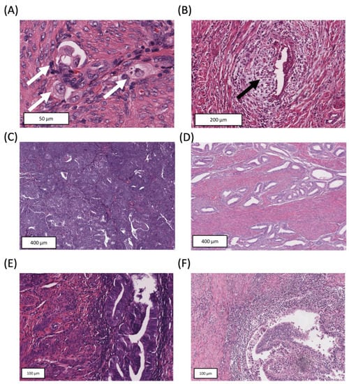
Independent Tissue-Based Biomarkers in Endometrioid Endometrial Cancer: Tumor Budding in Microsatellite Instability and WHO Grading in Copy-Number-Low Patients
by Fabian Stögbauer, Barbara Geß, Christine Brambs, Manuela Lautizi, Tim Kacprowski, Iordanis Ourailidis, Holger Bronger, Marion Kiechle, Aurelia Noske, Gisela Keller, Moritz Jesinghaus, Christopher Poremba, Wilko Weichert and Melanie Boxberg
Cancers 2023, 15(15), 3832; https://doi.org/10.3390/cancers15153832 - 28 Jul 2023
Cited by 1 | Viewed by 1577
Abstract
The molecular characterization of endometrial endometrioid adenocarcinomas has provided major advances in its prognostic stratification. However, risk assessment of microsatellite instability (MSI) and copy-number (CN)-low cases remains a challenge. Thus, we aimed to identify tissue-based morphologic biomarkers that might help in the prognostic [...] Read more.
The molecular characterization of endometrial endometrioid adenocarcinomas has provided major advances in its prognostic stratification. However, risk assessment of microsatellite instability (MSI) and copy-number (CN)-low cases remains a challenge. Thus, we aimed to identify tissue-based morphologic biomarkers that might help in the prognostic stratification of these cases. Histomorphologic parameters (WHO grading, tumor budding (TB), tumor–stroma ratio (as a quantitative description of stromal desmoplasia), tumor-infiltrating lymphocytes (TIL), “microcystic, elongated, fragmented” (MELF) pattern) were analyzed in resection specimens of the TCGA-UCEC cohort (n = 228). For each quantitative parameter, a two-tiered system was developed utilizing systematically determined cutoffs. Associations with survival outcomes were calculated in univariate and multivariate analysis and validated in two independent cohorts. In MSI tumors, only TB remained an independent prognostic factor. TB (≥3 buds/high-power field) was associated with inferior outcomes and with lymph node metastases. The prognostic significance of TB was confirmed in two validation cohorts. For CN-low tumors, established grading defined by the WHO was independently prognostic with inferior outcomes for high-grade tumors. The evaluation of TB might help in identifying MSI-patients with unfavorable prognosis who, e.g., could benefit from lymphadenectomy. WHO-based grading facilitates independent prognostic stratification of CN-low endometrioid adenocarcinomas. Therefore, we propose the utilization of TB and WHO-based grading, two tissue-based and easy-to-assess biomarkers, in MSI/CN-low endometrial carcinomas for improved clinical management. Full article
Show Figures

Figure 1

9 pages, 541 KiB  
Article
Are CT-Derived Muscle Measurements Prognostic, Independent of Systemic Inflammation, in Good Performance Status Patients with Advanced Cancer?
by Josh McGovern, Ross D. Dolan, Claribel Simmons, Louise E. Daly, Aoife M. Ryan, Derek G. Power, Marie T. Fallon, Barry J. Laird and Donald C. McMillan
Cancers 2023, 15(13), 3497; https://doi.org/10.3390/cancers15133497 - 5 Jul 2023
Cited by 3 | Viewed by 1458
Abstract
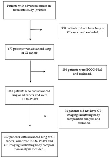
The present study examined the relationships between CT-derived muscle measurements, systemic inflammation, and survival in advanced cancer patients with good performance status (ECOG-PS 0/1). Data was collected prospectively from patients with advanced cancer undergoing anti-cancer therapy with palliative intent. The CT Sarcopenia score [...] Read more.
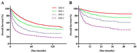
The present study examined the relationships between CT-derived muscle measurements, systemic inflammation, and survival in advanced cancer patients with good performance status (ECOG-PS 0/1). Data was collected prospectively from patients with advanced cancer undergoing anti-cancer therapy with palliative intent. The CT Sarcopenia score (CT-SS) was calculated by combining the CT-derived skeletal muscle index (SMI) and density (SMD). The systemic inflammatory status was determined using the modified Glasgow Prognostic Score (mGPS). The primary outcome of interest was overall survival (OS). Univariate and multivariate Cox regressions were used for survival analysis. Three hundred and seven patients met the inclusion criteria, out of which 62% (n = 109) were male and 47% (n = 144) were ≥65 years of age, while 38% (n = 118) were CT-SS ≥ 1 and 47% (n = 112) of patients with pre-study blood were inflamed (mGPS ≥ 1). The median survival from entry to the study was 11.1 months (1–68.1). On univariate analysis, cancer type (p < 0.05) and mGPS (p < 0.001) were significantly associated with OS. On multivariate analysis, only mGPS (p < 0.001) remained significantly associated with OS. In patients who were ECOG-PS 0, mGPS was significantly associated with CT-SS (p < 0.05). mGPS may dominate the prognostic value of CT-derived sarcopenia in good-performance-status patients with advanced cancer. Full article
Show Figures

Figure 1

17 pages, 10041 KiB  
Article
Molecular Subtyping and Survival Analysis of Osteosarcoma Reveals Prognostic Biomarkers and Key Canonical Pathways
by Siddesh Southekal, Sushil Kumar Shakyawar, Prachi Bajpai, Amr Elkholy, Upender Manne, Nitish Kumar Mishra and Chittibabu Guda
Cancers 2023, 15(7), 2134; https://doi.org/10.3390/cancers15072134 - 4 Apr 2023
Cited by 8 | Viewed by 2715
Abstract
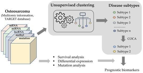
Osteosarcoma (OS) is a common bone malignancy in children and adolescents. Although histological subtyping followed by improved OS treatment regimens have helped achieve favorable outcomes, a lack of understanding of the molecular subtypes remains a challenge to characterize its genetic heterogeneity and subsequently [...] Read more.
Osteosarcoma (OS) is a common bone malignancy in children and adolescents. Although histological subtyping followed by improved OS treatment regimens have helped achieve favorable outcomes, a lack of understanding of the molecular subtypes remains a challenge to characterize its genetic heterogeneity and subsequently to identify diagnostic and prognostic biomarkers for developing effective treatments. In the present study, global analysis of DNA methylation, and mRNA and miRNA gene expression in OS patient samples were correlated with their clinical characteristics. The mucin family of genes, MUC6, MUC12, and MUC4, were found to be highly mutated in the OS patients. Results revealed the enrichment of molecular pathways including Wnt signaling, Calcium signaling, and PI3K-Akt signaling in the OS tumors. Survival analyses showed that the expression levels of several genes such as RAMP1, CRIP1, CORT, CHST13, and DDX60L, miRNAs and lncRNAs were associated with survival of OS patients. Molecular subtyping using Cluster-Of-Clusters Analysis (COCA) for mRNA, lncRNA, and miRNA expression; DNA methylation; and mutation data from the TARGET dataset revealed two distinct molecular subtypes, each with a distinctive gene expression profile. Between the two subtypes, three upregulated genes, POP4, HEY1, CERKL, and seven downregulated genes, CEACAM1, ABLIM1, LTBP2, ISLR, LRRC32, PTPRF, and GPX3, associated with OS metastasis were found to be differentially regulated. Thus, the molecular subtyping results provide a strong basis for classification of OS patients that could be used to develop better prognostic treatment strategies. Full article
Show Figures

Graphical abstract

37 pages, 1778 KiB  
Review
Mucins as Potential Biomarkers for Early Detection of Cancer
by Shailendra K. Gautam, Parvez Khan, Gopalakrishnan Natarajan, Pranita Atri, Abhijit Aithal, Apar K. Ganti, Surinder K. Batra, Mohd W. Nasser and Maneesh Jain
Cancers 2023, 15(6), 1640; https://doi.org/10.3390/cancers15061640 - 7 Mar 2023
Cited by 10 | Viewed by 5319
Abstract
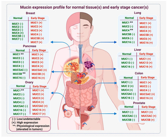
Early detection significantly correlates with improved survival in cancer patients. So far, a limited number of biomarkers have been validated to diagnose cancers at an early stage. Considering the leading cancer types that contribute to more than 50% of deaths in the USA, [...] Read more.
Early detection significantly correlates with improved survival in cancer patients. So far, a limited number of biomarkers have been validated to diagnose cancers at an early stage. Considering the leading cancer types that contribute to more than 50% of deaths in the USA, we discuss the ongoing endeavors toward early detection of lung, breast, ovarian, colon, prostate, liver, and pancreatic cancers to highlight the significance of mucin glycoproteins in cancer diagnosis. As mucin deregulation is one of the earliest events in most epithelial malignancies following oncogenic transformation, these high-molecular-weight glycoproteins are considered potential candidates for biomarker development. The diagnostic potential of mucins is mainly attributed to their deregulated expression, altered glycosylation, splicing, and ability to induce autoantibodies. Secretory and shed mucins are commonly detected in patients’ sera, body fluids, and tumor biopsies. For instance, CA125, also called MUC16, is one of the biomarkers implemented for the diagnosis of ovarian cancer and is currently being investigated for other malignancies. Similarly, MUC5AC, a secretory mucin, is a potential biomarker for pancreatic cancer. Moreover, anti-mucin autoantibodies and mucin-packaged exosomes have opened new avenues of biomarker development for early cancer diagnosis. In this review, we discuss the diagnostic potential of mucins in epithelial cancers and provide evidence and a rationale for developing a mucin-based biomarker panel for early cancer detection. Full article
Show Figures

Figure 1

23 pages, 6723 KiB  
Article
Mass Spectrometry-Based Biomarkers to Detect Prostate Cancer: A Multicentric Study Based on Non-Invasive Urine Collection without Prior Digital Rectal Examination
by Maria Frantzi, Zoran Culig, Isabel Heidegger, Marika Mokou, Agnieszka Latosinska, Marie C. Roesch, Axel S. Merseburger, Manousos Makridakis, Antonia Vlahou, Ana Blanca-Pedregosa, Julia Carrasco-Valiente, Harald Mischak and Enrique Gomez-Gomez
Cancers 2023, 15(4), 1166; https://doi.org/10.3390/cancers15041166 - 11 Feb 2023
Cited by 7 | Viewed by 2420
Abstract
(1) Background: Prostate cancer (PCa) is the most frequently diagnosed cancer in men. Wide application of prostate specific antigen test has historically led to over-treatment, starting from excessive biopsies. Risk calculators based on molecular and clinical variables can be of value to determine [...] Read more.
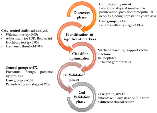
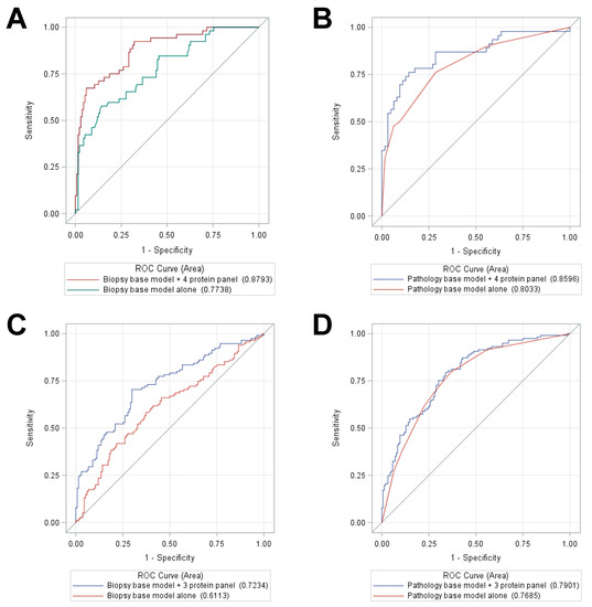
(1) Background: Prostate cancer (PCa) is the most frequently diagnosed cancer in men. Wide application of prostate specific antigen test has historically led to over-treatment, starting from excessive biopsies. Risk calculators based on molecular and clinical variables can be of value to determine the risk of PCa and as such, reduce unnecessary and invasive biopsies. Urinary molecular studies have been mostly focusing on sampling after initial intervention (digital rectal examination and/or prostate massage). (2) Methods: Building on previous proteomics studies, in this manuscript, we aimed at developing a biomarker model for PCa detection based on urine sampling without prior intervention. Capillary electrophoresis coupled to mass spectrometry was applied to acquire proteomics profiles from 970 patients from two different clinical centers. (3) Results: A case-control comparison was performed in a training set of 413 patients and 181 significant peptides were subsequently combined by a support vector machine algorithm. Independent validation was initially performed in 272 negative for PCa and 138 biopsy-confirmed PCa, resulting in an AUC of 0.81, outperforming current standards, while a second validation phase included 147 PCa patients. (4) Conclusions: This multi-dimensional biomarker model holds promise to improve the current diagnosis of PCa, by guiding invasive biopsies. Full article
Show Figures

Figure 1

13 pages, 1712 KiB  
Systematic Review
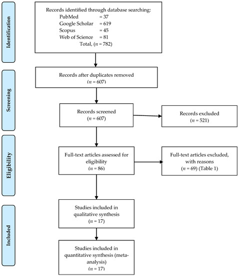
Association of Telomere Length with Colorectal Cancer Risk and Prognosis: A Systematic Review and Meta-Analysis
by Svenja Pauleck, Jennifer A. Sinnott, Yun-Ling Zheng, Shahinaz M. Gadalla, Richard Viskochil, Benjamin Haaland, Richard M. Cawthon, Albrecht Hoffmeister and Sheetal Hardikar
Cancers 2023, 15(4), 1159; https://doi.org/10.3390/cancers15041159 - 11 Feb 2023
Cited by 7 | Viewed by 1977
Abstract
(1) Background: Colorectal cancer risk and survival have previously been associated with telomere length in peripheral blood leukocytes and tumor tissue. A systematic review and meta-analysis of the literature was conducted. The PubMed, Embase, and Web of Science databases were searched through March [...] Read more.
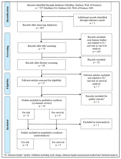
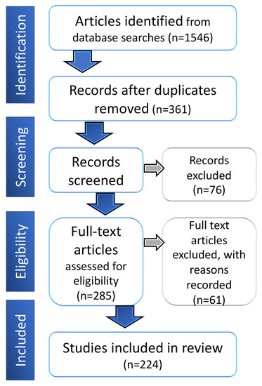
(1) Background: Colorectal cancer risk and survival have previously been associated with telomere length in peripheral blood leukocytes and tumor tissue. A systematic review and meta-analysis of the literature was conducted. The PubMed, Embase, and Web of Science databases were searched through March 2022. (2) Methods: Relevant studies were identified through database searching following PRISMA guidelines. Risk estimates were extracted from identified studies; meta-analyses were conducted using random effects models. (3) Results: Fourteen studies were identified (eight on risk; six on survival) through systematic review. While no association was observed between circulating leukocyte telomere length and the risk of colorectal cancer [overall OR (95% CI) = 1.01 (0.82–1.24)], a worse survival for those with shorter telomeres in leukocytes and longer telomeres in tumor tissues was observed [Quartile1/Quartile2–4 overall HR (95% CI) = 1.41 (0.26–7.59) and 0.82 (0.69–0.98), respectively]. (4) Conclusions: Although there was no association with colorectal cancer risk, a poorer survival was observed among those with shorter leukocyte telomere length. Future larger studies evaluating a potentially non-linear relationship between telomeres and colorectal cancer are needed. Full article
Show Figures

Figure 1

20 pages, 2496 KiB  
Article
Systematic Evaluation of Antigenic Stimulation in Chronic Lymphocytic Leukemia: Humoral Immunity as Biomarkers for Disease Evolution
by Alicia Landeira-Viñuela, Miguel Alcoceba-Sanchez, Almudena Navarro-Bailón, Carlota Arias-Hidalgo, Pablo Juanes-Velasco, José Manuel Sánchez-Santos, Quentin Lecrevisse, Carlos Eduardo Pedreira, Marina L. García-Vaquero, Ángela-Patricia Hernández, Enrique Montalvillo, Rafael Góngora, Javier De las Rivas, Marcos González-Díaz, Alberto Orfao and Manuel Fuentes
Cancers 2023, 15(3), 891; https://doi.org/10.3390/cancers15030891 - 31 Jan 2023
Viewed by 2588
Abstract
Chronic lymphocytic leukemia (CLL) is the most common leukemia in the Western world. Studies of CLL antibody reactivity have shown differential targets to autoantigens and antimicrobial molecular motifs that support the current hypothesis of CLL pathogenesis. Methods: In this study, we conducted a [...] Read more.
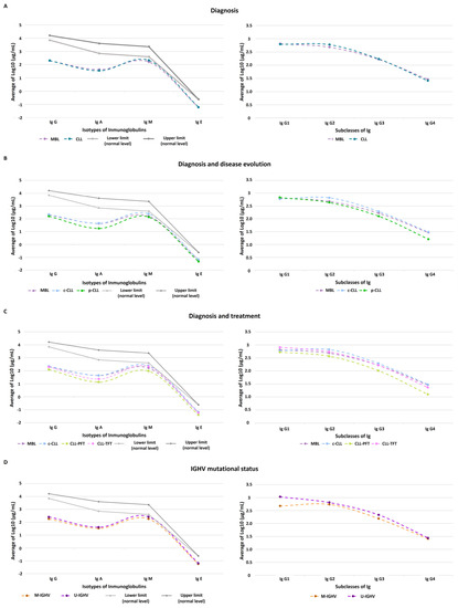
Chronic lymphocytic leukemia (CLL) is the most common leukemia in the Western world. Studies of CLL antibody reactivity have shown differential targets to autoantigens and antimicrobial molecular motifs that support the current hypothesis of CLL pathogenesis. Methods: In this study, we conducted a quantitative serum analysis of 7 immunoglobulins in CLL and monoclonal B-cell lymphocytosis (MBL) patients (bead-suspension protein arrays) and a serological profile (IgG and IgM) study of autoantibodies and antimicrobial antigens (protein microarrays). Results: Significant differences in the IgA levels were observed according to disease progression and evolution as well as significant alterations in IgG1 according to IGHV mutational status. More representative IgG autoantibodies in the cohort were against nonmutagenic proteins and IgM autoantibodies were against vesicle proteins. Antimicrobial IgG and IgM were detected against microbes associated with respiratory tract infections. Conclusions: Quantitative differences in immunoglobulin serum levels could be potential biomarkers for disease progression. In the top 5 tumoral antigens, we detected autoantibodies (IgM and IgG) against proteins related to cell homeostasis and metabolism in the studied cohort. The top 5 microbial antigens were associated with respiratory and gastrointestinal infections; moreover, the subsets with better prognostics were characterized by a reactivation of Cytomegalovirus. The viral humoral response could be a potential prognosis biomarker for disease progression. Full article
Show Figures

Figure 1

23 pages, 7438 KiB  
Article
EPHA3 Could Be a Novel Prognosis Biomarker and Correlates with Immune Infiltrates in Bladder Cancer
by Junpeng Liu, Zewen Zhou, Yifan Jiang, Yuzhao Lin, Yunzhi Yang, Chongjiang Tian, Jinwen Liu, Hao Lin and Bin Huang
Cancers 2023, 15(3), 621; https://doi.org/10.3390/cancers15030621 - 19 Jan 2023
Cited by 3 | Viewed by 2449
Abstract
Purpose: To assess the mechanism of EPH receptor A3 (EPHA3) and its potential value for immunotherapy in BLCA. Materials and Methods: The Cancer Genome Atlas (TCGA) bladder cancer (BLCA) database and the Gene Expression Omnibus (GEO) database were used for assessing whether EHPA3 [...] Read more.
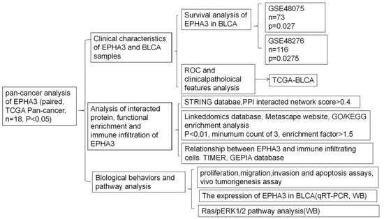
Purpose: To assess the mechanism of EPH receptor A3 (EPHA3) and its potential value for immunotherapy in BLCA. Materials and Methods: The Cancer Genome Atlas (TCGA) bladder cancer (BLCA) database and the Gene Expression Omnibus (GEO) database were used for assessing whether EHPA3 could be used to predict BLCA prognosis. This work carried out in vitro and in vivo assays for exploring how EPHA3 affected the biological behaviors. The downstream pathway was explored using a Western blotting technique. The CIBERSORT, ESTIMATE, TIMER, and TIDE tools were used to predict the immunotherapy value of EPHA3 in BLCA. Results: EPHA3 was poorly expressed in BLCA (p < 0.05), its high expression is related to a good survival prognosis (p = 0.027 and p = 0.0275), and it has a good predictive ability for the histologic grade and status of BLCA (area under curve = 0.787 and 0.904). Overexpressed EPHA3 could inhibit BLCA cell biological behaviors, and it be associated with the downregulation of the Ras/pERK1/2 pathway. EPHA3 was correlated with several immune-infiltrating cells and the corresponding marker genes. Conclusions: EPHA3 could be regarded as an acceptable anti-cancer biomarker in BLCA. EPHA3 plays an inhibiting role in BLCA, and it could be the candidate immunotherapeutic target for BLCA. Full article
Show Figures

Figure 1

13 pages, 594 KiB  
Article
Gene Screening for Prognosis of Non-Muscle-Invasive Bladder Carcinoma under Competing Risks Endpoints
by Chenlu Ke, Dipankar Bandyopadhyay and Devanand Sarkar
Cancers 2023, 15(2), 379; https://doi.org/10.3390/cancers15020379 - 6 Jan 2023
Cited by 1 | Viewed by 1578
Abstract
Background: Discovering clinically useful molecular markers for predicting the survival of patients diagnosed with non–muscle-invasive bladder cancer can provide insights into cancer dynamics and improve treatment outcomes. However, the presence of competing risks (CR) endpoints complicates the estimation and inferential framework. There is [...] Read more.
Background: Discovering clinically useful molecular markers for predicting the survival of patients diagnosed with non–muscle-invasive bladder cancer can provide insights into cancer dynamics and improve treatment outcomes. However, the presence of competing risks (CR) endpoints complicates the estimation and inferential framework. There is also a lack of statistical analysis tools and software for coping with the high-throughput nature of these data, in terms of marker screening and selection. Aims: To propose a gene screening procedure for proportional subdistribution hazards regression under a CR framework, and illustrate its application in using molecular profiling to predict survival for non-muscle invasive bladder carcinoma. Methods: Tumors from 300 patients diagnosed with bladder cancer were analyzed for genomic abnormalities while controlling for clinically important covariates. Genes with expression patterns that were associated with survival were identified through a screening procedure based on proportional subdistribution hazards regression. A molecular predictor of risk was constructed and examined for prediction accuracy. Results: A six-gene signature was found to be a significant predictor associated with survival of non–muscle-invasive bladder cancer, subject to competing risks after adjusting for age, gender, reevaluated WHO grade, stage and BCG/MMC treatment (p-value < 0.001). Conclusion: The proposed gene screening procedure can be used to discover molecular determinants of survival for non–muscle-invasive bladder cancer and in general facilitate high-throughput competing risks data analysis with easy implementation. Full article
Show Figures

Figure 1

2022

Jump to: 2023, 2021, 2020, 2019

13 pages, 2751 KiB  
Article
Dynamic Prediction of Resectability for Patients with Advanced Ovarian Cancer Undergoing Neo-Adjuvant Chemotherapy: Application of Joint Model for Longitudinal CA-125 Levels
by Koceila Amroun, Raphael Chaltiel, Fabien Reyal, Reza Kianmanesh, Aude-Marie Savoye, Marine Perrier, Zoubir Djerada and Olivier Bouché
Cancers 2023, 15(1), 231; https://doi.org/10.3390/cancers15010231 - 30 Dec 2022
Cited by 1 | Viewed by 1592
Abstract
In patients with advanced ovarian cancer (AOC) receiving neoadjuvant chemotherapy (NAC), predicting the feasibility of complete interval cytoreductive surgery (ICRS) is helpful and may avoid unnecessary laparotomy. A joint model (JM) is a dynamic individual predictive model. The aim of this study was [...] Read more.
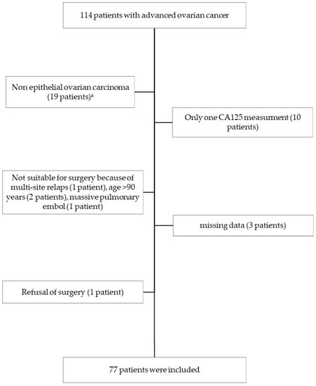
In patients with advanced ovarian cancer (AOC) receiving neoadjuvant chemotherapy (NAC), predicting the feasibility of complete interval cytoreductive surgery (ICRS) is helpful and may avoid unnecessary laparotomy. A joint model (JM) is a dynamic individual predictive model. The aim of this study was to develop a predictive JM combining CA-125 kinetics during NAC with patients’ and clinical factors to predict resectability after NAC in patients with AOC. A retrospective study included 77 patients with AOC treated with NAC. A linear mixed effect (LME) sub-model was used to describe the evolution of CA-125 during NAC considering factors influencing the biomarker levels. A Cox sub-model screened the covariates associated with resectability. The JM combined the LME sub-model with the Cox sub-model. Using the LME sub-model, we observed that CA-125 levels were influenced by the number of NAC cycles and the performance of paracentesis. In the Cox sub-model, complete resectability was associated with Performance Status (HR = 0.57, [0.34–0.95], p = 0.03) and the presence of peritoneal carcinomatosis in the epigastric region (HR = 0.39, [0.19–0.80], p = 0.01). The JM accuracy to predict complete ICRS was 88% [82–100] with a predictive error of 2.24% [0–2.32]. Using a JM of a longitudinal CA-125 level during NAC could be a reliable predictor of complete ICRS. Full article
Show Figures

Figure 1

25 pages, 3514 KiB  
Systematic Review
BRAF V600E Mutation in Ameloblastoma: A Systematic Review and Meta-Analysis
by Mohd Nazzary Mamat @ Yusof, Ewe Seng Ch’ng and Nawal Radhiah Abdul Rahman
Cancers 2022, 14(22), 5593; https://doi.org/10.3390/cancers14225593 - 14 Nov 2022
Cited by 11 | Viewed by 4011
Abstract
The discovery that ameloblastoma has a high mutation incidence of BRAF V600E may enable a better investigation of pathophysiology. However, there is inconsistent evidence regarding this mutation occurrence and its association with clinical information. This systematic review and meta-analysis aim to pool the [...] Read more.
The discovery that ameloblastoma has a high mutation incidence of BRAF V600E may enable a better investigation of pathophysiology. However, there is inconsistent evidence regarding this mutation occurrence and its association with clinical information. This systematic review and meta-analysis aim to pool the overall mutation prevalence of BRAF V600E in reported ameloblastoma cases and to determine its association with patient demographic and clinicopathological features. Following the PRISMA guidelines, a comprehensive article search was conducted through four databases (Scopus, Google Scholar, PubMed, and Web of Science). Seventeen articles between 2014 and 2022 met the inclusion criteria with 833 ameloblastoma cases. For each included study, the significance of BRAF V600E on the outcome parameters was determined using odd ratios and 95% confidence intervals. Meta-analysis prevalence of BRAF V600E in ameloblastoma was 70.49%, and a significant meta-analysis association was reported for those younger than 54 years old and in the mandible. On the contrary, other factors, such as sex, histological variants, and recurrence, were insignificant. As a result of the significant outcome of BRAF V600E mutation in ameloblastoma pathogenesis, targeted therapy formulation can be developed with this handful of evidence. Full article
Show Figures

Figure 1

16 pages, 864 KiB  
Article
Impact of Change in Body Composition during Follow-Up on the Survival of GEP-NET
by Fernando Sebastian-Valles, Nuria Sánchez de la Blanca Carrero, Víctor Rodríguez-Laval, Rebeca Martinez-Hernández, Ana Serrano-Somavilla, Carolina Knott-Torcal, José Luis Muñoz de Nova, Elena Martín-Pérez, Mónica Marazuela and Miguel Antonio Sampedro-Nuñez
Cancers 2022, 14(21), 5189; https://doi.org/10.3390/cancers14215189 - 22 Oct 2022
Cited by 4 | Viewed by 2357
Abstract
Background: Gastroenteropancreatic neuroendocrine tumors (GEP-NETs) are heterogeneous rare diseases causing malnutrition and cachexia in which the study of body composition may have an impact in prognosis. Aim: Evaluation of muscle and fat tissues by computed tomography (CT) at the level of the third [...] Read more.
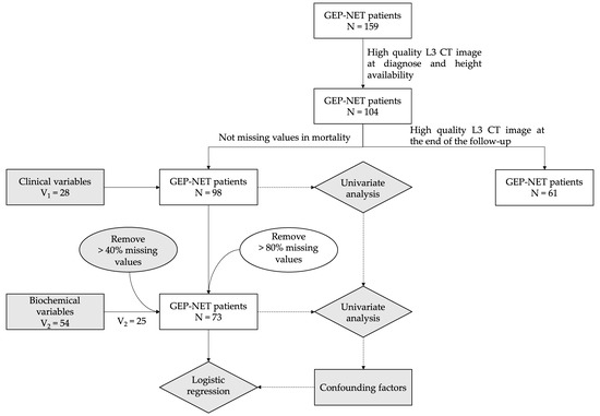
Background: Gastroenteropancreatic neuroendocrine tumors (GEP-NETs) are heterogeneous rare diseases causing malnutrition and cachexia in which the study of body composition may have an impact in prognosis. Aim: Evaluation of muscle and fat tissues by computed tomography (CT) at the level of the third lumbar (L3 level) at diagnosis and at the end of follow-up in GET-NET patients and their relationships with clinical and biochemical variables as predictors of survival. Methodology: Ninety-eight GEP-NET patients were included. Clinical and biochemical parameters were evaluated. Total body, subcutaneous, visceral and total fat areas and very low-density, low-density, normal density, high-density, very high-density and total muscle areas were obtained from CT images. Results: Body composition measures and overall mortality correlated with age, ECOG (Eastern Cooperative Oncology Group performance status) metastases, lactate dehydrogenase (LDH), albumin and urea levels. Although there was no relationship between body composition variables at diagnosis and overall and specific mortality, an increase in low-density muscle and a decrease in normal-density muscle during follow-up were independently correlated to overall (p <0.05) and tumor-cause mortality (p < 0.05). Conclusion: Although body composition measures obtained by CT at diagnosis did not impact survival of GEP-NET patients, a loss of good quality muscle during follow-up was associated with an increased overall and tumor-related mortality. Nutritional status should therefore be supervised by nutrition specialists and an increase in good quality muscle could improve prognosis. Full article
Show Figures

Figure 1

12 pages, 659 KiB  
Review
Molecular Biomarker Expression in Window of Opportunity Studies for Oestrogen Receptor Positive Breast Cancer—A Systematic Review of the Literature
by James W. M. Francis, Manmeet Saundh, Ruth M. Parks and Kwok-Leung Cheung
Cancers 2022, 14(20), 5027; https://doi.org/10.3390/cancers14205027 - 14 Oct 2022
Cited by 1 | Viewed by 1770
Abstract
Window of opportunity (WoO) trials create the opportunity to demonstrate pharmacodynamic parameters of a drug in vivo and have increasing use in breast cancer research. Most breast cancer tumours are oestrogen receptor-positive (ER+), leading to the development of multiple treatment options tailored towards [...] Read more.
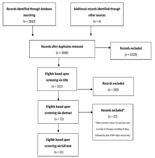
Window of opportunity (WoO) trials create the opportunity to demonstrate pharmacodynamic parameters of a drug in vivo and have increasing use in breast cancer research. Most breast cancer tumours are oestrogen receptor-positive (ER+), leading to the development of multiple treatment options tailored towards this particular tumour subtype. The aim of this literature review is to review WoO trials pertaining to the pharmacodynamic activity of drugs available for use in ER+ breast cancer in order to help guide treatment for patients receiving neoadjuvant and primary endocrine therapy. Five databases (EMBASE, Cochrane, MEDLINE, PubMed, Web of Science) were searched for eligible studies. Studies performed in treatment-naïve patients with histologically confirmed ER+ breast cancer were included if they acquired pre- and post-treatment biopsies, compared measurement of a proteomic biomarker between these two biopsies and delivered treatment for a maximum mean duration of 31 days. Fifteen studies were eligible for inclusion and covered six different drug classes: three endocrine therapies (ETs) including aromatase inhibitors (AIs), selective oestrogen receptor modulators (SERMs), selective oestrogen receptor degraders (SERDs) and three non-ETs including mTOR inhibitors, AKT inhibitors and synthetic oestrogens. Ki67 was the most frequently measured marker, appearing in all studies. Progesterone receptor (PR) and ER were the next most frequently measured markers, appearing five and four studies, respectively. All three of these markers were significantly downregulated in both AIs and SERDs; Ki67 alone was downregulated in SERMs. Less commonly assessed markers including pS6, pGSH3B, FSH and IGF1 were downregulated while CD34, pAKT and SHBG were significantly upregulated. There were no significant changes in the other biomarkers measured such as phosphate and tensin homolog (PTEN), Bax and Bcl-2.WoO studies have been widely utilised within the ER+ breast cancer subtype, demonstrating their worth in pharmacodynamic research. However, research remains focused upon routinely measured biomarkers such ER PR and Ki67, with an array of less common markers sporadically used. Full article
Show Figures

Figure 1

13 pages, 1988 KiB  
Article
The Expression of the Immunoproteasome Subunit PSMB9 Is Related to Distinct Molecular Subtypes of Uterine Leiomyosarcoma
by Raul Maia Falcão, Georgia Kokaraki, Wout De Wispelaere, Frédéric Amant, Gustavo Antônio De Souza, Jorge Estefano Santana de Souza, Joseph Woodward Carlson and Tirzah Braz Petta
Cancers 2022, 14(20), 5007; https://doi.org/10.3390/cancers14205007 - 13 Oct 2022
Cited by 4 | Viewed by 2328
Abstract
Background: Uterine leiomyosarcoma (uLMS) are rare and malignant tumors that arise in the myometrium cells and whose diagnosis is based on histopathological features. Identifying diagnostic biomarkers for uLMS is a challenge due to molecular heterogeneity and the scarcity of samples. In vivo and [...] Read more.
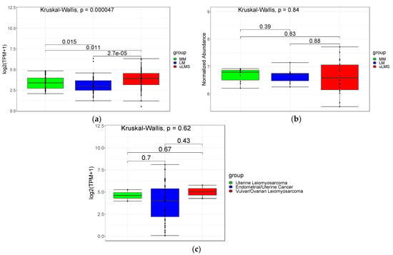
Background: Uterine leiomyosarcoma (uLMS) are rare and malignant tumors that arise in the myometrium cells and whose diagnosis is based on histopathological features. Identifying diagnostic biomarkers for uLMS is a challenge due to molecular heterogeneity and the scarcity of samples. In vivo and in vitro models for uLMS are urgently needed. Knockout female mice for the catalytic subunit of the immunoproteasome PSMB9 (MIM:177045) develop spontaneous uLMS. This study aimed to analyze the role of PSMB9 in uLMS tumorigenesis and patient outcome. Methods: Molecular data from 3 non-related uLMS cohorts were integrated and analyzed by proteotranscriptomic using gene expression and protein abundance levels in 68 normal adjacent myometrium (MM), 66 uterine leiomyoma (LM), and 67 uLMS. Results: the immunoproteasome pathway is upregulated and the gene PMSB9 shows heterogeneous expression values in uLMS. Quartile group analysis showed no significant difference between groups high and low PSMB9 expression groups at 3-years overall survival (OS). Using CYBERSORTx analysis we observed 9 out of 17 samples in the high group clustering together due to high M2 macrophages and CD4 memory resting, and high CD8+/PSMB9 ratio was associated with better OS. The main pathway regulated in the high group is IFNγ and in the low is the ECM pathway dependent on the proto-oncogene SRC. Conclusion: these findings suggest 2 subtypes of uLMS (immune-related and ECM-related) with different candidate mechanisms of malignancy. Full article
Show Figures

Figure 1

21 pages, 5736 KiB  
Article
Systematic Analysis of Genetic and Pathway Determinants of Eribulin Sensitivity across 100 Human Cancer Cell Lines from the Cancer Cell Line Encyclopedia (CCLE)
by Pallavi Sachdev, Roy Ronen, Janusz Dutkowski and Bruce A. Littlefield
Cancers 2022, 14(18), 4532; https://doi.org/10.3390/cancers14184532 - 19 Sep 2022
Cited by 3 | Viewed by 2410
Abstract
Eribulin, a natural product-based microtubule targeting agent with cytotoxic and noncytotoxic mechanisms, is FDA approved for certain patients with advanced breast cancer and liposarcoma. To investigate the feasibility of developing drug-specific predictive biomarkers, we quantified antiproliferative activities of eribulin versus paclitaxel and vinorelbine [...] Read more.
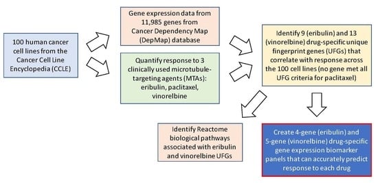
Eribulin, a natural product-based microtubule targeting agent with cytotoxic and noncytotoxic mechanisms, is FDA approved for certain patients with advanced breast cancer and liposarcoma. To investigate the feasibility of developing drug-specific predictive biomarkers, we quantified antiproliferative activities of eribulin versus paclitaxel and vinorelbine against 100 human cancer cell lines from the Cancer Cell Line Encyclopedia, and correlated results with publicly available databases to identify genes and pathways associated with eribulin response, either uniquely or shared with paclitaxel or vinorelbine. Mean expression ratios of 11,985 genes between the most and least sensitive cell line quartiles were sorted by p-values and drug overlaps, yielding 52, 29 and 80 genes uniquely associated with eribulin, paclitaxel and vinorelbine, respectively. Further restriction to minimum 2-fold ratios followed by reintroducing data from the middle two quartiles identified 9 and 13 drug-specific unique fingerprint genes for eribulin and vinorelbine, respectively; surprisingly, no gene met all criteria for paclitaxel. Interactome and Reactome pathway analyses showed that unique fingerprint genes of both drugs were primarily associated with cellular signaling, not microtubule-related pathways, although considerable differences existed in individual pathways identified. Finally, four-gene (C5ORF38, DAAM1, IRX2, CD70) and five-gene (EPHA2, NGEF, SEPTIN10, TRIP10, VSIG10) multivariate regression models for eribulin and vinorelbine showed high statistical correlation with drug-specific responses across the 100 cell lines and accurately calculated predicted mean IC50s for the most and least sensitive cell line quartiles as surrogates for responders and nonresponders, respectively. Collectively, these results provide a foundation for developing drug-specific predictive biomarkers for eribulin and vinorelbine. Full article
Show Figures

Graphical abstract

15 pages, 1345 KiB  
Review
Racial Disparity in Quadruple Negative Breast Cancer: Aggressive Biology and Potential Therapeutic Targeting and Prevention
by Nikita Jinna, Tijana Jovanovic-Talisman, Mark LaBarge, Rama Natarajan, Rick Kittles, Christopher Sistrunk, Padmashree Rida and Victoria L. Seewaldt
Cancers 2022, 14(18), 4484; https://doi.org/10.3390/cancers14184484 - 16 Sep 2022
Cited by 8 | Viewed by 2611
Abstract
Black/African-American (AA) women, relative to their White/European-American (EA) counterparts, experience disproportionately high breast cancer mortality. Central to this survival disparity, Black/AA women have an unequal burden of aggressive breast cancer subtypes, such as triple-negative breast cancer (ER/PR-, HER2-wild type; TNBC). While TNBC has [...] Read more.
Black/African-American (AA) women, relative to their White/European-American (EA) counterparts, experience disproportionately high breast cancer mortality. Central to this survival disparity, Black/AA women have an unequal burden of aggressive breast cancer subtypes, such as triple-negative breast cancer (ER/PR-, HER2-wild type; TNBC). While TNBC has been well characterized, recent studies have identified a highly aggressive androgen receptor (AR)-negative subtype of TNBC, quadruple-negative breast cancer (ER/PR-, HER2-wildtype, AR-; QNBC). Similar to TNBC, QNBC disproportionately impacts Black/AA women and likely plays an important role in the breast cancer survival disparities experienced by Black/AA women. Here, we discuss the racial disparities of QNBC and molecular signaling pathways that may contribute to the aggressive biology of QNBC in Black/AA women. Our immediate goal is to spotlight potential prevention and therapeutic targets for Black/AA QNBC; ultimately our goal is to provide greater insight into reducing the breast cancer survival burden experienced by Black/AA women. Full article
Show Figures

Figure 1

15 pages, 1558 KiB  
Article
Meta-Analysis Reveals Both the Promises and the Challenges of Clinical Metabolomics
by Heidi E. Roth and Robert Powers
Cancers 2022, 14(16), 3992; https://doi.org/10.3390/cancers14163992 - 18 Aug 2022
Cited by 15 | Viewed by 2447
Abstract
Clinical metabolomics is a rapidly expanding field focused on identifying molecular biomarkers to aid in the efficient diagnosis and treatment of human diseases. Variations in study design, metabolomics methodologies, and investigator protocols raise serious concerns about the accuracy and reproducibility of these potential [...] Read more.
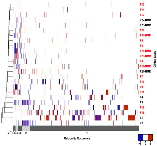
Clinical metabolomics is a rapidly expanding field focused on identifying molecular biomarkers to aid in the efficient diagnosis and treatment of human diseases. Variations in study design, metabolomics methodologies, and investigator protocols raise serious concerns about the accuracy and reproducibility of these potential biomarkers. The explosive growth of the field has led to the recent availability of numerous replicate clinical studies, which permits an evaluation of the consistency of biomarkers identified across multiple metabolomics projects. Pancreatic ductal adenocarcinoma (PDAC) is the third-leading cause of cancer-related death and has the lowest five-year survival rate primarily due to the lack of an early diagnosis and the limited treatment options. Accordingly, PDAC has been a popular target of clinical metabolomics studies. We compiled 24 PDAC metabolomics studies from the scientific literature for a detailed meta-analysis. A consistent identification across these multiple studies allowed for the validation of potential clinical biomarkers of PDAC while also highlighting variations in study protocols that may explain poor reproducibility. Our meta-analysis identified 10 metabolites that may serve as PDAC biomarkers and warrant further investigation. However, 87% of the 655 metabolites identified as potential biomarkers were identified in single studies. Differences in cohort size and demographics, p-value choice, fold-change significance, sample type, handling and storage, data collection, and analysis were all factors that likely contributed to this apparently large false positive rate. Our meta-analysis demonstrated the need for consistent experimental design and normalized practices to accurately leverage clinical metabolomics data for reliable and reproducible biomarker discovery. Full article
Show Figures

Figure 1

13 pages, 1104 KiB  
Article
Elevated Serotonin and NT-proBNP Levels Predict and Detect Carcinoid Heart Disease in a Large Validation Study
by Sonja Levy, Aoife B. Kilgallen, Catharina M. Korse, Marish I. F. J. Oerlemans, Joost P. G. Sluijter, Linda W. van Laake, Gerlof D. Valk and Margot E. T. Tesselaar
Cancers 2022, 14(10), 2361; https://doi.org/10.3390/cancers14102361 - 10 May 2022
Cited by 6 | Viewed by 2288
Abstract
Carcinoid heart disease (CHD) is a rare fibrotic cardiac complication of neuroendocrine tumors. Besides known biomarkers N-Terminal pro-B-type natriuretic peptide (NT-proBNP) and serotonin, activin A, connective tissue growth factor (CTGF), and soluble suppression of tumorigenicity 2 (sST2) have been suggested as potential biomarkers [...] Read more.
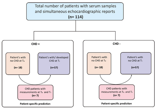
Carcinoid heart disease (CHD) is a rare fibrotic cardiac complication of neuroendocrine tumors. Besides known biomarkers N-Terminal pro-B-type natriuretic peptide (NT-proBNP) and serotonin, activin A, connective tissue growth factor (CTGF), and soluble suppression of tumorigenicity 2 (sST2) have been suggested as potential biomarkers for CHD. Here, we validated the predictive/diagnostic value of these biomarkers in a case-control study of 114 patients between 1990 and 2021. Two time-points were analyzed: T0: liver metastasis without CHD for all patients. T1: confirmed CHD in cases (CHD+, n = 57); confirmed absence of CHD five or more years after liver metastasis in controls (CHD–, n = 57). Thirty-one (54%) and 25 (44%) females were included in CHD+ and CHD– patients, respectively. Median age was 57.9 years for CHD+ and 59.7 for CHD- patients (p = 0.290). At T0: activin A was similar across both groups (p = 0.724); NT-proBNP was higher in CHD+ patients (17 vs. 6 pmol/L, p = 0.016), area under the curve (AUC) 0.84, and the most optimal cut-off at 6.5 pmol/L. At T1: activin A was higher in CHD+ patients (0.65 vs. 0.38 ng/mL, p = 0.045), AUC 0.62, without an optimal cut-off value. NT-pro-BNP was higher in CHD+ patients (63 vs. 11 pmol/L, p < 0.001), AUC 0.89, with an optimal cut-off of 27 pmol/L. Serotonin (p = 0.345), sST2 (p = 0.867) and CTGF (p = 0.232) levels were similar across groups. This large validation study identified NT-proBNP as the superior biomarker for CHD. Patients with elevated serotonin levels and NT-proBNP levels between 6.5 and 27 pmol/L, and specifically >27 pmol/L, should be monitored closely for the development of CHD. Full article
Show Figures

Figure 1

13 pages, 1651 KiB  
Article
Implication of COPB2 Expression on Cutaneous Squamous Cell Carcinoma Pathogenesis
by Taiqin Chen, Ki-Yeol Kim, Yeongjoo Oh, Hei Cheul Jeung, Kee Yang Chung, Mi Ryung Roh and Xianglan Zhang
Cancers 2022, 14(8), 2038; https://doi.org/10.3390/cancers14082038 - 18 Apr 2022
Cited by 3 | Viewed by 2230
Abstract
The underlying molecular mechanisms of cutaneous squamous cell carcinoma (cSCC) pathogenesis are largely unknown. In the present study, we aimed to evaluate the effect of coatomer protein complex subunit beta 2 (COPB2) expression on cSCC pathogenesis. Clinicopathological significance of COPB2 in cSCC was [...] Read more.
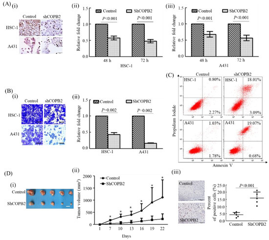
The underlying molecular mechanisms of cutaneous squamous cell carcinoma (cSCC) pathogenesis are largely unknown. In the present study, we aimed to evaluate the effect of coatomer protein complex subunit beta 2 (COPB2) expression on cSCC pathogenesis. Clinicopathological significance of COPB2 in cSCC was investigated by analyzing the Gene Expression Omnibus (GEO) database and through a retrospective cohort study of 95 cSCC patients. The effect of COPB2 expression on the biological behavior of cSCC cells was investigated both in vitro and in vivo. We found that COPB2 expression was significantly higher in cSCC samples than in normal skin samples. In our cohort, a considerable association was found between COPB2 expression and indicators of tumor immune microenvironment (TIME), such as histocompatibility complex class (MHC) I, and MHC II, CD4+/ CD8+ tumor-infiltrating lymphocytes. Additionally, COPB2 expression had an independent impact on worsened recurrence-free survival in our cohort. Furthermore, decreased proliferation, invasion, tumorigenic activities, and increased apoptosis were observed after COPB2 knockdown in cSCC cells. COPB2 may act as a potential oncogene and candidate modulator of the TIME in cSCC. Therefore, it can serve as a novel predictive prognostic biomarker and candidate immunotherapeutic target in cSCC patients. Full article
Show Figures

Figure 1

20 pages, 17547 KiB  
Review
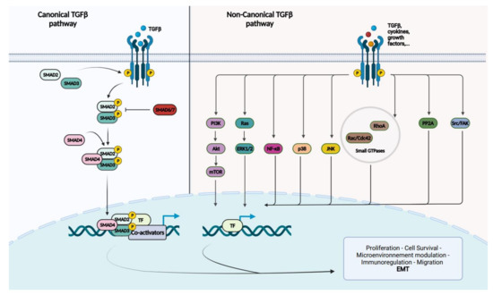
The Role of SMAD4 Inactivation in Epithelial–Mesenchymal Plasticity of Pancreatic Ductal Adenocarcinoma: The Missing Link?
by Marie-Lucie Racu, Laetitia Lebrun, Andrea Alex Schiavo, Claude Van Campenhout, Sarah De Clercq, Lara Absil, Esmeralda Minguijon Perez, Calliope Maris, Christine Decaestecker, Isabelle Salmon and Nicky D’Haene
Cancers 2022, 14(4), 973; https://doi.org/10.3390/cancers14040973 - 15 Feb 2022
Cited by 11 | Viewed by 4641
Abstract
Pancreatic ductal adenocarcinoma (PDAC) presents a five-year survival rate of 10% and its incidence increases over the years. It is, therefore, essential to improve our understanding of the molecular mechanisms that promote metastasis and chemoresistance in PDAC, which are the main causes of [...] Read more.
Pancreatic ductal adenocarcinoma (PDAC) presents a five-year survival rate of 10% and its incidence increases over the years. It is, therefore, essential to improve our understanding of the molecular mechanisms that promote metastasis and chemoresistance in PDAC, which are the main causes of death in these patients. SMAD4 is inactivated in 50% of PDACs and its loss has been associated with worse overall survival and metastasis, although some controversy still exists. SMAD4 is the central signal transducer of the transforming growth factor-beta (TGF-beta) pathway, which is notably known to play a role in epithelial–mesenchymal transition (EMT). EMT is a biological process where epithelial cells lose their characteristics to acquire a spindle-cell phenotype and increased motility. EMT has been increasingly studied due to its potential implication in metastasis and therapy resistance. Recently, it has been suggested that cells undergo EMT transition through intermediary states, which is referred to as epithelial–mesenchymal plasticity (EMP). The intermediary states are characterized by enhanced aggressiveness and more efficient metastasis. Therefore, this review aims to summarize and analyze the current knowledge on SMAD4 loss in patients with PDAC and to investigate its potential role in EMP in order to better understand its function in PDAC carcinogenesis. Full article
Show Figures

Figure 1

20 pages, 3643 KiB  
Article
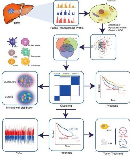
Novel Risk Classification Based on Pyroptosis-Related Genes Defines Immune Microenvironment and Pharmaceutical Landscape for Hepatocellular Carcinoma
by Jianye Wang, Ying Wang, Marcella Steffani, Christian Stöß, Donna Ankerst, Helmut Friess, Norbert Hüser and Daniel Hartmann
Cancers 2022, 14(2), 447; https://doi.org/10.3390/cancers14020447 - 17 Jan 2022
Cited by 6 | Viewed by 3285
Abstract
Growing evidence has indicated that pyroptosis functions in the development of cancer. Nonetheless, specific roles of pyroptosis-related genes in tumor progression, immune response, prognosis, and immunotherapy have not been thoroughly elucidated. After a comprehensive evaluation of pyroptosis genes, unsupervised clustering was performed to [...] Read more.
Growing evidence has indicated that pyroptosis functions in the development of cancer. Nonetheless, specific roles of pyroptosis-related genes in tumor progression, immune response, prognosis, and immunotherapy have not been thoroughly elucidated. After a comprehensive evaluation of pyroptosis genes, unsupervised clustering was performed to generate three distinct clusters from hepatocellular carcinoma (HCC) samples. Three distinct pyroptosis-related molecular subtypes comprising three gene clusters that had differential prognostic effects on patient survival were then identified. Immune characteristics analyses revealed diversified immune cell infiltration among the subtypes. Two clusters served as immune-hot phenotypes associated with significantly poorer survival compared to a remaining third immune-cold cluster. Among these, the immune-hot clusters were characterized by abundant adaptive immune cell infiltration, active CD4+ and CD8+ T cells, high total leukocyte counts and tumor growth status, and lower Th17 cell and M2 macrophage densities. Then, risk scores indicated that low-risk patients were more sensitive to anti-tumor therapy. Subsequently, we found a significant correlation between pyroptosis and prognosis in HCC and that pyroptosis genes drive the heterogeneity of the tumor microenvironment. The risk scoring system, based on pyroptosis-related differentially expressed genes, was established to evaluate the individual outcomes and contribute to new insights into the molecular characterization of pyroptosis-related subtypes. Full article
Show Figures

Graphical abstract

2021

Jump to: 2023, 2022, 2020, 2019

15 pages, 2290 KiB  
Article
GCC2 as a New Early Diagnostic Biomarker for Non-Small Cell Lung Cancer
by Hyesun Jeong, Byeong Hyeon Choi, JinA Park, Jik-Han Jung, Hyunku Shin, Ka-Won Kang, Yu Hua Quan, Jewon Yu, Ji-Ho Park, Yong Park, Yeonho Choi, Hyun Koo Kim and Sunghoi Hong
Cancers 2021, 13(21), 5482; https://doi.org/10.3390/cancers13215482 - 31 Oct 2021
Cited by 12 | Viewed by 4402
Abstract
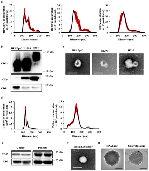
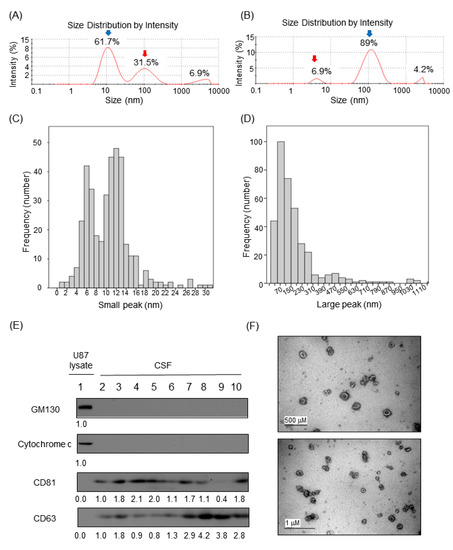
No specific markers have been identified to detect non-small cell lung cancer (NSCLC) cell-derived exosomes circulating in the blood. Here, we report a new biomarker that distinguishes between cancer and non-cancer cell-derived exosomes. Exosomes isolated from patient plasmas at various pathological stages of [...] Read more.
No specific markers have been identified to detect non-small cell lung cancer (NSCLC) cell-derived exosomes circulating in the blood. Here, we report a new biomarker that distinguishes between cancer and non-cancer cell-derived exosomes. Exosomes isolated from patient plasmas at various pathological stages of NSCLC, NSCLC cell lines, and human pulmonary alveolar epithelial cells isolated using size exclusion chromatography were characterized. The GRIP and coiled-coil domain-containing 2 (GCC2) protein, involved in endosome-to-Golgi transport, was identified by proteomics analysis of NSCLC cell line-derived exosomes. GCC2 protein levels in the exosomes derived from early-stage NSCLC patients were higher than those from healthy controls. Receiver operating characteristic curve analysis revealed the diagnostic sensitivity and specificity of exosomal GCC2 to be 90% and 75%, respectively. A high area under the curve, 0.844, confirmed that GCC2 levels could effectively distinguish between the exosomes. These results demonstrate GCC2 as a promising early diagnostic biomarker for NSCLC. Full article
Show Figures

Figure 1

15 pages, 2442 KiB  
Article
Human DLG1 and SCRIB Are Distinctly Regulated Independently of HPV-16 during the Progression of Oropharyngeal Squamous Cell Carcinomas: A Preliminary Analysis
by Lucija Lulić, Antonia Jakovčević, Luka Manojlović, Emil Dediol, Lawrence Banks and Vjekoslav Tomaić
Cancers 2021, 13(17), 4461; https://doi.org/10.3390/cancers13174461 - 4 Sep 2021
Cited by 5 | Viewed by 2897
Abstract
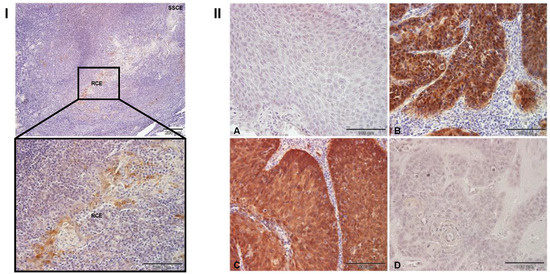
The major causative agents of head and neck squamous cell carcinomas (HNSCCs) are either environmental factors, such as tobacco and alcohol consumption, or infection with oncogenic human papillomaviruses (HPVs). An important aspect of HPV-induced oncogenesis is the targeting by the E6 oncoprotein of [...] Read more.
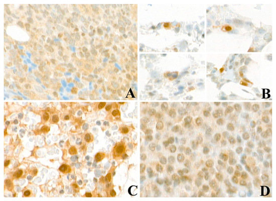
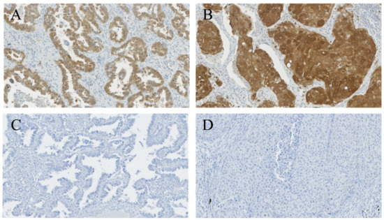
The major causative agents of head and neck squamous cell carcinomas (HNSCCs) are either environmental factors, such as tobacco and alcohol consumption, or infection with oncogenic human papillomaviruses (HPVs). An important aspect of HPV-induced oncogenesis is the targeting by the E6 oncoprotein of PDZ domain-containing substrates for proteasomal destruction. Tumor suppressors DLG1 and SCRIB are two of the principal PDZ domain-containing E6 targets. Both have been shown to play critical roles in the regulation of cell growth and polarity and in maintaining the structural integrity of the epithelia. We investigated how modifications in the cellular localization and protein expression of DLG1 and SCRIB in HPV16-positive and HPV-negative histologic oropharyngeal squamous cell carcinomas (OPSCC) might reflect disease progression. HPV presence was determined by p16 staining and HPV genotyping. Whilst DLG1 expression levels did not differ markedly between HPV-negative and HPV16-positive OPSCCs, it appeared to be relocated from cell–cell contacts to the cytoplasm in most samples, regardless of HPV16 positivity. This indicates that alterations in DLG1 distribution could contribute to malignant progression in OPSCCs. Interestingly, SCRIB was also relocated from cell–cell contacts to the cytoplasm in the tumor samples in comparison with normal tissue, regardless of HPV16 status, but in addition there was an obvious reduction in SCRIB expression in higher grade tumors. Strikingly, loss of SCRIB was even more pronounced in HPV16-positive OPSCCs. These alterations in SCRIB levels may contribute to transformation and loss of tissue architecture in the process of carcinogenesis and could potentially serve as markers in the development of OPSCCs. Full article
Show Figures

Figure 1

18 pages, 31187 KiB  
Article
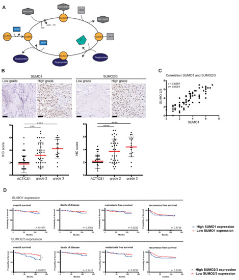
SUMOylation Is Associated with Aggressive Behavior in Chondrosarcoma of Bone
by Jessie S. Kroonen, Alwine B. Kruisselbrink, Inge H. Briaire-de Bruijn, Olaejirinde O. Olaofe, Judith V. M. G. Bovée and Alfred C. O. Vertegaal
Cancers 2021, 13(15), 3823; https://doi.org/10.3390/cancers13153823 - 29 Jul 2021
Cited by 7 | Viewed by 2786
Abstract
Multiple components of the SUMOylation machinery are deregulated in various cancers and could represent potential therapeutic targets. Understanding the role of SUMOylation in tumor progression and aggressiveness would increase our insight in the role of SUMO in cancer and clarify its potential as [...] Read more.
Multiple components of the SUMOylation machinery are deregulated in various cancers and could represent potential therapeutic targets. Understanding the role of SUMOylation in tumor progression and aggressiveness would increase our insight in the role of SUMO in cancer and clarify its potential as a therapeutic target. Here we investigate SUMO in relation to conventional chondrosarcomas, which are malignant cartilage forming tumors of the bone. Aggressiveness of chondrosarcoma increases with increasing histological grade, and a multistep progression model is assumed. High-grade chondrosarcomas have acquired an increased number of genetic alterations. Using immunohistochemistry on tissue microarrays (TMA) containing 137 chondrosarcomas, we showed that higher expression of SUMO1 and SUMO2/3 correlates with increased histological grade. In addition, high SUMO2/3 expression was associated with decreased overall survival chances (p = 0. 0312) in chondrosarcoma patients as determined by log-rank analysis and Cox regression. Various chondrosarcoma cell lines (n = 7), especially those derived from dedifferentiated chondrosarcoma, were sensitive to SUMO inhibition in vitro. Mechanistically, we found that SUMO E1 inhibition interferes with cell division and as a consequence DNA bridges are frequently formed between daughter cells. In conclusion, SUMO expression could potentially serve as a prognostic biomarker. Full article
Show Figures

Figure 1

15 pages, 2870 KiB  
Article
Combined Simplified Molecular Classification of Gastric Adenocarcinoma, Enhanced by Lymph Node Status: An Integrative Approach
by Till Daun, Ronny Nienhold, Aino Paasinen-Sohns, Angela Frank, Melanie Sachs, Inti Zlobec and Gieri Cathomas
Cancers 2021, 13(15), 3722; https://doi.org/10.3390/cancers13153722 - 24 Jul 2021
Cited by 7 | Viewed by 3310
Abstract
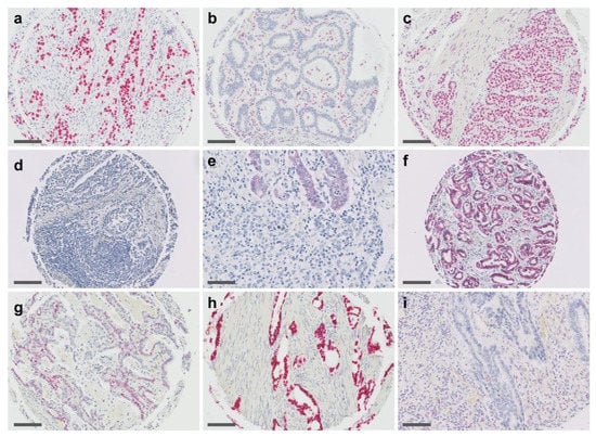
Gastric adenocarcinoma (GAC) is a heterogeneous disease and at least two major studies have recently provided a molecular classification for this tumor: The Cancer Genome Atlas (TCGA) and the Asian Cancer Research Group (ARCG). Both classifications quote four molecular subtypes, but these subtypes [...] Read more.
Gastric adenocarcinoma (GAC) is a heterogeneous disease and at least two major studies have recently provided a molecular classification for this tumor: The Cancer Genome Atlas (TCGA) and the Asian Cancer Research Group (ARCG). Both classifications quote four molecular subtypes, but these subtypes only partially overlap. In addition, the classifications are based on complex and cost-intensive technologies, which are hardly feasible for everyday practice. Therefore, simplified approaches using immunohistochemistry (IHC), in situ hybridization (ISH) as well as commercially available next generation sequencing (NGS) have been considered for routine use. In the present study, we screened 115 GAC by IHC for p53, MutL Homolog 1 (MLH1) and E-cadherin and performed ISH for Epstein–Barr virus (EBV). In addition, sequencing by NGS for TP53 and tumor associated genes was performed. With this approach, we were able to define five subtypes of GAC: (1) Microsatellite Instable (MSI), (2) EBV-associated, (3) Epithelial Mesenchymal Transition (EMT)-like, (4) p53 aberrant tumors surrogating for chromosomal instability and (5) p53 proficient tumors surrogating for genomics stable cancers. Furthermore, by considering lymph node metastasis in the p53 aberrant GAC, a better prognostic stratification was achieved which finally allowed us to separate the GAC highly significant in a group with poor and good-to-intermediate prognosis, respectively. Our data show that molecular classification of GAC can be achieved by using commercially available assays including IHC, ISH and NGS. Furthermore, we present an integrative workflow, which has the potential to overcome the uncertainty resulting from discrepancies from existing classification schemes. Full article
Show Figures

Figure 1

21 pages, 415 KiB  
Review
Molecular Pathways and Druggable Targets in Head and Neck Squamous Cell Carcinoma
by Farzaneh Kordbacheh and Camile S. Farah
Cancers 2021, 13(14), 3453; https://doi.org/10.3390/cancers13143453 - 9 Jul 2021
Cited by 10 | Viewed by 4223
Abstract
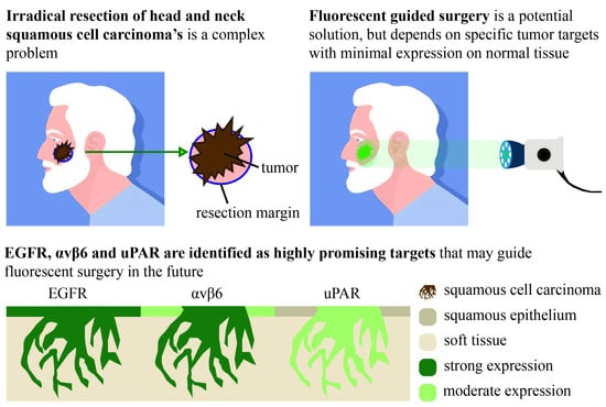
Head and neck cancers are a heterogeneous group of neoplasms, affecting an ever increasing global population. Despite advances in diagnostic technology and surgical approaches to manage these conditions, survival rates have only marginally improved and this has occurred mainly in developed countries. Some [...] Read more.
Head and neck cancers are a heterogeneous group of neoplasms, affecting an ever increasing global population. Despite advances in diagnostic technology and surgical approaches to manage these conditions, survival rates have only marginally improved and this has occurred mainly in developed countries. Some improvements in survival, however, have been a result of new management and treatment approaches made possible because of our ever-increasing understanding of the molecular pathways triggered in head and neck oncogenesis, and the growing understanding of the abundant heterogeneity of this group of cancers. Some important pathways are common to other solid tumours, but their impact on reducing the burden of head and neck disease has been less than impressive. Other less known and little-explored pathways may hold the key to the development of potential druggable targets. The extensive work carried out over the last decade, mostly utilising next generation sequencing has opened up the development of many novel approaches to head and neck cancer treatment. This paper explores our current understanding of the molecular pathways of this group of tumours and outlines associated druggable targets which are deployed as therapeutic approaches in head and neck oncology with the ultimate aim of improving patient outcomes and controlling the personal and economic burden of head and neck cancer. Full article
43 pages, 1988 KiB  
Review
Oncogenic Functions and Clinical Significance of Circular RNAs in Colorectal Cancer
by Maria Radanova, Galya Mihaylova, Neshe Nazifova-Tasinova, Mariya Levkova, Oskan Tasinov, Desislava Ivanova, Zhasmina Mihaylova and Ivan Donev
Cancers 2021, 13(14), 3395; https://doi.org/10.3390/cancers13143395 - 6 Jul 2021
Cited by 6 | Viewed by 3103
Abstract
Colorectal cancer (CRC) is ranked as the second most commonly diagnosed disease in females and the third in males worldwide. Therefore, the finding of new more reliable biomarkers for early diagnosis, for prediction of metastasis, and resistance to conventional therapies is an important [...] Read more.
Colorectal cancer (CRC) is ranked as the second most commonly diagnosed disease in females and the third in males worldwide. Therefore, the finding of new more reliable biomarkers for early diagnosis, for prediction of metastasis, and resistance to conventional therapies is an important challenge in overcoming the disease. The current review presents circular RNAs (circRNAs) with their unique features as potential prognostic and diagnostic biomarkers in CRC. The review highlights the mechanism of action and the role of circRNAs with oncogenic functions in the CRC as well as the association between their expression and clinicopathological characteristics of CRC patients. The comprehension of the role of oncogenic circRNAs in CRC pathogenesis is growing rapidly and the next step is using them as suitable new drug targets in the personalized treatment of CRC patients. Full article
Show Figures

Figure 1

16 pages, 5204 KiB  
Article
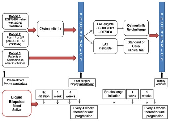
Longitudinal Circulating Tumor DNA Analysis in Blood and Saliva for Prediction of Response to Osimertinib and Disease Progression in EGFR-Mutant Lung Adenocarcinoma
by Chul Kim, Liqiang Xi, Constance M. Cultraro, Fang Wei, Gregory Jones, Jordan Cheng, Ahmad Shafiei, Trinh Hoc-Tran Pham, Nitin Roper, Elizabeth Akoth, Azam Ghafoor, Vikram Misra, Nina Monkash, Charles Strom, Michael Tu, Wei Liao, David Chia, Clive Morris, Seth M. Steinberg, Hadi Bagheri, David T. W. Wong, Mark Raffeld and Udayan Guhaadd Show full author list remove Hide full author list
Cancers 2021, 13(13), 3342; https://doi.org/10.3390/cancers13133342 - 3 Jul 2021
Cited by 15 | Viewed by 6026
Abstract
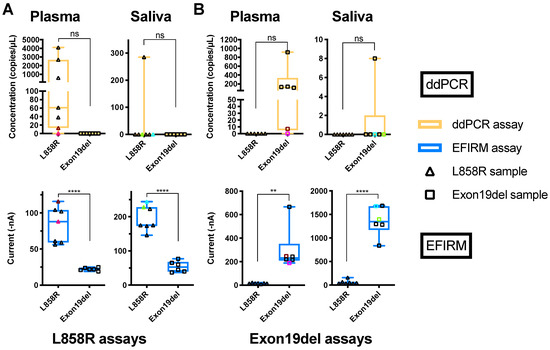
Background: We assessed whether serial ctDNA monitoring of plasma and saliva predicts response and resistance to osimertinib in EGFR-mutant lung adenocarcinoma. Three ctDNA technologies—blood-based droplet-digital PCR (ddPCR), next-generation sequencing (NGS), and saliva-based EFIRM liquid biopsy (eLB)—were employed to investigate their complementary roles. [...] Read more.
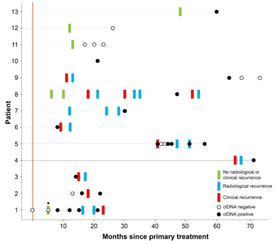
Background: We assessed whether serial ctDNA monitoring of plasma and saliva predicts response and resistance to osimertinib in EGFR-mutant lung adenocarcinoma. Three ctDNA technologies—blood-based droplet-digital PCR (ddPCR), next-generation sequencing (NGS), and saliva-based EFIRM liquid biopsy (eLB)—were employed to investigate their complementary roles. Methods: Plasma and saliva samples were collected from patients enrolled in a prospective clinical trial of osimertinib and local ablative therapy upon progression (NCT02759835). Plasma was analyzed by ddPCR and NGS. Saliva was analyzed by eLB. Results: A total of 25 patients were included. We analyzed 534 samples by ddPCR (n = 25), 256 samples by NGS (n = 24) and 371 samples by eLB (n = 22). Among 20 patients who progressed, ctDNA progression predated RECIST 1.1 progression by a median of 118 days (range: 61–272 days) in 11 (55%) patients. Of nine patients without ctDNA progression by ddPCR, two patients had an increase in mutant EGFR by eLB and two patients were found to have ctDNA progression by NGS. Levels of ctDNA measured by ddPCR and NGS at early time points, but not volumetric tumor burden, were associated with PFS. EGFR/ERBB2/MET/KRAS amplifications, EGFR C797S, PIK3CA E545K, PTEN V9del, and CTNNB1 S45P were key resistance mechanisms identified by NGS. Conclusion: Serial assessment of ctDNA in plasma and saliva predicts response and resistance to osimertinib, with each assay having supplementary roles. Full article
Show Figures

Figure 1

11 pages, 2257 KiB  
Article
Metastatic Risk Factors Associated with Class 1A Uveal Melanoma Patients
by Alexej Ballhausen, Elizabeth Urias, Stephen K. Gruschkus, Michelle Williams, Maura S. Glover, Yong Qin, Dan S. Gombos and Sapna P. Patel
Cancers 2021, 13(13), 3292; https://doi.org/10.3390/cancers13133292 - 30 Jun 2021
Cited by 7 | Viewed by 3489
Abstract
In uveal melanoma (UM), gene expression profiling (GEP) is commonly used to classify metastatic risk into three groups (Class 1A, 1B, and 2). Class 1A patients have a lower metastatic risk of 2% at 5 years compared to other groups. We aimed to [...] Read more.
In uveal melanoma (UM), gene expression profiling (GEP) is commonly used to classify metastatic risk into three groups (Class 1A, 1B, and 2). Class 1A patients have a lower metastatic risk of 2% at 5 years compared to other groups. We aimed to describe clinical features associated with the development of metastasis in this low-risk group. This single-center IRB-approved retrospective case series review included all UM patients between February 2006 and March 2019 with an archived or fresh specimen classified as Class 1A. Cox regression and receiver operating characteristics analyses were used to identify factors associated with metastasis development and OS. Among 73 UM patients with Class 1A, the 5-year cumulative incidence of local recurrence and distant metastasis was 4.2% and 17.0%, respectively. Stage III disease (HR 20.7; 95% confidence interval (95% CI) 1.4–300.6; p= 0.0264) was found to be independently associated with metastatic recurrence, while primary therapy was associated with OS (enucleation vs. brachytherapy, HR 13.5; 95% CI 1.3–147.6; p = 0.0348). Combined clinical decision-making utilizing factors such as GEP class, American Joint Committee on Cancer (AJCC) stage, and COMS size could have a significant clinical impact by improving risk stratification and adapting follow-up intervals in UM Class 1A patients. Full article
Show Figures

Figure 1

12 pages, 2353 KiB  
Article
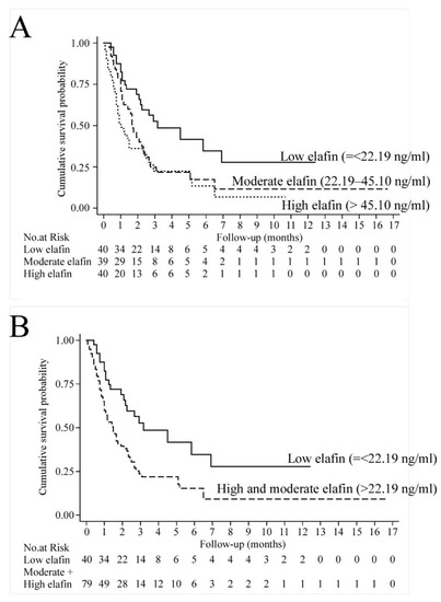
High Serum Elafin Prediction of Poor Prognosis of Locoregional Esophageal Squamous Cell Carcinoma
by I-Chen Wu, Yao-Kuang Wang, Yi-Hsun Chen, Chun-Chieh Wu, Meng-Chieh Wu, Wei-Chung Chen, Wen-Lun Wang, Hung-Shun Lin, Chou-Cheng Chen, Shah-Hwa Chou, Yu-Peng Liu and Ming-Tsang Wu
Cancers 2021, 13(12), 3082; https://doi.org/10.3390/cancers13123082 - 21 Jun 2021
Cited by 1 | Viewed by 2271
Abstract
Esophageal squamous cell carcinoma (ESCC) is a highly aggressive tumor known to have locally advanced and metastatic features which cause a dismal prognosis. We sought to determine whether elafin, a non-invasive and secretory small-molecule marker, could be used to predict prognosis in locoregional [...] Read more.
Esophageal squamous cell carcinoma (ESCC) is a highly aggressive tumor known to have locally advanced and metastatic features which cause a dismal prognosis. We sought to determine whether elafin, a non-invasive and secretory small-molecule marker, could be used to predict prognosis in locoregional ESCC patients in human and in vitro studies. In our human study, 119 subjects were identified as having incident and pathologically-proved ESCC with stage I-IIIA tumors from southern Taiwan between 2000 and 2016. We measured their serum elafin levels at baseline and followed them until the date of cancer death or until January 2020, the end of this study. Those with high serum elafin levels were found to have a 1.99-fold risk (95% confidence interval: 1.17–3.38) shorter survival than those who did not. In our in vitro experiments, elevated elafin levels were found to drive ESCC cell proliferation, migration and invasion, while attenuation of elafin level by shRNA abrogated those effects. We concluded that elafin promotes ESCC motility and invasion and leads to a worse clinical prognosis in ESCC patients without distant metastasis. Full article
Show Figures

Figure 1

17 pages, 970 KiB  
Article
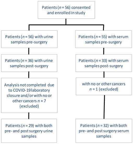
Metabolic Changes in Early-Stage Non–Small Cell Lung Cancer Patients after Surgical Resection
by Naseer Ahmed, Biniam Kidane, Le Wang, Zoann Nugent, Nataliya Moldovan, April McElrea, Shiva Shariati-Ievari, Gefei Qing, Lawrence Tan, Gordon Buduhan, Sadeesh K. Srinathan and Michel Aliani
Cancers 2021, 13(12), 3012; https://doi.org/10.3390/cancers13123012 - 16 Jun 2021
Cited by 11 | Viewed by 3154
Abstract
Metabolic alterations in malignant cells play a vital role in tumor initiation, proliferation, and metastasis. Biofluids from patients with non–small cell lung cancer (NSCLC) harbor metabolic biomarkers with potential clinical applications. In this study, we assessed the changes in the metabolic profile of [...] Read more.
Metabolic alterations in malignant cells play a vital role in tumor initiation, proliferation, and metastasis. Biofluids from patients with non–small cell lung cancer (NSCLC) harbor metabolic biomarkers with potential clinical applications. In this study, we assessed the changes in the metabolic profile of patients with early-stage NSCLC using mass spectrometry and nuclear magnetic resonance spectroscopy before and after surgical resection. A single cohort of 35 patients provided a total of 29 and 32 pairs of urine and serum samples, respectively, pre-and post-surgery. We identified a profile of 48 metabolites that were significantly different pre- and post-surgery: 17 in urine and 31 in serum. A higher proportion of metabolites were upregulated than downregulated post-surgery (p < 0.01); however, the median fold change (FC) was higher for downregulated than upregulated metabolites (p < 0.05). Purines/pyrimidines and proteins had a larger dysregulation than other classes of metabolites (p < 0.05 for each class). Several of the dysregulated metabolites have been previously associated with cancer, including leucyl proline, asymmetric dimethylarginine, isopentenyladenine, fumaric acid (all downregulated post-surgery), as well as N6-methyladenosine and several deoxycholic acid moieties, which were upregulated post-surgery. This study establishes metabolomic analysis of biofluids as a path to non-invasive diagnostics, screening, and monitoring in NSCLC. Full article
Show Figures

Figure 1

18 pages, 929 KiB  
Review
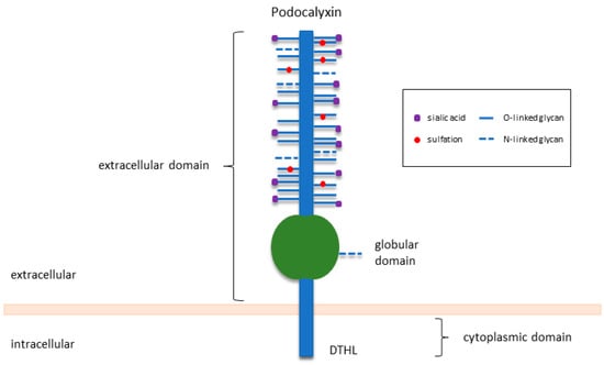
Podocalyxin in Normal Tissue and Epithelial Cancer
by Ngoc Le Tran, Yao Wang and Guiying Nie
Cancers 2021, 13(12), 2863; https://doi.org/10.3390/cancers13122863 - 8 Jun 2021
Cited by 22 | Viewed by 5650
Abstract
Podocalyxin (PODXL), a glycosylated cell surface sialomucin of the CD34 family, is normally expressed in kidney podocytes, vascular endothelial cells, hematopoietic progenitors, mesothelium, as well as a subset of neurons. In the kidney, PODXL functions primarily as an antiadhesive molecule in podocyte epithelial [...] Read more.
Podocalyxin (PODXL), a glycosylated cell surface sialomucin of the CD34 family, is normally expressed in kidney podocytes, vascular endothelial cells, hematopoietic progenitors, mesothelium, as well as a subset of neurons. In the kidney, PODXL functions primarily as an antiadhesive molecule in podocyte epithelial cells, regulating adhesion and cell morphology, and playing an essential role in the development and function of the organ. Outside the kidney, PODXL plays subtle roles in tissue remodelling and development. Furthermore, many cancers, especially those that originated from the epithelium, have been reported to overexpress PODXL. Collective evidence suggests that PODXL overexpression is linked to poor prognosis, more aggressive tumour progression, unfavourable treatment outcomes, and possibly chemoresistance. This review summarises our current knowledge of PODXL in normal tissue function and epithelial cancer, with a particular focus on its underlying roles in cancer metastasis, likely involvement in chemoresistance, and potential use as a diagnostic and prognostic biomarker. Full article
Show Figures

Figure 1

17 pages, 2820 KiB  
Article
Deep Learning Improves Pancreatic Cancer Diagnosis Using RNA-Based Variants
by Ali Al-Fatlawi, Negin Malekian, Sebastián García, Andreas Henschel, Ilwook Kim, Andreas Dahl, Beatrix Jahnke, Peter Bailey, Sarah Naomi Bolz, Anna R. Poetsch, Sandra Mahler, Robert Grützmann, Christian Pilarsky and Michael Schroeder
Cancers 2021, 13(11), 2654; https://doi.org/10.3390/cancers13112654 - 28 May 2021
Cited by 9 | Viewed by 4488
Abstract
For optimal pancreatic cancer treatment, early and accurate diagnosis is vital. Blood-derived biomarkers and genetic predispositions can contribute to early diagnosis, but they often have limited accuracy or applicability. Here, we seek to exploit the synergy between them by combining the biomarker CA19-9 [...] Read more.
For optimal pancreatic cancer treatment, early and accurate diagnosis is vital. Blood-derived biomarkers and genetic predispositions can contribute to early diagnosis, but they often have limited accuracy or applicability. Here, we seek to exploit the synergy between them by combining the biomarker CA19-9 with RNA-based variants. We use deep sequencing and deep learning to improve differentiating pancreatic cancer and chronic pancreatitis. We obtained samples of nucleated cells found in peripheral blood from 268 patients suffering from resectable, non-resectable pancreatic cancer, and chronic pancreatitis. We sequenced RNA with high coverage and obtained millions of variants. The high-quality variants served as input together with CA19-9 values to deep learning models. Our model achieved an area under the curve (AUC) of 96% in differentiating resectable cancer from pancreatitis using a test cohort. Moreover, we identified variants to estimate survival in resectable cancer. We show that the blood transcriptome harbours variants, which can substantially improve noninvasive clinical diagnosis. Full article
Show Figures

Figure 1

11 pages, 2138 KiB  
Article
Validation of the Combined Biomarker for Prediction of Response to Checkpoint Inhibitor in Patients with Advanced Cancer
by Jin-Chul Kim, You-Jeong Heo, So-Young Kang, Jeeyun Lee and Kyoung-Mee Kim
Cancers 2021, 13(10), 2316; https://doi.org/10.3390/cancers13102316 - 12 May 2021
Cited by 6 | Viewed by 2372
Abstract
Although immune checkpoint inhibitors can induce durable responses in patients with multiple types of advanced cancer, only a limited number of patients have a known reliable biomarker. This study aimed to validate the IMmunotherapy Against GastrIc Cancer (IMAGiC) model, which was developed based [...] Read more.
Although immune checkpoint inhibitors can induce durable responses in patients with multiple types of advanced cancer, only a limited number of patients have a known reliable biomarker. This study aimed to validate the IMmunotherapy Against GastrIc Cancer (IMAGiC) model, which was developed based on a previous study of four-gene and PD-L1 level, to predict immunotherapy response. We developed a clinical assay for formalin-fixed paraffin-embedded samples using quantitative real-time polymerase chain reaction to measure the expression level of the previously published four-gene set. The predictive performance was validated in a cohort of 89 patients with several advanced tumor types. The IMAGiC score was derived from tumor samples of 89 patients consisting of eight cancer types, and 73 out of 89 patients available for clinical response were analyzed with clinicopathological factors. The IMAGiC group (responder vs. non-responder) was determined with a specific value of the IMAGiC score as a cutoff, which was set by log-rank statistics for progression-free survival (PFS) divided the patients into 56 (76.7%) non-responders and 17 (23.3%) responders. Clinical responders (complete response/partial response) were higher in the IMAGiC responder group than in the non-responder group (70.6 vs. 21.4%). The median PFS of the IMAGiC responder group and non-responder was 20.8 months (95% CI 9.1-not reached) and 6.7 months (95% CI 4.9–11.1, p = 0.007), respectively. Among the 17 IMAGiC responders, 11 patients had tumor mutation burden-low and microsatellite-stable tumors. This study validated a predictive model based on a four-gene expression signature. Along with conventional biomarkers, our model could be useful for predicting response to immunotherapy in patients with advanced cancer. Full article
Show Figures

Figure 1

16 pages, 1167 KiB  
Article
Clinical Implication of Liquid Biopsy in Colorectal Cancer Patients Treated with Metastasectomy
by Soohyeon Lee, Young-Soo Park, Won-Jin Chang, Jung Yoon Choi, Ahreum Lim, Boyeon Kim, Saet-Byeol Lee, Jong-Won Lee, Seon-Hahn Kim, Jin Kim, Jung-Myun Kwak, Kyung-Chul Yoon, Sung-Ho Lee and Yeul Hong Kim
Cancers 2021, 13(9), 2231; https://doi.org/10.3390/cancers13092231 - 6 May 2021
Cited by 13 | Viewed by 2365
Abstract
Background & Aims: The application of circulating tumor DNA (ctDNA) has been studied for predicting recurrent disease after surgery and treatment response during systemic treatment. Metastasectomy can be curative for well-selected patients with metastatic colorectal cancer (mCRC). This prospective study investigated the ctDNA [...] Read more.
Background & Aims: The application of circulating tumor DNA (ctDNA) has been studied for predicting recurrent disease after surgery and treatment response during systemic treatment. Metastasectomy can be curative for well-selected patients with metastatic colorectal cancer (mCRC). This prospective study investigated the ctDNA level before and after metastasectomy in patients with mCRC to explore its potential as a predictive biomarker. Methods: We collected data on 98 metastasectomies for mCRC performed from March 2017 to February 2020. Somatic mutations in the primary and metastatic tumors were identified and tumor-informed ctDNAs were selected by ultra-deep targeted sequencing. Plasma samples were mandatorily collected before and 3–4 weeks after metastasectomy and serially, if patients agreed. Results: Data on 67 of 98 metastasectomies (58 patients) meeting the criteria were collected. ctDNA was detected in 9 (29%) of 31 cases treated with upfront metastasectomy and in 7 (19.4%) of 36 cases treated with metastasectomy after upfront chemotherapy. The detection rate of ctDNA was higher in liver metastasis (p = 0.0045) and tumors measuring ≥1 cm (p = 0.0183). ctDNA was less likely to be detected if the response to chemotherapy was good. After metastasectomy, ctDNA was found in 4 (6%) cases with rapid progressive disease. Conclusion: The biological factors affecting the ctDNA shedding from the tumor should be considered when applying ctDNA assays in a clinical setting. After metastasectomy for oligometastatic lesions in good responders of chemotherapy, most ctDNA was cleared or existed below the detection level. To assist clinical decision making after metastasectomy for mCRC using ctDNA, further studies for improving specific outcomes are needed. Full article
Show Figures

Figure 1

13 pages, 1189 KiB  
Article
Characterization of Cell-Bound CA125 on Immune Cell Subtypes of Ovarian Cancer Patients Using a Novel Imaging Platform
by Germán González, Kornél Lakatos, Jawad Hoballah, Roberta Fritz-Klaus, Lojain Al-Johani, Jeff Brooker, Sinyoung Jeong, Conor L. Evans, Petra Krauledat, Daniel W. Cramer, Robert A. Hoffman, W. Peter Hansen and Manish S. Patankar
Cancers 2021, 13(9), 2072; https://doi.org/10.3390/cancers13092072 - 25 Apr 2021
Cited by 8 | Viewed by 3223
Abstract
MUC16, a sialomucin that contains the ovarian cancer biomarker CA125, binds at low abundance to leucocytes via the immune receptor, Siglec-9. Conventional fluorescence-based imaging techniques lack the sensitivity to assess this low-abundance event, prompting us to develop a novel “digital” optical cytometry technique [...] Read more.
MUC16, a sialomucin that contains the ovarian cancer biomarker CA125, binds at low abundance to leucocytes via the immune receptor, Siglec-9. Conventional fluorescence-based imaging techniques lack the sensitivity to assess this low-abundance event, prompting us to develop a novel “digital” optical cytometry technique for qualitative and quantitative assessment of CA125 binding to peripheral blood mononuclear cells (PBMC). Plasmonic nanoparticle labeled detection antibody allows assessment of CA125 at the near-single molecule level when bound to specific immune cell lineages that are simultaneously identified using multiparameter fluorescence imaging. Image analysis and deep learning were used to quantify CA125 per each cell lineage. PBMC from treatment naïve ovarian cancer patients (N = 14) showed higher cell surface abundance of CA125 on the aggregate PBMC population as well as on NK (p = 0.013), T (p < 0.001) and B cells (p = 0.024) compared to circulating lymphocytes of healthy donors (N = 7). Differences in CA125 binding to monocytes or NK-T cells between the two cohorts were not significant. There was no correlation between the PBMC-bound and serum levels of CA125, suggesting that these two compartments are not in stoichiometric equilibrium. Understanding where and how subset-specific cell-bound surface CA125 takes place may provide guidance towards a new diagnostic biomarker in ovarian cancer. Full article
Show Figures

Figure 1

17 pages, 1661 KiB  
Article
Neo-Fs Index: A Novel Immunohistochemical Biomarker Panel Predicts Survival and Response to Anti-Angiogenetic Agents in Clear Cell Renal Cell Carcinoma
by Jisup Kim, Jee-Young Park, Su-Jin Shin, Beom Jin Lim and Heounjeong Go
Cancers 2021, 13(6), 1199; https://doi.org/10.3390/cancers13061199 - 10 Mar 2021
Cited by 3 | Viewed by 2617
Abstract
Background: Frameshift indels have emerged as a predictor of immunotherapy response but were not evaluated yet to predict anti-angiogenetic agent (AAA) response or prognosis in clear cell renal cell carcinoma (ccRCC). Methods: Here, to develop biomarkers that predict survival and response [...] Read more.
Background: Frameshift indels have emerged as a predictor of immunotherapy response but were not evaluated yet to predict anti-angiogenetic agent (AAA) response or prognosis in clear cell renal cell carcinoma (ccRCC). Methods: Here, to develop biomarkers that predict survival and response to AAA, we evaluated the immunohistochemical expression of proteins whose genes frequently harbor frameshift indels in 638 ccRCC patients and correlated the individual and integrated markers with prognosis and AAA response. The mutational landscape was evaluated using targeted next-generation sequencing in 12 patients concerning protein markers. Immune gene signatures were retrieved from TCGA RNA seq data. Results: Five proteins (APC, NOTCH1, ARID1A, EYS, and filamin A) were independent adverse prognosticators and were incorporated into the Neo-fs index. Better overall, disease-specific and recurrence-free survival were observed with high Neo-fs index in univariate and multivariate survival analyses. Better AAA responses were observed with a high Neo-fs index, which reflected increased MHC class I, CD8+ T cell, cytolytic activity, and plasmacytoid dendritic cell signatures and decreased type II-IFN response signatures, as well as greater single-nucleotide variant (SNV) and indel counts. Conclusions: Neo-fs index, reflecting antitumor immune signature and more SNVs. and indels, is a powerful predictor of survival and AAA response in ccRCC. Full article
Show Figures

Figure 1

25 pages, 2118 KiB  
Review
Resistance to Molecularly Targeted Therapies in Melanoma
by Meet Patel, Adam Eckburg, Shahina Gantiwala, Zachary Hart, Joshua Dein, Katie Lam and Neelu Puri
Cancers 2021, 13(5), 1115; https://doi.org/10.3390/cancers13051115 - 5 Mar 2021
Cited by 44 | Viewed by 6485
Abstract
Malignant melanoma is the most aggressive type of skin cancer with invasive growth patterns. In 2021, 106,110 patients are projected to be diagnosed with melanoma, out of which 7180 are expected to die. Traditional methods like surgery, radiation therapy, and chemotherapy are not [...] Read more.
Malignant melanoma is the most aggressive type of skin cancer with invasive growth patterns. In 2021, 106,110 patients are projected to be diagnosed with melanoma, out of which 7180 are expected to die. Traditional methods like surgery, radiation therapy, and chemotherapy are not effective in the treatment of metastatic and advanced melanoma. Recent approaches to treat melanoma have focused on biomarkers that play significant roles in cell growth, proliferation, migration, and survival. Several FDA-approved molecular targeted therapies such as tyrosine kinase inhibitors (TKIs) have been developed against genetic biomarkers whose overexpression is implicated in tumorigenesis. The use of targeted therapies as an alternative or supplement to immunotherapy has revolutionized the management of metastatic melanoma. Although this treatment strategy is more efficacious and less toxic in comparison to traditional therapies, targeted therapies are less effective after prolonged treatment due to acquired resistance caused by mutations and activation of alternative mechanisms in melanoma tumors. Recent studies focus on understanding the mechanisms of acquired resistance to these current therapies. Further research is needed for the development of better approaches to improve prognosis in melanoma patients. In this article, various melanoma biomarkers including BRAF, MEK, RAS, c-KIT, VEGFR, c-MET and PI3K are described, and their potential mechanisms for drug resistance are discussed. Full article
Show Figures

Figure 1

15 pages, 1765 KiB  
Article
HER2 Expression Is Predictive of Survival in Cetuximab Treated Patients with RAS Wild Type Metastatic Colorectal Cancer
by Said A. Khelwatty, Soozana Puvanenthiran, Sharadah Essapen, Izhar Bagwan, Alan M. Seddon and Helmout Modjtahedi
Cancers 2021, 13(4), 638; https://doi.org/10.3390/cancers13040638 - 5 Feb 2021
Cited by 12 | Viewed by 2994
Abstract
The overexpressed HER2 is an important target for treatment with monoclonal antibody (mAb) trastuzumab, only in patients with breast and gastric cancers, and is an emerging therapeutic biomarker in metastatic colorectal cancer (mCRC) treated with anti-epidermal growth factor receptor (EGFR) mAbs cetuximab and [...] Read more.
The overexpressed HER2 is an important target for treatment with monoclonal antibody (mAb) trastuzumab, only in patients with breast and gastric cancers, and is an emerging therapeutic biomarker in metastatic colorectal cancer (mCRC) treated with anti-epidermal growth factor receptor (EGFR) mAbs cetuximab and panitumumab. In this study, we investigated the relative expression and predictive value of all human epidermal growth factor receptor (HER) family members in 144 cetuximab-treated patients with wild type RAS mCRC. The relative expression of EGFR and HER2 have also been examined in 21-paired primary tumours and their metastatic sites by immunohistochemistry. Of the 144 cases examined, 25%, 97%, 79%, 48%, and 10% were positive for EGFR, HER2, HER3, and HER4 and all four HER family members, respectively. The expression of EGFR was an indicator of poorer overall survival and the membranous expression of HER2 and HER3 3+ intensity was associated with a shorter progression free survival (PFS). In contrast, the cytoplasmic expression of HER2 was associated with better PFS. In 48% and 71% of the cases, there were discordance in the expression of EGFR or one or more HER family members in paired primary and related metastatic tumours, respectively. Our results implicate the importance of a large prospective investigation of the expression level and predictive value of not only the therapeutic target (i.e., EGFR protein) but also HER2 and other HER family members as therapeutic targets, or for response to therapy with anti-EGFR mAbs and other forms of HER inhibitors, in both the primary tumours and metastatic sites in mCRC. Full article
Show Figures

Figure 1

12 pages, 1498 KiB  
Article
Early Prostate-Specific Antigen (PSA) Change at Four Weeks of the First-Line Treatment Using Abiraterone and Enzalutamide Could Predict Early/Primary Resistance in Metastatic Castration-Resistant Prostate Cancer
by Taizo Uchimoto, Kazumasa Komura, Wataru Fukuokaya, Takahiro Kimura, Kazuhiro Takahashi, Kazuki Nishimura, Keita Nakamori, Yuya Fujiwara, Tomohisa Matsunaga, Takeshi Tsutsumi, Takuya Tsujino, Ryoichi Maenosono, Yuki Yoshikawa, Kohei Taniguchi, Tomohito Tanaka, Hirofumi Uehara, Naokazu Ibuki, Hajime Hirano, Hayahito Nomi, Kiyoshi Takahara, Teruo Inamoto, Shin Egawa and Haruhito Azumaadd Show full author list remove Hide full author list
Cancers 2021, 13(3), 526; https://doi.org/10.3390/cancers13030526 - 30 Jan 2021
Cited by 6 | Viewed by 3620
Abstract
The identification of early or primary resistance to androgen signaling inhibitors (ASIs) is of great value for the treatment of metastatic castration-resistant prostate cancer (mCRPC). This study evaluates the predictive value of prostate-specific antigen (PSA) response at dour weeks of first-line ASIs treatment [...] Read more.
The identification of early or primary resistance to androgen signaling inhibitors (ASIs) is of great value for the treatment of metastatic castration-resistant prostate cancer (mCRPC). This study evaluates the predictive value of prostate-specific antigen (PSA) response at dour weeks of first-line ASIs treatment for mCRPC patients. A total of 254 patients treated with ASIs (abiraterone acetate: AA and enzalutamide: Enz) at the first-line treatment are retrospectively analyzed. Patients are stratified according to the achievement of >30% PSA decline at 4 and 12 weeks from the treatment initiation. At four weeks of the treatment, 157 patients (61.8%) achieved >30% PSA decline from the baseline. Thereafter, 177 patients (69.7%) achieved >30% PSA decline at 12 weeks of the treatment. A multivariate analysis exhibits >30% PSA decline at four weeks as an independent predictor for overall survival (OS). We note that 30 of 97 (30.9%) patients who did not achieve >30% PSA decline at four weeks consequently achieved >30% PSA decline at 12 weeks, and had a comparable favorable three years OS rate as the 147 patients achieving >30% PSA decline at both 4 and 12 weeks. To identify the variables that discriminate the patient survival in 97 patients without achieving >30% PSA decline at four weeks, a multivariate analysis is performed. The duration of androgen deprivation therapy before CRPC ≤ 12 months and Eastern Cooperative Oncology Group Performance Status ≥ 1 are identified as independent predictors for shorter OS for those patients. These data offer a concept of early treatment switch after four weeks of first-line ASIs when not observing >30% PSA decline at four weeks—particularly in patients with a modest effect of ADT and poor performance status. Full article
Show Figures

Figure 1

16 pages, 1787 KiB  
Article
Radiomics Features of 18F-Fluorodeoxyglucose Positron-Emission Tomography as a Novel Prognostic Signature in Colorectal Cancer
by Jeonghyun Kang, Jae-Hoon Lee, Hye Sun Lee, Eun-Suk Cho, Eun Jung Park, Seung Hyuk Baik, Kang Young Lee, Chihyun Park, Yunku Yeu, Jean R. Clemenceau, Sunho Park, Hongming Xu, Changjin Hong and Tae Hyun Hwang
Cancers 2021, 13(3), 392; https://doi.org/10.3390/cancers13030392 - 21 Jan 2021
Cited by 12 | Viewed by 3065
Abstract
The aim of this study was to investigate the prognostic value of radiomics signatures derived from 18F-fluorodeoxyglucose (18F-FDG) positron-emission tomography (PET) in patients with colorectal cancer (CRC). From April 2008 to Jan 2014, we identified CRC patients who underwent 18 [...] Read more.
The aim of this study was to investigate the prognostic value of radiomics signatures derived from 18F-fluorodeoxyglucose (18F-FDG) positron-emission tomography (PET) in patients with colorectal cancer (CRC). From April 2008 to Jan 2014, we identified CRC patients who underwent 18F-FDG-PET before starting any neoadjuvant treatments and surgery. Radiomics features were extracted from the primary lesions identified on 18F-FDG-PET. Patients were divided into a training and validation set by random sampling. A least absolute shrinkage and selection operator Cox regression model was applied for prognostic signature building with progression-free survival (PFS) using the training set. Using the calculated radiomics score, a nomogram was developed, and its clinical utility was assessed in the validation set. A total of 381 patients with surgically resected CRC patients (training set: 228 vs. validation set: 153) were included. In the training set, a radiomics signature labeled as a rad_score was generated using two PET-derived features, such as gray-level run length matrix long-run emphasis (GLRLM_LRE) and gray-level zone length matrix short-zone low-gray-level emphasis (GLZLM_SZLGE). Patients with a high rad_score in the training and validation set had a shorter PFS. Multivariable analysis revealed that the rad_score was an independent prognostic factor in both training and validation sets. A radiomics nomogram, developed using rad_score, nodal stage, and lymphovascular invasion, showed good performance in the calibration curve and comparable predictive power with the staging system in the validation set. Textural features derived from 18F-FDG-PET images may enable detailed stratification of prognosis in patients with CRC. Full article
Show Figures

Figure 1

13 pages, 1337 KiB  
Article
Prognostic Value of Plasma hPG80 (Circulating Progastrin) in Metastatic Renal Cell Carcinoma
by Manish Kohli, Winston Tan, Bérengère Vire, Pierre Liaud, Mélina Blairvacq, Frederic Berthier, Daniel Rouison, George Garnier, Léa Payen, Thierry Cousin, Dominique Joubert and Alexandre Prieur
Cancers 2021, 13(3), 375; https://doi.org/10.3390/cancers13030375 - 20 Jan 2021
Cited by 10 | Viewed by 2960
Abstract
Precise management of kidney cancer requires the identification of prognostic factors. hPG80 (circulating progastrin) is a tumor promoting peptide present in the blood of patients with various cancers, including renal cell carcinoma (RCC). In this study, we evaluated the prognostic value of [...] Read more.
Precise management of kidney cancer requires the identification of prognostic factors. hPG80 (circulating progastrin) is a tumor promoting peptide present in the blood of patients with various cancers, including renal cell carcinoma (RCC). In this study, we evaluated the prognostic value of plasma hPG80 in 143 prospectively collected patients with metastatic RCC (mRCC). The prognostic impact of hPG80 levels on overall survival (OS) in mRCC patients after controlling for hPG80 levels in non-cancer age matched controls was determined and compared to the International Metastatic Database Consortium (IMDC) risk model (good, intermediate, poor). ROC curves were used to evaluate the diagnostic accuracy of hPG80 using the area under the curve (AUC). Our results showed that plasma hPG80 was detected in 94% of mRCC patients. hPG80 levels displayed high predictive accuracy with an AUC of 0.93 and 0.84 when compared to 18–25 year old controls and 50–80 year old controls, respectively. mRCC patients with high hPG80 levels (>4.5 pM) had significantly lower OS compared to patients with low hPG80 levels (<4.5 pM) (12 versus 31.2 months, respectively; p = 0.0031). Adding hPG80 levels (score of 1 for patients having hPG80 levels > 4.5 pM) to the six variables of the IMDC risk model showed a greater and significant difference in OS between the newly defined good-, intermediate- and poor-risk groups (p = 0.0003 compared to p = 0.0076). Finally, when patients with IMDC intermediate-risk group were further divided into two groups based on hPG80 levels within these subgroups, increased OS were observed in patients with low hPG80 levels (<4.5 pM). In conclusion, our data suggest that hPG80 could be used for prognosticating survival in mRCC alone or integrated to the IMDC score (by adding a variable to the IMDC score or by substratifying the IMDC risk groups), be a prognostic biomarker in mRCC patients. Full article
Show Figures

Figure 1

2020

Jump to: 2023, 2022, 2021, 2019

21 pages, 2756 KiB  
Article
Prognostic Inflammatory Index Based on Preoperative Peripheral Blood for Predicting the Prognosis of Colorectal Cancer Patients
by Jinming Fu, Ji Zhu, Fenqi Du, Lijie Zhang, Dapeng Li, Hao Huang, Tian Tian, Yupeng Liu, Lei Zhang, Ying Liu, Yuanyuan Zhang, Jing Xu, Shuhan Meng, Chenyang Jia, Simin Sun, Xue Li, Liyuan Zhao, Ding Zhang, Lixin Kang, Lijing Gao, Ting Zheng, Sanjun Cai, Yanlong Liu and Yashuang Zhaoadd Show full author list remove Hide full author list
Cancers 2021, 13(1), 3; https://doi.org/10.3390/cancers13010003 - 22 Dec 2020
Cited by 8 | Viewed by 3131
Abstract
Host inflammation is a critical component of tumor progression and its status can be indicated by peripheral blood cell counts. We aimed to construct a comprehensively prognostic inflammatory index (PII) based on preoperative peripheral blood cell counts and further evaluate its prognostic value [...] Read more.
Host inflammation is a critical component of tumor progression and its status can be indicated by peripheral blood cell counts. We aimed to construct a comprehensively prognostic inflammatory index (PII) based on preoperative peripheral blood cell counts and further evaluate its prognostic value for patients with colorectal cancer (CRC). A total of 9315 patients with stage II and III CRC from training and external validation cohorts were included. The PII was constructed by integrating all the peripheral blood cell counts associated with prognosis in the training cohort. Cox analyses were performed to evaluate the association between PII and overall survival (OS) and disease-free survival (DFS). In the training cohort, multivariate Cox analyses indicated that high OS-PII (>4.27) was significantly associated with worse OS (HR: 1.330, 95% CI: 1.189–1.489, p < 0.001); and high DFS-PII (>4.47) was significantly associated with worse DFS (HR: 1.366, 95% CI: 1.206–1.548, p < 0.001). The prognostic values of both OS-PII and DFS-PII were validated in the external validation cohort. The nomograms achieved good accuracy in predicting both OS and DFS. Time-dependent ROC analyses showed that both OS-PII and DFS-PII have a stable prognostic performance at various follow-up times. The prognostic value of tumor-node-metastasis staging could be enhanced by combining it with either OS-PII or DFS-PII. We demonstrated that PIIs are independent prognostic predictors for CRC patients, and the nomograms based on PIIs can be recommended for personalized survival prediction of patients with CRC. Full article
Show Figures

Figure 1

26 pages, 2382 KiB  
Review
Targeting Glycans and Heavily Glycosylated Proteins for Tumor Imaging
by Ruben D. Houvast, Mireille Vankemmelbeke, Lindy G. Durrant, Manfred Wuhrer, Victor M. Baart, Peter J. K. Kuppen, Lioe-Fee de Geus-Oei, Alexander L. Vahrmeijer and Cornelis F. M. Sier
Cancers 2020, 12(12), 3870; https://doi.org/10.3390/cancers12123870 - 21 Dec 2020
Cited by 14 | Viewed by 5596
Abstract
Real-time tumor imaging techniques are increasingly used in oncological surgery, but still need to be supplemented with novel targeted tracers, providing specific tumor tissue detection based on intra-tumoral processes or protein expression. To maximize tumor/non-tumor contrast, targets should be highly and homogenously expressed [...] Read more.
Real-time tumor imaging techniques are increasingly used in oncological surgery, but still need to be supplemented with novel targeted tracers, providing specific tumor tissue detection based on intra-tumoral processes or protein expression. To maximize tumor/non-tumor contrast, targets should be highly and homogenously expressed on tumor tissue only, preferably from the earliest developmental stage onward. Unfortunately, most evaluated tumor-associated proteins appear not to meet all of these criteria. Thus, the quest for ideal targets continues. Aberrant glycosylation of proteins and lipids is a fundamental hallmark of almost all cancer types and contributes to tumor progression. Additionally, overexpression of glycoproteins that carry aberrant glycans, such as mucins and proteoglycans, is observed. Selected tumor-associated glyco-antigens are abundantly expressed and could, thus, be ideal candidates for targeted tumor imaging. Nevertheless, glycan-based tumor imaging is still in its infancy. In this review, we highlight the potential of glycans, and heavily glycosylated proteoglycans and mucins as targets for multimodal tumor imaging by discussing the preclinical and clinical accomplishments within this field. Additionally, we describe the major advantages and limitations of targeting glycans compared to cancer-associated proteins. Lastly, by providing a brief overview of the most attractive tumor-associated glycans and glycosylated proteins in association with their respective tumor types, we set out the way for implementing glycan-based imaging in a clinical practice. Full article
Show Figures

Graphical abstract

19 pages, 3964 KiB  
Article
Serum-Derived Exosomal MicroRNA Profiles Can Predict Poor Survival Outcomes in Patients with Extranodal Natural Killer/T-Cell Lymphoma
by Kyung Ju Ryu, Ji Young Lee, Myung Eun Choi, Sang Eun Yoon, Junhun Cho, Young Hyeh Ko, Joon Ho Shim, Won Seog Kim, Chaehwa Park and Seok Jin Kim
Cancers 2020, 12(12), 3548; https://doi.org/10.3390/cancers12123548 - 27 Nov 2020
Cited by 12 | Viewed by 2982
Abstract
Exosomes containing microRNAs (miRNAs) might have utility as biomarkers to predict the risk of treatment failure in extranodal NK/T-cell lymphoma (ENKTL) because exosomal cargo miRNAs could reflect tumor aggressiveness. We analyzed the exosomal miRNAs of patients in favorable (n = 22) and [...] Read more.
Exosomes containing microRNAs (miRNAs) might have utility as biomarkers to predict the risk of treatment failure in extranodal NK/T-cell lymphoma (ENKTL) because exosomal cargo miRNAs could reflect tumor aggressiveness. We analyzed the exosomal miRNAs of patients in favorable (n = 22) and poor outcome (n = 23) groups in a training cohort. Then, using the Nanostring nCounter® microRNA array, we compared them with miRNAs identified in human NK/T lymphoma (NKTL) cell line-derived exosomes to develop exosomal miRNA profiles. We validated the prognostic value of serum exosomal miRNA profiles with an independent cohort (n = 85) and analyzed their association with treatment resistance using etoposide-resistant cell lines. A comparison of the top 20 upregulated miRNAs in the training cohort with poor outcomes with 16 miRNAs that were upregulated in both NKTL cell lines, identified five candidate miRNAs (miR-320e, miR-4454, miR-222-3p, miR-21-5p, and miR-25-3p). Among these, increased levels of exosomal miR-4454, miR-21-5p, and miR-320e were associated with poor overall survival in the validation cohort. Increased levels were also found in relapsed patients post-treatment. These three miRNAs were overexpressed in NKTL cell lines that were resistant to etoposide. Furthermore, transfection of NKTL cell lines with miR-21-5p and miR-320e induced an increase in expression of the proinflammatory cytokines such as macrophage inflammatory protein 1 alpha. These studies show that serum levels of exosomal miR-21-5p, miR-320e, and miR-4454 are increased in ENKTL patients with poor prognosis. Upregulation of these exosomal miRNAs in treatment-resistant cell lines suggests they have a role as biomarkers for the identification of ENKTL patients at high risk of treatment failure. Full article
Show Figures

Figure 1

24 pages, 1050 KiB  
Review
Liquid Biopsy for Solid Ophthalmic Malignancies: An Updated Review and Perspectives
by Arnaud Martel, Stephanie Baillif, Sacha Nahon-esteve, Lauris Gastaud, Corine Bertolotto, Barnabé Roméo, Baharia Mograbi, Sandra Lassalle and Paul Hofman
Cancers 2020, 12(11), 3284; https://doi.org/10.3390/cancers12113284 - 6 Nov 2020
Cited by 17 | Viewed by 3026
Abstract
Tissue biopsy is considered the gold standard when establishing a diagnosis of cancer. However, tissue biopsies of intraocular ophthalmic malignancies are hard to collect and are thought to be associated with a non-negligible risk of extraocular dissemination. Recently, the liquid biopsy (LB) has [...] Read more.
Tissue biopsy is considered the gold standard when establishing a diagnosis of cancer. However, tissue biopsies of intraocular ophthalmic malignancies are hard to collect and are thought to be associated with a non-negligible risk of extraocular dissemination. Recently, the liquid biopsy (LB) has emerged as a viable, non-invasive, repeatable, and promising way of obtaining a diagnosis, prognosis, and theragnosis of patients with solid tumors. LB refers to blood, as well as any human liquid. The natural history of uveal melanoma (UM) and retinoblastoma (RB) are radically opposed. On the one hand, UM is known to disseminate through the bloodstream, and is, therefore, more accessible to systemic venous liquid biopsy. On the other hand, RB rarely disseminates hematogenous, and is, therefore, more accessible to local liquid biopsy by performing an anterior chamber puncture. In this review, we summarize the current knowledge concerning LB in UM, RB, conjunctival tumors, and choroidal metastases. We also develop the current limitations encountered, as well as the perspectives. Full article
Show Figures

Graphical abstract

16 pages, 2645 KiB  
Article
Prognostic Interactions between FAP+ Fibroblasts and CD8a+ T Cells in Colon Cancer
by Mercedes Herrera, Artur Mezheyeuski, Lisa Villabona, Sara Corvigno, Carina Strell, Christian Klein, Gabriele Hölzlwimmer, Bengt Glimelius, Giuseppe Masucci, Tobias Sjöblom and Arne Östman
Cancers 2020, 12(11), 3238; https://doi.org/10.3390/cancers12113238 - 3 Nov 2020
Cited by 15 | Viewed by 3351
Abstract
Inter-case variations in immune cell and fibroblast composition are associated with prognosis in solid tumors, including colon cancer. A series of experimental studies suggest immune-modulatory roles of marker-defined fibroblast populations, including FAP-positive fibroblasts. These studies imply that the fibroblast status of tumors might [...] Read more.
Inter-case variations in immune cell and fibroblast composition are associated with prognosis in solid tumors, including colon cancer. A series of experimental studies suggest immune-modulatory roles of marker-defined fibroblast populations, including FAP-positive fibroblasts. These studies imply that the fibroblast status of tumors might affect the prognostic significance of immune-related features. Analyses of a population-based colon cancer cohort demonstrated good prognosis associations of FAP intensity and CD8a density. Notably, a significant prognostic interaction was detected between these markers (p = 0.013 in nonadjusted analyses and p = 0.003 in analyses adjusted for cofounding factors) in a manner where the good prognosis association of CD8 density was restricted to the FAP intensity-high group. This prognostic interaction was also detected in an independent randomized trial-derived colon cancer cohort (p = 0.048 in nonadjusted analyses). In the CD8-high group, FAP intensity was significantly associated with a higher total tumor density of FoxP3-positive immune cells and a higher ratio of epithelial-to-stromal density of CD8a T cells. The study presents findings relevant for the ongoing efforts to improve the prognostic performance of CD8-related markers and should be followed by additional validation studies. Furthermore, findings support, in general, earlier model-derived studies implying fibroblast subsets as clinically relevant modulators of immune surveillance. Finally, the associations between FAP intensity and specific immune features suggest mechanisms of fibroblast-immune crosstalk with therapeutic potential. Full article
Show Figures

Figure 1

22 pages, 537 KiB  
Article
Effect of Citric Acid Cycle Genetic Variants and Their Interactions with Obesity, Physical Activity and Energy Intake on the Risk of Colorectal Cancer: Results from a Nested Case-Control Study in the UK Biobank
by Sooyoung Cho, Nan Song, Ji-Yeob Choi and Aesun Shin
Cancers 2020, 12(10), 2939; https://doi.org/10.3390/cancers12102939 - 12 Oct 2020
Cited by 8 | Viewed by 3689
Abstract
Colorectal cancer is a common malignancy worldwide. Physical activity and a healthy diet contribute to energy balance and have been recommended for the prevention of colorectal cancer. We suggest that the individual differences in energy balance can be explained by genetic polymorphisms involved [...] Read more.
Colorectal cancer is a common malignancy worldwide. Physical activity and a healthy diet contribute to energy balance and have been recommended for the prevention of colorectal cancer. We suggest that the individual differences in energy balance can be explained by genetic polymorphisms involved in mitochondria, which play a central role in energy metabolism at the cellular level. This study aimed to evaluate the association between genetic variants of the mitochondrial citric acid cycle and colorectal cancer. Study participants comprised 3523 colorectal cancer cases and 10,522 matched controls from the UK Biobank study. Odds ratios (ORs) and 95% confidence intervals (CIs) for colorectal cancer were estimated using a conditional logistic regression model. We found a significant association between the SUCLG2 gene rs35494829 and colon cancer (ORs [95% CIs] per increment of the minor allele, 0.82 [0.74–0.92]). Statistical significance was observed in the interactions of the citric acid cycle variants with obesity, energy intake, and vigorous physical activity in colorectal cancer. We also identified significant SNP-SNP interactions among citric acid cycle SNPs in colorectal cancer. The results of this study may provide evidence for bioenergetics in the development of colorectal cancer and for establishing a precise prevention strategy. Full article
Show Figures

Figure 1

16 pages, 3833 KiB  
Article
Screening for Early Gastric Cancer Using a Noninvasive Urine Metabolomics Approach
by Hyuk Nam Kwon, Hyuk Lee, Ji Won Park, Young-Ho Kim, Sunghyouk Park and Jae J. Kim
Cancers 2020, 12(10), 2904; https://doi.org/10.3390/cancers12102904 - 9 Oct 2020
Cited by 27 | Viewed by 3588
Abstract
The early detection of gastric cancer (GC) could decrease its incidence and mortality. However, there are currently no accurate noninvasive markers for GC screening. Therefore, we developed a noninvasive diagnostic approach, employing urine nuclear magnetic resonance (NMR) metabolomics, to discover putative metabolic markers [...] Read more.
The early detection of gastric cancer (GC) could decrease its incidence and mortality. However, there are currently no accurate noninvasive markers for GC screening. Therefore, we developed a noninvasive diagnostic approach, employing urine nuclear magnetic resonance (NMR) metabolomics, to discover putative metabolic markers associated with GC. Changes in urine metabolite levels during oncogenesis were evaluated using samples from 103 patients with GC and 100 age- and sex-matched healthy controls. Approximately 70% of the patients with GC (n = 69) had stage I GC, with the majority (n = 56) having intramucosal cancer. A multivariate statistical analysis of the urine NMR data well discriminated between the patient and control groups and revealed nine metabolites, including alanine, citrate, creatine, creatinine, glycerol, hippurate, phenylalanine, taurine, and 3-hydroxybutyrate, that contributed to the difference. A diagnostic performance test with a separate validation set exhibited a sensitivity and specificity of more than 90%, even with the intramucosal cancer samples only. In conclusion, the NMR-based urine metabolomics approach may have potential as a convenient screening method for the early detection of GC and may facilitate consequent endoscopic examination through risk stratification. Full article
Show Figures

Figure 1

15 pages, 1861 KiB  
Article
Genomic Instability Signature of Palindromic Non-Coding Somatic Mutations in Bladder Cancer
by Sophie Vacher, Voreak Suybeng, Elodie Girard, Julien Masliah Planchon, Grégory Thomson, Constance Le Goux, Simon Garinet, Anne Schnitzler, Walid Chemlali, Virginie Firlej, Diane Damotte, Yves Allory, Maud Kamal, Géraldine Pignot and Ivan Bieche
Cancers 2020, 12(10), 2882; https://doi.org/10.3390/cancers12102882 - 8 Oct 2020
Cited by 12 | Viewed by 3513
Abstract
Numerous pan-genomic studies identified alterations in protein-coding genes and signaling pathways involved in bladder carcinogenesis, while non-coding somatic alterations remain weakly explored. The goal of this study was to identify clinical biomarkers in non-coding regions for bladder cancer patients. We have previously identified [...] Read more.
Numerous pan-genomic studies identified alterations in protein-coding genes and signaling pathways involved in bladder carcinogenesis, while non-coding somatic alterations remain weakly explored. The goal of this study was to identify clinical biomarkers in non-coding regions for bladder cancer patients. We have previously identified in bladder tumors two non-coding mutational hotspots occurring at high frequencies (≥30%). These mutations are located close to the GPR126 and PLEKHS1 genes, at the guanine or the cytosine of a TGAACA core motif flanked, on both sides, by a stretch of palindromic sequences. Here, we hypothesize that such a pattern of recurrent non-coding mutations could be a signature of somatic genomic instability specifically involved in bladder cancer. We analyzed 26 additional mutable non-coding sites with the same core motif in a cohort of 103 bladder cancers composed of 44 NMIBC cases and 59 MIBC cases using high-resolution melting (HRM) and Sanger sequencing. Five bladder cancers were additionally analyzed for protein-coding gene mutations using a targeted NGS panel composed of 571 genes. Expression levels of three members of the APOBEC3 family genes were assessed using real-time quantitative RT-PCR. Non-coding somatic mutations were observed for at least one TGAACA core motif locus in 62.1% (64/103) of bladder tumor samples. These non-coding mutations co-occurred in the bladder tumors but were absent in prostate tumor, HPV-positive Head and Neck Squamous Cell Carcinoma, and high microsatellite instability (MSI-H) colorectal tumor series. This signature of palindromic non-coding somatic mutations, specific to bladder tumors, was not associated with patients’ outcome and was more frequent in females. Interestingly, this signature was associated with high tumor mutational burden (TMB) and high expression levels of APOBEC3B and interferon inducible genes. We identified a new type of somatic genomic instability targeting the TGAACA core motif loci flanked by palindromic sequences in bladder cancer. This mutational signature is a promising candidate clinical biomarker for the early detection of relapse and a major low-cost alternative to the TMB to monitor the response to immunotherapy for bladder cancer patients. Full article
Show Figures

Figure 1

19 pages, 3969 KiB  
Article
Nanoparticles in 472 Human Cerebrospinal Fluid: Changes in Extracellular Vesicle Concentration and miR-21 Expression as a Biomarker for Leptomeningeal Metastasis
by Kyue-Yim Lee, Ji Hye Im, Weiwei Lin, Ho-Shin Gwak, Jong Heon Kim, Byong Chul Yoo, Tae Hoon Kim, Jong Bae Park, Hyeon Jin Park, Ho-Jin Kim, Ji-Woong Kwon, Sang Hoon Shin, Heon Yoo and Changjin Lee
Cancers 2020, 12(10), 2745; https://doi.org/10.3390/cancers12102745 - 24 Sep 2020
Cited by 14 | Viewed by 3225
Abstract
Leptomeningeal metastasis (LM) has a poor prognosis and is difficult to diagnose and predict the response of treatment. In this study, we suggested that the monitoring of changes in the concentration of extracellular vesicles in cerebrospinal fluid could help diagnose or predict outcomes [...] Read more.
Leptomeningeal metastasis (LM) has a poor prognosis and is difficult to diagnose and predict the response of treatment. In this study, we suggested that the monitoring of changes in the concentration of extracellular vesicles in cerebrospinal fluid could help diagnose or predict outcomes for LM. We measured nanoparticles in 472 human cerebrospinal fluid (CSF) from patients including LM with both Dynamic Light Scattering (DLS) and Nanoparticle Tracking Analysis (NTA) after two-step centrifugations. NTA revealed that the concentration of CSF nanoparticles was significantly increased in LM compared to other groups (2.80 × 10⁸ /mL vs. 1.49 × 10⁸ /mL, p < 0.01). Changes in NTA-measured nanoparticles concentration after intra-CSF chemotherapy were further examined in 33 non-small cell lung cancer patients with LM. Overall survival was longer for patients with increased EV than the others (442 vs. 165 days, p < 0.001). Markers of extracellular vesicles (CD9/CD63/CD81) significantly decreased in the EV-decreased group. MicroRNA-21 expression decreased in this favorable prognostic group, whereas it increased in the EV-decreased group. In conclusion, the elevated concentration of extracellular vesicles in cerebrospinal fluid in patients with LM may be a predictive marker for survival duration. Moreover, EV changes combined with microRNA-21 might be a biomarker for monitoring the efficacy of intracranial chemotherapy of LM in non-small cell lung cancer patients. Full article
Show Figures

Figure 1

16 pages, 3647 KiB  
Article
The FMS like Tyrosine Kinase 3 (FLT3) Is Overexpressed in a Subgroup of Multiple Myeloma Patients with Inferior Prognosis
by Normann Steiner, Karin Jöhrer, Selina Plewan, Andrea Brunner-Véber, Georg Göbel, David Nachbaur, Dominik Wolf, Eberhard Gunsilius and Gerold Untergasser
Cancers 2020, 12(9), 2341; https://doi.org/10.3390/cancers12092341 - 19 Aug 2020
Cited by 4 | Viewed by 2742
Abstract
Therapy resistance remains a major challenge in the management of multiple myeloma (MM). We evaluated the expression of FLT3 tyrosine kinase receptor (FLT3, CD135) in myeloma cells as a possible clonal driver. FLT3 expression was analyzed in bone marrow biopsies of patients with [...] Read more.
Therapy resistance remains a major challenge in the management of multiple myeloma (MM). We evaluated the expression of FLT3 tyrosine kinase receptor (FLT3, CD135) in myeloma cells as a possible clonal driver. FLT3 expression was analyzed in bone marrow biopsies of patients with monoclonal gammopathy of undetermined significance or smoldering myeloma (MGUS, SMM), newly diagnosed MM (NDMM), and relapsed/refractory MM (RRMM) by immunohistochemistry (IHC). FLT3 gene expression was analyzed by RNA sequencing (RNAseq) and real-time PCR (rt-PCR). Anti-myeloma activity of FLT3 inhibitors (midostaurin, gilteritinib) was tested in vitro on MM cell lines and primary MM cells by 3H-tymidine incorporation assays or flow cytometry. Semi-quantitative expression analysis applying a staining score (FLT3 expression IHC-score, FES, range 1–6) revealed that a high FES (>3) was associated with a significantly shorter progression-free survival (PFS) in NDMM and RRMM patients (p = 0.04). RNAseq and real-time PCR confirmed the expression of FLT3 in CD138-purified MM samples. The functional relevance of FLT3 expression was corroborated by demonstrating the in vitro anti-myeloma activity of FLT3 inhibitors on FLT3-positive MM cell lines and primary MM cells. FLT3 inhibitors might offer a new targeted therapy approach in a subgroup of MM patients displaying aberrant FLT3 signaling. Full article
Show Figures

Figure 1

15 pages, 691 KiB  
Article
Novel Somatic Genetic Variants as Predictors of Resistance to EGFR-Targeted Therapies in Metastatic Colorectal Cancer Patients
by Pau Riera, Benjamín Rodríguez-Santiago, Adriana Lasa, Lidia Gonzalez-Quereda, Berta Martín, Juliana Salazar, Ana Sebio, Anna C. Virgili, Jordi Minguillón, Cristina Camps, Jordi Surrallés and David Páez
Cancers 2020, 12(8), 2245; https://doi.org/10.3390/cancers12082245 - 11 Aug 2020
Cited by 2 | Viewed by 3248
Abstract
Background: About 40% of RAS/BRAF wild-type metastatic colorectal cancer (mCRC) patients undergoing anti-EGFR-based therapy have poor outcomes. Treatment failure is not only associated with poorer prognosis but higher healthcare costs. Our aim was to identify novel somatic genetic variants in the primary tumor [...] Read more.
Background: About 40% of RAS/BRAF wild-type metastatic colorectal cancer (mCRC) patients undergoing anti-EGFR-based therapy have poor outcomes. Treatment failure is not only associated with poorer prognosis but higher healthcare costs. Our aim was to identify novel somatic genetic variants in the primary tumor and assess their effect on anti-EGFR response. Patients and Methods: Tumor (somatic) and blood (germline) DNA samples were obtained from two well-defined cohorts of mCRC patients, those sensitive and those resistant to EGFR blockade. Genetic variant screening of 43 EGFR-related genes was performed using targeted next-generation sequencing (NGS). Relevant clinical data were collected through chart review to assess genetic results. Results: Among 61 patients, 38 were sensitive and 23 were resistant to treatment. We identified eight somatic variants that predicted non-response. Three were located in insulin-related genes (I668N and E1218K in IGF1R, T1156M in IRS2) and three in genes belonging to the LRIG family (T152T in LRIG1, S697L in LRIG2 and V812M in LRIG3). The remaining two variants were found in NRAS (G115Efs*46) and PDGFRA (T301T). We did not identify any somatic variants related to good response. Conclusions: This study provides evidence that novel somatic genetic variants along the EGFR-triggered pathway could modulate the response to anti-EGFR drugs in mCRC patients. It also highlights the influence of insulin-related genes and LRIG genes on anti-EGFR efficacy. Our findings could help characterize patients who are resistant to anti-EGFR blockade despite harboring RAS/BRAF wild-type tumors. Full article
Show Figures

Figure 1

13 pages, 1751 KiB  
Brief Report
Utility of Circulating Tumor DNA for Detection and Monitoring of Endometrial Cancer Recurrence and Progression
by Esther L. Moss, Diviya N. Gorsia, Anna Collins, Pavandeep Sandhu, Nalini Foreman, Anupama Gore, Joey Wood, Christopher Kent, Lee Silcock and David S. Guttery
Cancers 2020, 12(8), 2231; https://doi.org/10.3390/cancers12082231 - 10 Aug 2020
Cited by 43 | Viewed by 5552
Abstract
Despite the increasing incidence of endometrial cancer (EC) worldwide and the poor overall survival of patients who recur, no reliable biomarker exists for detecting and monitoring EC recurrence and progression during routine follow-up. Circulating tumor DNA (ctDNA) is a sensitive method for monitoring [...] Read more.
Despite the increasing incidence of endometrial cancer (EC) worldwide and the poor overall survival of patients who recur, no reliable biomarker exists for detecting and monitoring EC recurrence and progression during routine follow-up. Circulating tumor DNA (ctDNA) is a sensitive method for monitoring cancer activity and stratifying patients that are likely to respond to therapy. As a pilot study, we investigated the utility of ctDNA for detecting and monitoring EC recurrence and progression in 13 patients, using targeted next-generation sequencing (tNGS) and personalized ctDNA assays. Using tNGS, at least one somatic mutation at a variant allele frequency (VAF) > 20% was detected in 69% (9/13) of patient tumors. The four patients with no detectable tumor mutations at >20% VAF were whole exome sequenced, with all four harboring mutations in genes not analyzed by tNGS. Analysis of matched and longitudinal plasma DNA revealed earlier detection of EC recurrence and progression and dynamic kinetics of ctDNA levels reflecting treatment response. We also detected acquired high microsatellite instability (MSI-H) in ctDNA from one patient whose primary tumor was MSI stable. Our study suggests that ctDNA analysis could become a useful biomarker for early detection and monitoring of EC recurrence. However, further research is needed to confirm these findings and to explore their potential implications for patient management. Full article
Show Figures

Figure 1

18 pages, 3469 KiB  
Article
The Roles of Imprinted SLC22A18 and SLC22A18AS Gene Overexpression Caused by Promoter CpG Island Hypomethylation as Diagnostic and Prognostic Biomarkers for Non-Small Cell Lung Cancer Patients
by José Francisco Noguera-Uclés, Laura Boyero, Ana Salinas, Juan Antonio Cordero Varela, Johana Cristina Benedetti, Reyes Bernabé-Caro, Amparo Sánchez-Gastaldo, Miriam Alonso, Luis Paz-Ares and Sonia Molina-Pinelo
Cancers 2020, 12(8), 2075; https://doi.org/10.3390/cancers12082075 - 27 Jul 2020
Cited by 11 | Viewed by 4529
Abstract
Genomic imprinting is a process that involves one gene copy turned-off in a parent-of-origin-dependent manner. The regulation of imprinted genes is broadly dependent on promoter methylation marks, which are frequently associated with both oncogenes and tumor suppressors. The purpose of this study was [...] Read more.
Genomic imprinting is a process that involves one gene copy turned-off in a parent-of-origin-dependent manner. The regulation of imprinted genes is broadly dependent on promoter methylation marks, which are frequently associated with both oncogenes and tumor suppressors. The purpose of this study was to assess the DNA methylation patterns of the imprinted solute-carrier family 22 member 18 (SLC22A18) and SLC22A18 antisense (SLC22A18AS) genes in non-small cell lung cancer (NSCLC) patients to study their relevance to the disease. We found that both genes were hypomethylated in adenocarcinoma and squamous cell carcinoma patients. Due to this imprinting loss, SLC22A18 and SLC22A18AS were found to be overexpressed in NSCLC tissues, which is significantly more evident in lung adenocarcinoma patients. These results were validated through analyses of public databases of NSCLC patients. The reversed gene profile of both genes was achieved in vitro by treatment with ademetionine. We then showed that high SLC22A18 and SLC22A18AS expression levels were significantly associated with worsening disease progression. In addition, low levels of SLC22A18AS were also correlated with better overall survival for lung adenocarcinoma patients. We found that SLC22A18 and SLC22A18AS knockdown inhibits cell proliferation in vitro. All these results suggest that both genes may be useful as diagnostic and prognostic biomarkers in NSCLC, revealing novel therapeutic opportunities. Full article
Show Figures

Figure 1

15 pages, 1937 KiB  
Article
Ultra-Short Circulating Tumor DNA (usctDNA) in Plasma and Saliva of Non-Small Cell Lung Cancer (NSCLC) Patients
by Feng Li, Fang Wei, Wei-Lun Huang, Chien-Chung Lin, Liang Li, Macy M. Shen, Qingxiang Yan, Wei Liao, David Chia, Michael Tu, Jason H. Tang, Ziding Feng, Yong Kim, Wu-Chou Su and David T. W. Wong
Cancers 2020, 12(8), 2041; https://doi.org/10.3390/cancers12082041 - 24 Jul 2020
Cited by 31 | Viewed by 5526
Abstract
Mutations identified in the epidermal growth factor receptor (EGFR) predict sensitivity to EGFR-targeted therapy for non-small cell lung carcinoma (NSCLC). We previously reported that Electric Field-Induced Release and Measurement (EFIRM)-based liquid biopsy could detect EGFR ctDNA with >94% concordance with tissue-based genotyping. A [...] Read more.
Mutations identified in the epidermal growth factor receptor (EGFR) predict sensitivity to EGFR-targeted therapy for non-small cell lung carcinoma (NSCLC). We previously reported that Electric Field-Induced Release and Measurement (EFIRM)-based liquid biopsy could detect EGFR ctDNA with >94% concordance with tissue-based genotyping. A side-by-side comparison of concordance of EFIRM and droplet digital PCR (ddPCR) for the detection of the two front-line actionable EFGR mutations was performed with paired plasma and saliva samples from 13 NSCLC patients. Deep sequencing analysis based on single-strand DNA library preparation was employed to determine the size distributions of EGFR L858R ctDNA in plasma and saliva samples. EFIRM detected both EGFR mutations with 100% sensitivity in both plasma and saliva samples, whereas ddPCR detected EGFR mutations with sensitivities of 84.6% and 15.4%, respectively. In saliva samples, the majority of EGFR L858R ctDNA fragments detected were <80 bp. Deep sequencing analysis of ctDNA enriched for the EGFR L858R mutation revealed the significant presence of EGFR L858R ctDNA as ultra-short circulating tumor DNA (usctDNA) with the size of 40–60 bp in patient plasma and saliva. Most of usctDNAs are not amplifiable with the current ddPCR assay. Further examination using cell lines and patient biofluids revealed that the majority of usctDNAs were predominately localized in the exosomal fraction. Our study revealed the abundant existence of EGFR ctDNA in the plasma and saliva of NSCLC patients is usctDNA. usctDNA is a novel type of targets for liquid biopsy that can be efficiently detected by EFIRM technology. Full article
Show Figures

Figure 1

8 pages, 710 KiB  
Communication
A Panel of Urinary Volatile Biomarkers for Differential Diagnosis of Prostate Cancer from Other Urological Cancers
by Ana Rita Lima, Joana Pinto, Carina Carvalho-Maia, Carmen Jerónimo, Rui Henrique, Maria de Lourdes Bastos, Márcia Carvalho and Paula Guedes de Pinho
Cancers 2020, 12(8), 2017; https://doi.org/10.3390/cancers12082017 - 23 Jul 2020
Cited by 20 | Viewed by 3307
Abstract
Our group recently developed a urinary 6-biomarker panel for the diagnosis of prostate cancer (PCa) which has a higher level of accuracy compared to the serum prostate specific antigen (PSA) test. Herein, urine from an independent cohort of PCa patients and cancer-free controls [...] Read more.
Our group recently developed a urinary 6-biomarker panel for the diagnosis of prostate cancer (PCa) which has a higher level of accuracy compared to the serum prostate specific antigen (PSA) test. Herein, urine from an independent cohort of PCa patients and cancer-free controls was analyzed to further validate the discriminative power of that panel. Additionally, urine from patients diagnosed with bladder cancer (BC) and renal cancer (RC) were included to evaluate the site-specificity of the panel. Results confirmed the ability of the 6-biomarker panel to discriminate PCa patients from controls, but not from other urological cancers. To overcome this limitation, an untargeted approach was performed to unveil discriminant metabolites among the three cancer types. A 10-biomarker panel comprising the original panel plus four new metabolites was established to discriminate PCa from controls, BC, and RC, with 76% sensitivity, 90% specificity, and 92% accuracy. This improved panel also disclosed better accuracy than serum PSA test and provides the basis for a new non-invasive early detection tool for PCa. Full article
Show Figures

Figure 1

30 pages, 1440 KiB  
Review
Circulating Tumor Cell Detection Technologies and Clinical Utility: Challenges and Opportunities
by Zeina Habli, Walid AlChamaa, Raya Saab, Humam Kadara and Massoud L. Khraiche
Cancers 2020, 12(7), 1930; https://doi.org/10.3390/cancers12071930 - 17 Jul 2020
Cited by 140 | Viewed by 10645
Abstract
The potential clinical utility of circulating tumor cells (CTCs) in the diagnosis and management of cancer has drawn a lot of attention in the past 10 years. CTCs disseminate from tumors into the bloodstream and are believed to carry vital information about tumor [...] Read more.
The potential clinical utility of circulating tumor cells (CTCs) in the diagnosis and management of cancer has drawn a lot of attention in the past 10 years. CTCs disseminate from tumors into the bloodstream and are believed to carry vital information about tumor onset, progression, and metastasis. In addition, CTCs reflect different biological aspects of the primary tumor they originate from, mainly in their genetic and protein expression. Moreover, emerging evidence indicates that CTC liquid biopsies can be extended beyond prognostication to pharmacodynamic and predictive biomarkers in cancer patient management. A key challenge in harnessing the clinical potential and utility of CTCs is enumerating and isolating these rare heterogeneous cells from a blood sample while allowing downstream CTC analysis. That being said, there have been serious doubts regarding the potential value of CTCs as clinical biomarkers for cancer due to the low number of promising outcomes in the published results. This review aims to present an overview of the current preclinical CTC detection technologies and the advantages and limitations of each sensing platform, while surveying and analyzing the published evidence of the clinical utility of CTCs. Full article
Show Figures

Figure 1

14 pages, 681 KiB  
Article
Study of Ras Mutations’ Prognostic Value in Metastatic Colorectal Cancer: STORIA Analysis
by Alessandro Ottaiano, Nicola Normanno, Sergio Facchini, Antonino Cassata, Anna Nappi, Carmela Romano, Lucrezia Silvestro, Alfonso De Stefano, Anna Maria Rachiglio, Cristin Roma, Monica R. Maiello, Stefania Scala, Paolo Delrio, Fabiana Tatangelo, Annabella Di Mauro, Gerardo Botti, Antonio Avallone and Guglielmo Nasti
Cancers 2020, 12(7), 1919; https://doi.org/10.3390/cancers12071919 - 16 Jul 2020
Cited by 28 | Viewed by 3523
Abstract
Background: Colorectal cancer (CRC) is the second most common cause of cancer-specific death in both sexes in Western countries. KRAS mutations occur in about 50% of metastatic CRCs (mCRCs). The prognostic value of specific KRAS mutations still remains unexplored and unclear. Methods: Two [...] Read more.
Background: Colorectal cancer (CRC) is the second most common cause of cancer-specific death in both sexes in Western countries. KRAS mutations occur in about 50% of metastatic CRCs (mCRCs). The prognostic value of specific KRAS mutations still remains unexplored and unclear. Methods: Two hundred and forty KRAS wild-type and 206 KRAS/NRAS mutant consecutive unresectable mCRC patients with PS Eastern Cooperative Oncology Group (ECOG) 0 or 1, aged < 80 years, and with a life expectancy >3 months entered into this study. DNA was extracted from paraffin-embedded formalin-fixed tumour tissues, and it was sequenced with the Oncomine Solid Tumour DNA kit (Thermo Fisher Scientific, Waltham, MA, USA). Data were analysed using the Torrent Suite Software v5.0 (Thermo Fisher Scientific). The primary outcome was the analysis of the prognostic role of different KRAS mutations in terms of overall survival (OS). Results: There were no significant differences among the most prevalent mutations (p.G12D, p.G12V, p.G13D, p.G12A, p.G12C, and p.G12S) in terms of age (<65 vs. ≥65 years), gender (male vs. female), grading (G1/G2 vs. G3), side of primary tumour (left vs. right), pT, and pN. At the median follow-up of 25.6 months, there were 77 deaths in KRAS-mutated patients and 94 in wild-type patients. Three homogeneous prognostic groups were identified: wild-type patients (group A, median survival: 27.5 months), p.G13D/p.G12A/p.G12V/p.G12D mutants (group B, median survival: 17.3 months), and p.G12C/p.G12S mutants (group C, median survival: 5.0 months, p < 0.0001 according to Log Rank test). Upon multivariate analysis, metastatic involvement and p.G12C/p.G12S KRAS mutation group C (vs. other mutations) emerged as independent prognostic variables for survival. Conclusions: We show that mutant KRAS is a negative prognostic factor and that p.G12C/p.G12S variants present the worst clinical courses. This information suggests a clear difference among KRAS mutations, and it will be useful to test potentiated and/or innovative therapeutic strategies in p.G12C/p.G12S metastatic CRC patients. Full article
Show Figures

Figure 1

31 pages, 4315 KiB  
Article
Combined Systematic Review and Transcriptomic Analyses of Mammalian Aquaporin Classes 1 to 10 as Biomarkers and Prognostic Indicators in Diverse Cancers
by Pak Hin Chow, Joanne Bowen and Andrea J Yool
Cancers 2020, 12(7), 1911; https://doi.org/10.3390/cancers12071911 - 15 Jul 2020
Cited by 25 | Viewed by 4096
Abstract
Aquaporin (AQP) channels enable regulated transport of water and solutes essential for fluid homeostasis, but they are gaining attention as targets for anticancer therapies. Patterns of AQP expression and survival rates for patients were evaluated by systematic review (PubMed and Embase) and transcriptomic [...] Read more.
Aquaporin (AQP) channels enable regulated transport of water and solutes essential for fluid homeostasis, but they are gaining attention as targets for anticancer therapies. Patterns of AQP expression and survival rates for patients were evaluated by systematic review (PubMed and Embase) and transcriptomic analyses of RNAseq data (Human Protein Atlas database). Meta-analyses confirmed predominantly negative associations between AQP protein and RNA expression levels and patient survival times, most notably for AQP1 in lung, breast and prostate cancers; AQP3 in esophageal, liver and breast cancers; and AQP9 in liver cancer. Patterns of AQP expression were clustered for groups of cancers and associated with risk of death. A quantitative transcriptomic analysis of AQP1-10 in human cancer biopsies similarly showed that increased transcript levels of AQPs 1, 3, 5 and 9 were most frequently associated with poor survival. Unexpectedly, increased AQP7 and AQP8 levels were associated with better survival times in glioma, ovarian and endometrial cancers, and increased AQP11 with better survival in colorectal and breast cancers. Although molecular mechanisms of aquaporins in pathology or protection remain to be fully defined, results here support the hypothesis that overexpression of selected classes of AQPs differentially augments cancer progression. Beyond fluid homeostasis, potential roles for AQPs in cancers (suggested from an expanding appreciation of their functions in normal tissues) include cell motility, membrane process extension, transport of signaling molecules, control of proliferation and apoptosis, increased mechanical compliance, and gas exchange. AQP expression also has been linked to differences in sensitivity to chemotherapy treatments, suggesting possible roles as biomarkers for personalized treatments. Development of AQP pharmacological modulators, administered in cancer-specific combinations, might inspire new interventions for controlling malignant carcinomas. Full article
Show Figures

Figure 1

15 pages, 5614 KiB  
Article
Loss of Function SETD2 Mutations in Poorly Differentiated Metastases from Two Hürthle Cell Carcinomas of the Thyroid
by Valeria Pecce, Antonella Verrienti, Luana Abballe, Raffaella Carletti, Giorgio Grani, Rosa Falcone, Valeria Ramundo, Cosimo Durante, Cira Di Gioia, Diego Russo, Sebastiano Filetti and Marialuisa Sponziello
Cancers 2020, 12(7), 1892; https://doi.org/10.3390/cancers12071892 - 14 Jul 2020
Cited by 11 | Viewed by 3595
Abstract
Hürthle cell carcinomas (HCC) are rare differentiated thyroid cancers that display low avidity for radioactive iodine and respond poorly to kinase inhibitors. Here, using next-generation sequencing, we analyzed the mutational status of primary tissue and poorly differentiated metastatic tissue from two HCC patients. [...] Read more.
Hürthle cell carcinomas (HCC) are rare differentiated thyroid cancers that display low avidity for radioactive iodine and respond poorly to kinase inhibitors. Here, using next-generation sequencing, we analyzed the mutational status of primary tissue and poorly differentiated metastatic tissue from two HCC patients. In both cases, metastatic tissues harbored a mutation of SETD2, each resulting in loss of the SRI and WW domains of SETD2, a methyltransferase that trimethylates H3K36 (H3K36me3) and also interacts with p53 to promote its stability. Functional studies of the novel p.D1890fs6* mutation (case 1) revealed significantly reduced H3K36me3 levels in SETD2-mutated tissue and primary cell cultures and decreased levels of the active form of p53. Restoration of SETD2-wildtype expression in the SETD2-mutant cells significantly reduced the expression of four well-known stemness markers (OCT-4, SOX2, IPF1, Goosecoid). These findings suggest potential roles for SETD2 loss-of-function mutations in HCC progression, possibly involving p53 destabilization and promotion of stemness. Their prevalence and potential treatment implications in thyroid cancer, especially HCC, require further study. Full article
Show Figures

Figure 1

14 pages, 2329 KiB  
Article
Identification of Novel microRNA Prognostic Markers Using Cascaded Wx, a Neural Network-Based Framework, in Lung Adenocarcinoma Patients
by Jeong Seon Kim, Sang Hoon Chun, Sungsoo Park, Sieun Lee, Sae Eun Kim, Ji Hyung Hong, Keunsoo Kang, Yoon Ho Ko and Young-Ho Ahn
Cancers 2020, 12(7), 1890; https://doi.org/10.3390/cancers12071890 - 14 Jul 2020
Cited by 7 | Viewed by 3252
Abstract
The evolution of next-generation sequencing technology has resulted in a generation of large amounts of cancer genomic data. Therefore, increasingly complex techniques are required to appropriately analyze this data in order to determine its clinical relevance. In this study, we applied a neural [...] Read more.
The evolution of next-generation sequencing technology has resulted in a generation of large amounts of cancer genomic data. Therefore, increasingly complex techniques are required to appropriately analyze this data in order to determine its clinical relevance. In this study, we applied a neural network-based technique to analyze data from The Cancer Genome Atlas and extract useful microRNA (miRNA) features for predicting the prognosis of patients with lung adenocarcinomas (LUAD). Using the Cascaded Wx platform, we identified and ranked miRNAs that affected LUAD patient survival and selected the two top-ranked miRNAs (miR-374a and miR-374b) for measurement of their expression levels in patient tumor tissues and in lung cancer cells exhibiting an altered epithelial-to-mesenchymal transition (EMT) status. Analysis of miRNA expression from tumor samples revealed that high miR-374a/b expression was associated with poor patient survival rates. In lung cancer cells, the EMT signal induced miR-374a/b expression, which, in turn, promoted EMT and invasiveness. These findings demonstrated that this approach enabled effective identification and validation of prognostic miRNA markers in LUAD, suggesting its potential efficacy for clinical use. Full article
Show Figures

Figure 1

12 pages, 1891 KiB  
Article
Prospective Assessment of Systemic MicroRNAs as Markers of Response to Neoadjuvant Chemotherapy in Breast Cancer
by Andrew McGuire, Maire-Caitlin Casey, Ronan M. Waldron, Helen Heneghan, Olga Kalinina, Emma Holian, Ailbhe McDermott, Aoife J. Lowery, John Newell, Róisín M. Dwyer, Nicola Miller, Maccon Keane, James A.L. Brown and Michael J. Kerin
Cancers 2020, 12(7), 1820; https://doi.org/10.3390/cancers12071820 - 7 Jul 2020
Cited by 31 | Viewed by 4636
Abstract
Neoadjuvant chemotherapy (NACT) is used in locally advanced breast cancer to reduce tumour burden prior to surgical resection. However, only a subset of NACT treated patients will respond to treatment or achieve a pathologic complete response (pCR). This multicenter, prospective study (CTRIAL-IE (ICORG) [...] Read more.
Neoadjuvant chemotherapy (NACT) is used in locally advanced breast cancer to reduce tumour burden prior to surgical resection. However, only a subset of NACT treated patients will respond to treatment or achieve a pathologic complete response (pCR). This multicenter, prospective study (CTRIAL-IE (ICORG) 10-11 study) evaluated circulating microRNA as novel non-invasive prognostic biomarkers of NACT response in breast cancer. Selected circulating microRNAs (Let-7a, miR-21, miR-145, miR-155, miR-195) were quantified from patients undergoing standard of care NACT treatment (n = 114) from whole blood at collected at diagnosis, and the association with NACT response and clinicopathological features evaluated. NACT responders had significantly lower levels of miR-21 (p = 0.036) and miR-195 (p = 0.017), compared to non-responders. Evaluating all breast cancer cases miR-21 was found to be an independent predictor of response (OR 0.538, 95% CI 0.308–0.943, p < 0.05). Luminal cancer NACT responders were found to have significantly decreased levels of miR-145 (p = 0.033) and miR-21 (p = 0.048), compared to non-responders. This study demonstrates the prognostic ability of miR-21, miR-195 and miR-145 as circulating biomarkers stratifying breast cancer patients by NACT response, identifying patients that will derive the maximum benefit from chemotherapy. Full article
Show Figures

Figure 1

31 pages, 2491 KiB  
Review
Exploring the Therapeutic Potential of Membrane Transport Proteins: Focus on Cancer and Chemoresistance
by Shekoufeh Almasi and Yassine El Hiani
Cancers 2020, 12(6), 1624; https://doi.org/10.3390/cancers12061624 - 19 Jun 2020
Cited by 15 | Viewed by 6555
Abstract
Improving the therapeutic efficacy of conventional anticancer drugs represents the best hope for cancer treatment. However, the shortage of druggable targets and the increasing development of anticancer drug resistance remain significant problems. Recently, membrane transport proteins have emerged as novel therapeutic targets for [...] Read more.
Improving the therapeutic efficacy of conventional anticancer drugs represents the best hope for cancer treatment. However, the shortage of druggable targets and the increasing development of anticancer drug resistance remain significant problems. Recently, membrane transport proteins have emerged as novel therapeutic targets for cancer treatment. These proteins are essential for a plethora of cell functions ranging from cell homeostasis to clinical drug toxicity. Furthermore, their association with carcinogenesis and chemoresistance has opened new vistas for pharmacology-based cancer research. This review provides a comprehensive update of our current knowledge on the functional expression profile of membrane transport proteins in cancer and chemoresistant tumours that may form the basis for new cancer treatment strategies. Full article
Show Figures

Figure 1

13 pages, 2126 KiB  
Article
Circulating Plasma Gelsolin: A Predictor of Favorable Clinical Outcomes in Head and Neck Cancer and Sensitive Biomarker for Early Disease Diagnosis Combined with Soluble Fas Ligand
by Chen-Tzu Chiu, Pei-Wen Wang, Meshach Asare-Werehene, Benjamin K. Tsang and Dar-Bin Shieh
Cancers 2020, 12(6), 1569; https://doi.org/10.3390/cancers12061569 - 13 Jun 2020
Cited by 12 | Viewed by 3047
Abstract
Head and neck cancer (HNC) accounts for more than 330,000 cancer deaths annually worldwide. Despite late diagnosis being a major factor contributing to HNC mortality, no satisfactory biomarkers exist for early disease detection. Cytoplasmic gelsolin (cGSN) was discovered to predict disease progression in [...] Read more.
Head and neck cancer (HNC) accounts for more than 330,000 cancer deaths annually worldwide. Despite late diagnosis being a major factor contributing to HNC mortality, no satisfactory biomarkers exist for early disease detection. Cytoplasmic gelsolin (cGSN) was discovered to predict disease progression in HNC and other malignancies, and circulating plasma gelsolin (pGSN) levels are significantly correlated with infectious and inflammatory disease prognoses. Here, the plasma levels of five candidate biomarkers (circulating pGSN, squamous cell carcinoma antigen, cytokeratin 19 fragment, soluble Fas, and soluble Fas ligand (sFasL)) in 202 patients with HNC and 45 healthy controls were measured using enzyme-linked immunosorbent assay or Millipore cancer multiplex assay. The results demonstrated that circulating pGSN levels were significantly lower in patients with HNC than in healthy controls. Moreover, circulating pGSN outperformed other candidate biomarkers as an independent diagnostic biomarker of HNC in both sensitivity (82.7%) and specificity (95.6%). Receiver operating characteristic curves indicated that combined pGSN and sFasL levels further augmented this sensitivity (90.6%) for early disease detection. Moreover, higher pGSN levels predicted improved prognosis at both 5-year overall survival and progression-free survival. In conclusion, circulating pGSN could be an independent predictor of favorable clinical outcomes and a novel biomarker for the early HNC detection in combination with sFasL. Full article
Show Figures

Figure 1

11 pages, 1261 KiB  
Review
Circulating Tumor DNA as a Novel Biomarker Optimizing Chemotherapy for Colorectal Cancer
by Hiroki Osumi, Eiji Shinozaki and Kensei Yamaguchi
Cancers 2020, 12(6), 1566; https://doi.org/10.3390/cancers12061566 - 13 Jun 2020
Cited by 17 | Viewed by 4250
Abstract
Liquid biopsy is a minimally invasive method for detecting soluble factors, including circulating tumor DNA (ctDNA), in body fluids. ctDNA carrying tumor-specific genetic or epigenetic alterations is released into circulation from tumor cells. ctDNA in the plasma contains somatic mutations that have occurred [...] Read more.
Liquid biopsy is a minimally invasive method for detecting soluble factors, including circulating tumor DNA (ctDNA), in body fluids. ctDNA carrying tumor-specific genetic or epigenetic alterations is released into circulation from tumor cells. ctDNA in the plasma contains somatic mutations that have occurred in the tumor, and reflects tumor progression and therapeutic effects promptly and accurately. Furthermore, ctDNA is useful for early detection of recurrence and estimation of prognosis and may be utilized for diagnosis and personalized medicine for treatment selection. Thus, in the near future, it will be possible to select the most appropriate treatment based on real-time genetic information using ctDNA. Full article
Show Figures

Figure 1

14 pages, 1229 KiB  
Article
Serum Type XIX Collagen is Significantly Elevated in Non-Small Cell Lung Cancer: A Preliminary Study on Biomarker Potential
by Jeppe Thorlacius-Ussing, Tina Manon-Jensen, Shu Sun, Diana J. Leeming, Jannie M. Sand, Morten Karsdal and Nicholas Willumsen
Cancers 2020, 12(6), 1510; https://doi.org/10.3390/cancers12061510 - 9 Jun 2020
Cited by 15 | Viewed by 2828
Abstract
Type XIX collagen is a poorly characterized collagen associated with the basement membrane. It is abnormally regulated during breast cancer progression and the NC1 (XIX) domain has anti-tumorigenic signaling properties. However, little is known about the biomarker potential of collagen XIX in cancer. [...] Read more.
Type XIX collagen is a poorly characterized collagen associated with the basement membrane. It is abnormally regulated during breast cancer progression and the NC1 (XIX) domain has anti-tumorigenic signaling properties. However, little is known about the biomarker potential of collagen XIX in cancer. In this study, we describe a competitive ELISA, named PRO-C19, targeting the C-terminus of collagen XIX using a monoclonal antibody. PRO-C19 was measured in serum of patients with a range of cancer types and was elevated in non-small cell lung cancer (NSCLC) (p < 0.0001), small cell lung cancer (p = 0.0081), breast (p = 0.0005) and ovarian cancer (p < 0.0001) compared to healthy controls. In a separate NSCLC cohort, PRO-C19 was elevated compared to controls when evaluating adenocarcinoma (AD) (p = 0.0003) and squamous cell carcinoma (SCC) (p < 0.0001) patients but was not elevated in chronic obstructive pulmonary disease patients. SCC also had higher PRO-C19 levels than AD (p = 0.0457). PRO-C19 could discriminate between NSCLC and healthy controls (AUROC:0.749 and 0.826 for AD and SCC, respectively) and maintained discriminatory performance in patients of tumor stages I+II (AUROC:0.733 and 0.818 for AD and SCC, respectively). Lastly, we confirmed the elevated type XIX collagen levels using gene expression data from The Cancer Genome Atlas (TCGA) and Genotype-Tissue Expression (GTEx) initiatives. In conclusion, type XIX collagen is released into circulation and is significantly elevated in the serum of cancer patients and PRO-C19 shows promise as a cancer biomarker. Full article
Show Figures

Figure 1

13 pages, 2918 KiB  
Article
Prognostic Significance of CHIP and RIPK3 in Non-Small Cell Lung Cancer
by Jisup Kim, Joon-Yong Chung, Young Soo Park, Se Jin Jang, Hyeong Ryul Kim, Chang-Min Choi and Joon Seon Song
Cancers 2020, 12(6), 1496; https://doi.org/10.3390/cancers12061496 - 8 Jun 2020
Cited by 8 | Viewed by 2662
Abstract
RIPK3 is a key regulator of necroptosis, which plays a double-edged sword role in tumor progression. CHIP is an E3 ubiquitin ligase that regulates necroptosis by degrading RIPK3. Here, we investigated the prognostic value of RIPK3 and CHIP expression in 404 patients with [...] Read more.
RIPK3 is a key regulator of necroptosis, which plays a double-edged sword role in tumor progression. CHIP is an E3 ubiquitin ligase that regulates necroptosis by degrading RIPK3. Here, we investigated the prognostic value of RIPK3 and CHIP expression in 404 patients with non-small cell lung cancer (NSCLC). Expressions of CHIP and RIPK3 showed opposite correlations with survival. CHIP expression was associated with the longer overall survival (OS), whereas RIPK3 expression was associated with the shorter OS. RIPK3 positivity showed marginal association with shorter OS and disease-free survival (DFS) in adjuvant radiotherapy recipients but not in non-recipients, suggesting that necroptosis may induce radioresistance. In multivariate analysis, CHIP expression was associated with longer OS. Compared with other patients, CHIP(−)/RIPK3(+) patients had shorter OS and DFS. In summary, in patients with NSCLC, the expression of CHIP was an independent favorable prognostic factor while that of RIPK3 was an adverse prognostic factor. Full article
Show Figures

Figure 1

13 pages, 5181 KiB  
Article
EGFR and αvβ6 as Promising Targets for Molecular Imaging of Cutaneous and Mucosal Squamous Cell Carcinoma of the Head and Neck Region
by Victor M. Baart, Chayenne van Duijn, Sylvia L. van Egmond, Willem A. Dijckmeester, Jeroen C. Jansen, Alexander L. Vahrmeijer, Cornelis F. M. Sier and Danielle Cohen
Cancers 2020, 12(6), 1474; https://doi.org/10.3390/cancers12061474 - 5 Jun 2020
Cited by 22 | Viewed by 4111
Abstract
R0 resection is paramount in cutaneous squamous cell carcinoma (CSCC) and head and neck squamous cell carcinoma (HNSCC). However, in the setting of recurrence, immunocompromised patients, or non-keratinizing squamous cell carcinoma (SCC) with a spindle growth pattern, tumor borders are difficult, if not [...] Read more.
R0 resection is paramount in cutaneous squamous cell carcinoma (CSCC) and head and neck squamous cell carcinoma (HNSCC). However, in the setting of recurrence, immunocompromised patients, or non-keratinizing squamous cell carcinoma (SCC) with a spindle growth pattern, tumor borders are difficult, if not impossible, to determine. Fluorescence-guided surgery (FGS) aids in this differentiation. Potential targets for FGS of CSCC and HNSCC were evaluated. Most sections stained intensely for αvβ6 and epidermal growth factor receptor (EGFR) on tumor cells. Normal epithelium stained less for αvβ6 than for EGFR. In addition, soft tissue and stroma stained negative for both, allowing for clear discrimination of the soft tissue margin. Tumor cells weakly expressed urokinase plasminogen activator receptor (uPAR) while expression on stromal cells was moderate. Normal epithelium rarely expressed uPAR, resulting in clear discrimination of superficial margins. Tumors did not consistently express integrin β3, carcinoembryonic antigen, epithelial cell adhesion molecule, or vascular endothelial growth factor A. In conclusion, αvβ6 and EGFR allowed for precise discrimination of SSC at the surgically problematic soft tissue margins. Superficial margins are ideally distinguished with uPAR. In the future, FGS in the surgically challenging setting of cutaneous and mucosal SCC could benefit from a tailor-made approach, with EGFR and αvβ6 as targets. Full article
Show Figures

Graphical abstract

13 pages, 1116 KiB  
Article
A Germline Mutation in the POT1 Gene Is a Candidate for Familial Non-Medullary Thyroid Cancer
by Aayushi Srivastava, Beiping Miao, Diamanto Skopelitou, Varun Kumar, Abhishek Kumar, Nagarajan Paramasivam, Elena Bonora, Kari Hemminki, Asta Försti and Obul Reddy Bandapalli
Cancers 2020, 12(6), 1441; https://doi.org/10.3390/cancers12061441 - 1 Jun 2020
Cited by 30 | Viewed by 5394
Abstract
Non-medullary thyroid cancer (NMTC) is a common endocrine malignancy with a genetic basis that has yet to be unequivocally established. In a recent whole-genome sequencing study of five families with occurrence of NMTCs, we shortlisted promising variants with the help of bioinformatics tools. [...] Read more.
Non-medullary thyroid cancer (NMTC) is a common endocrine malignancy with a genetic basis that has yet to be unequivocally established. In a recent whole-genome sequencing study of five families with occurrence of NMTCs, we shortlisted promising variants with the help of bioinformatics tools. Here, we report in silico analyses and in vitro experiments on a novel germline variant (p.V29L) in the highly conserved oligonucleotide/oligosaccharide binding domain of the Protection of Telomeres 1 (POT1) gene in one of the families. The results showed a reduction in telomere-bound POT1 levels in the mutant protein as compared to its wild-type counterpart. HEK293T cells carrying POT1 p.V29L showed increased telomere length in comparison to wild-type cells, suggesting that the mutation causes telomere dysfunction and may play a role in predisposition to NMTC in this family. While one germline mutation in POT1 has already been reported in a melanoma-prone family with prevalence of thyroid cancers, we report the first of such mutations in a family affected solely by NMTCs, thus expanding current knowledge on shelterin complex-associated cancers. Full article
Show Figures

Figure 1

17 pages, 2682 KiB  
Article
BAX Redistribution Induces Apoptosis Resistance and Selective Stress Sensitivity in Human HCC
by Kathrin Funk, Carolin Czauderna, Ramona Klesse, Diana Becker, Jovana Hajduk, Aline Oelgeklaus, Frank Reichenbach, Franziska Fimm-Todt, Joachim Lauterwasser, Peter R. Galle, Jens U. Marquardt and Frank Edlich
Cancers 2020, 12(6), 1437; https://doi.org/10.3390/cancers12061437 - 31 May 2020
Cited by 13 | Viewed by 4023
Abstract
Cancer therapies induce differential cell responses, ranging from efficient cell death to complete stress resistance. The BCL-2 proteins BAX and BAK govern the cellular decision between survival and mitochondrial apoptosis. Therefore, the status of BAX/BAK regulation can predict the cellular apoptosis predisposition. Relative [...] Read more.
Cancer therapies induce differential cell responses, ranging from efficient cell death to complete stress resistance. The BCL-2 proteins BAX and BAK govern the cellular decision between survival and mitochondrial apoptosis. Therefore, the status of BAX/BAK regulation can predict the cellular apoptosis predisposition. Relative BAX/BAK localization was analyzed in tumor and corresponding non-tumor samples from 34 hepatocellular carcinoma (HCC) patients. Key transcriptome changes and gene expression profiles related to the status of BAX regulation were applied to two independent cohorts including over 500 HCC patients. The prediction of apoptotic response was tested using cell lines and polyclonal tumor isolates. Cellular protection from BAX was confirmed by challenging cells with mitochondrial BAX. We discovered a subgroup of HCC with selective protection from BAX-dependent apoptosis. BAX-protected tumors showed enrichment of signaling pathways associated with oxidative stress response and DNA repair as well as increased genetic heterogeneity. Gene expression profiles characteristic to BAX-specific protection are enriched in poorly differentiated HCCs and show significant association to the overall survival of HCC patients. Consistently, addiction to DNA repair of BAX-protected cancer cells caused selective sensitivity to PARP inhibition. Molecular characteristics of BAX-protected HCC were enriched in cells challenged with mitochondrial BAX. Our results demonstrate that predisposition to BAX activation impairs tumor biology in HCC. Selective BAX inhibition or lack thereof delineates distinct subgroups of HCC patients with molecular features and differential response pattern to apoptotic stimuli and inhibition of DNA repair mechanisms. Full article
Show Figures

Figure 1

15 pages, 1274 KiB  
Article
Clinical Significance of Systemic Inflammation Markers in Newly Diagnosed, Previously Untreated Hepatocellular Carcinoma
by Jeong Il Yu, Hee Chul Park, Gyu Sang Yoo, Seung Woon Paik, Moon Seok Choi, Hye-Seung Kim, Insuk Sohn and Heerim Nam
Cancers 2020, 12(5), 1300; https://doi.org/10.3390/cancers12051300 - 21 May 2020
Cited by 17 | Viewed by 2684
Abstract
This study aimed to investigate the clinical significance of systemic inflammation markers (SIMs)—including neutrophil-to-lymphocyte ratio (NLR), platelet-to-lymphocyte ratio (PLR), and lymphocyte-to-monocyte ratio (LMR)—in patients with newly diagnosed, previously untreated hepatocellular carcinoma (HCC). The present study was performed using prospectively collected registry data of [...] Read more.
This study aimed to investigate the clinical significance of systemic inflammation markers (SIMs)—including neutrophil-to-lymphocyte ratio (NLR), platelet-to-lymphocyte ratio (PLR), and lymphocyte-to-monocyte ratio (LMR)—in patients with newly diagnosed, previously untreated hepatocellular carcinoma (HCC). The present study was performed using prospectively collected registry data of newly diagnosed, previously untreated HCC from a single institution. The training set included 6619 patients from 2005 to 2013 and the validation set included 2084 patients from 2014 to 2016. The SIMs as continuous variables significantly affected the overall survival (OS), and the optimal cut-off value of NLR, PLR, and LMR was 3.0, 100.0, and 3.0, respectively. There were significant correlations between SIMs and the albumin-bilirubin grade/Child-Turcotte-Pugh class (indicative of liver function status) and the staging system/portal vein invasion (indicative of the tumor burden). The OS curves were well stratified according to the prognostic model of SIMs and validated using the bootstrap method (1000 times, C-index 0.6367, 95% confidence interval (CI) 0.6274–0.6459) and validation cohort (C-index 0.6810, 95% CI 0.6570–0.7049). SIMs showed significant prognostic ability for OS, independent of liver function and tumor extent, although these factors were significantly correlated with SIMs in patients with newly diagnosed, previously untreated HCC. Full article
Show Figures

Figure 1

21 pages, 3377 KiB  
Article
Circulating MicroRNAs Regulating DNA Damage Response and Responsiveness to Cisplatin in the Prognosis of Patients with Non-Small Cell Lung Cancer Treated with First-Line Platinum Chemotherapy
by Chara Papadaki, Alexia Monastirioti, Konstantinos Rounis, Dimitrios Makrakis, Konstantinos Kalbakis, Christoforos Nikolaou, Dimitrios Mavroudis and Sofia Agelaki
Cancers 2020, 12(5), 1282; https://doi.org/10.3390/cancers12051282 - 19 May 2020
Cited by 18 | Viewed by 3358
Abstract
The expression of microRNA (miR)-21, miR-128, miR-155, and miR-181a involved in DNA damage response (DDR) and tumor responsiveness to platinum was assessed by RT-qPCR in the plasma of patients with non-small cell lung cancer (NSCLC; n = 128) obtained prior to initiation of [...] Read more.
The expression of microRNA (miR)-21, miR-128, miR-155, and miR-181a involved in DNA damage response (DDR) and tumor responsiveness to platinum was assessed by RT-qPCR in the plasma of patients with non-small cell lung cancer (NSCLC; n = 128) obtained prior to initiation of first-line platinum chemotherapy. U6 small nuclear RNA (snRNA) was used for normalization, and fold change of each miRNA expression relative to the expression in healthy controls was calculated by the 2−ΔΔCt method. MicroRNA expression levels were correlated with patients’ outcomes. Integrated function and pathway enrichment analysis was performed to identify putative target genes. MiR-128, miR-155, and miR-181a expressions were higher in patients compared to healthy donors. MiRNA expression was not associated with response to treatment. High miR-128 and miR-155 were correlated with shorter overall survival (OS), whereas performance status (PS) 2 and high miR-128 independently predicted for decreased OS. In the squamous (SqCC) subgroup (n = 41), besides miR-128 and miR-155, high miR-21 and miR-181a expressions were also associated with worse survival and high miR-155 independently predicted for shorter OS. No associations of miRNA expression with clinical outcomes were observed in patients with non-SqCC (n = 87). Integrated function and pathway analysis on miRNA targets revealed significant enrichments in hypoxia-related pathways. Our study shows for the first time that plasma miR-128 and miR-155 hold independent prognostic implications in NSCLC patients treated with platinum-based chemotherapy possibly related to their involvement in tumor response to hypoxia. Further studies are needed to investigate the potential functional role of these miRNAs in an effort to exploit their therapeutic potential. Full article
Show Figures

Figure 1

21 pages, 1661 KiB  
Article
Proteomic Tissue-Based Classifier for Early Prediction of Prostate Cancer Progression
by Yuqian Gao, Yi-Ting Wang, Yongmei Chen, Hui Wang, Denise Young, Tujin Shi, Yingjie Song, Athena A. Schepmoes, Claire Kuo, Thomas L. Fillmore, Wei-Jun Qian, Richard D. Smith, Sudhir Srivastava, Jacob Kagan, Albert Dobi, Isabell A. Sesterhenn, Inger L. Rosner, Gyorgy Petrovics, Karin D. Rodland, Shiv Srivastava, Jennifer Cullen and Tao Liuadd Show full author list remove Hide full author list
Cancers 2020, 12(5), 1268; https://doi.org/10.3390/cancers12051268 - 17 May 2020
Cited by 11 | Viewed by 4281
Abstract
Although ~40% of screen-detected prostate cancers (PCa) are indolent, advanced-stage PCa is a lethal disease with 5-year survival rates around 29%. Identification of biomarkers for early detection of aggressive disease is a key challenge. Starting with 52 candidate biomarkers, selected from existing PCa [...] Read more.
Although ~40% of screen-detected prostate cancers (PCa) are indolent, advanced-stage PCa is a lethal disease with 5-year survival rates around 29%. Identification of biomarkers for early detection of aggressive disease is a key challenge. Starting with 52 candidate biomarkers, selected from existing PCa genomics datasets and known PCa driver genes, we used targeted mass spectrometry to quantify proteins that significantly differed in primary tumors from PCa patients treated with radical prostatectomy (RP) across three study outcomes: (i) metastasis ≥1-year post-RP, (ii) biochemical recurrence ≥1-year post-RP, and (iii) no progression after ≥10 years post-RP. Sixteen proteins that differed significantly in an initial set of 105 samples were evaluated in the entire cohort (n = 338). A five-protein classifier which combined FOLH1, KLK3, TGFB1, SPARC, and CAMKK2 with existing clinical and pathological standard of care variables demonstrated significant improvement in predicting distant metastasis, achieving an area under the receiver-operating characteristic curve of 0.92 (0.86, 0.99, p = 0.001) and a negative predictive value of 92% in the training/testing analysis. This classifier has the potential to stratify patients based on risk of aggressive, metastatic PCa that will require early intervention compared to low risk patients who could be managed through active surveillance. Full article
Show Figures

Figure 1

14 pages, 1025 KiB  
Article
Methylation-Based Signatures for Gastroesophageal Tumor Classification
by Nikolay Alabi, Dropen Sheka, Ashar Siddiqui and Edwin Wang
Cancers 2020, 12(5), 1208; https://doi.org/10.3390/cancers12051208 - 11 May 2020
Cited by 4 | Viewed by 3423
Abstract
Contention exists within the field of oncology with regards to gastroesophageal junction (GEJ) tumors, as in the past, they have been classified as gastric cancer, esophageal cancer, or a combination of both. Misclassifications of GEJ tumors ultimately influence treatment options, which may be [...] Read more.
Contention exists within the field of oncology with regards to gastroesophageal junction (GEJ) tumors, as in the past, they have been classified as gastric cancer, esophageal cancer, or a combination of both. Misclassifications of GEJ tumors ultimately influence treatment options, which may be rendered ineffective if treating for the wrong cancer attributes. It has been suggested that misclassification rates were as high as 45%, which is greater than reported for junctional cancer occurrences. Here, we aimed to use the methylation profiles of GEJ tumors to improve classifications of GEJ tumors. Four cohorts of DNA methylation profiles, containing ~27,000 (27k) methylation sites per sample, were collected from the Gene Expression Omnibus and The Cancer Genome Atlas. Tumor samples were assigned into discovery (nEC = 185, nGC = 395; EC, esophageal cancer; GC gastric cancer) and validation (nEC = 179, nGC = 369) sets. The optimized Multi-Survival Screening (MSS) algorithm was used to identify methylation biomarkers capable of distinguishing GEJ tumors. Three methylation signatures were identified: They were associated with protein binding, gene expression, and cellular component organization cellular processes, and achieved precision and recall rates of 94.7% and 99.2%, 97.6% and 96.8%, and 96.8% and 97.6%, respectively, in the validation dataset. Interestingly, the methylation sites of the signatures were very close (i.e., 170–270 base pairs) to their downstream transcription start sites (TSSs), suggesting that the methylations near TSSs play much more important roles in tumorigenesis. Here we presented the first set of methylation signatures with a higher predictive power for characterizing gastroesophageal tumors. Thus, they could improve the diagnosis and treatment of gastroesophageal tumors. Full article
Show Figures

Figure 1

18 pages, 5277 KiB  
Article
USP47 Promotes Tumorigenesis by Negative Regulation of p53 through Deubiquitinating Ribosomal Protein S2
by Jinhong Cho, Jinyoung Park, Sang Chul Shin, Mihue Jang, Jae-Hong Kim, Eunice EunKyeong Kim and Eun Joo Song
Cancers 2020, 12(5), 1137; https://doi.org/10.3390/cancers12051137 - 1 May 2020
Cited by 19 | Viewed by 4969
Abstract
p53 is activated in response to cellular stresses such as DNA damage, oxidative stress, and especially ribosomal stress. Although the regulations of p53 by E3 ligase and deubiquitinating enzymes (DUBs) have been described, the cellular roles of DUB associated with ribosomal stress have [...] Read more.
p53 is activated in response to cellular stresses such as DNA damage, oxidative stress, and especially ribosomal stress. Although the regulations of p53 by E3 ligase and deubiquitinating enzymes (DUBs) have been described, the cellular roles of DUB associated with ribosomal stress have not been well studied. In this study, we report that Ubiquitin Specific Protease 47 (USP47) functions as an important regulator of p53. We show that ubiquitinated ribosomal protein S2 (RPS2) by Mouse double minute 2 homolog (MDM2) is deubiquitinated by USP47. USP47 inhibits the interaction between RPS2 and MDM2 thereby alleviating RPS2-mediated suppression of MDM2 under normal conditions. However, dissociation of USP47 leads to RPS2 binding to MDM2, which is required for the suppression of MDM2, consequently inducing up-regulation of the p53 level under ribosomal stress. Finally, we show that depletion of USP47 induces p53 and therefore inhibits cell proliferation, colony formation, and tumor progression in cancer cell lines and a mouse xenograft model. These findings suggest that USP47 could be a potential therapeutic target for cancer. Full article
Show Figures

Figure 1

22 pages, 2176 KiB  
Article
Molecular and Clinical Relevance of ZBTB38 Expression Levels in Prostate Cancer
by Maud de Dieuleveult, Claire Marchal, Anne Jouinot, Anne Letessier and Benoit Miotto
Cancers 2020, 12(5), 1106; https://doi.org/10.3390/cancers12051106 - 29 Apr 2020
Cited by 7 | Viewed by 3713
Abstract
Prostate cancer is one of the most commonly diagnosed cancers in men. A number of genomic and clinical studies have led to a better understanding of prostate cancer biology. Still, the care of patients as well as the prediction of disease aggressiveness, recurrence [...] Read more.
Prostate cancer is one of the most commonly diagnosed cancers in men. A number of genomic and clinical studies have led to a better understanding of prostate cancer biology. Still, the care of patients as well as the prediction of disease aggressiveness, recurrence and outcome remain challenging. Here, we showed that expression of the gene ZBTB38 is associated with poor prognosis in localised prostate cancer and could help discriminate aggressive localised prostate tumours from those who can benefit only from observation. Analysis of different prostate cancer cohorts indicates that low expression levels of ZBTB38 associate with increased levels of chromosomal abnormalities and more aggressive pathological features, including higher rate of biochemical recurrence of the disease. Importantly, gene expression profiling of these tumours, complemented with cellular assays on prostate cancer cell lines, unveiled that tumours with low levels of ZBTB38 expression might be targeted by doxorubicin, a compound generating reactive oxygen species. Our study shows that ZBTB38 is involved in prostate cancer pathogenesis and may represent a useful marker to identify high risk and highly rearranged localised prostate cancer susceptible to doxorubicin. Full article
Show Figures

Figure 1

18 pages, 4230 KiB  
Article
S100A4 Is a Biomarker of Tumorigenesis, EMT, Invasion, and Colonization of Host Organs in Experimental Malignant Mesothelioma
by Joëlle S. Nader, Jordan Guillon, Coralie Petit, Alice Boissard, Florence Franconi, Stéphanie Blandin, Sylvia Lambot, Marc Grégoire, Véronique Verrièle, Béatrice Nawrocki-Raby, Philippe Birembaut, Olivier Coqueret, Catherine Guette and Daniel L. Pouliquen
Cancers 2020, 12(4), 939; https://doi.org/10.3390/cancers12040939 - 10 Apr 2020
Cited by 16 | Viewed by 3888
Abstract
Recent findings suggest that S100A4, a protein involved in communication between stromal cells and cancer cells, could be more involved than previously expected in cancer invasiveness. To investigate its cumulative value in the multistep process of the pathogenesis of malignant mesothelioma (MM), SWATH-MS [...] Read more.
Recent findings suggest that S100A4, a protein involved in communication between stromal cells and cancer cells, could be more involved than previously expected in cancer invasiveness. To investigate its cumulative value in the multistep process of the pathogenesis of malignant mesothelioma (MM), SWATH-MS (sequential window acquisition of all theoretical fragmentation spectra), an advanced and robust technique of quantitative proteomics, was used to analyze a collection of 26 preneoplastic and neoplastic rat mesothelial cell lines and models of MM with increasing invasiveness. Secondly, proteomic and histological analyses were conducted on formalin-fixed paraffin-embedded sections of liver metastases vs. primary tumor, and spleen from tumor-bearing rats vs. controls in the most invasive MM model. We found that S100A4, along with 12 other biomarkers, differentiated neoplastic from preneoplastic mesothelial cell lines, and invasive vs. non-invasive tumor cells in vitro, and MM tumors in vivo. Additionally, S100A4 was the only protein differentiating preneoplastic mesothelial cell lines with sarcomatoid vs. epithelioid morphology in relation to EMT (epithelial-to-mesenchymal transition). Finally, S100A4 was the most significantly increased biomarker in liver metastases vs. primary tumor, and in the spleen colonized by MM cells. Overall, we showed that S100A4 was the only protein that showed increased abundance in all situations, highlighting its crucial role in all stages of MM pathogenesis. Full article
Show Figures

Figure 1

11 pages, 247 KiB  
Article
Long Leukocyte Telomere Length Is Associated with Increased Risks of Soft Tissue Sarcoma: A Mendelian Randomization Study
by Yifan Xu, Junfeng Xu, Haidee Chancoco, Maosheng Huang, Keila E. Torres and Jian Gu
Cancers 2020, 12(3), 594; https://doi.org/10.3390/cancers12030594 - 5 Mar 2020
Cited by 11 | Viewed by 2934
Abstract
Background: Leukocyte telomere length (LTL) has been associated with the risks of several cancers in observational studies. Mendelian randomization (MR) studies, using genetic variants as instrumental variables, have also shown associations of genetically predicted LTL with cancer risks. In this study, we performed [...] Read more.
Background: Leukocyte telomere length (LTL) has been associated with the risks of several cancers in observational studies. Mendelian randomization (MR) studies, using genetic variants as instrumental variables, have also shown associations of genetically predicted LTL with cancer risks. In this study, we performed the first MR analysis on soft tissue sarcoma (STS) to investigate the causal relationship between LTL and the risk of STS. Methods: Genotypes from eleven LTL-associated single nucleotide polymorphisms (SNPs) in 821 STS cases and 851 cancer-free controls were aggregated into a weighted genetic risk score (GRS) to predict LTL. Multivariate logistic regression was used to assess the association of STS risk with individual SNPs and aggregated GRS. Results: Four SNPs displayed evidence for an individual association between long LTL-conferring allele and increased STS risk: rs7675998 (odds ratio (OR) = 1.21, 95% confidence interval (CI) = 1.02–1.43), rs9420907 (OR = 1.31, 95% CI = 1.08–1.59), rs8105767 (OR = 1.18, 95% CI = 1.02–1.37), and rs412658 (OR = 1.18, 95% CI = 1.02–1.36). Moreover, longer genetically predicted LTL, calculated as GRS, was strongly associated with an increased risk of STS (OR = 1.44, 95% CI = 1.18–1.75, p < 0.001), and there was a significant dose-response association (p for trend <0.001 in tertile and quartile analyses). The association of longer LTL with higher STS risk was more evident in women than in men. In stratified analyses by major STS subtypes, longer LTL was significantly associated with higher risks of leiomyosarcoma and gastrointestinal stromal tumors. Conclusions: Longer LTL is associated with increased risks of STS. Full article
19 pages, 2154 KiB  
Article
Plasma Next Generation Sequencing and Droplet Digital-qPCR-Based Quantification of Circulating Cell-Free RNA for Noninvasive Early Detection of Cancer
by Martin Metzenmacher, Renáta Váraljai, Balazs Hegedüs, Igor Cima, Jan Forster, Alexander Schramm, Björn Scheffler, Peter A. Horn, Christoph A. Klein, Tibor Szarvas, Hennig Reis, Nicola Bielefeld, Alexander Roesch, Clemens Aigner, Volker Kunzmann, Marcel Wiesweg, Jens T. Siveke, Martin Schuler and Smiths S. Lueong
Cancers 2020, 12(2), 353; https://doi.org/10.3390/cancers12020353 - 4 Feb 2020
Cited by 26 | Viewed by 7452
Abstract
Early detection of cancer holds high promise for reducing cancer-related mortality. Detection of circulating tumor-specific nucleic acids holds promise, but sensitivity and specificity issues remain with current technology. We studied cell-free RNA (cfRNA) in patients with non-small cell lung cancer (NSCLC; n = [...] Read more.
Early detection of cancer holds high promise for reducing cancer-related mortality. Detection of circulating tumor-specific nucleic acids holds promise, but sensitivity and specificity issues remain with current technology. We studied cell-free RNA (cfRNA) in patients with non-small cell lung cancer (NSCLC; n = 56 stage IV, n = 39 stages I-III), pancreatic cancer (PDAC, n = 20 stage III), malignant melanoma (MM, n = 12 stage III-IV), urothelial bladder cancer (UBC, n = 22 stage II and IV), and 65 healthy controls by means of next generation sequencing (NGS) and real-time droplet digital PCR (RT-ddPCR). We identified 192 overlapping upregulated transcripts in NSCLC and PDAC by NGS, more than 90% of which were noncoding. Previously reported transcripts (e.g., HOTAIRM1) were identified. Plasma cfRNA transcript levels of POU6F2-AS2 discriminated NSCLC from healthy donors (AUC = 0.82 and 0.76 for stages IV and I–III, respectively) and significantly associated (p = 0.017) with the established tumor marker Cyfra 21-1. cfRNA yield and POU6F2-AS transcript abundance discriminated PDAC patients from healthy donors (AUC = 1.0). POU6F2-AS2 transcript was significantly higher in MM (p = 0.044). In summary, our findings support further validation of cfRNA detection by RT-ddPCR as a biomarker for early detection of solid cancers. Full article
Show Figures

Figure 1

2019

Jump to: 2023, 2022, 2021, 2020

15 pages, 2381 KiB  
Article
Notch Signaling Molecules as Prognostic Biomarkers for Acute Myeloid Leukemia
by Paul Takam Kamga, Giada Dal Collo, Federica Resci, Riccardo Bazzoni, Angela Mercuri, Francesca Maria Quaglia, Ilaria Tanasi, Pietro Delfino, Carlo Visco, Massimiliano Bonifacio and Mauro Krampera
Cancers 2019, 11(12), 1958; https://doi.org/10.3390/cancers11121958 - 6 Dec 2019
Cited by 23 | Viewed by 4619
Abstract
The role of Notch signaling in acute myeloid leukemia (AML) is still under investigation. We have previously shown that high levels of Notch receptors and ligands could interfere with drug response. In this study, the protein expression of 79 AML blast samples collected [...] Read more.
The role of Notch signaling in acute myeloid leukemia (AML) is still under investigation. We have previously shown that high levels of Notch receptors and ligands could interfere with drug response. In this study, the protein expression of 79 AML blast samples collected from newly diagnosed patients was examined through flow cytometry. Gamma-secretase inhibitors were used in AML mouse xenograft models to evaluate the contribution of Notch pharmacological inhibition to mouse survival. We used univariate analysis for testing the correlation and/or association between protein expression and well-known prognostics markers. All the four receptors (Notch1–4) and some ligands (Jagged2, DLL-3) were highly expressed in less mature subtypes (M0–M1). Notch3, Notch4, and Jagged2 were overexpressed in an adverse cytogenetic risk group compared to good cytogenetic risk patients. Chi-square analysis revealed a positive association between the complete remission rate after induction therapy and weak expression of Notch2 and Notch3. We also found an association between low levels of Notch4 and Jagged2 and three-year remission following allogeneic stem cell transplantation (HSCT). Accordingly, Kaplan–Meier analysis showed improved OS for patients lacking significant expression of Notch4, Jagged2, and DLL3. In vivo experiments in an AML mouse model highlighted both improved survival and a significant reduction of leukemia cell burden in the bone marrow of mice treated with the combination of Notch pan-inhibitors (GSIs) plus chemotherapy (Ara-C). Our results suggest that Notch can be useful as a prognostic marker and therapeutic target in AML. Full article
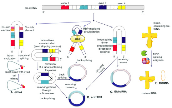
Show Figures

Figure 1

14 pages, 1567 KiB  
Article
A Circulating miRNA Signature for Stratification of Breast Lesions among Women with Abnormal Screening Mammograms
by Sau Yeen Loke, Prabhakaran Munusamy, Geok Ling Koh, Claire Hian Tzer Chan, Preetha Madhukumar, Jee Liang Thung, Kiat Tee Benita Tan, Kong Wee Ong, Wei Sean Yong, Yirong Sim, Chung Lie Oey, Sue Zann Lim, Mun Yew Patrick Chan, Teng Swan Juliana Ho, Boon Kheng James Khoo, Su Lin Jill Wong, Choon Hua Thng, Bee Kiang Chong, Ern Yu Tan, Veronique Kiak-Mien Tan and Ann Siew Gek Leeadd Show full author list remove Hide full author list
Cancers 2019, 11(12), 1872; https://doi.org/10.3390/cancers11121872 - 26 Nov 2019
Cited by 13 | Viewed by 4619
Abstract
Although mammography is the gold standard for breast cancer screening, the high rates of false-positive mammograms remain a concern. Thus, there is an unmet clinical need for a non-invasive and reliable test to differentiate between malignant and benign breast lesions in order to [...] Read more.
Although mammography is the gold standard for breast cancer screening, the high rates of false-positive mammograms remain a concern. Thus, there is an unmet clinical need for a non-invasive and reliable test to differentiate between malignant and benign breast lesions in order to avoid subjecting patients with abnormal mammograms to unnecessary follow-up diagnostic procedures. Serum samples from 116 malignant breast lesions and 64 benign breast lesions were comprehensively profiled for 2,083 microRNAs (miRNAs) using next-generation sequencing. Of the 180 samples profiled, three outliers were removed based on the principal component analysis (PCA), and the remaining samples were divided into training (n = 125) and test (n = 52) sets at a 70:30 ratio for further analysis. In the training set, significantly differentially expressed miRNAs (adjusted p < 0.01) were identified after correcting for multiple testing using a false discovery rate. Subsequently, a predictive classification model using an eight-miRNA signature and a Bayesian logistic regression algorithm was developed. Based on the receiver operating characteristic (ROC) curve analysis in the test set, the model could achieve an area under the curve (AUC) of 0.9542. Together, this study demonstrates the potential use of circulating miRNAs as an adjunct test to stratify breast lesions in patients with abnormal screening mammograms. Full article
Show Figures

Figure 1

18 pages, 5914 KiB  
Article
ALDH1A2 Is a Candidate Tumor Suppressor Gene in Ovarian Cancer
by Jung-A Choi, Hyunja Kwon, Hanbyoul Cho, Joon-Yong Chung, Stephen M. Hewitt and Jae-Hoon Kim
Cancers 2019, 11(10), 1553; https://doi.org/10.3390/cancers11101553 - 14 Oct 2019
Cited by 20 | Viewed by 4410
Abstract
Aldehyde dehydrogenase 1 family member A2 (ALDH1A2) is a rate-limiting enzyme involved in cellular retinoic acid synthesis. However, its functional role in ovarian cancer remains elusive. Here, we found that ALDH1A2 was the most prominently downregulated gene among ALDH family members in ovarian [...] Read more.
Aldehyde dehydrogenase 1 family member A2 (ALDH1A2) is a rate-limiting enzyme involved in cellular retinoic acid synthesis. However, its functional role in ovarian cancer remains elusive. Here, we found that ALDH1A2 was the most prominently downregulated gene among ALDH family members in ovarian cancer cells, according to complementary DNA microarray data. Low ALDH1A2 expression was associated with unfavorable prognosis and shorter disease-free and overall survival for ovarian cancer patients. Notably, hypermethylation of ALDH1A2 was significantly higher in ovarian cancer cell lines when compared to that in immortalized human ovarian surface epithelial cell lines. ALDH1A2 expression was restored in various ovarian cancer cell lines after treatment with the DNA methylation inhibitor 5-aza-2′-deoxycytidine. Furthermore, silencing DNA methyltransferase 1 (DNMT1) or 3B (DNMT3B) restored ALDH1A2 expression in ovarian cancer cell lines. Functional studies revealed that forced ALDH1A2 expression significantly impaired the proliferation of ovarian cancer cells and their invasive activity. To the best of our knowledge, this is the first study to show that ALDH1A2 expression is regulated by the epigenetic regulation of DNMTs, and subsequently that it might act as a tumor suppressor in ovarian cancer, further suggesting that enhancing ALDH1A2-linked signaling might provide new opportunities for therapeutic intervention in ovarian cancer. Full article
Show Figures

Figure 1

14 pages, 593 KiB  
Review
Plasma Biomarkers and Immune Checkpoint Inhibitors in Non-Small Cell Lung Cancer: New Tools for Better Patient Selection?
by Adrien Costantini, Paul Takam Kamga, Coraline Dumenil, Thierry Chinet, Jean-François Emile and Etienne Giroux Leprieur
Cancers 2019, 11(9), 1269; https://doi.org/10.3390/cancers11091269 - 29 Aug 2019
Cited by 23 | Viewed by 5418
Abstract
Immune checkpoint inhibitors (ICIs) have transformed the treatment landscape for patients with non-small cell lung cancer (NSCLC). Although some patients can experience important response rates and improved survival, many others do not benefit from ICIs developing hyper-progressive disease or immune-related adverse events. This [...] Read more.
Immune checkpoint inhibitors (ICIs) have transformed the treatment landscape for patients with non-small cell lung cancer (NSCLC). Although some patients can experience important response rates and improved survival, many others do not benefit from ICIs developing hyper-progressive disease or immune-related adverse events. This underlines the need to select biomarkers for ICIs use in order to better select patients. There is currently no universally validated robust biomarker for daily use of ICIs. Programmed death-ligand 1 (PD-L1) or tumor mutational burden (TMB) are sometimes used but still have several limitations. Plasma biomarkers are a promising approach in ICI treatment. This review will describe the development of novel plasma biomarkers such as soluble proteins, circulating tumor DNA (ctDNA), blood TMB, and blood microbiome in NSCLC patients treated with ICIs and their potential use in predicting response and toxicity. Full article
Show Figures

Figure 1

14 pages, 1307 KiB  
Article
Circulating Tumor Cell Enumeration and Characterization in Metastatic Castration-Resistant Prostate Cancer Patients Treated with Cabazitaxel
by Ingeborg E. de Kruijff, Anieta M. Sieuwerts, Wendy Onstenk, Jaco Kraan, Marcel Smid, Mai N. Van, Michelle van der Vlugt-Daane, Esther Oomen-de Hoop, Ron H.J. Mathijssen, Martijn P. Lolkema, Ronald de Wit, Paul Hamberg, Hielke J. Meulenbeld, Aart Beeker, Geert-Jan Creemers, John W.M. Martens and Stefan Sleijfer
Cancers 2019, 11(8), 1212; https://doi.org/10.3390/cancers11081212 - 20 Aug 2019
Cited by 21 | Viewed by 4085
Abstract
(1) Background: Markers identifying which patients with metastatic, castration-resistant prostate cancer (mCRPC) will benefit from cabazitaxel therapy are currently lacking. Therefore, the aim of this study was to identify markers associated with outcome to cabazitaxel therapy based on counts and gene expression profiles [...] Read more.
(1) Background: Markers identifying which patients with metastatic, castration-resistant prostate cancer (mCRPC) will benefit from cabazitaxel therapy are currently lacking. Therefore, the aim of this study was to identify markers associated with outcome to cabazitaxel therapy based on counts and gene expression profiles of circulating tumor cells (CTCs). (2) Methods: From 120 mCRPC patients, CellSearch enriched CTCs were obtained at baseline and after 6 weeks of cabazitaxel therapy. Furthermore, 91 genes associated with prostate cancer were measured in mRNA of these CTCs. (3) Results: In 114 mCRPC patients with an evaluable CTC count, the CTC count was independently associated with poor progression-free survival (PFS) and overall survival (OS) in multivariable analysis with other commonly used variables associated with outcome in mCRPC (age, prostate specific antigen (PSA), alkaline phosphatase, lactate dehydrogenase (LDH), albumin, hemoglobin), together with alkaline phosphatase and hemoglobin. A five-gene expression profile was generated to predict for outcome to cabazitaxel therapy. However, even though this signature was associated with OS in univariate analysis, this was not the case in the multivariate analysis for OS nor for PFS. (4) Conclusion: The established five-gene expression profile in CTCs was not independently associated with PFS nor OS. However, along with alkaline phosphatase and hemoglobin, CTC-count is independently associated with PFS and OS in mCRPC patients who are treated with cabazitaxel. Full article
Show Figures

Figure 1

17 pages, 2861 KiB  
Article
Kinome-Wide Screening with Small Interfering RNA Identified Polo-like Kinase 1 as a Key Regulator of Proliferation in Oral Cancer Cells
by Yih-Gang Goan, Pei-Feng Liu, Hsueh-Wei Chang, Hung-Chih Chen, Wen-Chi Chen, Shyh-Ming Kuo, Cheng-Hsin Lee and Chih-Wen Shu
Cancers 2019, 11(8), 1117; https://doi.org/10.3390/cancers11081117 - 5 Aug 2019
Cited by 15 | Viewed by 3648
Abstract
Oral squamous cell carcinoma (OSCC) is one of the major leading causes of cancer-related death worldwide, with limited effective markers for diagnosis and therapy, which has caused a low overall survival rate in the past decades. Kinases play important roles in tumor development [...] Read more.
Oral squamous cell carcinoma (OSCC) is one of the major leading causes of cancer-related death worldwide, with limited effective markers for diagnosis and therapy, which has caused a low overall survival rate in the past decades. Kinases play important roles in tumor development and malignancy in various types of cancer. However, little is known about the role of kinases in OSCC cells. In this study, an arrayed kinome small interfering RNA (siRNA) library was used to screen oral cancer cell lines and counter assayed with normal fibroblast cells to identify the genes required for cancer cell proliferation. We found that polo-like kinase 1 (PLK1) was one of the most potent genes required for OSCC cell proliferation. The knockdown of PLK1 with a siRNA or antisense oligonucleotide (ASO) consistently diminished cyclin-B1 (CCNB1) expression/phosphorylation and the G2-M phase transition. Similar effects were observed in cells treated with the PLK1 kinase inhibitor BI6727. Besides, The Cancer Genome Atlas (TCGA) analysis revealed that PLK1 was elevated in tumor tissues and associated with short survival in patients with OSCC. We also found that PLK1 expression was highly correlated with the expression of its downstream effector, CCNB1, in patients with OSCC. Coexpression of the two genes resulted in a poor prognosis of OSCC patients, particularly those in the advanced stages of OSCC. Taken together, our results suggest that PLK1 might be a diagnostic or therapeutic marker for OSCC. Full article
Show Figures

Figure 1

9 pages, 1361 KiB  
Brief Report
Prognostic Role of Androgen Receptor in Triple Negative Breast Cancer: A Multi-Institutional Study
by Shristi Bhattarai, Sergey Klimov, Karuna Mittal, Uma Krishnamurti, Xiaoxian (Bill) Li, Gabriela Oprea-Ilies, Ceyda Sonmez Wetherilt, Ansa Riaz, Mohammed A. Aleskandarany, Andrew R. Green, Ian O. Ellis, Guilherme Cantuaria, Meenakshi Gupta, Upender Manne, Johnson Agboola, Brett Baskovich, Emiel A. M. Janssen, Grace Callagy, Elaine M. Walsh, Anurag Mehta, Atika Dogra, Tanuja Shet, Pooja Gajaria, Tiffany Traina, Haruna A. Nggada, Abidemi Omonisi, Saad A. Ahmed, Emad A. Rakha, Padmashree Rida and Ritu Anejaadd Show full author list remove Hide full author list
Cancers 2019, 11(7), 995; https://doi.org/10.3390/cancers11070995 - 17 Jul 2019
Cited by 54 | Viewed by 7103
Abstract
Background: The androgen receptor (AR) has emerged as a potential therapeutic target for AR-positive triple-negative breast cancer (TNBC). However, conflicting reports regarding AR’s prognostic role in TNBC are putting its usefulness in question. Some studies conclude that AR positivity indicates a good prognosis [...] Read more.
Background: The androgen receptor (AR) has emerged as a potential therapeutic target for AR-positive triple-negative breast cancer (TNBC). However, conflicting reports regarding AR’s prognostic role in TNBC are putting its usefulness in question. Some studies conclude that AR positivity indicates a good prognosis in TNBC, whereas others suggest the opposite, and some show that AR status has no significant bearing on the patients’ prognosis. Methods: We evaluated the prognostic value of AR in resected primary tumors from TNBC patients from six international cohorts {US (n = 420), UK (n = 239), Norway (n = 104), Ireland (n = 222), Nigeria (n = 180), and India (n = 242); total n = 1407}. All TNBC samples were stained with the same anti-AR antibody using the same immunohistochemistry protocol, and samples with ≥1% of AR-positive nuclei were deemed AR-positive TNBCs. Results: AR status shows population-specific patterns of association with patients’ overall survival after controlling for age, grade, population, and chemotherapy. We found AR-positive status to be a marker of good prognosis in US and Nigerian cohorts, a marker of poor prognosis in Norway, Ireland and Indian cohorts, and neutral in UK cohort. Conclusion: AR status, on its own, is not a reliable prognostic marker. More research to investigate molecular subtype composition among the different cohorts is warranted. Full article
Show Figures

Figure 1

13 pages, 2134 KiB  
Article
Role of Bruton’s Tyrosine Kinase in Stage III Colorectal Cancer
by Debora Basile, Lorenzo Gerratana, Angela Buonadonna, Silvio Ken Garattini, Tiziana Perin, Emanuela Grassilli, Gianmaria Miolo, Maria Grazia Cerrito, Claudio Belluco, Giulio Bertola, Antonino De Paoli, Renato Cannizzaro, Marialuisa Lavitrano, Fabio Puglisi and Vincenzo Canzonieri
Cancers 2019, 11(6), 880; https://doi.org/10.3390/cancers11060880 - 24 Jun 2019
Cited by 10 | Viewed by 3495
Abstract
Background: Bruton’s tyrosine kinase (BTK) is involved in the immune response and its deficiency impairs B cell maturation. We evaluated the expression of a novel BTK isoform, p65BTK, in colorectal cancer (CRC), to identify its impact on survival. Materials and Methods: This retrospective [...] Read more.
Background: Bruton’s tyrosine kinase (BTK) is involved in the immune response and its deficiency impairs B cell maturation. We evaluated the expression of a novel BTK isoform, p65BTK, in colorectal cancer (CRC), to identify its impact on survival. Materials and Methods: This retrospective study evaluated 87 consecutive stage III CRC patients treated at the National Cancer Institute of Aviano (1999–2017). Multiple specimens were collected and analyzed for staining intensity and percentage of tumor cells positive for p65BTK. Prognostic impact was tested by univariate Cox regression analysis. Results: After a median follow-up of 82.59 months, median disease-free survival (DFS) and overall survival (OS) were 11.67 months and 31.33 months, respectively. Interestingly, 10% of patients did not express p65BTK. For the immunohistochemistry IHC intensity 1, the best cutoff point was 1% of p65BTK positivity; for IHC intensity 2, it was 50%; and for IHC intensity 3, it was 80%. Through univariate analysis, patients with highly expressed p65BTK (IHC intensity 3 and ≥80%) were shown to have the worst prognosis in terms of DFS (HR: 6.23; p = 0.005; 95% C.I. 1.75–22.79) and OS (HR: 2.54; p = 0.025; 95% C.I. 1.12–5.76). Conclusions: p65BTK is frequently expressed in CRC and, if highly expressed, is an unfavourable prognostic factor. However, further confirmation is needed and its potential targeting needs to be studied. Full article
Show Figures

Figure 1

16 pages, 4690 KiB  
Article
CHI3L1, NTRK2, 1p/19q and IDH Status Predicts Prognosis in Glioma
by Elise Deluche, Barbara Bessette, Stephanie Durand, François Caire, Valérie Rigau, Sandrine Robert, Alain Chaunavel, Lionel Forestier, François Labrousse, Marie-Odile Jauberteau, Karine Durand and Fabrice Lalloué
Cancers 2019, 11(4), 544; https://doi.org/10.3390/cancers11040544 - 15 Apr 2019
Cited by 18 | Viewed by 5127
Abstract
The aim of this study was to identify relevant biomarkers for the prognosis of glioma considering current molecular changes such as IDH mutation and 1p19q deletion. Gene expression profiling was performed using the TaqMan Low Density Array and hierarchical clustering using 96 selected [...] Read more.
The aim of this study was to identify relevant biomarkers for the prognosis of glioma considering current molecular changes such as IDH mutation and 1p19q deletion. Gene expression profiling was performed using the TaqMan Low Density Array and hierarchical clustering using 96 selected genes in 64 patients with newly diagnosed glioma. The expression dataset was validated on a large independent cohort from The Cancer Genome Atlas (TCGA) database. A differential expression panel of 26 genes discriminated two prognostic groups regardless of grade and molecular groups of tumors: Patients having a poor prognosis with a median overall survival (OS) of 23.0 ± 9.6 months (group A) and patients having a good prognosis with a median OS of 115.0 ± 6.6 months (group B) (p = 0.007). Hierarchical clustering of the glioma TCGA cohort supported the prognostic value of these 26 genes (p < 0.0001). Among these genes, CHI3L1 and NTRK2 were identified as factors that can be associated with IDH status and 1p/19q co-deletion to distinguish between prognostic groups of glioma from the TCGA cohort. Therefore, CHI3L1 associated with NTRK2 seemed to be able to provide new information on glioma prognosis. Full article
Show Figures

Figure 1

18 pages, 5024 KiB  
Article
Automated Quantification of Extranuclear ERα Using Phosphor-Integrated Dots for Predicting Endocrine Therapy Resistance in HR+/HER2 Breast Cancer
by Zhaorong Guo, Hiroshi Tada, Narufumi Kitamura, Yoh Hamada, Minoru Miyashita, Narumi Harada-Shoji, Akiko Sato, Yohei Hamanaka, Kouki Tsuboi, Nobuhisa Harada, Mayumi Takano-Kasuya, Hisatake Okada, Yasushi Nakano, Noriaki Ohuchi, Shin-ichi Hayashi, Takanori Ishida and Kohsuke Gonda
Cancers 2019, 11(4), 526; https://doi.org/10.3390/cancers11040526 - 12 Apr 2019
Cited by 7 | Viewed by 3716
Abstract
In addition to genomic signaling, Estrogen receptor alpha (ERα) is associated with cell proliferation and survival through extranuclear signaling contributing to endocrine therapy (ET) resistance. However, the relationship between extranuclear ERα and ET resistance has not been extensively studied. We sought to measure [...] Read more.
In addition to genomic signaling, Estrogen receptor alpha (ERα) is associated with cell proliferation and survival through extranuclear signaling contributing to endocrine therapy (ET) resistance. However, the relationship between extranuclear ERα and ET resistance has not been extensively studied. We sought to measure extranuclear ERα expression by immunohistochemistry using phosphor-integrated dots (IHC-PIDs) and to assess its predictive value for ET resistance. After quantitative detection of ERα by IHC-PIDs in vitro, we developed “the nearest-neighbor method” to calculate the extranuclear ERα. Furthermore, tissue sections from 65 patients with HR+/HER2- BC were examined by IHC-PIDs, and the total ERα, nuclear ERα, extranuclear ERα PIDs score, and ratio of extranuclear-to-nuclear ERα (ENR) were measured using the novel method. We demonstrate that quantification of ERα using IHC-PIDs exhibited strong correlations to real-time qRT-PCR (r2 = 0.94) and flow cytometry (r2 = 0.98). High ERα ENR was significantly associated with poor overall survival (p = 0.048) and disease-free survival (DFS) (p = 0.007). Multivariate analysis revealed that the ERα ENR was an independent prognostic factor for DFS [hazard ratio, 3.8; 95% CI, 1.4–11.8; p = 0.006]. Our automated measurement has high accuracy to localize and assess extranuclear ERα. A high ERα ENR in HR+/HER2 BC indicates decreased likelihood of benefiting from ET. Full article
Show Figures

Figure 1

13 pages, 245 KiB  
Review
Diagnostic and Prognostic Potential of AKR1B10 in Human Hepatocellular Carcinoma
by Johanna K. DiStefano and Bethany Davis
Cancers 2019, 11(4), 486; https://doi.org/10.3390/cancers11040486 - 5 Apr 2019
Cited by 75 | Viewed by 6718
Abstract
Hepatocellular carcinoma (HCC) is a leading cause of cancer-related death worldwide. Although diagnostic measures and surgical interventions have improved in recent years, the five-year survival rate for patients with advanced HCC remains bleak—a reality that is largely attributable to an absence of early [...] Read more.
Hepatocellular carcinoma (HCC) is a leading cause of cancer-related death worldwide. Although diagnostic measures and surgical interventions have improved in recent years, the five-year survival rate for patients with advanced HCC remains bleak—a reality that is largely attributable to an absence of early stage symptoms, lack of adequate diagnostic and prognostic biomarkers, and the common occurrence of acquired resistance to chemotherapeutic agents during HCC treatment. A limited understanding of the molecular mechanisms underlying HCC pathogenesis also presents a challenge for the development of specific and efficacious pharmacological strategies to treat, halt, or prevent progression to advanced stages. Over the past decade, aldo-keto reductase family 1 member 10 (AKR1B10) has emerged as a potential biomarker for the diagnosis and prognosis of HCC, and experimental studies have demonstrated roles for this enzyme in biological pathways underlying the development and progression of HCC and acquired resistance to chemotherapeutic agents used in the treatment of HCC. Here we provide an overview of studies supporting the diagnostic and prognostic utility of AKR1B10, summarize the experimental evidence linking AKR1B10 with HCC and the induction of chemoresistance, and discuss the clinical value of AKR1B10 as a potential target for HCC-directed drug development. We conclude that AKR1B10-based therapies in the clinical management of specific HCC subtypes warrant further investigation. Full article
15 pages, 2352 KiB  
Article
A Simple and Highly Specific MassARRAY-Based Stool DNA Assay to Prioritize Follow-up Decisions in Fecal Immunochemical Test-Positive Individuals
by Pi-Yueh Chang, Chia-Chun Chen, Jy-Ming Chiang, Shih-Cheng Chang, Mei-Chia Wang, Jinn-Shiun Chen, Wen-Sy Tsai, Jeng Fu You and Jang-Jih Lu
Cancers 2019, 11(3), 423; https://doi.org/10.3390/cancers11030423 - 25 Mar 2019
Cited by 4 | Viewed by 4339
Abstract
Background: Seventy-five percent of fecal immunochemical test (FIT)-positive individuals are false positives and undergo unnecessary colonoscopies. Here, we established a stool DNA (sDNA) test that uses the Single Allele Base Extension Reaction (SABER) MassARRAY platform to improve the accuracy of FIT-based CRC detection. [...] Read more.
Background: Seventy-five percent of fecal immunochemical test (FIT)-positive individuals are false positives and undergo unnecessary colonoscopies. Here, we established a stool DNA (sDNA) test that uses the Single Allele Base Extension Reaction (SABER) MassARRAY platform to improve the accuracy of FIT-based CRC detection. Methods: Twenty-one variants in five CRC-associated genes were selected for the sDNA panel. Cell line DNA and matched mutation-confirmed tissue and stool samples from 34 patients were used for accuracy assessment (cohort 1). The clinical performance of the sDNA assay was further evaluated in 101 independent FIT-positive stool samples (cohort 2). Results: In cohort 1, we obtained a 62% mutation concordance rate in paired tissue and stool samples of the CRC group, regardless of the FIT status. In cohort 2, 100% specificity in normal controls with positive FIT results was observed. By weighting the FIT value and the presence of a given variant type in stool and then summing the two scores, we found that a one-increment increase in the score was associated with a 4.538-fold risk (95% CI = 2.121–9.309) for malignancy in the FIT-positive setting. Conclusions: Our highly specific sDNA assay can help prioritize the most at-risk FIT-positive persons to receive prompt colonoscopic confirmation of CRC. Full article
Show Figures

Figure 1

21 pages, 1019 KiB  
Review
Circulating MicroRNA Biomarkers for Lung Cancer Detection in East Asian Populations
by Haixin Yu, Zhong Guan, Katarina Cuk, Yan Zhang and Hermann Brenner
Cancers 2019, 11(3), 415; https://doi.org/10.3390/cancers11030415 - 23 Mar 2019
Cited by 33 | Viewed by 4953
Abstract
Background: Lung cancer (LC) is the leading cause of cancer-related death in Eastern Asia. The prognosis of LC highly depends on tumor stages and early detection could substantially reduce LC mortality. Accumulating evidence suggested that circulating miRNAs in plasma or serum may have [...] Read more.
Background: Lung cancer (LC) is the leading cause of cancer-related death in Eastern Asia. The prognosis of LC highly depends on tumor stages and early detection could substantially reduce LC mortality. Accumulating evidence suggested that circulating miRNAs in plasma or serum may have applications in early LC detection. We thus conducted a systematic literature review on the diagnostic value of miRNAs markers for LC in East Asian populations. Methods: PubMed and ISI Web of Knowledge were searched to retrieve relevant articles published up to 17 September 2018. Information on study design, population characteristics, investigated miRNAs and diagnostic accuracy (including sensitivity, specificity and area under the curve (AUC)) were independently extracted by two reviewers. Results: Overall, 46 studies that evaluated a total of 88 miRNA markers for LC diagnosis in East Asian populations were identified. Sixteen of the 46 studies have incorporated individual miRNA markers as panels (with 2–20 markers). Three promising miRNA panels with ≥90% sensitivity and ≥90% specificity were discovered, two of which were externally validated. Diagnostic performance of circulating miRNAs in East Asian populations was comparable to previously summarized performance in Western populations. Forty-four miRNAs were reported in both populations. No major differences in diagnostic performance by ethnicity of the same miRNA was observed. Conclusions: Circulating miRNAs or miRNA panels, possibly in combination with other promising molecular markers including epigenetic and genetic markers, may be promising candidates for noninvasive LC early detection. However, large studies with samples collected prospectively in true screening settings are required to validate the promising markers or marker panels. Full article
Show Figures

Figure 1

11 pages, 1145 KiB  
Brief Report
Clinical Characteristics of Osimertinib Responder in Non-Small Cell Lung Cancer Patients with EGFR-T790M Mutation
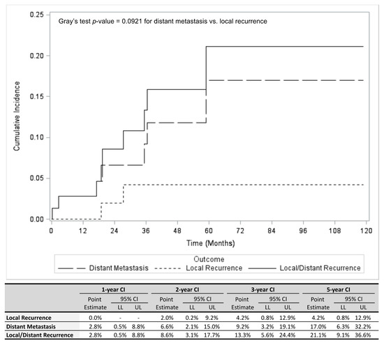
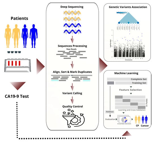
by Akihiro Yoshimura, Tadaaki Yamada, Naoko Okura, Takayuki Takeda, Kazuki Hirose, Yutaka Kubota, Shinsuke Shiotsu, Osamu Hiranuma, Yusuke Chihara, Nobuyo Tamiya, Yoshiko Kaneko, Junji Uchino and Koichi Takayama
Cancers 2019, 11(3), 365; https://doi.org/10.3390/cancers11030365 - 15 Mar 2019
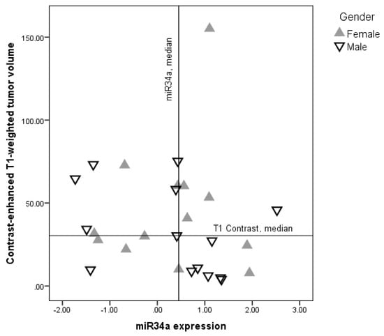
Cited by 9 | Viewed by 4127
Abstract
Osimertinib is a mutant-selective EGFR inhibitor that is effective against non-small cell lung cancer (NSCLC) in patients with the EGFR-T790M mutation, who are resistant to EGFR-tyrosine kinase inhibitors (EGFR-TKIs). However, the factors affecting response to osimertinib treatment are unknown. In this retrospective [...] Read more.
Osimertinib is a mutant-selective EGFR inhibitor that is effective against non-small cell lung cancer (NSCLC) in patients with the EGFR-T790M mutation, who are resistant to EGFR-tyrosine kinase inhibitors (EGFR-TKIs). However, the factors affecting response to osimertinib treatment are unknown. In this retrospective study, 27 NSCLC patients with the EGFR-T790M mutation were enrolled at five institutions in Japan. Among several parameters tested, the progression-free survival (PFS) associated with the initial EGFR-TKIs was positively correlated with the PFS after osimertinib treatment (p = 0.021). The median PFS following osimertinib treatment and the overall survival (OS) were longer in patients who responded to osimertinib than in those who did not (17.7 months versus 3.5 months, p = 0.009 and 24.2 months versus 13.5 months, p = 0.021, respectively). A multivariate analysis demonstrated that the PFS with initial EGFR-TKIs was significantly related to the PFS with osimertinib treatment (p = 0.035), whereas osimertinib response was significantly related to the PFS and OS with osimertinib treatment (p = 0.016 and p = 0.006, respectively). Our retrospective observations indicate that PFS following the initial EGFR-TKI treatment and the response rate to osimertinib might be promising predictors for effective osimertinib treatment in NSCLC patients with the EGFR-T790M mutation. Full article
Show Figures

Figure 1

11 pages, 614 KiB  
Article
Association of miR-34a Expression with Quality of Life of Glioblastoma Patients: A Prospective Study
by Paulina Vaitkiene, Aiste Pranckeviciene, Rytis Stakaitis, Giedrius Steponaitis, Arimantas Tamasauskas and Adomas Bunevicius
Cancers 2019, 11(3), 300; https://doi.org/10.3390/cancers11030300 - 4 Mar 2019
Cited by 11 | Viewed by 3156
Abstract
MiR-34a acts as tumor-suppressor by targeting many oncogenes related to proliferation, apoptosis, and invasion of gliomas. We studied the relationships between health-related quality of life (HRQOL), depression, and miR-34a expression status in patients with newly diagnosed glioblastoma (GBM). A comprehensive HRQOL assessment was [...] Read more.
MiR-34a acts as tumor-suppressor by targeting many oncogenes related to proliferation, apoptosis, and invasion of gliomas. We studied the relationships between health-related quality of life (HRQOL), depression, and miR-34a expression status in patients with newly diagnosed glioblastoma (GBM). A comprehensive HRQOL assessment was completed by 38 patients with glioblastoma prior to surgical resection and included the European Organization for Research and Treatment of Cancer (EORTC) questionnaire for cancer patients (QLQ-C30) and the Brain Cancer-Specific Quality of Life Questionnaire (QLQ-BN20), the Patient Health Questionnaire-9 (PHQ-9), the Karnofsky performance index (KPS), and The Glasgow Outcome Scale (GOS). The miR-34a expression in glioblastoma tissue was measured using quantitative reverse transcription PCR. Our findings show that lower miR-34a expression is significantly associated with higher tumor volume, worse physical functioning, lower KPS, and greater depressive symptom severity of GBM patients. Moreover, analysis reveals that miR-34a effects might be gender specific, as stronger relationships between miR-34a and patient functioning measures were observed in males when compared to females. Despite the fact that, due to small sample size, our results should be considered as preliminary, our study suggests that miR-34a is associated with tumor burden and can be important for health-related quality of life, functional status, and mood symptoms of glioblastoma patients. Full article
Show Figures

Figure 1

Back to TopTop