Article

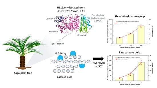
Functional Characterization of Recombinant Raw Starch Degrading α-Amylase from Roseateles terrae HL11 and Its Application on Cassava Pulp Saccharification

by
Daran Prongjit
1,
Hataikarn Lekakarn
1,*,
Benjarat Bunterngsook
2,
Katesuda Aiewviriyasakul
2,
Wipawee Sritusnee
2 and
Verawat Champreda
2
1
Department of Biotechnology, Faculty of Science and Technology, Thammasat University, Rangsit Campus, Khlong Nueng, Khlong Luang 12120, Pathum Thani, Thailand
2
Enzyme Technology Research Team, Biorefinery Technology and Bioproduct Research Group, National Center for Genetic Engineering and Biotechnology, 113 Thailand Science Park, Phahonyothin Road, Khlong Nueng, Khlong Luang 12120, Pathum Thani, Thailand
*
Author to whom correspondence should be addressed.
Catalysts 2022, 12(6), 647; https://doi.org/10.3390/catal12060647
Submission received: 13 May 2022 / Revised: 30 May 2022 / Accepted: 8 June 2022 / Published: 13 June 2022

Abstract

:
Exploring new raw starch-hydrolyzing α-amylases and understanding their biochemical characteristics are important for the utilization of starch-rich materials in bio-industry. In this work, the biochemical characteristics of a novel raw starch-degrading α-amylase (HL11 Amy) from Roseateles terrae HL11 was firstly reported. Evolutionary analysis revealed that HL11Amy was classified into glycoside hydrolase family 13 subfamily 32 (GH13_32). It contains four protein domains consisting of domain A, domain B, domain C and carbohydrate-binding module 20 (CMB20). The enzyme optimally worked at 50 °C, pH 4.0 with a specific activity of 6270 U/mg protein and 1030 raw starch-degrading (RSD) U/mg protein against soluble starch. Remarkably, HL11Amy exhibited activity toward both raw and gelatinized forms of various substrates, with the highest catalytic efficiency (kcat/Km) on starch from rice, followed by potato and cassava, respectively. HL11Amy effectively hydrolyzed cassava pulp (CP) hydrolysis, with a reducing sugar yield of 736 and 183 mg/g starch from gelatinized and raw CP, equivalent to 72% and 18% conversion based on starch content in the substrate, respectively. These demonstrated that HL11Amy represents a promising raw starch-degrading enzyme with potential applications in starch modification and cassava pulp saccharification.

Graphical Abstract

1. Introduction

Cassava (Manihot esculenta) is a carbohydrate-rich crop that has been widely utilized in the 4Fs: Food, Feed, Fuel and Factories. Thailand is the world’s second largest cassava producer and was ranked as the world’s largest cassava product exporter [1,2], resulting in large amounts of wastes, especially cassava pulp (5.15–7.30 million metric tons/year) from cassava processing [3,4]. This leads to a negative impact on environmental issues, particularly air pollution caused by the fermentation of large amounts of cassava pulp, which generates an unpleasant odor.
The predominant component of cassava pulp is starch granules (approximately 50–70% of dry matter) [5,6]. The rest comprises cell wall polysaccharides containing cellulose and hemicellulose (14–23% and 21% of dry matter, respectively) [7,8]. The high residual starch content makes it a unique feedstock for biorefineries compared with other second-generation cellulosic substrates. Furthermore, unlike lignocellulosic biomass, cassava pulp does not require chemical pre-treatment, allowing the development of a cost-effective process for biomass conversion into valuable bio-based products. Therefore, an enzymatic process using starch-degrading enzymes is a crucial step in starch conversion.
Starch-degrading enzymes are enzymes that proficiently hydrolyze α-1, 4 glycosidic linkages and α-1, 6 glycosidic linkages in starch. The α-amylases (EC 3.2.1.1) are predominant enzymes in starch hydrolysis. The α-amylases are endo-acting enzymes that catalyze the random hydrolysis of α-1, 4 glycosidic linkages in starch and glycogen. In general, α-amylases are large members belonging to the glycoside hydrolase (GH) class, which is distributed in many families in the CAZy database (http://www.cazy.org/), containing GH13, GH57, GH119 and GH126. GH13 is the biggest family of amylolytic enzymes, which is further classified into 44 subfamilies, of which 16 subfamilies (GH13_1, 5, 6, 7, 15, 19, 24, 27, 28, 32, 36, 37, 39, 41, 42 and 43) exhibit α-amylase activity [9,10,11,12,13,14,15,16,17].
The conventional starch conversion process is an energy-intensive process that requires the disruption of crystalline starch granules through the gelatinization process under high temperature conditions. Raw starch-degrading α-amylases (RSDAs) are capable of starch granule hydrolysis below gelatinization temperature [18]. RSDAs contain a starch-binding domain (SBD) as a crucial part, which belongs to the carbohydrate-binding modules (CBMs), frequently located at the C-terminus and serving the function of attaching to starch granules. Currently, among 90 established CBM families in CAZy (http://www.cazy.org), SBDs have been reported and classified into 15 families, including CBM20, CBM21, CBM25, CBM26, CBM34, CBM41, CBM45, CBM48, CBM53, CBM58, CBM68, CBM69, CBM74, CBM82 and CBM83 [19,20]. The potential of raw starch-degrading enzymes has received great attention in the starch industry as a non-thermal process strategy to substitute the traditional heating process in liquefaction [21,22].
The α-amylases are fundamental starch-degrading enzymes found in a wide variety of microorganisms that utilize oligosaccharides or glucose as a carbon source. Soil is the source of various microorganisms that show a novel biochemical characteristic of starch-degrading activities [23]. Roseateles terrae is a Gram-negative bacteria isolated from soil [24]. In this genus, R. depolymerans is the most studied and reported for its capability to degrade different polymers and aliphatic polyester [25,26]. However, R. terrae is still less studied and investigated than R. depolymerans. Furthermore, reports on the biochemical characteristics of starch-degrading enzymes from R. terrae are limited.
Therefore, this study firstly investigates the biochemical properties of a raw starch-degrading α-amylase isolated from R. terrae HL11, as a detailed examination of α-amylase isolated from R. terrae has never been reported. The aims also included the evaluation of the HL11 amylase’s potential for application in an enzyme mixture for cassava starch and cassava pulp saccharification. R. terrae α-amylase is a potent candidate enzyme for application in the bioconversion of starch-based agricultural waste into high value-added bio-based products.

2. Results

2.1. Amylase Gene Identification from R. terrae and Sequence Analysis

The amylolytic enzyme-producing bacteria was isolated from soil attached to sago palm root (Cycas revoluta), collected from Trang Province, Thailand (GPS 7°31′21.0″ N 99°43′47.1″ E) using minimal medium agar supplemented with 1% (w/v) soluble starch. According to 16s rDNA analysis, the HL11 strain was identified as R. terrae with 98% identity. The HL11AMY gene (1875 bp) encoding for α-amylase was isolated from R. terrae HL11. The gene encodes a protein of 625 amino acids with an estimated molecular weight of approximately 65 kDa and a pI value of 6.32. HL11Amy shared a close identity with the α-amylases from various species closely related to Proteobacteria, for instance, R. terrae (WP_088453876.1; 97.28%), followed by Roseateles depolymerans (WP_058933710.1; 92.64%), Rubrivivax sp. (RZI57913.1; 88.32%), Mitsuaria noduli (OWQ45775.1; 88.80%), Pelomonas puraquae (WP_088484981.1; 87.00%) and Burkholderiales bacterium (MBI3349245.1; 85.12%). The phylogenetic analysis of HL11Amy and 268 sequences of α-amylases obtained from Janíčková and Janeček (2021) clearly demonstrated that HL11Amy was identified as a member of subfamily 32 of glycoside hydrolase family 13 (GH13_32) (Figure 1).
In-depth analysis of HL11Amy revealed that 32 amino acids apart from the N-terminus were predicted as a signal peptide (SP) for protein secretion. The conserved domain annotation using the Conserved Domains Database (CDD) revealed that HL11Amy contained three protein families, including the α-amylase catalytic domain (E-value 3.36 × 10−125), alpha amylase C domain (E-value 4.90 × 10⁻²¹) and carbohydrate-binding module 20 (CMB20) (E-value 8.11 × 10⁻²³). Furthermore, based on the predicted 3D structure analysis, the α-amylase catalytic domain in HL11Amy was predicted as domains A and B (Figure 2a,b) [27,28]. Therefore, in conclusion, HL11Amy comprised four domains: (i) domain A, which is the catalytic TIM barrel structure (residues 38–120 and 189–413); (ii) domain B, protruding out of the TIM barrel (residues 121–188); (iii) domain C, with all β-fold structure (residues 414–507); (iv) CMB20 (residues 520–625). Two potential surface binding sites (SBSs) observed in CBM20 were SBS1, containing two critical tryptophan residues (Trp550 and Trp595), and SBS2, containing one tryptophan residue (Trp569) (Figure 2c). Based on sequence logo prediction from the alignment of all 140 amylase sequences classified as the GH13_32 subfamily, HL11Amy contained eight conserved sequence regions (CSRs), similar to the GH13_32 subfamily that was previously reported [14]. Although HL11Amy showed almost invariant residue within bacterial and fungal members of GH13_32 subfamilies, there were some residues that have been identically shown only in members of either bacteria or fungi. Moreover, His, Gly and Pro are located in positions 18 (CSR-I), 47 (CSR-VII) and 49 (CSR-VII), respectively. These amino acid residues, present in all 140 amylases studied here, also belong to the most highly conserved residues in both bacterial and fungal α-amylase GH13_32 subfamilies. However, these positions were replaced by a Gln, Ala and Ala in HL11Amy (Figure 3). The proposed catalytic residues were the catalytic nucleophile (position 28; Asp213 in CSR-II), the proton donor (position 37; Glu248 in CSR-III) and the transition-state stabilizer (position 46; Asp320 in CSR-IV) (Figure 3). Based on the alignment of all eight CSRs from 20 bacterial and 120 fungal α-amylase sequences, the sequence logo of HL11Amy also contains the residues involved in binding the chloride anion at positions 26 (Arg211), 44 (Asn255) and 50 (Gln324). On the other hand, the third residue involved in chloride binding presents as a lysine in a few bacteria, but it is not found in the fungal GH13_32 subfamily (Figure 3). Moreover, based on sequence alignment, HL11Amy shared many amino acid motifs with true animal α-amylases (GH13_24 subfamily) and animal-like α-amylases (GH13_32 subfamily), e.g., Drosophila melanogaster and Thermobifida fusca, respectively (Table S3 and Figure S1).

2.2. Heterologous Expression and Purification of Amylase

The full length of the HL11AMY gene was heterologously expressed in the E. coli BL21(DE3) expression system as an intracellular soluble protein with a molecular weight of approximately 65 kDa after induction with 1 mM IPTG at 25 °C for 3 h. After the purification step was carried out using an immobilized metal ion affinity chromatography HisTrap® column and ultra-certification for desalting, the purified HL11Amy exhibited the single protein band with more than 90% homogeneity (65 kDa) with typical yields of approximately 127 mg/L culture broth (Figure 4a,b).

2.3. Biochemical Characterization

The enzyme activity of HL11Amy was measured against soluble starch to examine the optimum condition of the enzyme. The effect of temperature was evaluated at temperatures ranging from 30–80 °C. Under the defined condition, HL11Amy showed the highest activity at 50 °C and retained relative activity of more than 80% and 60% at 40 °C and 30 °C, respectively (Figure 5a). However, the enzyme activity dramatically decreased at temperatures ranging 60–80 °C, with residual activity less than 20%. The effect of pH on α-amylase activity was investigated under different pH ranging from 3.0–9.0. HL11Amy presented the highest α-amylase activity in 50 mM sodium citrate buffer pH 4.0 (Figure 5b). The enzyme activity retained more than 50% of its maximum activity in pH 5.0–6.0, while less than 20% remaining activity was observed in pH 7.0–9.0. Therefore, HL11Amy provided the specific activity of 6270 ± 70 U/mg protein against soluble starch as a substrate under the optimum condition (pH 4.0, 50 °C).
HL11Amy did not only digest gelatinized starch but also presented raw starch digestion activity. The specificity of HL11Amy against different kinds of starch in gelatinized and raw forms was investigated. The specificity of HL11Amy on various kinds of gelatinized starch showed the highest specific activity on potato starch (10,570 ± 290 U/mg protein) and amylopectin (10,530 ± 250 U/mg protein), followed by rice starch, cassava starch, amylose, soluble starch, cassava pulp and pullulan (Table 1). For raw substrate digestion, HL11Amy distinguishably showed higher activity toward amylose than amylopectin from potato, with 5470 ± 20 and 4770 ± 60 U/mg protein, respectively. The highest specific activity was observed on raw rice starch (1970 ± 9 U/mg protein), followed by raw soluble starch, raw potato starch and raw cassava starch.
In order to gain in-depth insight into the amylolytic performance and substrate specificity relevant to HL11Amy, the kinetics parameters were thus studied. Among gelatinized substrates, the enzyme presented the highest Vmax and kcat values against soluble starch, with 19,800 ± 1650 U/mg protein and 25,720 1/s, respectively (Table 2). These values were slightly higher than the kinetics parameters on rice starch (Vmax 16,620 ± 550 and kcat 21,590 1/s), whereas the kinetic parameters on potato starch and cassava starch were in the same range. On the other hand, in the case of raw starch, raw rice starch provided the highest kinetic values (kcat 6340 1/s), followed by raw potato starch and raw cassava starch. The data agreed well with the substrate specificity shown in Table 1.

2.4. Cleavage Pattern on Maltooligosaccharides

In this study, the specific cleavage pattern of recombinant HL11Amy on maltooligosaccharides with different degrees of polymerization ranging from two to six was investigated. Based on the product profile analyzed using TLC and HPLC, HL11Amy was able to cleave only maltooligosaccharides with chain lengths of four to six (Figure 6a,b). However, the cleavage on maltose (M2) and maltotriose (M3) was not observed under the experimental conditions. The results demonstrated that maltotetraose (M4) represented the smallest substrate for HL11Amy by releasing maltose (M2) as a final product. In addition, maltose (M2), maltotriose (M3) and maltotetraose (M4) were detected as final products from the hydrolysis of maltopentaose (M5) and maltohexaose (M6).

2.5. Hydrolysis of Cassava Pulp

The efficiency of HL11Amy in cassava pulp (CP) hydrolysis was then evaluated. The highest reducing sugar yield of 736 ± 77 mg/g starch in CP (equivalent to 72% ± 7.5 conversion) was obtained after incubation of gelatinized cassava pulp with 0.18 μg protein/g CP of HL11Amy at the optimal condition (50 °C, pH 4.0) for 24 h (Figure 7a). The reaction mixture containing 0.09 μg protein/g CP of HL11Amy generated 412 ± 11 mg of reducing sugars/g starch in CP (40% ± 1.1 conversion). Furthermore, the ability of the enzyme to digest raw cassava pulp was thus investigated to describe the distinguishing feature of HL11Amy in raw starch digestion. The measured reducing sugar product was 183 ± 23 mg/g starch in CP, which represented 18% ± 2.2 conversion yield at 8 μg protein/g CP (Figure 7b). These demonstrated the high efficacy of HL11Amy in gelatinized and raw cassava pulp hydrolysis, which could be applied in starch biorefinery processes.

3. Discussion

The full length of the HL11AMY gene was successfully isolated from R. terrae HL11, an amylolytic enzyme-producing bacteria isolated from soil associated with sago stem. The sequence and biochemical characteristics of α-amylase from R. terrae were firstly described in this work. HL11Amy exhibited the highest identity proximity to α-amylase from R. terrae (97.28%) and was classified into subfamily 32 of glycoside hydrolase family 13 (GH13_32), in the same clade as GH13_32 from bacterial origin. The modular structure of HL11Amy consisted of four domains, including (i) domain A, (ii) domain B, (iii) domain C and (iv) CMB20. HL11Amy comprised eight CSRs, including seven CSRs (I-VII) that are unique features of GH13 and CSR VIII (FEW), similar to the GH13_32 subfamily that was previously reported [14,31]. The proposed catalytic residues of HL11Amy (Asp213/Glu248/Asp320) are conserved invariantly in all bacterial and fungal α-amylases in members of the GH13_32 subfamily as the catalytic nucleophile (Asp; β4), proton donor (Glu; β5) and transition-state stabilizer (Asp; β7), respectively [14]. Even though chloride-activated amylases are specific to all animals, some bacterial amylases in the subfamily GH13_32, such as Pseudoalteromonas haloplanktis, have already been confirmed to have a dependence on chloride anion [32,33]. Based on the sequence logo, there are only five bacterial GH13_32 subfamilies, Halomonas meridiana, Pseudomonas sp., T. fusca, Thermomonospora_curvata and Pseudoalteromonas_haloplanktis, that contain three residues involved in chloride binding: Arg (strand β4), Asn (strand β7) and Lys (strand β8), located in the CSR-II, CSR-IV and CSR-VII, respectively [14,34,35,36,37,38]. In HL11Amy, this lysine is replaced by a glutamine corresponding to the residue organization found in chloride-independent bacterial α-amylases, such as those from Bacillus amyloliquefaciens and B. licheniformis [39]. This kind of replacement produces active, chloride-independent α-amylases [32,33]. Regarding sequence similarity and phylogenetic distribution, interestingly, Gram-negative bacteria R. terrae HL11Amy, containing several typical animal motifs, is proposed as a new animal-like α-amylase with a starch-binding domain classified as CBM20, similar to Gram-positive bacteria T. fusca, Gram-negative bacteria Microbulbifer degradans and Actinobacteria Streptomyces limosus, probably resulting from repeated horizontal gene transfer from animals [40,41]. The alpha amylase C domain has been reported as a non-catalytic region important for the activity and starch-binding ability of several studied α-amylases [42,43]. Regarding sequence analysis, CBM20 contained two surface binding sites, including SBS1 and SBS2. The SBS1, forming from two critical tryptophan residues (Trp550 and Trp595), has been reported as an important part for raw starch-binding ability [20,44]. The two conserved tryptophan residues, for example, Trp543 and Trp590 found in the glucoamylase, form a hydrophobic region on the surface of CBM20, providing glucose ring interaction [20]. While SBS1 contains one tryptophan residue (Trp569), it may function to guide the starch chains to the active site [20]. CBM20 is a starch-binding domain (SBD) that has been reported as a key element of amylolytic enzyme-facilitated raw starch granule binding [30,45,46]. The SBDs exhibited a binding preference toward raw starch based on many previous reports. Deletion of the SBD usually results in a decrease in raw starch hydrolysis [47,48,49]. The removal of the SBD in the C-terminal region of an α-amylase from Bacillus sp. No. 195 led to a dramatic decrease in the binding and degradation of insoluble starches [49]. The adsorption evaluation of a truncated mutant (AmyPSBD) demonstrated that the truncated α-amylase without CBM69 lost its binding ability on raw starch. Furthermore, the SDB not only has influence on binding potential but also on enzymatic hydrolysis [47]. On the other hand, the addition of CBM20 in the α-amylase from Cryptococcus sp. S-2 enhanced the catalytic efficiency and thermostability of α-amylase (AmyP) toward raw rice starch [50].
HL11Amy was firstly expressed in E. coli BL21(DE3) and characterized. The recombinant HL11Amy was highly expressed as a soluble protein (127 mg/L culture medium) and showed high specificity towards gelatinized soluble starch (6270 ± 70 U/mg protein). In addition, HL11Amy could perform under a broad range of moderate temperatures (30–55 °C) and a wide range of pH (4.0–6.0), which could be applied to develop an enzyme mixture for starch saccharification. Considering the optimal temperature regarding raw starch digestion efficiency, the optimal working temperature of HL11Amy (50 °C), close to the pasting temperature of starch, resulted in a high raw starch digestibility due to the partial gelatinization of the starch. The degree of gelatinization is the main determinate for the binding affinity and catalytic efficiency of α-amylase, and in turn, the digestion rate [51].
The cleavage pattern of enzymes is the specific ability of each enzyme that provides specific products. According to the cleavage pattern, the recombinant HL11Amy acted on the tetramer (M4) to hexamer (M6) of maltooligosaccharides and released maltose (M2), maltotriose (M3) and maltotetraose (M4) as the products. However, M2 and M3 were not hydrolyzed by HL11Amy, suggesting that HL11Amy acted on oligosaccharides containing at least four glucose units. Compared with α-amylases from published data, its cleavage patterns were similar to the raw starch-degrading α-amylase from Gram-negative bacteria Corallococcus sp. EGB, as well as the Indonesian marine bacterium Bacillus sp. ALSHL3 [52].
Based on its biochemical characteristics, HL11Amy showed high specificity against raw starch and gelatinized starch. Intriguingly, HL11Amy showed a higher specificity constant (kcat/Km) against several substrates than several α-amylases from published data (Table S1), which demonstrates that it is a great potential starch-degrading enzyme. It is noteworthy that HL11Amy showed a higher specificity against amylopectin than amylose. This incident is unusual for α-amylases because they generally display higher activities toward amylose than amylopectin [53,54,55,56]. However, this phenomenon is similar to the α-amylase from the marine bacterium Pontibacillus sp. ZY (AmyZ1), which exhibited high activity toward raw starches. Interestingly, HL11Amy showed activity on pullulan that represents rich α-1,6 glycosidic linkages. In contrast, AmyZ1 showed negligible activity toward pullulan [57]. The raw starch-degrading α-amylase from Gram-negative bacteria Corallococcus sp. (AmyM) showed negligible activity toward pullulan [57,58]. These indicate that HL11Amy exhibited broader hydrolytic activity against branched substrates than previously reported α-amylases. According to previous reports, the SBS2 of barley α-amylase 1 (AMY1) was particularly important for the recognition and depolymerization of amylopectin and pullulan, which have a high content of α-1,6 glycosidic linkages [44,59,60]. Moreover, a dramatically decreasing binding ability to starch granule surface was observed in SBS2 mutants [44], demonstrating the importance of SBS2 for substrate recognition. Therefore, in case of HL11Amy, SBS2 containing one conserved tryptophan residue (Trp569), located in the CBM20 domain, could play an important role for recognition, with α-1,6 glycosidic linkages at the branch point of the substrates.
Several factors are related to raw starch granule digestibility, for example, surface area, number of pores on the granule, crystalline organization, degree of crystallinity, shape and granule size, which affects the binding of enzymes via the starch-binding domain [52,53,61,62,63,64]. Among three starches from different botanical sources, HL11Amy exhibited the highest specific activity and kinetic parameters (Vmax, kcat and kcat/Km) toward raw rice starch, followed by raw potato starch and raw cassava starch. These could result from the smaller granule size and lower crystallinity index of rice starch relative to the others (Table 3) [65]. Furthermore, the crystalline type of starch is an important factor influencing the efficiency of raw starch digestion. Several reports demonstrated that the A-types of crystalline were more readily hydrolyzed by enzymatic reaction than B-type and C-type, respectively [58,66,67,68]. In addition, HL11Amy acts more efficiently on rice starch (A-type) than potato starch (B-type) and cassava starch (C-type). For gelatinized starch digestion, a major determinant is the ratio of amylose and amylopectin that results from starch-degrading enzyme specificity [69]. Based on the results, the enzyme specific activity values of HL11Amy against rice starch and potato starch were similar. These could be due to their similar ratio of amylose and amylopectin [65,70,71,72] (Table 3).
Interestingly, HL11Amy demonstrated remarkable amylolytic enzyme properties in gelatinized cassava pulp saccharification using a single amylase enzyme, which gave 736 mg/g starch in CP, equivalent to 72% conversion yield. These indicated that HL11Amy is suitable to use as the main single amylolytic enzyme, combined with other fiber degradation enzymes, to achieve complete cassava pulp saccharification. Attractively, HL11Amy exhibited raw cassava pulp hydrolysis by itself. The reducing sugar yield was 183 mg/g starch in CP (18% conversion) at 8 μg protein/g CP. The lower efficiency of raw cassava pulp hydrolysis than that of the gelatinized form could be due to more rigidity and complexity of the starch granule in the untreated material. Typically, raw substrate hydrolysis requires the synergistic interaction of enzyme composites to achieve complete saccharification (conversion yield ≥80%), such as cooperation of α-amylase and glucoamylase in starch hydrolysis [73]. In addition, saccharification of raw cassava pulp needs the synergistic interaction of various starch-degrading and fiber-degrading enzymes to expose the complex structure of pulp to facilitate enzyme accessibility [5,74,75]. In summary, HL11Amy exhibited effective raw cassava pulp hydrolysis, which could be appropriate to promote further amylolytic enzymes in the non-thermal starch saccharification process by releasing solubilized starch. Furthermore, it could be used as a candidate enzyme in an enzyme mixture to create a one-step enzymatic hydrolysis for the biological decomposition of starch-containing materials.

4. Materials and Methods

4.1. Chemicals, Bacterial Strains and Plasmids

pJET1.2/blunt vector (Thermo Fisher Scientific, Waltham, MA, USA) and pET28a vector (Novagen, Darmstadt, Germany) were used for cloning and expression of the recombinant enzyme, respectively. E. coli DH5α was used as a host strain for DNA cloning. E. coli BL21(DE3) (Novagen, Darmstadt, Germany) was used for recombinant protein expression. Amylose, pullulan and maltooligosaccharides (M2–M6) were purchased from Megazyme (Wicklow, Ireland). Soluble starch was purchased from Carlo-Erba (Cornaredo, Italy). Amylopectin, rice starch and potato starch were purchased from Sigma-Aldrich (St. Louis, MO, USA). Sago starch was obtained locally. Cassava starch was purchased from Thai Wah Food Products Public Co., Ltd. (Bangkok, Thailand). Cassava pulp was kindly supplied by Chorchaiwat Industry Co. Ltd. (Bangkok, Thailand). All chemicals and reagents were analytical-grade and obtained from major chemical suppliers (Sigma, Merck and Fluka).

4.2. Identification of HL11Amy Amylase Gene from R. terrae HL11

R. terrae HL11 was cultured in 5 mL LB broth and incubated at 37 °C, 200 rpm for 16 h. Genomic DNA of R. terrae HL11 was extracted using the GeneJET Genomic DNA Purification Kit (Thermo Fisher Scientific, Waltham, MA, USA) and used as a template. The full-length HL11AMY gene was isolated from genomic DNA of R. terrae HL11 using amy11/F (5′-ATGCCCTTGCTGCCCCTGTCGAGC-3′) and amy11/R (5′-GAACTTGAAGTTGCCG TCCGACCG-3′) primers with Phusion DNA polymerase (Thermo Fisher Scientific, Waltham, MA, USA). The primer set was designed based on the α-amylase gene from the R. terrae (accession number OWQ84587.1) genome sequence deposited in the NCBI database. The temperature profile consisted of pre-denaturation at 95 °C for 5 min, followed by 30 cycles of denaturation at 95 °C for 30 s, annealing at 55 °C for 30 s and extension at 72 °C for 2 min, with a final extension step at 72 °C for 10 min. According to the manufacturer’s protocol, the PCR product was purified by a GeneJET Gel Extraction Kit (Thermo Fisher Scientific, Waltham, MA, USA) and cloned into pJET1.2/blunt vector, resulting in recombinant plasmid pJET1.2-HL11AMY, which was then transformed into E. coli DH5α. The inserted gene sequence was confirmed by Sanger sequencing (Macrogen, Seoul, Korea). The amino acid sequence of HL11Amy was compared to amino acid sequences in the Protein database available from NCBI using BLAST (https://blast.ncbi.nlm.nih.gov/Blast.cgi; accessed on 14 March 2022). The signal peptide was predicted using SignalP 5.0 (https://services.healthtech.dtu.dk/service.php?SignalP-5.0; accessed on 27 February 2022). Molecular weight and isoelectric point (pI) were calculated using the Benchling server (https://www.benchling.com/; accessed on 7 January 2021). The conserved domain annotation was performed against the Conserved Domains Database (CDD) (https://www.ncbi.nlm.nih.gov/Structure/cdd/cdd.shtml; accessed on 14 March 2022) [76]. The phylogenetic tree was constructed based on the full sequence of HL11Amy and 268 α-amylases from GH13 subfamilies that were previously described in [14]. The selected 268 α-amylases include 39 sequences from GH13_1, 35 sequences from GH13_5, 28 sequences from GH13_15, 23 sequences from GH13_24, 140 sequences GH13_32 and 3 sequences from GH13_42 [14]. Sequence alignment was performed using ClustalW [77]. The phylogenetic tree was constructed using the neighbor-joining method (bootstrap test 1000 replicates) using MEGA 11 software [78] and was visualized by iTOL [79]. The sequence logos of HL11Amy and 140 sequences classified in GH13_32 were created based on 8 conserved regions using the WebLogo 3.5.0 server [80]. The three-dimensional structure of HL11Amy was predicted by AlphaFold Colab v2.052 using the default parameters, displayed using UCSF ChimeraX v1.3. [27] and visualized using Chimera software [28].

4.3. Heterologous Expression and Purification of Amylase

The HL11AMY gene was amplified by PCR using pJET1.2 harboring HL11AMY gene as a template with specific primers HL11Amy/F (5′ CCATGGCAATGCCCTTGCTGCCCCTG TCGAGC 3′) and HL11Amy/R (5′ CTCGAGGAACTTGAAGTTGCCGTCCGACCG 3′). The underlined letters demonstrated NcoI and XhoI restriction sites. After digestion with NcoI and XhoI, the gene fragment was ligated into pET28a expression vector at the NcoI and XhoI sites to generate pET28a-HL11AMY. The recombinant plasmids were transformed into E. coli DH5α propagation host using a heat-shock method [81]. The transformants were selected on LB agar containing 50 µg/mL kanamycin and incubated at 37 °C, 200 rpm for 18 h. The selected transformants were then verified by colony PCR and DNA sequencing using HL11Amy/F and HL11Amy/R specific primers. The plasmid harboring the HL11AMY gene was introduced into the E. coli BL21(DE3) expression host using heat-shock transformation. The transformants were selected on LB agar containing 50 µg/mL kanamycin at 37 °C, 200 rpm for 18 h. The transformants were confirmed by colony PCR using HL11Amy/F and HL11Amy/R specific primers.
The recombinant E. coli BL21(DE3)-HL11AMY was cultivated in LB broth supplemented with 50 µg/mL kanamycin at 37 °C on an orbital shaker at 200 rpm shaking speed for 18 h. The 1% (v/v) starter was then transferred into 800 mL LB broth supplemented with 50 µg/mL kanamycin. The culture was incubated at 37 °C with 200 rpm for 3 h for cell accumulation. Protein production was induced by adding 1 mM isopropyl β-D-1-thiogalactopyranoside (IPTG) into the culture. The culture was further incubated at 25 °C on an orbital shaker at 200 rpm for 3 h. The cell was harvested by centrifugation at 8000× g at 4 °C for 10 min, and the supernatant was then discarded. The cell pellet was resuspended with cold 50 mM sodium phosphate buffer pH 7.4 and further disrupted by sonication using 60% amplitude for 15 min with 10 s pulse on and off (Sonics Ultrasonic Vibra Cell). The cell debris and inclusion bodies were precipitated by centrifugation at 12,000× g at 4 °C for 30 min, and the crude protein extract from the soluble fraction was collected. The protein bands were evaluated on 12% sodium dodecyl sulfate polyacrylamide gel electrophoresis (SDS-PAGE) at 15 mA for 65 min and visualized with Coomassie Blue staining. In addition, HL11Amy was verified by Western blot analysis using a monoclonal anti-His tag antibody.

4.4. Purification of Recombinant HL11Amy

HL11Amy in the soluble fraction was purified by affinity chromatography using HisTrap™ HP column (GE healthcare, Uppsala, Sweden). The column was equilibrated with 10 column volumes (CVs) of binding buffer (20 mM sodium phosphate buffer pH 7.4, 0.5 M NaCl and 20 mM imidazole) before applying soluble fraction. After that, the unbound protein was washed with 10 CVs of binding and washing buffer (20 mM and 50 mM imidazole), respectively. Elution of HL11Amy was performed by gradient elution steps using 5 CVs of elution buffer (20 mM sodium phosphate buffer pH 7.4, 0.5 M NaCl) consisting of 100, 200, 300 and 400 mM imidazole, respectively. Fractions with HL11Amy were pooled and concentrated using 10 kDa Macrosep® Centrifugal Filters (Pall, MI, USA) and dialyzed against 50 mM sodium phosphate buffer pH 7.4. Protein concentration was determined according to Bradford assay protocol (Bio-Rad, Hercules, CA, USA).

4.5. Enzyme Activity Assay

The amylase activity was analyzed based on the amount of reducing sugars liberated from substrates by enzymatic reaction according to the 3,5-dinitrosalicylic acid (DNS) method [82]. The reaction contained 1% (w/v) of substrate dissolved in 50 mM sodium citrate buffer pH 4.0 and purified HL11Amy. The gelatinized and raw soluble starches were used as substrates for activity assay. For the insoluble substrates, reactions were incubated at 50 °C for 10 min in ThermoMixer® C (Eppendorf, Hamburg, Germany) at 1000 rpm shaking speed. The supernatant containing the product was then collected for total reducing sugar measurement. The amount of reducing sugar was determined from the absorbance measurement at 540 nm. One unit of enzyme activity was defined as the amount of enzyme that liberated 1 µmole of product per 1 min.

4.6. Determination of Biochemical Properties of Recombinant HL11Amy

In order to identify the optimum condition of HL11Amy, the effect of temperature and pH were examined. The effect of temperature was carried out by varying temperatures in the range of 30 to 80 °C. The reaction contained 1% (w/v) gelatinized soluble starch in 50 mM sodium citrate buffer pH 4.0 at temperatures ranging from 30 to 80 °C for 10 min. The effect of pH was performed by activity measurement under the pH range of 3.0 to 9.0 using 50 mM sodium citrate buffer (pH 3.0–6.0), 50 mM sodium acetate buffer (pH 4.0–5.0), 50 mM sodium phosphate buffer (pH 6.0–8.0) and 50 mM Glycine-NaOH buffer (pH 9.0) at 50 °C for 10 min.
For substrate specificity analysis, both raw and gelatinized forms of several polysaccharides and starches were used as substrates, including amylose, amylopectin, pullulan, soluble starch, rice starch, potato starch, cassava starch and cassava pulp. The reaction mixture, containing 1% (w/v) substrate in 50 mM sodium citrate buffer pH 4.0 and appropriate dilution of purified HL11Amy, was incubated at 50 °C for 10 min. Subsequently, the mixture was centrifuged, and the amount of reducing sugar released during the enzymatic activity was determined according to the DNS method.

4.7. Determination of Kinetic Parameters (Vmax and Km) on Various Substrates

The kinetic parameters of HL11Amy, including Km, Vmax, kcat and kcat/Km were determined toward various gelatinized and raw substrates based on DNS assay. The determination was performed with substrate concentrations ranging from 1.25–20.0 mg/mL in 50 mM sodium citrate buffer pH 4.0 at 50 °C with incubation times ranging from 2.5–60 min. The kinetic values (Km and Vmax) for each substrate were calculated according to Michaelis–Menten’s equation. Standard deviation was determined from three replicate experiments.

4.8. Analysis of Cleavage Pattern on Maltooligosaccharides

The specific cleavage pattern of the recombinant HL11Amy was evaluated from the final products released from maltooligosaccharides (M2–M6) hydrolysis. The 1% (w/v) maltooligosaccharides (M2–M6) prepared in 50 mM sodium acetate buffer pH 5.0 was mixed with 6 µg of purified HL11Amy. Then, the hydrolysis reaction was incubated at 50 °C for 24 h. The hydrolysis reaction was inactivated by boiling at 100 °C for 10 min. The hydrolysate was filtered using 0.2 µm filter membrane before product analysis. The product profile was analyzed using Dionex3000 high-performance liquid chromatography (HPLC) using an Aminex HPX-87H ion exclusion column equipped with a refractive index (RI) detector using 5 mM sulfuric acid as mobile phase with 0.5 mL/min flow rate at 65 °C. In addition, the cleavage pattern of HL11Amy has been confirmed using thin-layer chromatography (TLC) compared to standards (glucose and M2–M6). The sugar profile released from the hydrolysis reaction was analyzed using TLC Silica gel 60 F254 (Merck, Darmstadt, Germany) as a stationary phase, and using the mixture of isopropanol (propan-2-ol), acetic acid and deionized water with ratio 4:1:1 as a mobile phase. The TLC plate was spotted with 1 μL of end products of reaction, and then thoroughly sprayed with a visualization reagent consisting of 0.1% (w/v) orcinol dissolved in a mixture of 95% (v/v) absolute ethanol and 5% (v/v) sulfuric acid. The TLC plate was dried and heated until the spots appeared.

4.9. Hydrolysis of Raw and Gelatinized Cassava Pulp

The appraisement of HL11Amy efficiency on raw cassava starch and raw cassava pulp hydrolysis was determined by measuring reducing sugar released from hydrolysate and calculated conversion yield. The evaluation was performed using 10 mg/mL of raw and gelatinized cassava pulp as substrates. The hydrolysis reactions contained 1% (w/v) of substrates dissolved in 50 mM sodium citrate buffer pH 4.0 with varying enzyme dosages of 0.03, 0.09 and 0.18 μg protein/g biomass for gelatinized CP and 2, 4 and 8 μg protein/g biomass for raw CP. The mixture was incubated at 50 °C, 1000 rpm by ThermoMixer® C (Eppendorf, Hamburg, Germany) for 24 h. The supernatant used for hydrolysis product analysis was obtained by centrifugation at 12,000× g for 5 min and was then boiled for 10 min. The performances of cassava pulp and cassava starch saccharification were evaluated by measuring reducing sugar released from the reaction using the DNS method [35]. The conversion yield was calculated from the amount of glucose final product compared with the initiation substrate.
%   C o n v e r s i o n   y i e l d = Reducing   sugar   yield   mg / mL Initiation   weight   of   biomass   mg / mL × 100

5. Conclusions

Raw starch-degrading α-amylase identified from R. terrae HL11 was firstly heterologously expressed and characterized for its biochemical properties. The enzyme exhibited high enzyme specificity and catalytic efficiency toward both raw and gelatinized forms of various starches. Regarding its biochemical characteristics, the enzyme represents a promising amylolytic enzyme with great potential in starch processing and saccharification.

Supplementary Materials

The following supporting information can be downloaded at: https://www.mdpi.com/article/10.3390/catal12060647/s1, Table S1. Summary on characteristics of reported raw starch-degrading α-amylase; Table S2. List of GH13_32 α-amylases for sequence alignment of CBM20; Table S3. List of selected animals and animal-like α-amylases for sequence alignment; Figure S1. The alignment of α-amylases from animal species and animal-like α-amylases from fungal and bacterial species. References [83,84,85,86,87,88,89] are cited in the supplementary materials.

Author Contributions

D.P., H.L., B.B., K.A., W.S. and V.C.: conceptualization and investigation. D.P., H.L. and B.B.: methodology. D.P., H.L., B.B., K.A., W.S. and V.C.: writing original draft, reviewing and editing. H.L. and D.P.: funding acquisition. All authors have read and agreed to the published version of the manuscript.

Funding

This work was financially supported by the Thailand Graduate Institute of Science and Technology (TGIST) (Grant number: SCA-CO-2563-12209-TH), the National Science and Technology Development Agency and Thammasat University Research Fund (Contract No. TUFT 042/2563).

Data Availability Statement

Not applicable.

Acknowledgments

The authors acknowledge the support provided by Enzyme Technology Research Team, National Center for Genetic Engineering and Biotechnology and Department of Biotechnology, Faculty of Science and Technology, Thammasat University. The authors acknowledge to Natinee Junmat for collection of soil sample from sago tree.

Conflicts of Interest

The authors declare that they have no conflicts of interest with the contents of this article.

References

  1. Food and Agriculture Organization of the United Nations. Food and agriculture organization of the united nations. Food Outlook: Biannual report on global food markets 2016. In Food Outlook; Food and Agriculture Organization of the United Nations: Rome, Italy, 2016. [Google Scholar]
  2. Sowcharoensuk, C. Thailand Industry Outlook 2020–2022; Krungsri Research: Bangkok, Thailand, 2020. [Google Scholar]
  3. Ghimire, A.; Sen, R.; Annachhatre, A.P. Biosolid management options in cassava starch industries of Thailand: Present practice and future possibilities. Procedia Chem. 2015, 14, 66–75. [Google Scholar] [CrossRef] [Green Version]
  4. Trakulvichean, S.; Chaiprasert, P.; Otmakhova, J.; Songkasiri, W. Comparison of fermented animal feed and mushroom growth media as two value-added options for waste cassava pulp management. Waste Manag. Res. 2017, 35, 1210–1219. [Google Scholar] [CrossRef]
  5. Bunterngsook, B.; Laothanachareon, T.; Natrchalayuth, S.; Lertphanich, S.; Fujii, T.; Inoue, H.; Youngthong, C.; Chantasingh, D.; Eurwilaichitr, L.; Champreda, V. Optimization of a minimal synergistic enzyme system for hydrolysis of raw cassava pulp. RSC Adv. 2017, 7, 48444–48453. [Google Scholar] [CrossRef] [Green Version]
  6. Hermiati, E.; Azuma, J.-i.; Mangunwidjaja, D.; Sunarti, T.C.; Suparno, O.; Prasetya, B. Hydrolysis of carbohydrates in cassava pulp and tapioca flour under microwave irradiation. Indones. J. Chem. 2011, 11, 238–245. [Google Scholar] [CrossRef]
  7. Leite, A.; Zanon, C.D.; Menegalli, F.C. Isolation and characterization of cellulose nanofibers from cassava root bagasse and peelings. Carbohydr. Polym. 2017, 157, 962–970. [Google Scholar] [CrossRef]
  8. Widiarto, S.; Pramono, E.; Rochliadi, A.; Arcana, I.M. Cellulose nanofibers preparation from cassava peels via mechanical disruption. Fibers 2019, 7, 44. [Google Scholar] [CrossRef] [Green Version]
  9. Janecek, S.; Gabrisko, M. Remarkable evolutionary relatedness among the enzymes and proteins from the alpha-amylase family. Cell. Mol. Life Sci. 2016, 73, 2707–2725. [Google Scholar] [CrossRef]
  10. Janecek, S.; Kuchtova, A. In silico identification of catalytic residues and domain fold of the family GH119 sharing the catalytic machinery with the alpha-amylase family GH57. FEBS Lett. 2012, 586, 3360–3366. [Google Scholar] [CrossRef] [Green Version]
  11. Janeček, Š.; Svensson, B. How many α-amylase GH families are there in the CAZy database? Amylase 2022, 6, 1–10. [Google Scholar] [CrossRef]
  12. Janecek, S.; Svensson, B.; MacGregor, E.A. alpha-Amylase: An enzyme specificity found in various families of glycoside hydrolases. Cell. Mol. Life Sci. 2014, 71, 1149–1170. [Google Scholar] [CrossRef]
  13. Janecek, S.; Zamocka, B. A new GH13 subfamily represented by the alpha-amylase from the halophilic archaeon Haloarcula hispanica. Extremophiles 2020, 24, 207–217. [Google Scholar] [CrossRef] [PubMed]
  14. Janíčková, Z.; Janeček, Š. In silico analysis of fungal and chloride-dependent α-amylases within the family GH13 with identification of possible secondary surface-binding sites. Molecules 2021, 26, 5704. [Google Scholar] [CrossRef] [PubMed]
  15. Lombard, V.; Golaconda Ramulu, H.; Drula, E.; Coutinho, P.M.; Henrissat, B. The carbohydrate-active enzymes database (CAZy) in 2013. Nucleic Acids Res. 2014, 42, D490–D495. [Google Scholar] [CrossRef] [PubMed] [Green Version]
  16. Sarian, F.D.; Janecek, S.; Pijning, T.; Nurachman, Z.; Radjasa, O.K.; Dijkhuizen, L.; Natalia, D.; van der Maarel, M.J. A new group of glycoside hydrolase family 13 alpha-amylases with an aberrant catalytic triad. Sci. Rep. 2017, 7, 44230. [Google Scholar] [CrossRef] [PubMed] [Green Version]
  17. Stam, M.R.; Danchin, E.G.; Rancurel, C.; Coutinho, P.M.; Henrissat, B. Dividing the large glycoside hydrolase family 13 into subfamilies: Towards improved functional annotations of alpha-amylase-related proteins. Protein Eng. Des. Sel. 2006, 19, 555–562. [Google Scholar] [CrossRef] [Green Version]
  18. Božić, N.; Lončar, N.; Slavić, M.Š.; Vujčić, Z. Raw starch degrading α-amylases: An unsolved riddle. Amylase 2017, 1, 12–25. [Google Scholar] [CrossRef]
  19. Hedin, N.; Velazquez, M.B.; Barchiesi, J.; Gomez-Casati, D.F.; Busi, M.V. CBM20CP, a novel functional protein of starch metabolism in green algae. Plant Mol. Biol. 2022, 108, 363–378. [Google Scholar] [CrossRef]
  20. Janecek, S.; Marecek, F.; MacGregor, E.A.; Svensson, B. Starch-binding domains as CBM families-history, occurrence, structure, function and evolution. Biotechnol. Adv. 2019, 37, 107451. [Google Scholar] [CrossRef]
  21. Gohel, V.; Duan, G. Conventional process for ethanol production from Indian broken rice and pearl millet. Bioprocess Biosyst. Eng. 2012, 35, 1297–1308. [Google Scholar] [CrossRef]
  22. Sun, H.; Zhao, P.; Ge, X.; Xia, Y.; Hao, Z.; Liu, J.; Peng, M. Recent advances in microbial raw starch degrading enzymes. Appl. Biochem. Biotechnol. 2010, 160, 988–1003. [Google Scholar] [CrossRef]
  23. Serin, B.; Akcan, N.; Uyar, F. Production and optimization of α-amylase from Bacillus circulans ATCC 4516 with solid state fermentation. J. Biol. Chem. 2012, 40, 393–400. [Google Scholar]
  24. Gomila, M.; Bowien, B.; Falsen, E.; Moore, E.R.; Lalucat, J. Description of Roseateles aquatilis sp. nov. and Roseateles terrae sp. nov., in the class Betaproteobacteria, and emended description of the genus Roseateles. Int. J. Syst. Evol. Microbiol. 2008, 58, 6–11. [Google Scholar] [CrossRef] [PubMed] [Green Version]
  25. Shah, A.A.; Eguchi, T.; Mayumi, D.; Kato, S.; Shintani, N.; Kamini, N.R.; Nakajima-Kambe, T. Purification and properties of novel aliphatic-aromatic co-polyesters degrading enzymes from newly isolated Roseateles depolymerans strain TB-87. Polym. Degrad. Stab. 2013, 98, 609–618. [Google Scholar] [CrossRef]
  26. Suyama, T.; Shigematsu, T.; Takaichi, S.; Nodasaka, Y.; Fujikawa, S.; Hosoya, H.; Tokiwa, Y.; Kanagawa, T.; Hanada, S. Roseateles depolymerans gen. nov., sp. nov., a new bacteriochlorophyll a-containing obligate aerobe belonging to the beta-subclass of the Proteobacteria. Int. J. Syst. Bacteriol. 1999, 49, 449–457. [Google Scholar] [CrossRef]
  27. Jumper, J.; Evans, R.; Pritzel, A.; Green, T.; Figurnov, M.; Ronneberger, O.; Tunyasuvunakool, K.; Bates, R.; Zidek, A.; Potapenko, A.; et al. Highly accurate protein structure prediction with AlphaFold. Nature 2021, 596, 583–589. [Google Scholar] [CrossRef]
  28. Pettersen, E.F.; Goddard, T.D.; Huang, C.C.; Couch, G.S.; Greenblatt, D.M.; Meng, E.C.; Ferrin, T.E. UCSF Chimera--a visualization system for exploratory research and analysis. J. Comput. Chem. 2004, 25, 1605–1612. [Google Scholar] [CrossRef] [Green Version]
  29. Svensson, B.; Jespersen, H.; Sierks, M.R.; MacGregor, E.A. Sequence homology between putative raw-starch binding domains from different starch-degrading enzymes. Biochem. J. 1989, 264, 309–311. [Google Scholar] [CrossRef] [Green Version]
  30. Christiansen, C.; Abou Hachem, M.; Janecek, S.; Vikso-Nielsen, A.; Blennow, A.; Svensson, B. The carbohydrate-binding module family 20--diversity, structure, and function. FEBS J. 2009, 276, 5006–5029. [Google Scholar] [CrossRef]
  31. Janecek, S. Sequence similarities and evolutionary relationships of microbial, plant and animal alpha-amylases. Eur. J. Biochem. 1994, 224, 519–524. [Google Scholar] [CrossRef]
  32. Aghajari, N.; Feller, G.; Gerday, C.; Haser, R. Structural basis of alpha-amylase activation by chloride. Protein Sci. 2002, 11, 1435–1441. [Google Scholar] [CrossRef]
  33. Feller, G.; Bussy, O.; Houssier, C.; Gerday, C. Structural and functional aspects of chloride binding to Alteromonas haloplanctis alpha-amylase. J. Biol. Chem. 1996, 271, 23836–23841. [Google Scholar] [CrossRef] [PubMed] [Green Version]
  34. Feller, G.; Lonhienne, T.; Deroanne, C.; Libioulle, C.; Van Beeumen, J.; Gerday, C. Purification, characterization, and nucleotide sequence of the thermolabile alpha-amylase from the antarctic psychrotroph Alteromonas haloplanctis A23. J. Biol. Chem. 1992, 267, 5217–5221. [Google Scholar] [CrossRef]
  35. Coronado, M.A.; Vargas, C.; Mellado, E.; Tegos, G.; Drainas, C.; Nieto, J.N.J.; Ventosa, A. The alpha-amylase gene amyH of the moderate halophile Halomonas meridiana: Cloning and molecular characterization. Microbiology 2000, 146, 861–868. [Google Scholar] [CrossRef] [PubMed] [Green Version]
  36. Kim, E.-S.; Na, H.-K.; Jhon, D.-Y.; Yoo, O.J.; Chun, S.-B.; Wui, I.-S. Cloning, sequencing and expression of the amylase isozyme gene from Pseudomonas sp. KFCC 10818. Biotechnol. Lett. 1996, 18, 169–174. [Google Scholar] [CrossRef]
  37. Petříček, M.; Tichý, P.; Kuncová, M. Characterization of the α-amylase-encoding gene from Thermomonospora curvata. Gene 1992, 112, 77–83. [Google Scholar] [CrossRef]
  38. Yang, C.H.; Liu, W.H. Cloning and characterization of a maltotriose-producing alpha-amylase gene from Thermobifida fusca. J. Ind. Microbiol. Biotechnol. 2007, 34, 325–330. [Google Scholar] [CrossRef]
  39. Machius, M.; Declerck, N.; Huber, R.; Wiegand, G. Activation of Bacillus licheniformis alpha-amylase through a disorder-->order transition of the substrate-binding site mediated by a calcium-sodium-calcium metal triad. Structure 1998, 6, 281–292. [Google Scholar] [CrossRef] [Green Version]
  40. Da Lage, J.-L.; Feller, G.; Janecek, S. Horizontal gene transfer from Eukarya to bacteria and domain shuffling: The α-amylase model. Cell. Mol. Life Sci. 2004, 61, 97–109. [Google Scholar] [CrossRef]
  41. Da Lage, J.L.; Binder, M.; Hua-Van, A.; Janeček, S.; Casane, D. Gene make-up: Rapid and massive intron gains after horizontal transfer of a bacterial α-amylase gene to Basidiomycetes. BMC Evol. Biol. 2013, 13, 40. [Google Scholar] [CrossRef] [Green Version]
  42. Mehta, D.; Satyanarayana, T. Domain C of thermostable alpha-amylase of Geobacillus thermoleovorans mediates raw starch adsorption. Appl. Microbiol. Biotechnol. 2014, 98, 4503–4519. [Google Scholar] [CrossRef]
  43. Sahnoun, M.; Jemli, S.; Trabelsi, S.; Ayadi, L.; Bejar, S. Aspergillus Oryzae S2 alpha-amylase domain C involvement in activity and specificity: In vivo proteolysis, molecular and docking studies. PLoS ONE 2016, 11, e0153868. [Google Scholar] [CrossRef] [PubMed] [Green Version]
  44. Cockburn, D.; Nielsen, M.M.; Christiansen, C.; Andersen, J.M.; Rannes, J.B.; Blennow, A.; Svensson, B. Surface binding sites in amylase have distinct roles in recognition of starch structure motifs and degradation. Int. J. Biol. Macromol. 2015, 75, 338–345. [Google Scholar] [CrossRef] [PubMed]
  45. Baroroh, U.; Yusuf, M.; Rachman, S.D.; Ishmayana, S.; Syamsunarno, M.; Levita, J.; Subroto, T. The importance of surface-binding site towards starch-adsorptivity level in alpha-amylase: A review on structural point of view. Enzyme Res. 2017, 2017, 4086845. [Google Scholar] [CrossRef] [PubMed] [Green Version]
  46. Tawil, G.; Vikso-Nielsen, A.; Rolland-Sabate, A.; Colonna, P.; Buleon, A. Hydrolysis of concentrated raw starch: A new very efficient alpha-amylase from Anoxybacillus flavothermus. Carbohydr. Polym. 2012, 87, 46–52. [Google Scholar] [CrossRef]
  47. Peng, H.; Zheng, Y.; Chen, M.; Wang, Y.; Xiao, Y.; Gao, Y. A starch-binding domain identified in alpha-amylase (AmyP) represents a new family of carbohydrate-binding modules that contribute to enzymatic hydrolysis of soluble starch. FEBS Lett. 2014, 588, 1161–1167. [Google Scholar] [CrossRef] [Green Version]
  48. Rodriguez Sanoja, R.; Morlon-Guyot, J.; Jore, J.; Pintado, J.; Juge, N.; Guyot, J.P. Comparative characterization of complete and truncated forms of Lactobacillus amylovorus alpha-amylase and role of the C-terminal direct repeats in raw-starch binding. Appl. Environ. Microbiol. 2000, 66, 3350–3356. [Google Scholar] [CrossRef] [Green Version]
  49. Sumitani, J.; Tottori, T.; Kawaguchi, T.; Arai, M. New type of starch-binding domain: The direct repeat motif in the C-terminal region of Bacillus sp. no. 195 alpha-amylase contributes to starch binding and raw starch degrading. Biochem. J. 2000, 350, 477–484. [Google Scholar] [CrossRef]
  50. Peng, H.; Li, R.; Li, F.; Zhai, L.; Zhang, X.; Xiao, Y.; Gao, Y. Extensive hydrolysis of raw rice starch by a chimeric alpha-amylase engineered with alpha-amylase (AmyP) and a starch-binding domain from Cryptococcus sp. S-2. Appl. Microbiol. Biotechnol. 2018, 102, 743–750. [Google Scholar] [CrossRef]
  51. Liu, X.; Li, L.; Yu, J.; Copeland, L.; Wang, S.; Wang, S. In vitro digestibility of starches with different crystalline polymorphs at low alpha-amylase activity to substrate ratio. Food Chem. 2021, 349, 129170. [Google Scholar] [CrossRef]
  52. Vidilaseris, K.; Hidayat, K.; Retnoningrum, D.S.; Nurachman, Z.; Noer, A.S.; Natalia, D. Biochemical characterization of a raw starch degrading α-amylase from the Indonesian marine bacterium Bacillus sp. ALSHL3. Biologia 2009, 64, 1047–1052. [Google Scholar] [CrossRef]
  53. Gangadharan, D.; Nampoothiri, K.M.; Sivaramakrishnan, S.; Pandey, A. Biochemical characterization of raw-starch-digesting alpha amylase purified from Bacillus amyloliquefaciens. Appl. Biochem. Biotechnol. 2009, 158, 653–662. [Google Scholar] [CrossRef] [PubMed]
  54. Lei, Y.; Peng, H.; Wang, Y.; Liu, Y.; Han, F.; Xiao, Y.; Gao, Y. Preferential and rapid degradation of raw rice starch by an alpha-amylase of glycoside hydrolase subfamily GH13_37. Appl. Microbiol. Biotechnol. 2012, 94, 1577–1584. [Google Scholar] [CrossRef] [PubMed]
  55. Maktouf, S.; Kamoun, A.; Moulis, C.; Remaud-Simeon, M.; Ghribi, D.; Chaabouni, S.E. A new raw-starch-digesting alpha-amylase: Production under solid-state fermentation on crude millet and biochemical characterization. J. Microbiol. Biotechnol. 2013, 23, 489–498. [Google Scholar] [CrossRef] [PubMed] [Green Version]
  56. Shivlata, L.; Satyanarayana, T. Characteristics of raw starch-digesting alpha-amylase of Streptomyces badius DB-1 with transglycosylation activity and its applications. Appl. Biochem. Biotechnol. 2017, 181, 1283–1303. [Google Scholar] [CrossRef] [PubMed]
  57. Fang, W.; Xue, S.; Deng, P.; Zhang, X.; Wang, X.; Xiao, Y.; Fang, Z. AmyZ1: A novel alpha-amylase from marine bacterium Pontibacillus sp. ZY with high activity toward raw starches. Biotechnol. Biofuels 2019, 12, 95. [Google Scholar] [CrossRef]
  58. Li, Z.; Wu, J.; Zhang, B.; Wang, F.; Ye, X.; Huang, Y.; Huang, Q.; Cui, Z. AmyM, a novel maltohexaose-forming alpha-amylase from Corallococcus sp. strain EGB. Appl. Environ. Microbiol. 2015, 81, 1977–1987. [Google Scholar] [CrossRef] [Green Version]
  59. Nielsen, J.W.; Kramhoft, B.; Bozonnet, S.; Abou Hachem, M.; Stipp, S.L.; Svensson, B.; Willemoes, M. Degradation of the starch components amylopectin and amylose by barley alpha-amylase 1: Role of surface binding site 2. Arch. Biochem. Biophys. 2012, 528, 1–6. [Google Scholar] [CrossRef]
  60. Nielsen, M.M.; Bozonnet, S.; Seo, E.S.; Motyan, J.A.; Andersen, J.M.; Dilokpimol, A.; Abou Hachem, M.; Gyemant, G.; Naested, H.; Kandra, L.; et al. Two secondary carbohydrate binding sites on the surface of barley alpha-amylase 1 have distinct functions and display synergy in hydrolysis of starch granules. Biochemistry 2009, 48, 7686–7697. [Google Scholar] [CrossRef]
  61. Edwards, C.H.; Maillot, M.; Parker, R.; Warren, F.J. A comparison of the kinetics of in vitro starch digestion in smooth and wrinkled peas by porcine pancreatic alpha-amylase. Food Chem. 2018, 244, 386–393. [Google Scholar] [CrossRef] [Green Version]
  62. Martens, B.M.J.; Gerrits, W.J.J.; Bruininx, E.; Schols, H.A. Amylopectin structure and crystallinity explains variation in digestion kinetics of starches across botanic sources in an in vitro pig model. J. Anim. Sci. Biotechnol. 2018, 9, 91. [Google Scholar] [CrossRef] [Green Version]
  63. Sarian, F.D.; van der Kaaij, R.M.; Kralj, S.; Wijbenga, D.J.; Binnema, D.J.; van der Maarel, M.J.; Dijkhuizen, L. Enzymatic degradation of granular potato starch by Microbacterium aurum strain B8.A. Appl. Microbiol. Biotechnol. 2012, 93, 645–654. [Google Scholar] [CrossRef] [PubMed] [Green Version]
  64. Liu, Y.; Yu, J.; Li, F.; Peng, H.; Zhang, X.; Xiao, Y.; He, C. Crystal structure of a raw-starch-degrading bacterial alpha-amylase belonging to subfamily 37 of the glycoside hydrolase family GH13. Sci. Rep. 2017, 7, 44067. [Google Scholar] [CrossRef] [PubMed] [Green Version]
  65. Ogunsona, E.; Ojogbo, E.; Mekonnen, T. Advanced material applications of starch and its derivatives. Eur. Polym. J. 2018, 108, 570–581. [Google Scholar] [CrossRef]
  66. Gallant, D.J.; Bouchet, B.; Buleon, A.; Perez, S. Physical characteristics of starch granules and susceptibility to enzymatic degradation. Eur. J. Clin. Nutr. 1992, 46, S3–S16. [Google Scholar]
  67. Shofiyah, S.S.; Yuliani, D.; Widya, N.; Sarian, F.D.; Puspasari, F.; Radjasa, O.K.; Natalia, D. Isolation, expression, and characterization of raw starch degrading alpha-amylase from a marine lake Bacillus megaterium NL3. Heliyon 2020, 6, e05796. [Google Scholar] [CrossRef]
  68. Dome, K.; Podgorbunskikh, E.; Bychkov, A.; Lomovsky, O. Changes in the crystallinity degree of starch having different types of crystal structure after mechanical pretreatment. Polymers 2020, 12, 641. [Google Scholar] [CrossRef] [Green Version]
  69. Lin, L.; Zhang, L.; Cai, X.; Liu, Q.; Zhang, C.; Wei, C. The relationship between enzyme hydrolysis and the components of rice starches with the same genetic background and amylopectin structure but different amylose contents. Food Hydrocoll. 2018, 84, 406–413. [Google Scholar] [CrossRef]
  70. Alcázar-Alay, S.C.; Meireles, M.A.A. Physicochemical properties, modifications and applications of starches from different botanical sources. Food Sci. Technol. 2015, 35, 215–236. [Google Scholar] [CrossRef] [Green Version]
  71. Hii, S.L.; Tan, J.S.; Ling, T.C.; Ariff, A.B. Pullulanase: Role in starch hydrolysis and potential industrial applications. Enzyme Res. 2012, 2012, 921362. [Google Scholar] [CrossRef] [Green Version]
  72. Yuan, Y.; Zhang, L.; Dai, Y.; Yu, J. Physicochemical properties of starch obtained from Dioscorea nipponica Makino comparison with other tuber starches. J. Food Eng. 2007, 82, 436–442. [Google Scholar] [CrossRef]
  73. Bai, Y.; Huang, H.; Meng, K.; Shi, P.; Yang, P.; Luo, H.; Luo, C.; Feng, Y.; Zhang, W.; Yao, B. Identification of an acidic α-amylase from Alicyclobacillus sp. A4 and assessment of its application in the starch industry. Food Chem. 2012, 131, 1473–1478. [Google Scholar] [CrossRef]
  74. Djuma’ali, D.a.; Soewarno, N.; Sumarno, S.; Primarini, D.; Sumaryono, W. Cassava pulp as a biofuel feedstock of an enzymatic hydrolysis process. Makara J. Technol. 2011, 15, 183–192. [Google Scholar] [CrossRef] [Green Version]
  75. Virunanon, C.; Ouephanit, C.; Burapatana, V.; Chulalaksananukul, W. Cassava pulp enzymatic hydrolysis process as a preliminary step in bio-alcohols production from waste starchy resources. J. Clean. Prod. 2013, 39, 273–279. [Google Scholar] [CrossRef]
  76. Lu, S.; Wang, J.; Chitsaz, F.; Derbyshire, M.K.; Geer, R.C.; Gonzales, N.R.; Gwadz, M.; Hurwitz, D.I.; Marchler, G.H.; Song, J.S.; et al. CDD/SPARCLE: The conserved domain database in 2020. Nucleic Acids Res. 2020, 48, D265–D268. [Google Scholar] [CrossRef] [Green Version]
  77. Thompson, J.D.; Higgins, D.G.; Gibson, T.J. CLUSTAL W: Improving the sensitivity of progressive multiple sequence alignment through sequence weighting, position-specific gap penalties and weight matrix choice. Nucleic Acids Res. 1994, 22, 4673–4680. [Google Scholar] [CrossRef] [Green Version]
  78. Tamura, K.; Stecher, G.; Kumar, S. MEGA11: Molecular Evolutionary Genetics Analysis Version 11. Mol. Biol. Evol. 2021, 38, 3022–3027. [Google Scholar] [CrossRef]
  79. Letunic, I.; Bork, P. Interactive Tree Of Life (iTOL) v5: An online tool for phylogenetic tree display and annotation. Nucleic Acids Res. 2021, 49, W293–W296. [Google Scholar] [CrossRef]
  80. Crooks, G.E.; Hon, G.; Chandonia, J.M.; Brenner, S.E. WebLogo: A sequence logo generator. Genome Res. 2004, 14, 1188–1190. [Google Scholar] [CrossRef] [Green Version]
  81. Sambrook, J. Molecular Cloning: A Laboratory Manual, 3rd ed.; Cold Spring Harbor Laboratory Press: Cold Spring Harbor, NY, USA, 2001. [Google Scholar]
  82. Miller, G.L. Use of dinitrosalicylic acid reagent for determination of reducing sugar. Anal. Chem. 2002, 31, 426–428. [Google Scholar] [CrossRef]
  83. Peng, H.; Chen, M.; Yi, L.; Zhang, X.; Wang, M.; Xiao, Y.; Zhang, N. Identification and characterization of a novel raw-starch-degrading α-amylase (AmyASS) from the marine fish pathogen Aeromonas salmonicida ssp. salmonicida. J. Mol. Catal. B Enzym. 2015, 119, 71–77. [Google Scholar] [CrossRef]
  84. Koropatkin, N.M.; Smith, T.J. SusG: A Unique cell-membrane-associated α-amylase from a prominent human gut symbiont targets complex starch molecules. Structure 2010, 18, 200–215. [Google Scholar] [CrossRef] [PubMed] [Green Version]
  85. Puspasari, F.; Radjasa, O.K.; Noer, A.S.; Nurachman, Z.; Syah, Y.M.; van der Maarel, M.; Dijkhuizen, L.; Janeček, Š.; Natalia, D. Raw starch–degrading α-amylase from Bacillus aquimaris MKSC 6.2: Isolation and expression of the gene, bioinformatics and biochemical characterization of the recombinant enzyme. J. Appl. Microbiol. 2013, 114, 108–120. [Google Scholar] [CrossRef] [PubMed] [Green Version]
  86. Roy, J.K.; Borah, A.; Mahanta, C.L.; Mukherjee, A.K. Cloning and overexpression of raw starch digesting α-amylase gene from Bacillus subtilis strain AS01a in Escherichia coli and application of the purified recombinant α-amylase (AmyBS-I) in raw starch digestion and baking industry. J. Mol. Catal. B Enzym. 2013, 97, 118–129. [Google Scholar] [CrossRef]
  87. Attanayaka, D.P.S.T.G.; Silva, S.N.T.D.; Nirosha, S.F.; Aththanayaka, A.M.W.S. Isolation of raw starch hydrolysing fungi and purification of α-amylase from Geotrichum candidum CMSS06. J. Natl. Sci. Found. Sri. Lanka 2009, 37, 93–98. [Google Scholar] [CrossRef]
  88. Tawil, G.; Viksø-Nielsen, A.; Rolland-Sabaté, A.; Colonna, P.; Buléon, A. In depth study of a new highly efficient raw starch hydrolyzing α-amylase from Rhizomucor sp. Biomacromolecules 2011, 12, 34–42. [Google Scholar] [CrossRef] [PubMed]
  89. Hostinová, E.; Janeček, Š.; Gašperík, J. Gene Sequence, Bioinformatics and enzymatic characterization of α-amylase from Saccharomycopsis fibuligera KZ. Protein J. 2010, 29, 355–364. [Google Scholar] [CrossRef]
Figure 1. Phylogenetic tree based on amino sequence alignment of HL11Amy and 268 related α-amylase members in various subfamilies of GH13 obtained from Janíčková and Janeček (2021). The neighbor-joining tree was constructed using the MEGA version 11, using the bootstrap method for estimation of phylogenetic evolution via number of bootstrap replications, 1000.
Catalysts 12 00647 g001
Figure 2. Domain organization and overall structure of HL11Amy. (a) Schematic representation of domain organization in HL11Amy. (b) The predicted three-dimensional structure of HL11Amy from R. terrae HL11. (c) Sequence alignment of CBM20 in HL11Amy and α-amylases in GH13_32. The 11 conserved amino acid residues in CBM20 based on [29,30] are highlighted. Two tryptophans of binding site 1 and one tryptophan of binding site 2 are highlighted in blue and red, respectively. The remaining eight residues are highlighted in yellow. The additional well-conserved phenylalanine in CBM20 is in black. The list of proteins is given in Table S2.
Catalysts 12 00647 g002
Figure 3. Sequence logos of eight conserved sequence regions (CSRs) found in HL11Amy and α-amylases from studied GH13_32 subfamily. The catalytic triad including aspartic acid in CSR-II, glutamic acid in CSR-III and aspartic acid in CSR-IV are indicated by asterisks. The unique amino acid residues only found in HL11Amy are indicated by black circles.
Catalysts 12 00647 g003
Figure 4. Investigation of the intracellular HL11Amy produced recombinantly in E. coli BL21(DE3). (a) Purified HL11Amy on SDS-PAGE analysis. (b) Purified HL11Amy on Western blot analysis. Lane M represents pre-stained protein marker (Thermo Scientific). Lane 1 represents purified HL11Amy fraction.
Catalysts 12 00647 g004
Figure 5. The effect of temperature and pH on amylase activity. (a) The effect of temperature on amylase activity was determined at temperatures ranging 30–80 °C (b) The effect of pH was measured in various buffers ranging pH 3.0–9.0.
Catalysts 12 00647 g005
Figure 6. Specific cleavage pattern of HL11Amy on maltooligosaccharides with different degrees of polymerization 2–6. The obtained final products analyzed using (a) TLC technique. (b) Schematic diagram of specific cleavage pattern of HL11Amy represented mode of action of HL11Amy on maltooligosaccharides with different degrees of polymerization and the obtained final products. G1 represents glucose, M2 represents maltose, M3 represents maltotriose, M4 represents maltotetraose, M5 represents maltopentaose and M6 represents maltohexaose.
Catalysts 12 00647 g006
Figure 7. The effect of HL11Amy dosage on reducing sugar yield from cassava pulp hydrolysis. (a) Gelatinized cassava pulp hydrolysis efficiency determined based on starch content in biomass and total biomass; (b) raw cassava pulp hydrolysis efficiency determined based on starch content in biomass and total biomass. The reaction contained 1% (w/v) cassava pulp in 50 mM sodium citrate buffer pH 4.0 with the varying enzyme dosage and incubated at 50 °C for 24 h.
Catalysts 12 00647 g007
Table 1. Substrate specificity of HL11Amy against various gelatinized and raw substrates.
SubstratesGelatinized SubstratesRaw Substrates
Relative Activity (%)Specific Activity (U/mg)Relative Activity (%)Specific Activity (U/mg)
Soluble starch100 ± 1.86270 ± 70100 ± 3.51030 ± 40
Amylose from potato147 ± 2.68890 ± 160531 ± 1.75470 ± 20
Amypectin from potato174 ± 4.110,530 ± 250463 ± 5.74770 ± 60
Cassava starch166 ± 5.910,000 ± 3608.44 ± 0.7590 ± 8
Potato starch175 ± 4.810,570 ± 29013.0 ± 0.75130 ± 8
Rice starch169 ± 4.210,220 ± 250191 ± 0.831970 ± 9
Cassava pulp103 ± 3.26200 ± 1900.93 ± 0.0410 ± 0.4
Pullulan3.58 ± 0.7220 ± 40ND aND
a ND means not determined. Pullulan is completely dissolved. Therefore, there was no insoluble form for enzyme activity assay.
Table 2. Km and Vmax values of HL11Amy to different gelatinized and raw starches.
Soluble StarchAmyloseAmylopectinCassava StarchPotato StarchRice Starch
Gelatinized substrates
Km (mg/mL)4.82 ± 0.862.40 ± 0.545.01 ± 0.435.17 ± 1.403.84 ± 0.655.38 ± 0.36
Vmax (U/mg)19,800 ± 16505660 ± 46010,750 ± 43011,670 ± 15108660 ± 63016,620 ± 550
kcat (1/s)25,720735013,96015,17011,25021,590
kcat/Km (mL/mg·sec)533030502780293029304010
Raw substrates
Km (mg/mL)13.51 ± 3.7312.42 ± 2.084.28 ± 1.4140.88 ± 2.5830.88 ± 7.4419.0 ± 3.00
Vmax (U/mg)800 ± 15.66050 ± 6506120 ± 9002.66 ± 0.30560 ± 0.66 4880 ± 1100
kcat (1/s)1040786079503.467206340
kcat/Km (mL/mg·sec)76.963018500.0823.4330
Table 3. Physical properties of starch from different botanical sources [67,68,70,71,72].
StarchGranule ShapeGranule Size (µM)Amylose Content (%)Amylopectin Content (%)Pasting Temperature (°C)Crystalline TypeDegree of
Crystallinity (%)
Cassava starchRound with a truncated end5–3519.498362–72C48.0
Potato starchSpherical
or oval
5–100217952–72B45.9
Rice starchPolygonal, angular3–8178355–79A30.7–35.7
Amylose
from potato
--98----
Amylopectin from potato---98---
Publisher’s Note: MDPI stays neutral with regard to jurisdictional claims in published maps and institutional affiliations.

