Antimicrobial Abietane-Type Diterpenoids from Plectranthus punctatus


1
Department of Chemistry, Organic and Bioorganic Chemistry, Bielefeld University, P.O. Box 100131, Bielefeld 33501, Germany
2
Department of Chemistry, College of Natural Sciences, Jimma University, P.O. Box 378, Jimma 251, Ethiopia
*
Authors to whom correspondence should be addressed.
Molecules 2017, 22(11), 1919; https://doi.org/10.3390/molecules22111919
Submission received: 7 October 2017
/
Revised: 2 November 2017
/
Accepted: 3 November 2017
/
Published: 7 November 2017
(This article belongs to the Collection Bioactive Compounds)
Abstract
:Four new para-benzoquinone containing abietane-type diterpenoids (1–4) along with thirteen known diterpenoids (5–17) were isolated from the roots of Plectranthus punctatus. The structures of the compounds were established by detailed spectroscopic analyses and comparison with literature data. The compounds were tested for their antibacterial and cytotoxic activity and showed significant inhibitory activity against all bacterial strains used, with compounds 6, 8, 10, and 11 showing an inhibition zone for Staphylococcus warneri even greater than the reference drug, gentamycin.

1. Introduction
Plectranthus (family Lamiaceae; subfamily Nepetoideae) is a widespread genus comprising about 300 species worldwide [1]. Although Plectranthus species are being recognized for their ornamental value and cultivated for their attractive foliage [2], some are also known for their use in traditional medicinal practice [3] and for their economic value, as they are rich in essential oils, having applications in cosmetics and pharmaceutical products [4]. Plectranthus species produce the abietane-type diterpenoids as common secondary metabolites with reported antibacterial [5,6,7], antitumoral [6,7], antifungal [8], insecticidal [9], and antiplasmodial [7] activities.
Plectranthus punctatus is a herbaceous perennial plant growing from a tuberous rootstock [10]. It has been reported to be used traditionally as pain killer, anthelmintic, and for medicine after vomiting [10,11,12]. The root of P. punctatus is being commonly applied by traditional healers in the eastern part of Ethiopia for treatment of stomach pain and wound healings. However, the plant has no previous record of phytochemical analysis, and reports on its biological activity are only limited to ovicidal and larvicidal activity of its crude extract [12].
2. Results and Discussion
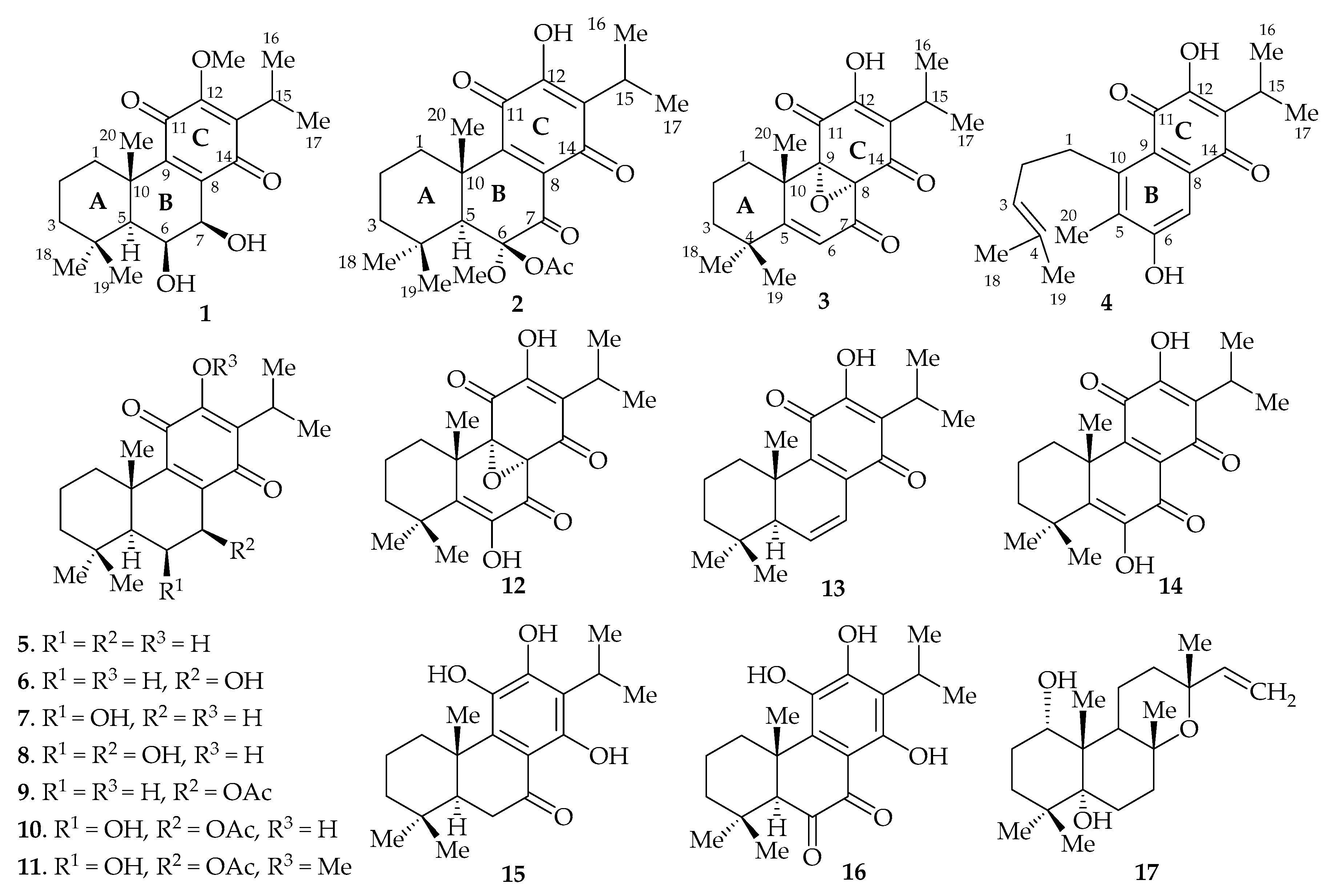
We report the isolation of four new abietane-type diterpenoids (1–4), together with thirteen previously known compounds (Figure 1) from the methanol/dichloromethane (1:1) extract of the root of P. punctatus, and the evaluation of the cytotoxic and antibacterial activities of the compounds. The known diterpenoids were identified as royleanone (5) [13], 7β-hydroxyroyleanone (6) [14], 6β-hydroxyroyleanone (7) [14], 6β,7β-dihydroxyroyleanone (8) [15], 7β-acetoxyroyleanone (9) [13], 7β-acetoxy-6β-hydroxyroyleanone (10) [15], 7β-acetoxy-6β-hydroxy-12-O-methylroyleanone (11) [15], 8α,9α-epoxycoleon-U-quinone (12) [16], 6,7-dehydroroyleanone (13) [14], coleon-U-quinone (14) [17], demethylinuroyleanol (15) [18], coleon V (16) [17], 1α,5α-dihydroxymanoyl oxide (17) [19] by comparison with reported spectroscopic data.
Compound 1 was isolated as a yellow amorphous solid. The HR-ESI-MS ion peaks at m/z 363.2170 for [M + H]+ and 385.1981 for [M + Na]+ are in agreement with the molecular formula C21H30O5, indicating seven double bond equivalents. The sharp IR absorptions at 1655 and 1640 cm−1 are attributed to the presence of conjugated carbonyls and the broad band at 3432 cm−1 to hydroxyl groups. The maximum absorption bands at 192 and 270 nm in UV spectrum and 13C-NMR signals at δC 184.9 and 189.3 are in agreement with the presence of a para-benzoquinone moiety [15]. The 1H-NMR spectrum (Supplementary Material) suggests the presence of an isopropyl substituent at the para-benzoquinone as evidenced from the characteristic downfield shifted signal at δH 3.18 (1H, qq, J = 7.1, 7.1 Hz, H-15) and two doublet methyl groups at δH 1.21 (3H, d, J = 7.1 Hz, CH3-16), and 1.73 (3H, d, J = 7.1 Hz, CH3-17), which is consistent with an oxidized C-ring as in abietane-type diterpenoids [15]. The 1H-NMR spectrum further shows two oxygenated methine signals at δH 4.35 (1H, dd, J = 3.7, 2.2 Hz) and 4.56 (1H, d, J = 2.2 Hz), a tertiary methine signal at δH 1.54 (1H, d, J = 3.7 Hz), three singlet methyl groups (δH 1.00, 1.27, 1.66), and a set of signals between 1.12 and 2.51, for three mutually coupled (as deduced from COSY experiments) vicinal methylene groups. The presence of one singlet at δH 3.91, for a methoxy group, was also evident. The 13C-NMR spectrum (Supplementary Material) shows 21 carbon signals corresponding to the para-benzoquinone moiety (δC 136.2, 140.5, 150.3, 157.8, 184.9, and 189.3) of ring C, together with two aliphatic quaternary carbons (δC 34.5, 39.9), four methine groups (δC 25.3, 49.6, 67.9, 69.9), three methylene groups (δC 24.4, 39.2, 43.3), five methyl (δC 19.9, 20.7, 21.0, 22.3, 34.2), and one methoxy (δC 61.1) carbon atoms. The 1H- and 13C-NMR spectra of 1 closely resemble those of compound 8, an abietane-type diterpenoid previously isolated from P. zeylanicus [15]. The only notable difference is the absence of a signal for a hydroxyl group which resonates at δH 7.04 in 8; instead, a signal for a methoxy group (δH 3.91; δC 61.1) is observed in 1, indicating the presence of a methoxy group at C-12.
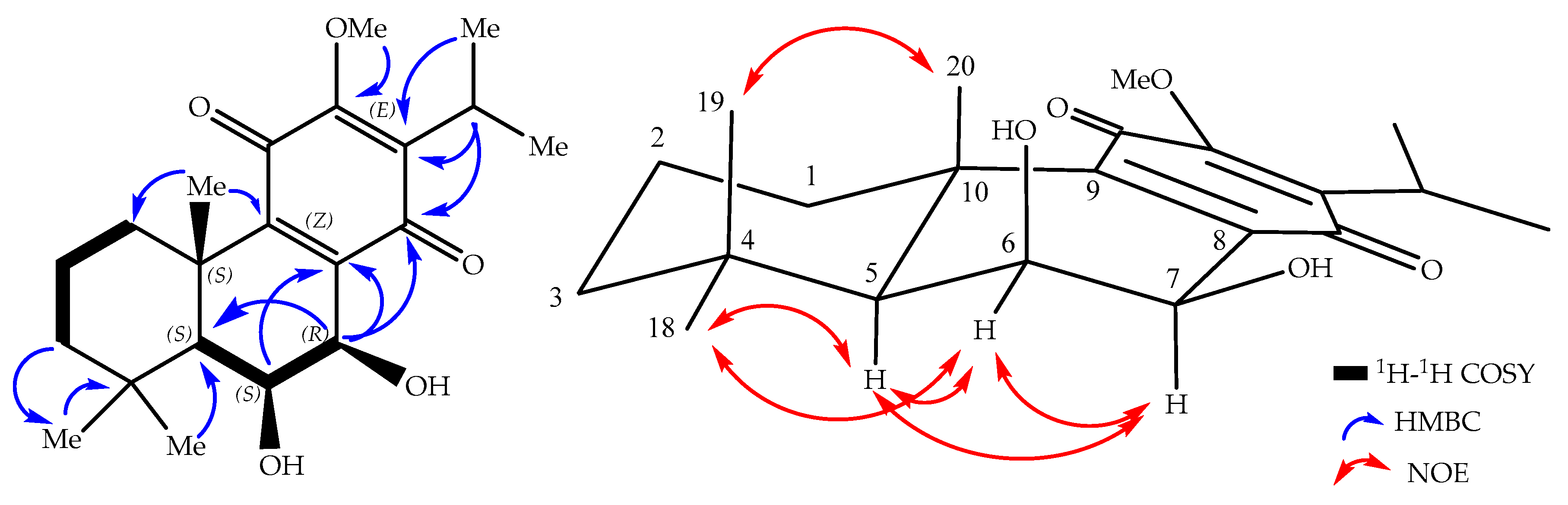
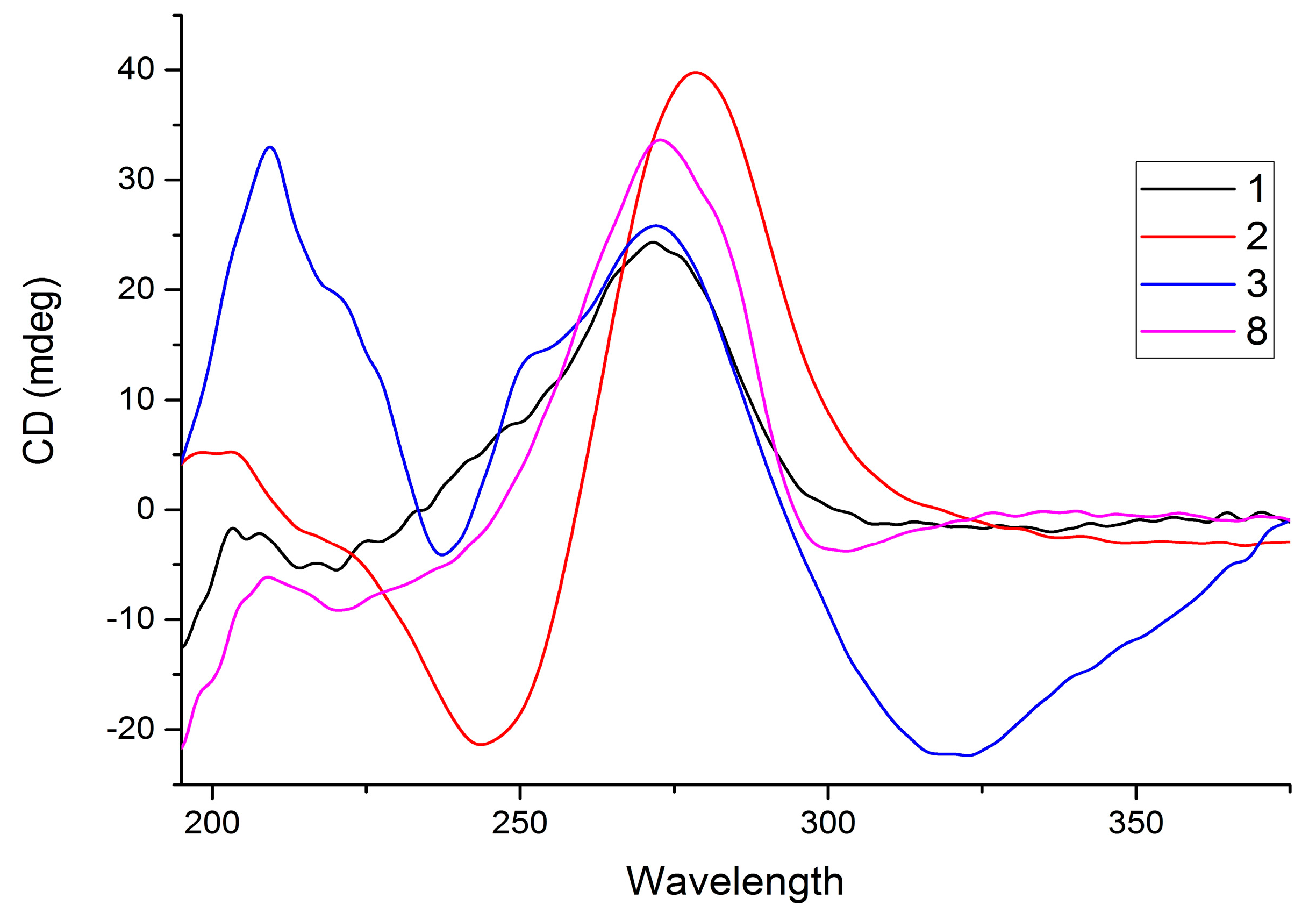
The relative configuration was established based on the NOE experimental data and coupling constants. The NOESY cross peaks from H-6 (δH 4.35) to H-7 (δH 4.56), H-5 (δH 1.45), and H-18 (δH 1.00) suggest that these hydrogens are cofacial. This is confirmed by the small 1H–1H coupling constants of H-5 (d, J5,6 = 3.7 Hz), H-6 (dd, J5,6 = 3.7 Hz; J6,7 = 2.2 Hz), and H-7 (d, J6,7 = 2.2 Hz), indicating cis-interactions of the vicinal hydrogens with H-5 and H-7 oriented axially (Figure 2). Therefore, the two methyl groups δH 1.66 and 1.27, at C-10 and C-4, respectively, were assigned as being oriented axially (β), which is further confirmed by the chemical shift value as a result of 1,3-diaxal interactions, with the hydroxyl group at C-6 [15]. The absolute configuration of abietane-type diterpenoids at C-5 and C-10 is 5S and 10S, based on biosynthetic consideration [20], which in combination with the already established relative configurations, allowed the absolute configuration of the compound to be assigned as 5S,6S,7R,10S. Compound 1 has similar optical activity ( = +24 (c 0.5, CH2Cl2)) and CD Cotton effects (Figure 3) as compound 8 [15]. Based on the above spectroscopic evidence, compound 1 was identified as 6,7-dihydroxy-12-methoxy-11,14-dioxoabieta-8,12-diene, which was given the trivial name 6β,7β-dihydroxy-12-methylroyleanone.
The second compound (2) was isolated as a yellow powder. Its HR-ESI-MS data reveal a peak for [M + H]+ at m/z 419.2080 and for [M + Na]+ at m/z 441.1905, both corresponding to a molecular formula of C23H30O7. The UV (λmax 193, 270 nm) and IR (νmax 1736, 1652, 1646 cm−1) spectra revealed absorptions for a conjugated carbonyl moiety. The 1H-NMR spectrum (Supplementary Material) displays signals for a hydroxyl group at δH 7.03 (s), an isopropyl group at δH 3.15 (1H, qq, J = 7.3, 7.3 Hz), 1.20 (3H, d, J = 7.3 Hz), and 1.19 (3H, d, J = 7.3 Hz), an acetyl group at δH 2.13 (s), a methoxy group at δH 3.42 (s), a methine at δH 3.00 (s), three methyl groups at δH 1.00 (s), 1.36 (s), 1.37 (s), and three mutually coupled (deduced from COSY experiment) methylene groups at δH 1.27–2.83 (m).
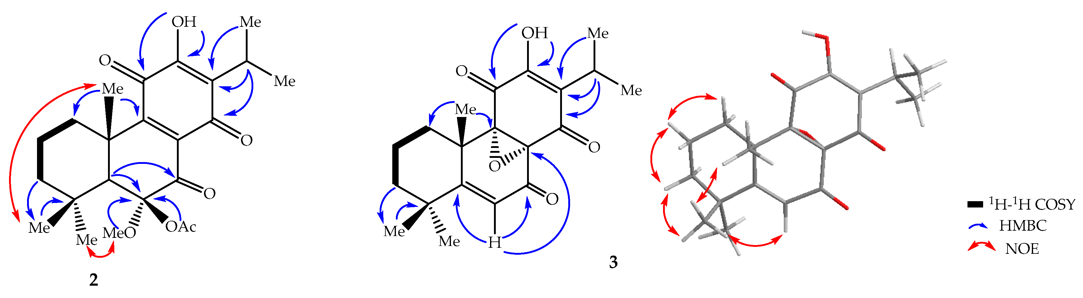
The 13C-NMR spectrum (Supplementary Material) displays signals for 23 carbon atoms assigned to the para-benzoquinone moiety (183.8, 183.5, 150.5, 147.6, 138.1, 125.2), a carbonyl group (199.7), three quaternary aliphatic carbons (98.3, 45.2, 32.7), an isopropyl group (24.4, 19.9, 18.8), a methoxy group (52.7), an acetyl residue (20.9, 169.3), three methyl (32.8, 22.0, 18.6), and three methylene (41.6, 36.7, 18.9) groups. These spectroscopic features are similar to those of compound 1, except for the absence of signals at δH 4.35 and 4.56 for the two oxygenated methines. Instead, two singlets are observed at δH 3.42 and 2.13, both integrating for three protons, corresponding to a methoxy and an acetyl group. These are consistent with the presence of extra carbon signals for a methoxy (δC 52.7) and an acetyl (20.9 and 169.3) group in the 13C-NMR spectrum. The upfield chemical shift (δH 3.42) of the methoxy group is an indication of its attachment to an sp3 carbon. The protons of the methoxy and the acetyl groups show HMBC correlations to C-6 (δC 98.3). A carbonyl signal is present at δC 199.7 in the 13C-NMR spectrum, together with a signal at δC 98.3, corresponding to a quarternary carbon. This indicates that the two alcohol moieties in 1 resonating at δC 69.9 (C-6) and 67.9 (C-7) are oxidized to a ketal (δC 98.3) and a ketone (δC 199.7), respectively, in compound 2. The weak HMBC correlation of H-5 (δH 3.00) with C-6 (δC 98.3) and strong correlation with C-7 (δC 199.7) confirm the presence of a ketal and keto moiety at C-6 and C-7, respectively. The relative configuration of 2 was determined to be the same for 1 based on NOESY correlations (Figure 4) observed from 6-OCH3 (δH 3.42) to H-5 (δH 3.00) and H-18 (δH 1.00). The latter cross peak also confirms the position of the ketal at C-6. Compound 2 shows a positive specific rotation (( = +13 (c 0.5, CH2Cl2)) and has a similar CD Cotton effect (Figure 3). Based on the above spectroscopic evidence, the second compound was identified as 6-acetoxy-12-hydroxy-6-methoxy-7,11,14-trioxoabieta-8,12-diene, which was given the trivial name 6β-acetoxy-6α-methoxy-7-oxoroyleanone (2).
Compound 3 was obtained as yellow amorphous solid, and its molecular formula C20H24O5 was deduced from HR-ESI-MS (m/z 345.1745 [M + H]+, calcd. for C20H25O5, 345.1702), indicating nine double bond equivalents. The IR spectrum revealed the presence of a hydroxyl group (3379 cm−1) and conjugated carbonyls (1729, 1634 cm−1). The UV absorptions at 223, 314 nm and the 13C-NMR signals at δC 187.0 and 185.5 support the presence of conjugated carbonyls. The 1H-NMR spectrum (Supplementary Material) exhibits an olefinic proton at δH 6.04 (1H, s, H-6), an isopropyl group at δH 3.19 (1H, qq, J = 7.2, 7.2 Hz, H-15); 1.23 (3H, d, J = 7.2 Hz, H-16); 1.21 (3H, d, J = 7.2 Hz, H-17), three methyl groups at 1.26 (3H, s, H-18); 1.18 (3H, s, H-19); 1.50 (3H, s, H-20), one hydroxy group at 7.04 (1H, s, 12-OH), and three mutually coupled (according to COSY) methylene groups at δH 1.44–2.98 (m) (Table 1). The 13C-NMR spectrum (Supplementary Material) shows signals corresponding to 20 carbon atoms, including three carbonyl carbons, seven quaternary carbons (three olefinic ones, two oxygenated ones, and two aliphatic ones), two methine groups (one olefinic), three methylene groups and five methyl carbons similar to the abietane-type diterpenoids. However, in this case, it shows signals for conjugated 1,4-dione (187.0, 185.5, 151.2, 128.4) with two oxygen-bearing tertiary carbons at δC 60.0 and 67.8 that would constitute the para-benzoquinone moiety, with the latter two corresponding to epoxy carbon atoms, similar to compound 12 [16]. The HMBC correlations of 12-OH (δH 7.04) with its neighbors C-11, C-12, C-13, and H-15, with C-12, C-13, C-14, establish the 1,4-dione. The oxygen-bearing tertiary carbons were assigned to C-8 (δC 60.0) and C-9 (67.8), respectively. This is supported by the HMBC correlation (Figure 4) of H-6, with one of the oxygen bearing carbon, C-8 (60.0), and a carbonyl carbon, C-7 (189.0).
The stereochemistry of 3 was elucidated on the basis of the circular dichroism (CD). The spectrum exhibits a strongly negative Cotton effect at longer wavelength (320 nm) and a strongly positive Cotton effect at shorter wavelength close to 220 and 270 nm, which is similar with the literature precedent for related compounds (12 and 8α,9α-epoxy-7-oxoroyleanon) having an epoxide ring at a similar position [16,21]. The absolute configurations at the chiral centers are then assigned as 8S,9S,10S in comparison with compound 12 [16,21]. This compound was, therefore, identified as 12-hydroxy-8α,9α-epoxy-7,11,14-trioxoabieta-5,12-diene, and given the trivial name 8α,9α-epoxy-6-deoxycoleon U (3).
Compound 4 was isolated as yellow solid with a molecular formula of C20H24O4, as deduced from the HR-ESI-MS ion at m/z 329.1754 ([M + H]+, calcd. for 329.1753), which corresponds to nine double bond equivalents. The UV (λmax 214, 272, 305) and IR (1637, 1570) absorptions indicate the presence of conjugated quinoid moiety [22]. The 13C-NMR shows signals for 20 carbon atoms assignable to two carbonyl (181.4, 185.4), eight quaternary (119.9, 125.4, 129.1, 132.7, 135.2, 148.1, 154.2, 159.6), two methylene (28.0, 30.7), three methine (24.6, 123.8, 112.8), and five methyl (11.5, 17.8, 20.0, 20.0, 25.9) groups. The 1H-NMR spectrum shows signals typical for an isopropyl group at δH 3.35 (1H, qq, J = 7.1, 7.1 Hz), 1.30 (3H, d, J = 7.1 Hz) and 1.30 (3H, d, J = 7.1 Hz), together with a singlet corresponding to hydroxy group at δH 8.05 on the para-benzoquinone ring at C-13 and C-12, respectively, as in abietane-type diterpenoids [13]. These substitutions were further confirmed by HMBC correlations of H-15 (δH 3.35) to C-12, C-13, C-14, C-16, C-17, and 12-OH (δH 8.05) to C-11, C-12, C-13. It further shows signals for a singlet aromatic proton (δH 7.69), an olefinic proton (δH 5.31), two mutually coupled methylene (δH 2.19, 3.23), one aromatic methyl (δH 2.31), and two aliphatic methyl (δH 1.64, 1.75) groups, which is similar to the rearranged abietane-type diterpenoid, 12-hydroxysapriparaquinone [22]. The downfield shifted singlet aromatic proton signal at δH 7.69 indicates its position peri to the carbonyl carbon, and was assigned to H-7, which otherwise substituted with methyl and hydroxyl groups at C-5 (δC 129.1) and C-6 (δC 159.6), respectively, confirmed from the HMBC correlation of H-7 with C-5, C-6, C-9, C-14, and H-20 with C-10, C-5, C-6. This substitution is further supported by biosynthetic consideration, that the rearranged diterpenoid is formed by migration of the C-l0 methyl group to C-5 accompanied by fission of ring A [22]. The other substituents with the two mutually coupled methylene protons (from COSY experiment) at δH 3.23 and 2.19 were assigned to H-1 and H-2, respectively, which are accompanied by an olefinic proton (H-3) showing HMBC correlation to C-1, C-2, C-4, C-18, and C-19. Therefore, the structure of compound 4 was identified as the rearranged abietane-type diterpenoid, 6,12-dihydroxy-sapriparaquinone.
The compounds were evaluated for their antibacterial and cytotoxic activities. The disk diffusion assay was employed to determine the antibacterial activities of the isolated compounds against five bacterial species: Escherichia coli (DSMZ1058), Bacillus subtilis (DSMZ704), Micrococcus luteus (DSMZ1605), Pseudomonas agarici (DSMZ11810), and Staphylococcus warneri (DSMZ20036) (Table 2). These strains are non-pathogenic correlates of bacteria resistant to most of the first line antibiotics [23,24]. Hence, any substance toxic to these strains can be assumed to be active on the pathogenic relatives, too. The antibacterial activity test (Table 2) indicates that the compounds have antibacterial activities against all the bacterial strains with compounds 1, 6, 8, 10, 11, and 14 displaying superior activity. The antibiotic activity (zone of inhibition) of compounds 1, 6, 8, 10, and 11 is even greater than that of the reference drug (gentamycin) against S. warneri (Table 2). Interestingly, compound 9, which has similar structural features except for the presence of acetyl group at C-7, exhibits less antibiotic activity. This indicates that the presence of aliphatic free hydroxyl groups at C-6 and C-7 may have a positive effect in inhibiting the growth of these bacteria. Compounds 10, 11, and 14 also show better antibiotic activity against M. luteus in comparison to the reference drug. In general, the activities of these diterpenoids from the traditional medicinal plant P. punctatus against both Gram negative and Gram positive bacteria are good, with variable degree of potency between the tested compounds, and provide a scientific basis for the traditional use of the plant.
The human cervix carcinoma cell line KB-3-1 was used for cytotoxicity test with cryptophycin-52 (IC50 = 1.3 × 10⁻⁵ µM) and griseofulvin (IC50 = 19 µM) as positive controls, as described in previous reports [25]. Compounds 6, 7, 9, 10, 11, and 13 showed marginal cytotoxic activity with IC50 values of 50, 49, 13, 42, 52, and 30 µM, respectively, whereas the other compounds showed little or none inhibitory activities.
3. Materials and Methods
3.1. General Information
Column chromatography was carried out on silica gel (0.06–0.2 mm, Merck, Darmstadt, Germany). Gel filtration was performed on Sephadex LH-20 (GE Healthcare, Uppsala, Sweden). Analytical TLC was performed on Merck pre-coated silica gel 60 F254 plates (Merck, Darmstadt, Germany). Preparative HPLC, LaChrom System (Merck Hitachi) equipped with a Phenomenex Jupiter column (10 mm, C18, 300 Å, 250 × 21.1 mm) was used for purification of the compounds. Melting points were measured on B-540 melting point apparatus (Büchi, Flawil, Switzerland). UV spectra were recorded on a UV-3100PC spectrophotometer (VWR International GmbH, Darmstadt, Germany). IR spectra were recorded on a Nicolet 380 FT-IR spectrometer (Thermo Electron Corporation, Madison, WI, USA). High Resolution ESI-MS was done on a Micromass AC-TOFmicro mass spectrometer (Micromass, Agilent Technologies 1200 series, Tokyo, Japan). CD spectra were measured on a JASCO J-810 CD spectrometer (JASCO, Tokyo, Japan). Optical rotations were measured on a P-1020 polarimeter (JASCO, Tokyo, Japan). 1D (1H, 13C) NMR and 2D (COSY, HSQC, HMBC, NOESY) NMR spectra were recorded on an Avance 500 MHz spectrometer (Bruker, Billerica, MA, USA, at 500 MHz (1H) and 125 MHz (13C) at 298 K, using the residual solvent peaks (acetone: δH 2.05, δC 29.84; CDCl3: δH 7.26, δC 77.16) as a reference.
3.2. Plant Materials
The roots of P. punctatus were collected from Gulufa, Amuru District, Horro Guduru Wolega zone, Oromia regional state, Ethiopia in September 2016. The plant material was identified and the voucher specimen (voucher number NA-07) has been deposited in Jimma University Herbarium.
3.3. Extraction and Isolation
The air-dried roots (620 g) of P. punctatus were milled into powder and then extracted using CH2Cl2/MeOH (1:1) four times for 24 h at room temperature. The extract was concentrated under vacuum using a rotary evaporator to yield a dark brown residue (37 g, 5.97%). A 35 g portion of the extract was subjected to column chromatography on silica gel (300 g), eluting with petroleum ether containing increasing amounts of ethyl acetate, to afford 36 major fractions of ca. 250 mL each. Fractions 4–10 (5% EtOAc in petroleum ether) were combined and purified by Sephadex LH-20 (eluting with CH2Cl2/MeOH; 1:1) to give royleanone (5, 4.9 mg), 7β-hydroxyroyleanone (6, 84.2 mg) and 6,7-dehydroroyleanone (13, 7.3 mg); while fractions 11–15 (10% EtOAc in petroleum ether) showed a yellow precipitate that was washed with 100% petroleum ether and further purified on Sephadex LH-20 (eluting with CH2Cl2/MeOH; 1:1, v/v) to give 7β-acetoxyroyleanone (9, 27.3 mg), 7β-acetoxy-6β-hydroxyroyleanone (10, 24.7 mg), and 7β-acetoxy-6β-hydroxy-12-methylroyleanone (11, 4.7 mg). Fractions 16–20 (15% ethyl acetate in petroleum ether) showed mixtures of seven compounds, which were combined and subjected to column chromatography (column size: 80 cm length and 4 cm diameter) on silica gel (250 g; eluent: increasing gradient of EtOAc in petroleum ether) followed by Sephadex LH-20 (eluting with CH2Cl2/MeOH; 1:1, v/v) yielding compound 1 (4.8 mg), 6β-hydroxyroyleanone (7, 5.1 mg) and 1α, 5α-dihydroxymanoyl oxide (17, 4.3 mg). The remaining compounds of the same fraction were further purified by RP-HPLC, with a linear gradient solvent system of 20% acetonitrile/water to 100% acetonitrile at a flow rate of 10 mL/min over 60 min, to afford compound 2 (3.8 mg, tR 42 min), 8α,9α-epoxycoleon-U-quinone (12, 3.7 mg, tR 36 min), coleon-U-quinone (14, 6.2 mg, tR 46 min), and demethylinuroyleanol (15, 3.9 mg, tR 39 min). Fractions 25–30 (35% EtOAc in petroleum ether) were combined and chromatographed on Sephadex LH-20 (eluting with CH2Cl2/MeOH; 1:1, v/v) followed by further purification using the same RP-HPLC protocol to give compound 3 (4.3 mg, tR 32 min), 4 (6.1 mg, tR 28 min), 6β,7β-dihydroxyroyleanone (8, 11.8 mg, tR 38 min), and coleon V (16, 4.7 mg, tR 41 min).
6β,7β-Dihydroxy-12-methylroyleanone (1): Yellow amorphous solid. m.p. 189–191 °C. UV (CH3CN): λmax (logε) = 192 (2.65), 270 (2.46) nm. IR (CH2Cl2) νmax cm−1 3432, 1655, 1640. = +24 (c 0.5, CH2Cl2). 1H- and 13C-NMR (Table 1). ESI-MS (rel. int.): m/z = 385 (32, [M + Na]+), 363 (65, [M + H]+). HR-ESI-MS m/z = 363.2170, [M + H]+ (calcd. for C21H30O5, 363.2172).
6β-Acetoxy-6α-methoxy-7-oxoroyleanone (2): Yellow amorphous solid. m.p. 147–149 °C. UV (CH3CN): λmax (log ε) = 193 (2.71), 270 (2.53) nm. IR (CH2Cl2) νmax cm−1 3342, 1736, 1652, 1646. = +13 (c 0.5, CH2Cl2).1H- and 13C-NMR (Table 1). ESI-MS (rel. int.) m/z = 441 (13, [M + Na]+), 419 (97, [M + H]+). HR-ESI-MS m/z = 419.2080, [M + H]+ (calcd. for C23H30O7, 419.2070); 441.1905, [M + Na]+.
8α,9α-Epoxy-6-deoxycoleon U (3): Yellow solid. m.p. 159–161 °C. UV (CH3CN): λmax (logε) = 223 (2.63), 314 (2.21) nm. IR (CH2Cl2) νmax cm−1 3379, 1729, 1634. = +8 (c 0.5, CH2Cl2). 1H- and 13C-NMR (Table 1). ESI-MS (rel.int.): m/z = 711 (22, [2M + Na]+), 367 (37, [M + Na]+), 345 (100, [M + H]+). HR-ESI-MS m/z = 345.1745, [M + H]+ (calcd. for C20H24O5, 345.1702).
6,12-Dihydroxysapriparaquinone (4): Yellow solid. m.p. 234–236 °C. UV (CH3CN): λmax (logε) = 214 (2.67), 272 (2.62), 30.5 (2.19) nm. IR (CH2Cl2) νmax cm−1 3332, 1637, 1570. 1H- and 13C-NMR (Table 1). ESI-MS (rel. int.): m/z = 351 (21, [M + Na]+), 329 (100, [M + H]+). HR-ESI-MS m/z = 329.1754, [M + H]+ (calcd. for C20H24O4, 329.1753).
3.4. Antimicrobial Assay Using Agar Diffusion Test
The antimicrobial activity of the isolated compounds was investigated in a paper-disk diffusion assay [26] with some modifications: the bacterial strains Escherichia coli (DSMZ1058), Bacillus subtilis (DSMZ704), Micrococcus luteus (DSMZ1605), Pseudomonas agarici (DSMZ11810), and Staphylococcus warneri (DSMZ20036) were grown on a nutrient agar medium (3 g L−1 beef extract, 10 g L−1 peptone, and 20 g L−1 agar) and the pH was adjusted to 7.2. Twenty milliliters of medium seeded with test organism was poured into sterile Petri dishes (diameter 9 cm). The paper disks (diameter 6 mm) were placed on inoculated agar plates after solidification. Subsequently, the test samples (5 µg/mL each) were applied to the Petri dishes and kept in a refrigerator at 4 °C for 2 h, to allow the loaded substances to diffuse the into the microbial culture. The plates were then incubated for 24 h at 35 °C, and the diameters of the inhibition zones were then measured.
3.5. Cytotoxicity Assay
The human cervix carcinoma cell line KB-3-1 was used in the cytotoxicity assay, as described previously [25]. Briefly, the cell line was cultivated as a monolayer in DMEM (Dulbecco’s modified Eagle medium) with glucose (4.5 g/L), l-glutamine, sodium pyruvate, and phenol red, supplemented with 10% fetal bovine serum (FBS) and were maintained at 5.3% CO2 and 37 °C in humidified air. The cells (70% confluence) were detached with trypsin-ethylenediaminetetraacetic acid solution (0.05%; 0.02% in DPBS), and placed in sterile 96-well plates in a density of 10,000 cells in 100 μL medium per well. The dilution series of the compounds were prepared from stock solutions in DMSO of concentrations of 100 mM, 50 mM, or 25 mM, and the stock solutions were diluted with culture medium down to pM concentration. The dilution prepared from stock solution was added to the wells and each concentration was tested in at least six replicates. The control contained the same concentration of DMSO as the first dilution. After incubation for 72 h at 37 °C and 5.3% CO2-humidified air, 30 μL of an aqueous resazurin solution (175 μM) was added to each well. The cells were incubated at the same conditions for 5 h. Subsequently, the fluorescence was recorded at a wavelength of 588 nm. The IC50 values were calculated as a sigmoidal dose response curve using GRAPHPAD PRISM 4.03.
4. Conclusions
Four new diterpenoids were isolated together with thirteen known compounds from the dried roots of P. punctatus. Some of the compounds show marginal cytotoxic activity against the human cervix carcinoma cell line KB-3-1. However, almost all compounds display interesting antibacterial activity against both Gram positive and Gram negative microorganisms. In particular, six compounds (1, 6, 8, 10, 11, and 14) show greater zones of inhibition than the reference drug (gentamycin). This could give insight about the potentials of these compounds as lead structures in development of antibacterial drugs.
Supplementary Materials
The mass spectra and NMR data of compounds 1–4 are available.
Acknowledgments
Negera Abdissa gratefully acknowledges the Alexander von Humboldt foundation for a Georg Forster postdoctoral fellowship. We are grateful to Marco Wißbrock, Anke Nieß, and Carmela Michalek, Organic and Bioorganic Chemistry, Bielefeld University, for their technical assistance and bioactivity tests. We also acknowledge financial support from the German Research Foundation (DFG) and the Open Access Publication Fund of Bielefeld University for the article processing charge.
Author Contributions
N.A. and N.S. designed the experiments; N.A. performed the extraction and isolation; N.A. carried out the spectroscopic characterization of the compounds; M.F. analyzed the antibacterial and cytotoxicity data; all authors participated in manuscript preparation.
Conflicts of Interest
The authors declare that there is no conflict of interest.
References
- Abdel Khalik, K.N. A Systematic Revision of the Genus Plectranthus L. (Lamiaceae) in Saudi Arabia Based on Morphological, Palynological, and Micromorphological Characters of Trichomes. Am. J. Plant Sci. 2016, 7, 1429–1444. [Google Scholar] [CrossRef]
- Van Jaarsveld, E.J. The Plectranthus Handbook; National Botanic Gardens of South Africa, CTP Book Printers: Cape Town, South Africa, 1988; p. 23. [Google Scholar]
- Lukhoba, C.W.; Simmonds, M.S.J.; Paton, A.J. Plectranthus: A review of ethnobotanical uses. J. Ethnopharmacol. 2006, 103, 1–24. [Google Scholar] [CrossRef] [PubMed]
- Khan, M.; Al-Saleem, M.S.M.; Alkhathlan, H.Z. A detailed study on chemical characterization of essential oil components of two Plectranthus species grown in Saudi Arabia. J. Saudi Chem. Soc. 2016, 20, 711–721. [Google Scholar] [CrossRef]
- Stavri, M.; Paton, A.J.; Skelton, B.W.; Gibbons, S. Antibacterial diterpenes from Plectranthus ernstii. J. Nat. Prod. 2009, 72, 1191–1194. [Google Scholar] [CrossRef] [PubMed]
- Gaspar-Marques, C.; Rijo, P.; Simões, M.F.; Duarte, M.A.; Rodriguez, B. Abietanes from Plectranthus grandidentatus and Plectranthus hereroensis against methicillin and vancomycin-resistant bacteria. Phytomedicine 2006, 13, 267–271. [Google Scholar] [CrossRef] [PubMed]
- Van Zyl, R.L.; Khan, F.; Edwards, T.J.; Drewes, S.E. Antiplasmodial activities of some abietane diterpenes from the leaves of five Plectranthus species. S. Afr. J. Sci. 2008, 104, 62–64. [Google Scholar]
- Simões, M.F.; Rijo, P.; Duarte, A.; Barbosa, D.; Matias, D.; Delgado, J.; Cirilo, N.; Rodriguez, B. Two new diterpenoids from Plectranthus species. Phytochem. Lett. 2010, 3, 221–225. [Google Scholar] [CrossRef]
- Grayer, R.J.; Eckert, M.R.; Lever, A.; Veitch, N.C.; Kite, G.C.; Paton, A.J. Distribution of exudate flavonoids in the genus Plectranthus. Biochem. Sys. Ecol. 2010, 38, 335–341. [Google Scholar] [CrossRef]
- Fichtl, R.; Adi, A. Honeybee Flora of Ethiopia; Margraf: Weikersheim, Germany, 1994; pp. 118–121. [Google Scholar]
- Allemann, J.; Laurie, S.M.; Thiart, S.; Vorster, H.J.; Bornman, C.H. Sustainable production of root and tuber crops (potato, sweet potato, indigenous potato, cassava) in southern Africa. S. Afr. J. Bot. 2004, 70, 60–66. [Google Scholar] [CrossRef]
- Tadesse, D.; Eguale, T.; Giday, M.; Mussa, A. Ovicidal and larvicidal activity of crude extracts of Maesa lanceolata and Plectranthus punctatus against Haemonchus contortus. J. Ethnopharmacol. 2009, 122, 240–244. [Google Scholar] [CrossRef] [PubMed]
- Edwards, O.E.; Feniak, G.; Los, M. Diterpenoid quinones of Inula royleana. Can. J. Chem. 1962, 40, 1540–1546. [Google Scholar]
- Hensch, M.; Rüedi, P.; Eugster, C.H. Horminon, Taxochinon und weitere Royleanone aus 2 abessinischen Plectranthus-Spezies (Labiatae). Helv. Chim. Acta 1975, 58, 1921–1934. [Google Scholar] [CrossRef]
- Mehrotra, R.; Vishwakarma, R.A.; Thakur, R.S. Abietane diterpenoids from Coleus zeylanicus. Phytochemistry 1989, 28, 3135–3137. [Google Scholar] [CrossRef]
- Alder, A.C.; Rüedi, P.; Eugster, C.H. Drüsenfarbstoffe aus Labiaten: Die polaren Diterpenoide aus Plectranthus argentatus S.T. BLAKE. Helv. Chim. Acta 1984, 67, 1523–1530. [Google Scholar] [CrossRef]
- Miyase, T.; Rüedi, P.; Eugster, C.H. Diterpenoide Drüsenfarbstoffe aus Labiaten: Coleone U, V, W und 14-O-Formyl-coleon-V sowie 2 Royleanone aus Plectranthus myrianthus BRIQ.; cis- und trans-A/B-6, 7-Dioxoroyleanon. Helv. Chim. Acta 1977, 60, 2770–2779. [Google Scholar] [CrossRef]
- Chang, C.I.; Tseng, M.H.; Kuo, Y.H. Five new diterpenoids from the bark of Taiwania cryptomerioides. Chem. Pharm. Bull. 2005, 53, 286–289. [Google Scholar] [CrossRef] [PubMed]
- Labbé, C.; Castillo, M.; Fainia, F.; Coll, J.; Connolly, J.D. Rearranged isopimarenes and other diterpenoids from Satureja gilliesii. Phytochemistry 1994, 36, 735–738. [Google Scholar] [CrossRef]
- Inabuy, F.S.; Fischedick, J.T.; Lange, I.; Hartmann, M.; Srividya, N.; Parrish, A.N.; Xu, M.; Peters, R.J.; Lange, B.M. Biosynthesis of diterpenoids in Tripterygium adventitious root cultures. Plant Physiol. 2017, 175, 92–103. [Google Scholar] [CrossRef] [PubMed]
- Wu, L.; Lu, Y.; Zheng, Q.T.; Xiao-Li, X.L.; Zhao, Q.S. 8α,9α-Epoxy-7-oxoroyleanon. Acta Cryst. E 2006, 62, 3269–3270. [Google Scholar] [CrossRef]
- Topcu, G.; Eriş, C.; Ulubelen, A. Rearranged abietane diterpenes from Salvia limbata. Phytochemistry 1996, 41, 1143–1147. [Google Scholar] [CrossRef]
- Mehta, J.P.; Davariya, V.S.; Parmar, P.H. An antimicrobial activity of anthraquinones from Cassia occidentalis. Int. J. Chem. Sci. 2012, 10, 413–419. [Google Scholar]
- Rasheed, M.U.; Thajuddin, N.; Ahamed, P.; Teklemariam, Z.; Jamil, K. Antimicrobial drug resistance in strains of Escherichia coli isolated from food sources. Rev. Inst. Med. Trop. Sao Paulo 2014, 56, 341–346. [Google Scholar] [CrossRef] [PubMed]
- Sammet, B.; Bogner, T.; Nahrwold, M.; Weiss, C.; Sewald, N. Approaches for the synthesis of functionalized cryptophycins. J. Org. Chem. 2010, 75, 6953–6960. [Google Scholar] [CrossRef] [PubMed]
- Baur, A.W.; Kirby, W.M.; Sherris, J.C.; Truck, M. Antibiotic susceptibility testing by a standardized single disk method. Am. J. Clin. Pathol. 1966, 45, 493. [Google Scholar]
Sample Availability: Samples of the compounds 7, 8, 9, 10 and 13 are available from the authors. |
Figure 1.
Structures of the isolated compounds.

Figure 2.
Key 1H–1H COSY (bold lines), HMBC (blue arrows) and NOE (red arrows) correlations of 1.

Figure 3.
CD spectra for compounds 1–3 and 8 (in acetonitrile).

Figure 4.
Key 1H–1H COSY (bold lines); HMBC (blue arrows) and NOE (red arrows) correlations of 2 and 3.
Figure 4.
Key 1H–1H COSY (bold lines); HMBC (blue arrows) and NOE (red arrows) correlations of 2 and 3.


Table 1.
1H (500 MHz) and 13C (125 MHz) NMR data of compound 1 (in acetone-d6) and compounds 2–4 (in CDCl3).
Table 1.
1H (500 MHz) and 13C (125 MHz) NMR data of compound 1 (in acetone-d6) and compounds 2–4 (in CDCl3).
| Position | 1 | 2 | 3 | 4 | ||||
|---|---|---|---|---|---|---|---|---|
| δH (m, J in Hz) | δC | δH (m, J in Hz) | δC | δH (m, J in Hz) | δC | δH (m, J in Hz) | δC | |
| 1 | 1.18 (m), 2.51 (m) | 39.2 | 1.36 (m), 2.83 (m) | 36.7 | 1.73 (m), 2.98 (m) | 33.9 | 3.23 (m) | 30.7 |
| 2 | 1.12 (m), 1.44 (m) | 24.4 | 1.34 (m), 1.64 (m) | 18.9 | 1.23 (m), 1.70 (m) | 17.4 | 2.19 (m) | 28.0 |
| 3 | 1.47 (m), 1.50 (m) | 43.3 | 1.42 (m), 1.27 (m) | 41.6 | 1.58 (m), 1.44 (m) | 39.1 | 5.31 (br t, 3.4) | 123.8 |
| 4 | 34.5 | 32.7 | 37.7 | 132.7 | ||||
| 5 | 1.54 (d, 3.7) | 49.6 | 3.00 (s) | 58.7 | 169.5 | 129.1 | ||
| 6 | 4.35 (dd, 3.7, 2.2) | 69.9 | 98.3 | 6.04 (s) | 120.9 | 159.6 | ||
| 7 | 4.56 (d, 2.2) | 67.9 | 199.7 | 189.0 | 7.69 (s) | 112.8 | ||
| 8 | 140.5 | 138.1 | 60.0 | 135.2 | ||||
| 9 | 150.3 | 147.6 | 67.7 | 119.9 | ||||
| 10 | 39.9 | 45.2 | 41.6 | 148.1 | ||||
| 11 | 184.9 | 183.8 | 187.0 | 181.4 | ||||
| 12 | 157.8 | 150.5 | 151.2 | 154.2 | ||||
| 13 | 136.2 | 125.2 | 128.4 | 125.4 | ||||
| 14 | 189.3 | 183.5 | 185.5 | 185.4 | ||||
| 15 | 3.18 (qq, 7.1, 7.1) | 25.3 | 3.15 (qq, 7.3, 7.3) | 24.4 | 3.19 (qq, 7.2, 7.2) | 24.9 | 3.35 (qq, 7.1, 7.1) | 24.6 |
| 16 | 1.73 (d, 7.1) | 20.7 | 1.19 (d, 7.3) | 19.9 | 1.23 (d, 7.2) | 19.2 | 1.30 (d, 7.1) | 20.0 |
| 17 | 1.21 (d, 7.1) | 19.9 | 1.20 (d, 7.3) | 19.8 | 1.21 (d, 7.2) | 19.4 | 1.30 (d, 7.1) | 20.0 |
| 18 | 1.00 (s) | 34.2 | 1.00 (s) | 32.8 | 1.26 (s) | 30.7 | 1. 64 (s) | 17.8 |
| 19 | 1.27 (s) | 22.3 | 1.37 (s) | 22.0 | 1.18 (s) | 32.2 | 1.75 (s) | 25.9 |
| 20 | 1.66 (s) | 21.0 | 1.36 (s) | 18.6 | 1.50 (s) | 24.0 | 2.31 (s) | 11.5 |
| 12-OMe/OH | 3.91 (s) | 61.1 | 7.03 (s) | 7.04 (s) | 8.05 (s) | |||
| 6-OMe | 3.42 (s) | 52.7 | ||||||
| 6-OAc | 2.13 (s) | 20.9, 169.3 | ||||||
Table 2.
Diameter of zone of bacterial growth inhibition (in mm) of the isolated compounds.
| Compound | E. coli DSMZ1058 | S. warneri DSMZ20036 | B. subtlis DSMZ704 | M. luteus DSMZ1605 | P. agarici DSMZ11810 |
|---|---|---|---|---|---|
| 1 | n.a. | 22 | 20 | 23 | 21 |
| 2 | n.a. | 9 | 10 | 14 | 9 |
| 3 | n.a. | n.a. | 7 | n.a. | 14 |
| 4 | n.a. | n.a. | 7 | n.a. | 8 |
| 6 | 8 | 26 | 20 | 18 | 23 |
| 7 | 8 | 20 | 19 | 21 | 19 |
| 8 | 7 | 28 | 15 | 8 | 20 |
| 9 | 7 | 9 | 12 | 9 | 9 |
| 10 | 7 | 27 | 25 | 25 | 21 |
| 11 | 8 | 26 | 25 | 24 | 21 |
| 13 | 7 | 7 | 9 | 9 | n.a. |
| 14 | 8 | 19 | 13 | 24 | 14 |
| 15 | n.a. | 18 | 13 | 23 | 16 |
| 16 | 8 | 19 | 14 | 23 | 16 |
| Gentamycin | 23 | 21 | 26 | 23 | 24 |
n.a.: not active; all values are mean values ± standard deviation of three replicates.
© 2017 by the authors. Licensee MDPI, Basel, Switzerland. This article is an open access article distributed under the terms and conditions of the Creative Commons Attribution (CC BY) license (http://creativecommons.org/licenses/by/4.0/).
Share and Cite
MDPI and ACS Style
Abdissa, N.; Frese, M.; Sewald, N. Antimicrobial Abietane-Type Diterpenoids from Plectranthus punctatus. Molecules 2017, 22, 1919. https://doi.org/10.3390/molecules22111919
AMA Style
Abdissa N, Frese M, Sewald N. Antimicrobial Abietane-Type Diterpenoids from Plectranthus punctatus. Molecules. 2017; 22(11):1919. https://doi.org/10.3390/molecules22111919
Chicago/Turabian StyleAbdissa, Negera, Marcel Frese, and Norbert Sewald. 2017. "Antimicrobial Abietane-Type Diterpenoids from Plectranthus punctatus" Molecules 22, no. 11: 1919. https://doi.org/10.3390/molecules22111919