6′-Sialyllactose Ameliorates In Vivo and In Vitro Benign Prostatic Hyperplasia by Regulating the E2F1/pRb–AR Pathway

 and
and 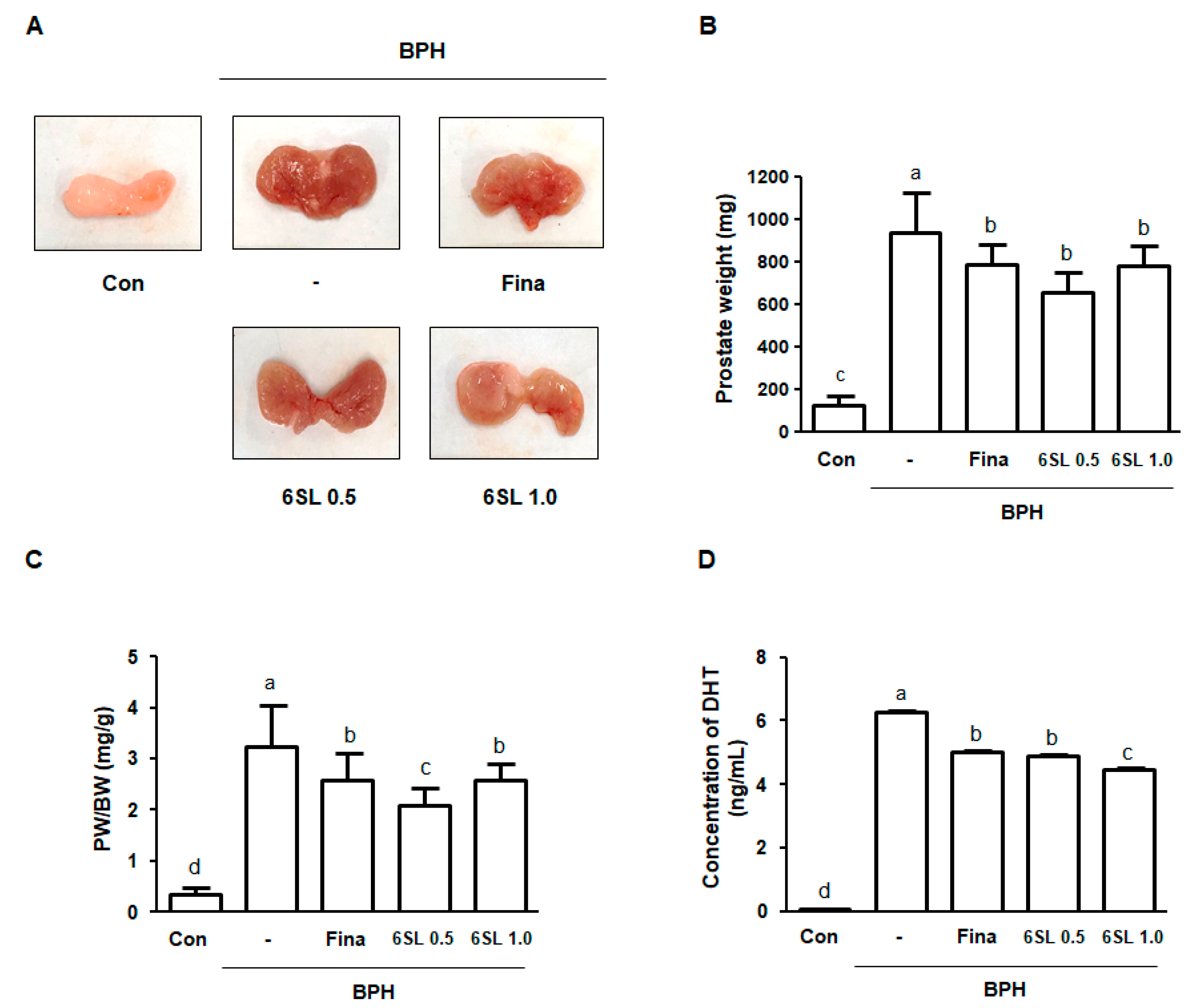
Abstract
:1. Introduction
2. Materials and Methods
2.1. Animals
2.2. Induction of BPH and Agent Administration
2.3. Serum Level of DHT Analysis
2.4. Histological Analysis and Immunohistochemistry
2.5. Western Blot Analysis
2.6. Cell Culture and Sample Treatment
2.7. MTT Assay
2.8. Statistical Analyses
3. Results
3.1. 6SL Attenuated Prostate Enlargement in a TP-Induced BPH Rat Model
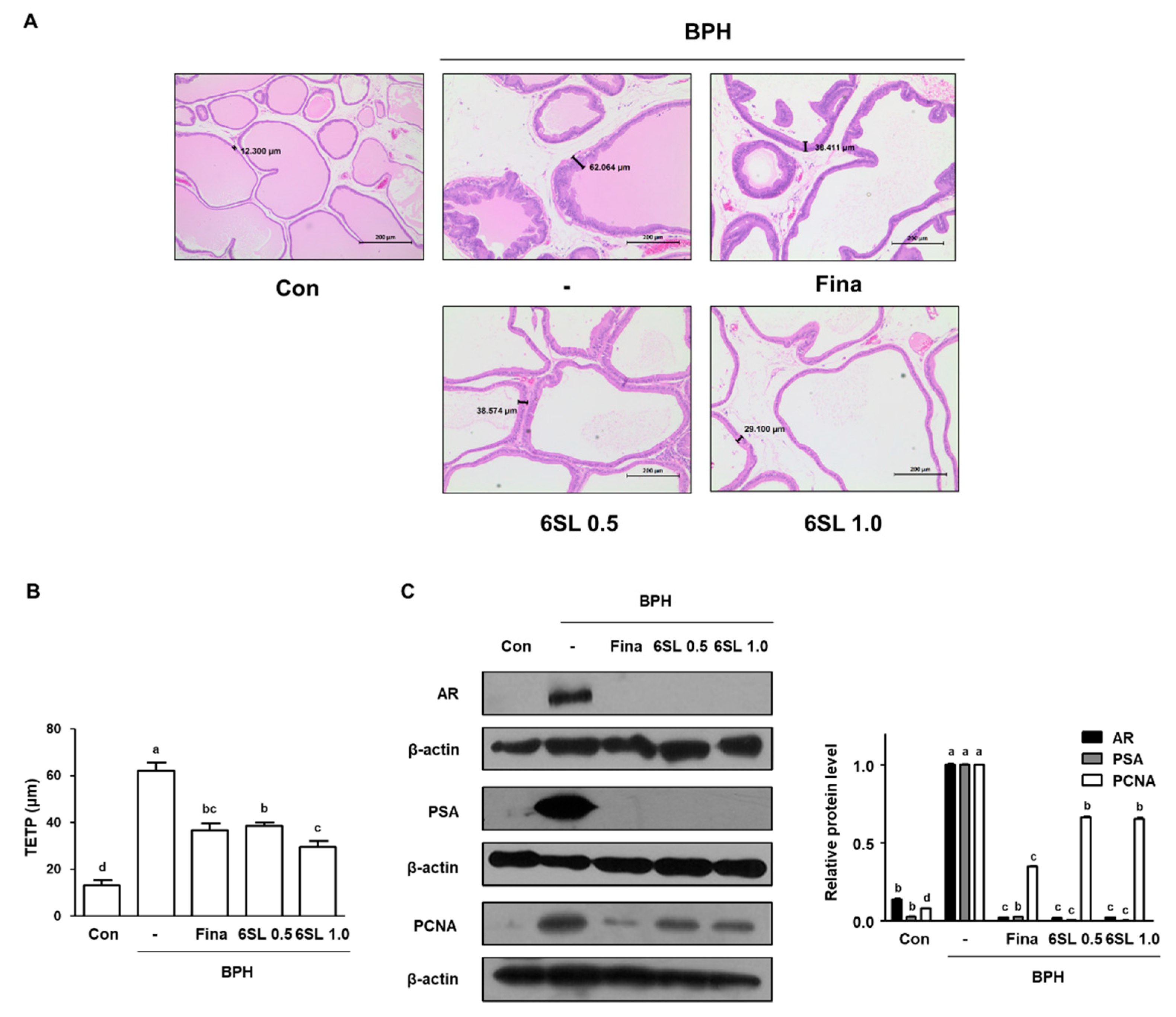
3.2. 6SL Ameliorated Prostatic Hyperplasia by Regulating AR Signaling in a TP-Induced BPH Rat Model
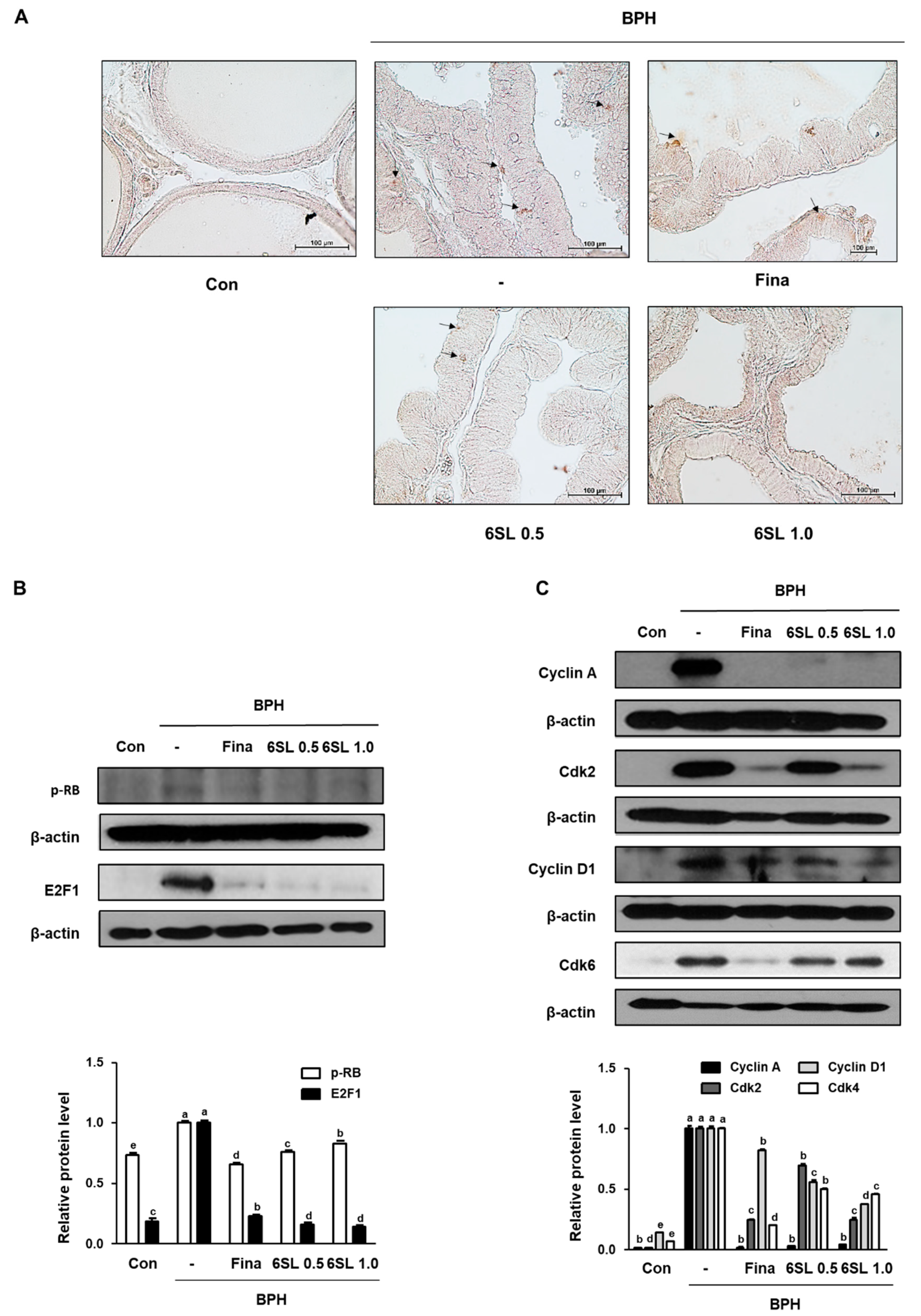
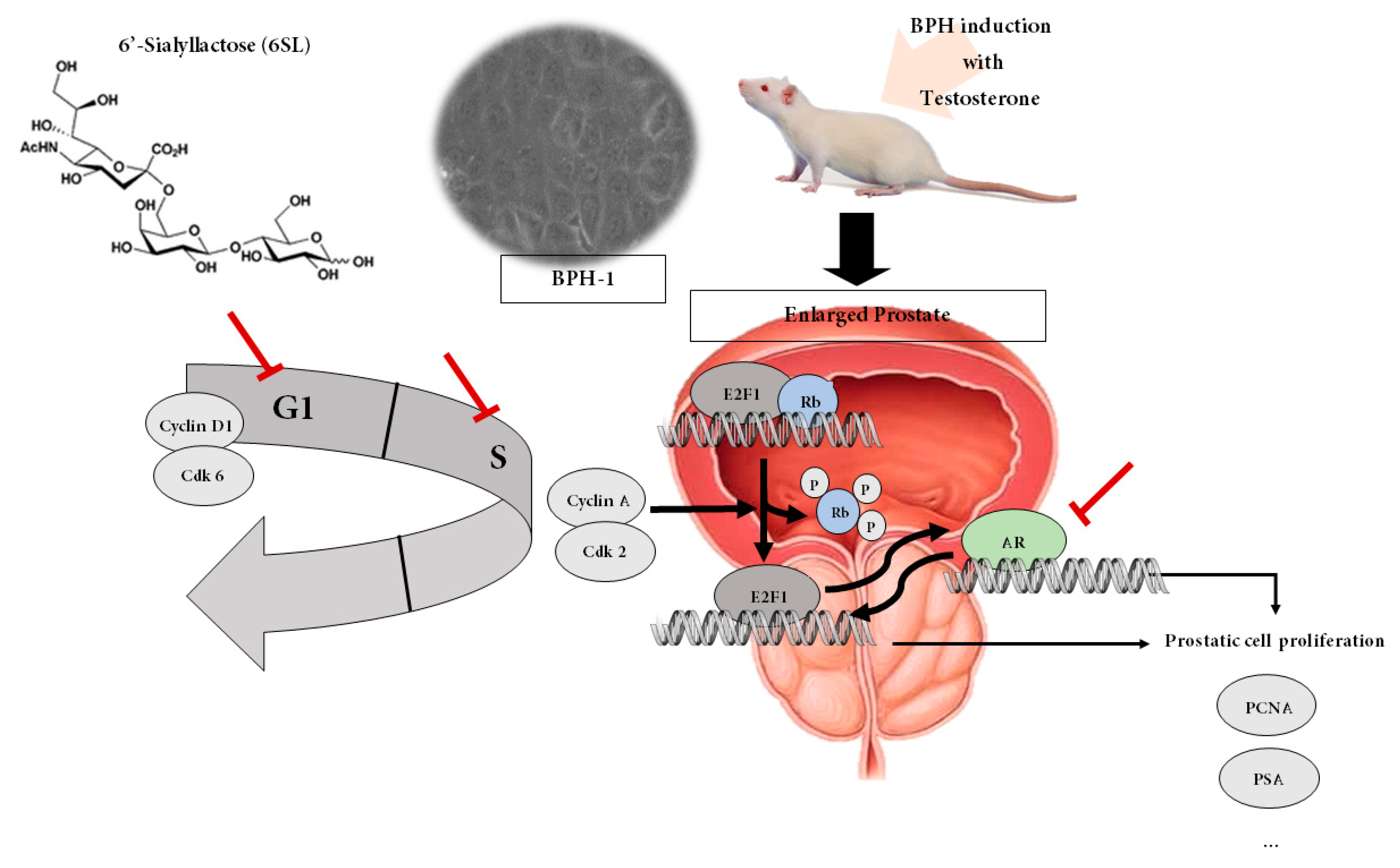
3.3. 6SL Suppressed E2F1–pRb Pathway Signaling and Induced Cell Cycle Arrest at the G1 and S Phases in a TP-Induced BPH Rat Model
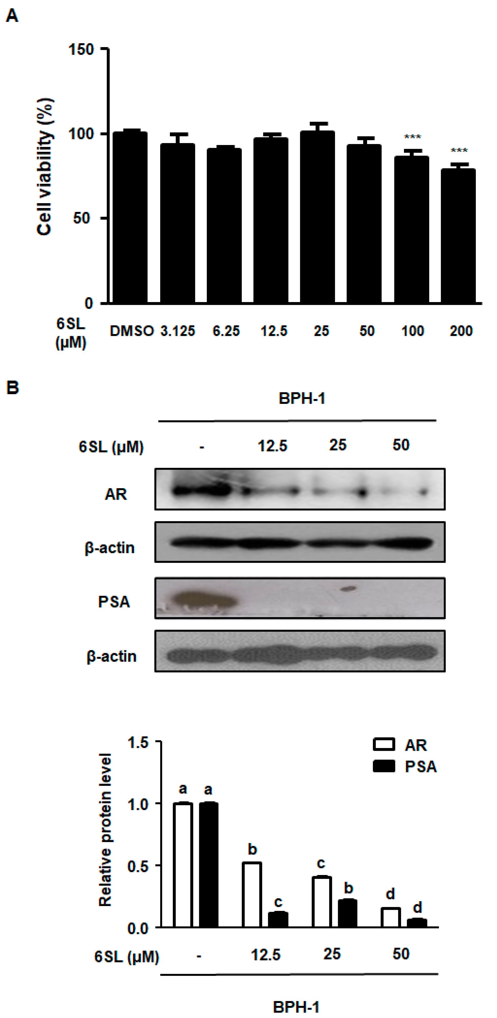
3.4. 6SL Abrogated Androgen-Relative Protein Expression in Human BPH Epithelial Cells
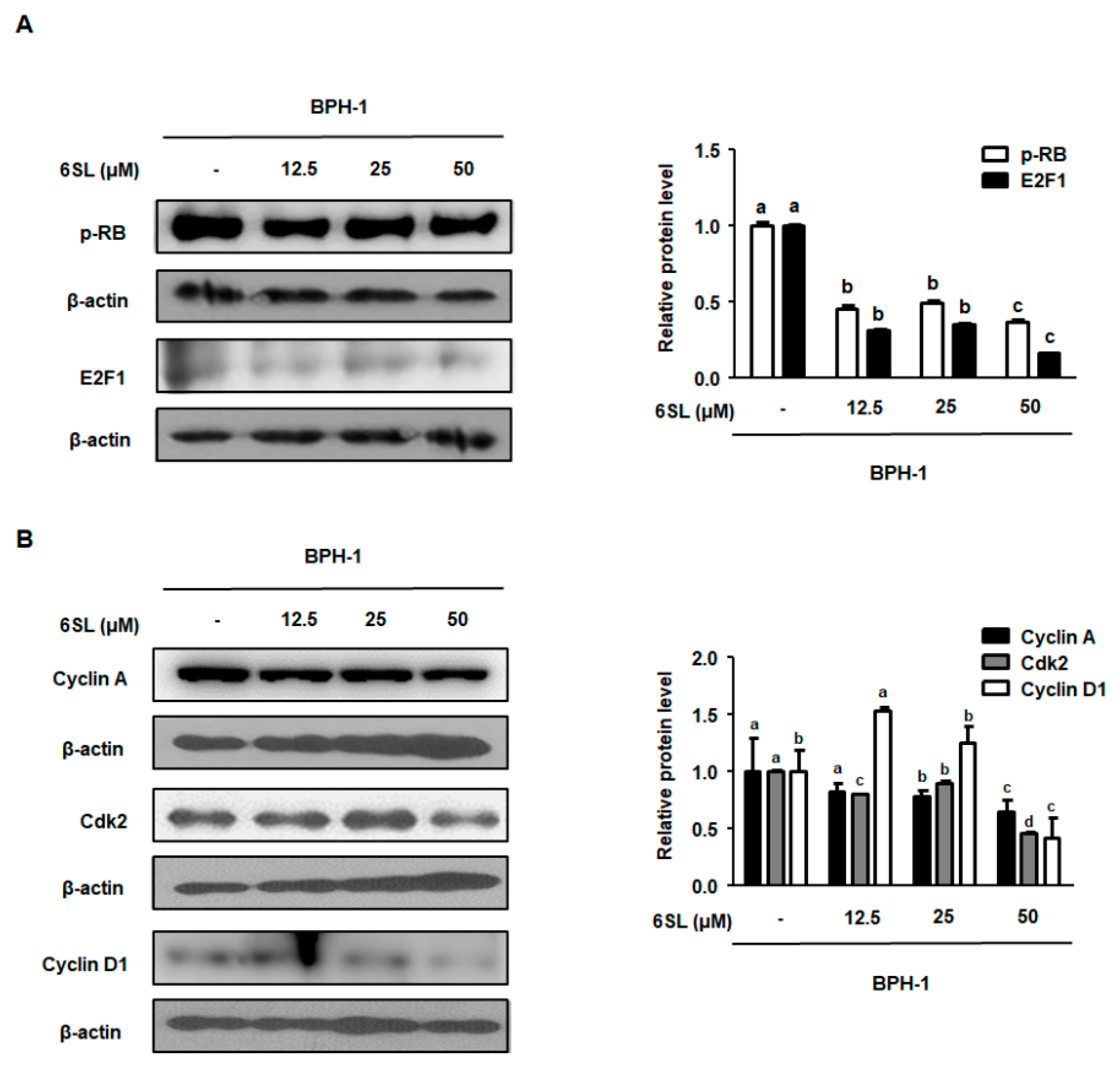
3.5. 6SL Repressed E2F1/pRb Signaling by Regulating G1 and S Checkpoint Protein
4. Discussion
5. Conclusions
Supplementary Materials
Author Contributions
Funding
Conflicts of Interest
Abbreviations
| AR | androgen receptor |
| BPH | benign prostatic hyperplasia |
| Con | control animals |
| DHT | dihydrotestosterone |
| DLP | dorsolateral prostate |
| E2F1 | E2F transcription factor 1 |
| HMOs | Human milk oligosaccharides |
| Fina | rats with BPH orally administrated 5 mg/kg finasteride |
| PCNA | proliferating cell nuclear antigen |
| PSA | prostate-specific antigen |
| Rb | retinoblastoma protein |
| 6SL 0.5 and 6SL 1.0 | rats with BPH intraperitoneally administrated 0.5 or 1.0 mg/kg 6SL |
| TP | testosterone propionate |
| TUMT | transurethral microwave thermotherapy |
| VP | ventral prostate |
| 6SL | 6′-Sialyllactose |
| TETP | the epithelium tissue from prostate |
References
- Bode, L. Recent advances on structure, metabolism, and function of human milk oligosaccharides. J. Nutr. 2006, 136, 2127–2130. [Google Scholar] [CrossRef] [PubMed]
- Bode, L.; Kunz, C.; Muhly-Reinholz, M.; Mayer, K.; Seeger, W.; Rudloff, S. Inhibition of monocyte, lymphocyte, and neutrophil adhesion to endothelial cells by human milk oligosaccharides. Thromb. Haemost. 2004, 92, 1402–1410. [Google Scholar] [CrossRef]
- Oliveros, E.; Vazquez, E.; Barranco, A.; Ramirez, M.; Gruart, A.; Delgado-Garcia, J.M.; Buck, R.; Rueda, R.; Martin, M.J. Sialic acid and sialylated oligosaccharide supplementation during lactation improves learning and memory in rats. Nutrients 2018, 10, 1519. [Google Scholar] [CrossRef] [PubMed]
- Ten Bruggencate, S.J.; Bovee-Oudenhoven, I.M.; Feitsma, A.L.; van Hoffen, E.; Schoterman, M.H. Functional role and mechanisms of sialyllactose and other sialylated milk oligosaccharides. Nutr. Rev. 2014, 72, 377–389. [Google Scholar] [CrossRef] [PubMed] [Green Version]
- Burns, A.J.; Rowland, I.R. Anti-carcinogenicity of probiotics and prebiotics. Curr. Issues Intest. Microbiol. 2000, 1, 13–24. [Google Scholar] [PubMed]
- Kuntz, S.; Rudloff, S.; Kunz, C. Oligosaccharides from human milk influence growth-related characteristics of intestinally transformed and non-transformed intestinal cells. Br. J. Nutr. 2008, 99, 462–471. [Google Scholar] [CrossRef] [PubMed] [Green Version]
- Chung, T.W.; Kim, E.Y.; Kim, S.J.; Choi, H.J.; Jang, S.B.; Kim, K.J.; Ha, S.H.; Abekura, F.; Kwak, C.H.; Kim, C.H.; et al. Sialyllactose suppresses angiogenesis by inhibiting vegfr-2 activation, and tumor progression. Oncotarget 2017, 8, 58152–58162. [Google Scholar] [CrossRef]
- Patel, N.D.; Parsons, J.K. Epidemiology and etiology of benign prostatic hyperplasia and bladder outlet obstruction. Indian J. Urol. IJU J. Urol. Soc. India 2014, 30, 170–176. [Google Scholar]
- Donnell, R.F. Benign prostate hyperplasia: A review of the year’s progress from bench to clinic. Curr. Opin. Urol. 2011, 21, 22–26. [Google Scholar] [CrossRef]
- Sasagawa, I.; Nakada, T.; Kazama, T.; Satomi, S.; Terada, T.; Katayama, T. Volume change of the prostate and seminal vesicles in male hypogonadism after androgen replacement therapy. Int. Urol. Nephrol. 1990, 22, 279–284. [Google Scholar] [CrossRef]
- Izumi, K.; Mizokami, A.; Lin, W.J.; Lai, K.P.; Chang, C. Androgen receptor roles in the development of benign prostate hyperplasia. Am. J. Pathol. 2013, 182, 1942–1949. [Google Scholar] [CrossRef] [PubMed]
- DeGregori, J. The genetics of the e2f family of transcription factors: Shared functions and unique roles. Biochim. Biophys. Acta 2002, 1602, 131–150. [Google Scholar] [CrossRef]
- Goodrich, D.W.; Wang, N.P.; Qian, Y.W.; Lee, E.Y.; Lee, W.H. The retinoblastoma gene product regulates progression through the g1 phase of the cell cycle. Cell 1991, 67, 293–302. [Google Scholar] [CrossRef]
- Phillips, S.M.; Barton, C.M.; Lee, S.J.; Morton, D.G.; Wallace, D.M.; Lemoine, N.R.; Neoptolemos, J.P. Loss of the retinoblastoma susceptibility gene (rb1) is a frequent and early event in prostatic tumorigenesis. Br. J. Cancer 1994, 70, 1252–1257. [Google Scholar] [CrossRef] [PubMed]
- Oelke, M.; Bachmann, A.; Descazeaud, A.; Emberton, M.; Gravas, S.; Michel, M.C.; N’Dow, J.; Nordling, J.; de la Rosette, J.J. European Association of Urology. EAU guidelines on the treatment and follow-up of non-neurogenic male lower urinary tract symptoms including benign prostatic obstruction. Eur. Urol. 2013, 64, 118–140. [Google Scholar] [CrossRef] [PubMed]
- Greco, K.A.; McVary, K.T. The role of combination medical therapy in benign prostatic hyperplasia. Int. J. Impot. Res. 2008, 20 (Suppl. 3), S33–S43. [Google Scholar] [CrossRef]
- Thompson, I.M.; Goodman, P.J.; Tangen, C.M.; Lucia, M.S.; Miller, G.J.; Ford, L.G.; Lieber, M.M.; Cespedes, R.D.; Atkins, J.N.; Lippman, S.M.; et al. The influence of finasteride on the development of prostate cancer. N. Engl. J. Med. 2003, 349, 215–224. [Google Scholar] [CrossRef] [PubMed]
- Li, J.; Tian, Y.; Guo, S.; Gu, H.; Yuan, Q.; Xie, X. Testosterone-induced benign prostatic hyperplasia rat and dog as facile models to assess drugs targeting lower urinary tract symptoms. PLoS ONE 2018, 13, e0191469. [Google Scholar] [CrossRef]
- Lee, M.Y.; Shin, I.S.; Seo, C.S.; Lee, N.H.; Ha, H.K.; Son, J.K.; Shin, H.K. Effects of Melandrium firmum methanolic extract on testosterone-induced benign prostatic hyperplasia in Wistar rats. Asian J. Androl. 2012, 14, 320–324. [Google Scholar] [CrossRef] [Green Version]
- Jin, B.R.; Chung, K.S.; Kim, H.J.; An, H.J. Chinese skullcap (scutellaria baicalensis georgi) inhibits inflammation and proliferation on benign prostatic hyperplasia in rats. J. Ethnopharmacol. 2019, 235, 481–488. [Google Scholar] [CrossRef]
- Altintas, D.M.; Shukla, M.S.; Goutte-Gattat, D.; Angelov, D.; Rouault, J.P.; Dimitrov, S.; Samarut, J. Direct cooperation between androgen receptor and e2f1 reveals a common regulation mechanism for androgen-responsive genes in prostate cells. Mol. Endocrinol. 2012, 26, 1531–1541. [Google Scholar] [CrossRef] [PubMed]
- Bendris, N.; Lemmers, B.; Blanchard, J.M.; Arsic, N. Cyclin a2 mutagenesis analysis: A new insight into cdk activation and cellular localization requirements. PLoS ONE 2011, 6, e22879. [Google Scholar] [CrossRef] [PubMed]
- Tempany, C.M.; Partin, A.W.; Zerhouni, E.A.; Zinreich, S.J.; Walsh, P.C. The influence of finasteride on the volume of the peripheral and periurethral zones of the prostate in men with benign prostatic hyperplasia. Prostate 1993, 22, 39–42. [Google Scholar] [CrossRef] [PubMed]
- Kim, T.H.; Lim, H.J.; Kim, M.S.; Lee, M.S. Dietary supplements for benign prostatic hyperplasia: An overview of systematic reviews. Maturitas 2012, 73, 180–185. [Google Scholar] [CrossRef] [PubMed]
- Chen, X. Human milk oligosaccharides (HMOS): Structure, function, and enzyme-catalyzed synthesis. Adv. Carbohydr. Chem. Biochem. 2015, 72, 113–190. [Google Scholar] [PubMed]
- Kunz, C.; Rudloff, S. Potential anti-inflammatory and anti-infectious effects of human milk oligosaccharides. Adv. Exp. Med. Biol. 2008, 606, 455–465. [Google Scholar] [PubMed]
- Boquien, C.Y. Human milk: An ideal food for nutrition of preterm newborn. Front. Pediatr. 2018, 6, 295. [Google Scholar] [CrossRef]
- Zehra, S. Direct Effects of Milk Oligosaccharides on the Inflammatory Response in Relation to Allergy. Master’s Thesis, McMaster University, Hamilton, ON, Canada, 21 November 2015. [Google Scholar]
- Zhong, W.; Peng, J.; He, H.; Wu, D.; Han, Z.; Bi, X.; Dai, Q. Ki-67 and pcna expression in prostate cancer and benign prostatic hyperplasia. Clin. Investig. Med. 2008, 31, E8–E15. [Google Scholar] [CrossRef]
- Mallik, I.; Davila, M.; Tapia, T.; Schanen, B.; Chakrabarti, R. Androgen regulates cdc6 transcription through interactions between androgen receptor and e2f transcription factor in prostate cancer cells. Biochim. Biophys. Acta 2008, 1783, 1737–1744. [Google Scholar] [CrossRef] [PubMed]
- Buchkovich, K.; Duffy, L.A.; Harlow, E. The retinoblastoma protein is phosphorylated during specific phases of the cell cycle. Cell 1989, 58, 1097–1105. [Google Scholar] [CrossRef]
- Gopinathan, L.; Tan, S.L.; Padmakumar, V.C.; Coppola, V.; Tessarollo, L.; Kaldis, P. Loss of cdk2 and cyclin a2 impairs cell proliferation and tumorigenesis. Cancer Res. 2014, 74, 3870–3879. [Google Scholar] [CrossRef] [PubMed]
- Hofman, K.; Swinnen, J.V.; Claessens, F.; Verhoeven, G.; Heyns, W. The retinoblastoma protein-associated transcription repressor rbak interacts with the androgen receptor and enhances its transcriptional activity. J. Mol. Endocrinol. 2003, 31, 583–596. [Google Scholar] [CrossRef] [PubMed]


© 2019 by the authors. Licensee MDPI, Basel, Switzerland. This article is an open access article distributed under the terms and conditions of the Creative Commons Attribution (CC BY) license (http://creativecommons.org/licenses/by/4.0/).
Share and Cite
Jin, B.-R.; Kim, H.-J.; Kim, E.-Y.; Chung, T.-W.; Ha, K.-T.; An, H.-J. 6′-Sialyllactose Ameliorates In Vivo and In Vitro Benign Prostatic Hyperplasia by Regulating the E2F1/pRb–AR Pathway. Nutrients 2019, 11, 2203. https://doi.org/10.3390/nu11092203
Jin B-R, Kim H-J, Kim E-Y, Chung T-W, Ha K-T, An H-J. 6′-Sialyllactose Ameliorates In Vivo and In Vitro Benign Prostatic Hyperplasia by Regulating the E2F1/pRb–AR Pathway. Nutrients. 2019; 11(9):2203. https://doi.org/10.3390/nu11092203
Chicago/Turabian StyleJin, Bo-Ram, Hyo-Jung Kim, Eun-Yeong Kim, Tae-Wook Chung, Ki-Tae Ha, and Hyo-Jin An. 2019. "6′-Sialyllactose Ameliorates In Vivo and In Vitro Benign Prostatic Hyperplasia by Regulating the E2F1/pRb–AR Pathway" Nutrients 11, no. 9: 2203. https://doi.org/10.3390/nu11092203