Review

The Gut-Brain Axis in Alzheimer’s Disease and Omega-3. A Critical Overview of Clinical Trials

1
IRCCS Fondazione Don Carlo Gnocchi, 20100 Milan, Italy
2
Department of Physiopathology and Transplants, University of Milano, 20100 Milan, Italy
3
Department of Biology and Biotechnology, University of Pavia, 27100 Pavia, Italy
4
Istituto per lo Studio e la Cura del Diabete [ISCD], Casagiove, 81022 Caserta, Italy
*
Author to whom correspondence should be addressed.
Nutrients 2018, 10(9), 1267; https://doi.org/10.3390/nu10091267
Submission received: 24 August 2018 / Revised: 5 September 2018 / Accepted: 6 September 2018 / Published: 8 September 2018
(This article belongs to the Special Issue Brain Aging and Gut-Brain Axis)

Abstract

:
Despite intensive study, neurodegenerative diseases remain insufficiently understood, precluding rational design of therapeutic interventions that can reverse or even arrest the progressive loss of neurological function. In the last decade, several theories investigating the causes of neurodegenerative diseases have been formulated and a condition or risk factor that can contribute is described by the gut-brain axis hypothesis: stress, unbalanced diet, and drugs impact altering microbiota composition which contributes to dysbiosis. An altered gut microbiota may lead to a dysbiotic condition and to a subsequent increase in intestinal permeability, causing the so-called leaky-gut syndrome. Herein, in this review we report recent findings in clinical trials on the risk factor of the gut-brain axis in Alzheimer’s disease and on the effect of omega-3 supplementation, in shifting gut microbiota balance towards an eubiosis status. Despite this promising effect, evidences reported in selected randomized clinical trials on the effect of omega-3 fatty acid on cognitive decline in Alzheimer’s disease are few. Only Mild Cognitive Impairment, a prodromal state that could precede the progress to Alzheimer’s disease could be affected by omega-3 FA supplementation. We report some of the critical issues which emerged from these studies. Randomized controlled trials in well-selected AD patients considering the critical points underlined in this review are warranted.

1. Introduction

The gut microbiota is defined as the microbial community found within the gut. This microbial ecosystem is an essential endocrine “organ” involved in critical functions such as influencing metabolism and the absorption of food, providing trophic and protective functions, instructing innate immunity, and acting as a dynamic filter of environmental exposure in the host [1,2,3,4].
In healthy people, a study of 16S rRNA genes from human faecal microbial communities has shown that the three million bacteria of the gut microbiota belong to two large phyla, the Bacteroidetes and the Firmicutes, but this includes several hundred species-level phylotypes [1,5].
The process of human gut colonization is initiated in utero by different microbial communities present in the amniotic fluid and placenta [6] and is stably maintained over the lifespan of an individual: as an example, the Bacteroidetes/Firmicutes phylum ratio remains remarkably stable over time [5]. The change of the relative abundance of each species in the intestinal microbial community can influence immune performance, physiological homeostasis, and healthy ageing [7,8,9].
In the past, microorganisms have not been considered as particularly relevant to brain development and function, nor in the pathophysiology of chronic brain diseases, such as Alzheimer’s disease (AD), Parkinson’s disease (PD), multiple sclerosis (MS) or mood disorders. The characterization of the healthy human microbiome and, in particular, of the gut microbiome (Human Microbiome Project Consortium, 2012) revealed that the gut is involved in regulating brain function and in shifting the balance between protective and pathogenic immune responses [10,11].
Despite evidences demonstrating tight interactions between gut-associated immune, enteric, endocrine, and nervous systems [12], psychiatric and neurological communities have long ignored gut–brain interactions as a possible target to treat brain diseases.
In the first part of this review, we discuss several lines of evidence that highlight the role of the gut microbiota in the development of neurodegenerative diseases, and in particular AD, focusing on intestinal wellbeing as a possible therapeutic approach. About that, two recent reviews describe how microbiota influences brain function, cognitive decline and behaviour via gut microbiota-brain axis in dementia and AD [13,14]. Although different neurophatological hallmarks of AD, including amyloid plaques, genetic and environment identified, this research proposed a new context to investigate the risk factors to AD development: inflammation and gut–pathogens colonization.
In the second part, we first critically overview the effect of omega-3 fatty acid (FA) supplementation on microbiota composition and then in AD. Recent works underline that both diet [15] and/or prebiotics supplementation [16,17] act in a positive changing microbiota composition and increase anti-inflammatory immune responses. We report different evidences that demonstrate the effects docosahexaenoic acid (DHA) and eicosapentaenoic acid (EPA) supplementation in increasing short chain fatty acids production (SCFA) shifting gut microbiota balance towards eubiosis condition. Despite this promising omega-3 effect, in AD the only evidence of healthy effect of omega-3 FA supplementation is a delay in the cognitive decline in Mild Cognitive Impairment (MCI) [18], also reviewed by Fiala et al. [19]. Indeed, MCI is a prodromal clinical state that could precede the progress to Alzheimer’s disease, and, in particular, elderly patients with MCI are at high risk of developing dementia and AD [20].
In conclusion, current literature offers several ideas to speculate about an important involvement of gut-microorganisms in neurodegenerative diseases and in particular in AD. This critical overview of clinical trials suggests that randomized placebo-controlled double-blinded clinical trials will be necessary to clarify the effect of omega-3 FA supplementation in well-selected AD patients.

2. Gut Microbiota–Brain Axis in Alzheimer’s Disease

2.1. The Gut Ecosystem: Profile and Immunity

In 2002, Hooper et al. described the importance of the unknown world of the bacterial ecosystem: 1013–1014 inhabit our bodies and the great majority of these microorganisms are hidden in the gastrointestinal tract [21]. Initially, little was known about this heterogeneous bacterial population; with more than 1000 bacterial species, six phyla are highly expressed: Firmicutes and Bacteroidetes make up 90%, while Actinobacteria, Proteobacteria, Fusobacteria and Verrucomicrobia represent the remaining 10%. In addition, Rampelli et al. described the genetic profile of such complex communities and their relationship with viruses by means of metagenomic analysis [22].
The human microbiota plays a crucial role in the structural integrity and metabolic functions of the colonic mucosa through the production of short chain fatty acids (SCFAs). Acetate, propionate, and butyrate are the most abundant SCFAs present in the gut lumen, reaching millimolar concentration levels in the colon.
Butyrate is synthesized in the colon via the fermentation of otherwise non-digestible fibers introduced with the diet. A condensation process of two acetyl-CoA molecules is required to produce butyryl-CoA and then butyrate [23,24]. This process also leads to the formation of intermediate molecules such as lactate, succinate or formate. Such intermediate molecules, as well as butyrate, are absorbed from the gut lumen and used by bacteria to survive and proliferate [25,26,27]. Butyrate plays a pivotal role in maintaining human intestinal health by being the major energy source for the colonic mucosa and is fundamental in regulating gene expression, differentiation, inflammation, apoptosis in host cells and cross-feeding interactions with Bifidobacteria [28,29,30,31].
In a recent review, Bourassa discussed the role of bacteria butyrate producers suggesting that a high-fiber diet may improve brain health because it increases Firmicutes bacteria; butyrate may exert a neuroprotective effect as a histone deacetylase inhibitor, but also as a ligand for G-protein–coupled receptors and as a stimulus for mitochondrial activity [27].
In Figure 1, we list the major bacterial genera associated with butyrate production.
Intestinal microbiota colonization is essential in balancing immune responses in a homeostatic manner: studies conducted on germ-free mice suggest that the absence of an intestinal microbiota leads to impaired immune function (for a review see Round and Mazmanian, 2009, [32]) and that microbiota influences the specific phenotypes and physiological functions of T and B cells in the gut mucosa layer (for a review see Honda and Littman, 2016, [33]). These cells play a pivotal role in immune homeostasis by defending against foreign antigens and by maintaining the integrity of the gut mucosal barrier. A balanced gut-microbiota stimulates resident macrophages to release large amounts of interleukin (IL)-10 and transforming growth factor (TGF)-beta [34], thus promoting the induction of regulatory T cells (Tregs) and preventing an increase in the number of proinflammatory T helper 17 (Th17) cells in the gut [35]. In fact, a peripheral tolerance is maintained by the correct balance between gut-bacteria population and responses by the host.
An altered microbiota, including a reduction in health-promoting bacteria such as Lactobacilli and Bifidobacteria, may lead to a dysbiotic condition generated by Gram-negative-bacteria producing immunogenic endotoxins such as lipopolysaccharide (LPS) which increase intestinal permeability [33,36,37]. This condition changes the microbiota taxa profile and switches Treg to T-inflammatory cells (i.e., Th17) triggering a toll-like receptor (TLRs) mediated inflammatory response [38]. Very recently, the intestinal microbiota has emerged as a potential triggering factor in central nervous system–specific auto-immunity disease, as demonstrated in an experimental animal model for MS [39,40,41]. Gut microbiota from patients with MS enabled a spontaneous experimental autoimmune encephalomyelitis in germ-free transgenic mice, devoid of resident microbes, demonstrating that the MS-derived microbiota has factors that may precipitate an MS-like autoimmune disease in a transgenic mouse model [39]. Brain autoimmunity critically depends on the presence of an intact gut flora. Some members of the intestinal microbiota may ignite innate immune responses with the activation of dormant brain-specific T cells within gut-associated lymphatic tissues. Upon activation, such lymphocytes travel to the central nervous system (CNS) and trigger a cascade of events that culminate in the formation of brain-specific autoantibodies. Brain-specific autoantibodies together with T-cell activation produce demyelinating lesions, similar to active multiple sclerosis (MS) plaques [41].
As demonstrated in a recent pilot study conducted in our laboratory, dietary components, and in particular fruit, vegetables and fibers, allow the longtime maintenance of gut–microbiota homeostasis. It was observed that patients with MS who followed a controlled diet showed a significant decrease of Th1 and Th17 cells compared to those that followed a Western diet. This evidence correlated with an increase in the gut microbiota population of Lachnospiraceae family (phylum Firmicutes, see Figure 1), which are butyrate producers [15].
Therefore, these results link the gut microbiota to a variety of brain disorders. Studies of the mechanisms elucidating this transition could have important consequences both for developing new therapeutic strategies for treatment and the diagnosis of human neurodegenerative diseases [33]. It is still unclear whether a dysbiotic microbial profile exists a priori or whether changes in the composition of microbiota occur after the onset of disease [42].

2.2. The Gut Microbiota and Neuroinflammation

Increasing clinical findings [13,14] reveal that the pathogenesis of many neurodegenerative diseases may depend on the gut microbiota and that the resident commensal microbiota modulates CNS autoimmunity [43,44], beyond neuroinflammation [45,46].
A common trait between theories investigating the causes of neurodegenerative diseases is the presence of neuroinflammation [47,48] that has been associated with activation of microglia and peripheral monocytes that cross the BBB. These cells produce inflammatory cytokines and several neurotoxic molecules, such as TNF-α and IL-1β [49,50,51,52,53,54] in an attempt to counteract the formation and/or extend misfolding of neuronal proteins and the formation of insoluble fibrillary aggregates [55,56,57].
AD is associated with impaired cognition and cerebral accumulation of amyloid-β peptides (Aβ). Indeed, recent studies published, respectively, by Italian and American researchers described interactions between brain protein misfolding and microbiota [58,59].
Larsen et al. [60,61] and Jordal et al. [62] have described an abundance of functional bacterial amyloids; amyloid is used by bacteria such as Proteobacteria, Bacteroidetes, Chloroflexi, Actinobacteria and Firmicutes as structural and adhesive material, toxin, and protection against host innate immunological defenses [63]. The human innate immune system recognizes bacterial amyloid proteins by using several pathways involving TLR 1/2, Nod-like receptor-3 protein (NLRP3), nuclear factor kappa-B (NF-kb), CD14, and inducible nitric oxide synthase [57,64,65]. Other misfolded proteins produced by bacteria could predispose to tissue damage and to the production of proinflammatory cytokines associated with the development of dementia [66].
Other Authors [67,68] have shown that in a mouse model of Huntington’s disease treatment with butyrate-like analogues such as phenylbutyrate, histone deacetylase inhibitor or sodium butyrate used to modulate transcription prevents neuronal death and lengthens the life of mice in a dose-dependent manner.
As described above, the gut microbiota could influence the integrity of the blood-brain barrier (BBB). For example, this can be observed in a microbiota dysbiotic condition that induces increased permeability of the gut (the so-called “leaky gut”) in both foetal and adult mouse brains. Braniste et al. [69] conducted a study in adult and embryonic animals showing the relationship between the lack of a normal gut flora in germ-free mice and increased BBB permeability. This study highlighted the importance of gut microbiota integrity on the CNS, especially in the progression of neurodegenerative diseases. Studies by Diaz et al. in mice have shown that the normal gut microbiota modulates brain development and subsequent adult behaviour, investigated as motor control and anxiety behaviours, suggesting that the microbial colonization process triggers cellular pathways that affect specific neuronal circuits [11]. Other authors have shown that the disruption or absence of the microbiota in mice impaired the function of the BBB and altered cortical myelination and hippocampal neurogenesis, decreased cognitive function and memory formation, and decreased social behaviour [70].
According to Alkasir et al. [13] that proposed a schematic representation of the gut-microbiota and neuroinflammation axis in AD, we can speculate that dysfunction of the gut epithelial barrier results in peripheral inflammation. This condition is linked with dementia onset because the production of inflammatory cytokines, like IL-1β, reduces Aβ phagocytosis, induces NLRP3-inflammasome activation with consequent release of NLRP3-related cytokines such as caspase-1 and IL-18 [71], thus increasing amyloid-beta deposits.
Therefore, modulation of innate immune responses through changes in microbiota composition may exert healthy effects especially on cognitive decline in MCI patients, possibly slowing down or avoiding the progression to AD.

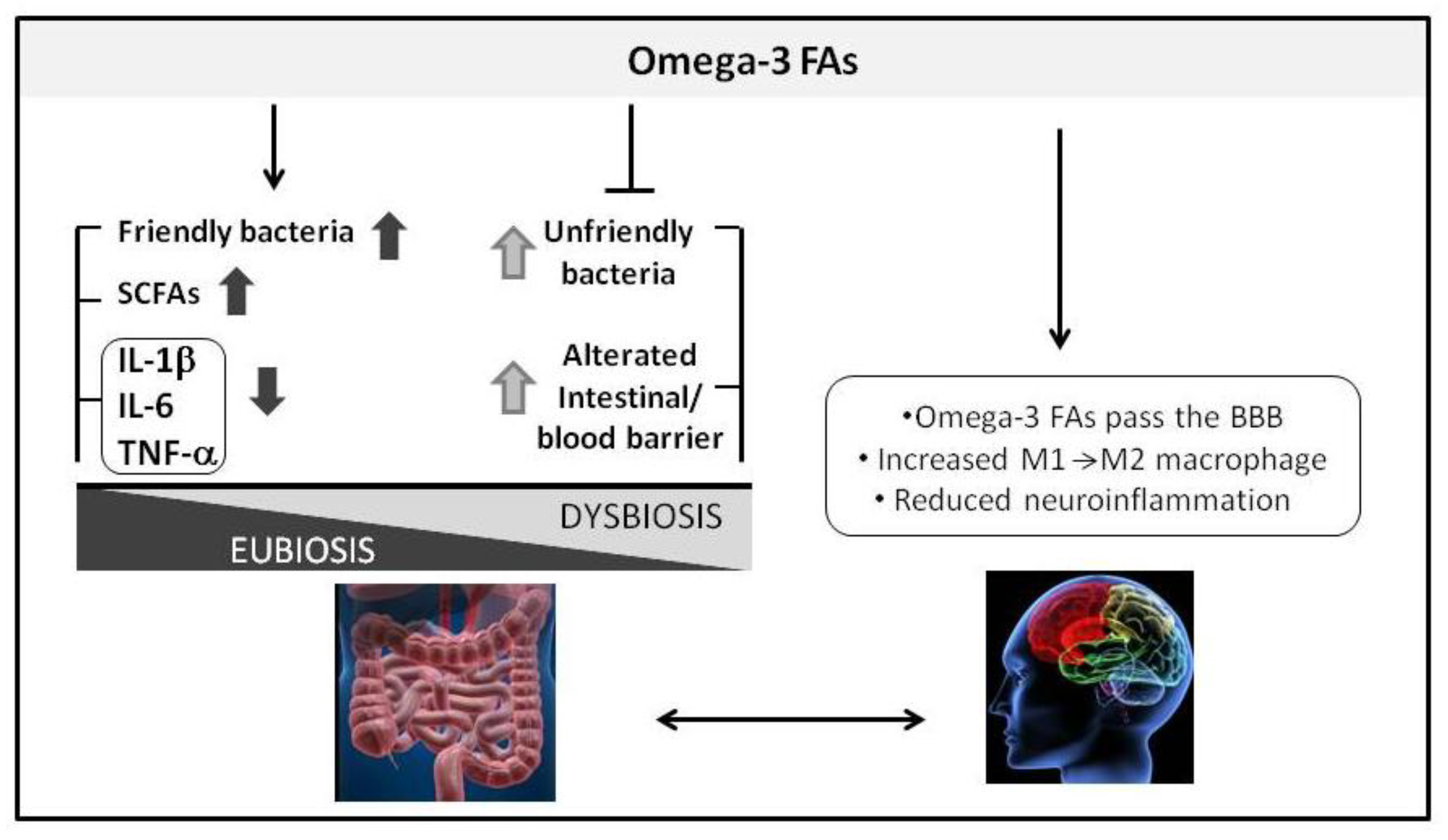
3. Omega-3 Fatty Acids and Gut-Brain Axis in Alzheimer’s Disease

3.1. Omega-3 Fatty Acids and Microbiota

Omega-3 FAs are a class of polyunsaturated fatty acids derived from the double bond in third position, starting the count from the terminal carbon of the chain. The three most important physiological omega-3 FAs are described: EPA, alpha-linolenic acid (ALA), and DHA. FAs are precursors for neuronal components membranes, fluidity of cell membranes, signalling, neurotransmission and modulation of enzymatic activities [72,73].
Recent studies addressing the correlation between omega-3 FAs and the human gut-microbiota [74,75,76,77,78] provided new insight into the microbiota composition, evidencing how this depends from several environmental factors, such as diet, exposure to microorganisms and antibiotic or pharmacologic therapies (Table 1). However, in spite of this, it is still a highly debated issue. Below we reported systematic reviews that are currently shared by the scientific community regarding the impact of omega-3 on microbiota.
Very recently, Watson et al., by next-generation sequencing (NGS) technology, described the effect of 4 g a day of mixed DHA/EPA supplement for eight weeks on the microbiota composition of 20 middle-aged healthy individuals [73]. Differences were not observed in the Firmicutes/Bacteroidetes phyla ratio, but differences were reported at the family and genus levels. This study highlighted the increased abundance of butyrate-producing bacterial genera (phylum Firmicutes, see Figure 2) after omega-3 FAs supplementation.
The Canola Oil Multicenter Intervention Trial was a double-blinded randomised crossover clinical study carried out on 25 volunteers who were enrolled based on the presence of at least one of the metabolic syndrome risk factors [74]. Five different unsaturated oil blends were used for 30 days, among these conventional canola oil and DHA-enriched high oleic canola oil (containing about 3.5 g DHA). Results, in line with previous work [73], showed no significant differences at the phylum level. However, after DHA dosing, a reversible increase in the abundance of so-called ‘beneficial’ bacterial genera, such as Bifidobacterium, Lachnospira, Roseburia and Lactobacillus (Figure 1), was observed during one or both types of omega-3 FAsA interventions. This is consistent with data obtained in mice.
In a case report, Noriega et al., using NGS, described the results of a diet treatment in a 45-year-old healthy man after two weeks of daily supplementation with 600 mg of omega-3 PUFA using fish protein: the Firmicutes/Bacteroidetes phyla ratio increased and Actinobacteria decreased. At the genera level, a reduction in Faecalibacterium versus an increase in Blautia, Coprococcus, Roseburia, Subdoligranulum and Ruminococcus genera was observed. This change in microbiota composition led to greater SCFA production. After two washout weeks, the authors observed a reversed trend, confirming that the gut microbiota was susceptible to dietary change, as a dynamic ecosystem [75]. This result highlighted a temporal shift in the composition of microbial communities: an omega-3 FA dietary supplementation determined a marked and notable increase in genera associated with beneficial effects in the host (Figure 2) [75].
Recently, Menni et al. correlated DHA circulating levels with DHA dietary intake determined by a Food Frequency Questionnaire using a cohort of 876 middle-aged and elderly women. The DHA intake of 350 mg/day was found to positively correlate with SCFA-producing bacteria as demonstrated by using next-generation DNA sequencing (i.e., Lachnospiraceae family) [76].
The Pilchardus Study was a multicentre randomised clinical trial in patients diagnosed with type 2 diabetes who had not received any kind of treatment with antidiabetic drugs (using quantitative real-time polymerase chain reaction on target bacterial indicators). After a sardine-enriched diet in drug-naïve patients, the authors described a change in the gut microbiota at the phylum level, with a decrease in the Firmicutes/Bacteroidetes ratio and an increase in the Prevotella genus [77]. This evidence was also described by Costantini et al., 2017 [16].
In conclusion, omega-3 FAs supplementation increases SCFA production shifting gut microbiota balance towards a healthy status (eubiosis). The mechanism that underlies bacterial selection of omega-3 FAs has not yet been determined.

3.2. Omega-3 Fatty Acids: A Nutritional Strategy in Alzheimer’s Disease Treatment?

As previously described, investigations into dietary impact on the gut microbiota show an interactive relationship between altered microbiota composition and the development of neurodegenerative diseases [46,78,79]. Given the effect of omega-3 FA in microbiota and given its anti-inflammatory effects, omega-3 FAs supplementation has emerged as a candidate for the prevention and management of AD.
Furthermore, meta-analyses studies described significantly lowered DHA levels in the blood as well as lower brain/cerebrospinal fluid (CSF) concentrations in patients with AD [77]. Several groups also reported that a reduction in cerebral omega-3 was linked to cognitive aging and to an increase in the progression of AD [80,81].
To analyse the impact of omega-3 FAs supplementation on AD cognitive impairment, we systematically collected articles found in MEDLINE. The search strategy combined the term “omega-3” AND “Alzheimer” and identified 394 papers. After screening, full-text examination, eligibility analysis we finally selected six papers, reported in Table 2. We selected only randomized control trials (RCT) and we included only studies where omega-3 FA supplementation combined DHA and EPA. Studies describing the effects of EPA and DHA used in combination with other substances were excluded (Table 2). All studies selected reported the effects on mild to moderate AD patients of an omega-3 FA supplement duration between 4 and 18 months.
Sample size varied from 17 to 402 women and men, 55–90 years old with mild-to-moderate AD, according to the Mini-Mental State Examination (MMSE) questionnaire (Table 3). The supplementation time varied from four months to 18 months. In all studies patients were subjected to the medication for AD prior to the beginning of intervention. In all studies, placebo groups received soy oil, olive oil, corn oil, or an isocaloric product.
The most frequent questionnaires that were used in these studies to assess cognitive performances were Alzheimer’s Disease Assessment Scale (ADAS-cog) and MMSE (Table 3).
Freund-Levi et al. [18] in a randomized, double-blind placebo-controlled clinical trial assessed omega-3 FAs supplementation in 174 patients (mean age: 74 ± 9 years) with mild-to moderate AD. The treated group received each day 1720 mg DHA and 600 mg EPA for six months, whereas the placebo group received each day 4 g of corn oil, containing 2.4 g of linoleic acid. ADAS-cog, MMSE and Clinical Dementia Rating Scale (CDR) did not show a statistically significant difference between the two groups. A significant difference was measured in the first six months only in a small number of patients with very mild AD.
In a second paper published from the same research group using the same sample [82], neuropsychiatric behaviour was assessed by means of Neuropsychiatric inventory (NPI), Disability Assessment for Dementia Scale (DAD) and Montgomey Asberg Depression Rating Scale (MADRS). No significant differences were observed in the depression scale between the omega-3FAs group vs. the placebo group (NPI or MADRS score) and no statistically significant difference was seen in DAD score changes.
Quinn et al. [83] assessed 402 patients with mild-to moderate AD (mean age: 76 ± 8.7 years). The intervention group received each day 2 g of DHA and the placebo group received soy or corn oil for 18 months. No significant effect was observed in cognitive and functional decline in ADAS-cog, CDR, Alzheimer Disease Cooperative Study—Activities of Daily Living (ADCS-ADL), NPI, and in MMSE on the 18-month follow-up. Some patients (n = 102) received volumetric magnetic resonance imaging (MRI) and there was no change in brain atrophy decline between the two groups. ADCS-ADL assess basic living skills such as eating and bathing. A variant of this method is Alzheimer Disease Cooperative Study—Instrumental Activities of Daily Living (ADCS-IADL) that measure more complex daily functions such as using the telephone, shopping or cooking (Table 3). Quinn et al. [83] used the full scale and did not find statistically significant differences between the placebo and the intervention group.
Shinto et al. [84] studied 34 subjects older than 55 years with probable AD in a pilot study organized in a three-arm, parallel group, double-blind placebo-controlled trial. One group received only omega-3 FAs (975 mg EPA and 675 mg DHA), a second group received the same supplement with the addition of alpha lipoic acid (600 mg ALA) while the placebo group only received soy oil. Supplementation lasted 12 months. As a result, there were no differences in ADAS-cog and ADCS-ADL between placebo and both supplemented groups. There was no difference between placebo and omega 3 FA group in MMSE scores, whereas a statistically significant difference was found between the placebo group and the omega-3 FAs + ALA group. Shinto et al. [84] used a modified version of the ADCS-ADL scores separating the two skills: basic living skills, by means of ADCS-ADL, and more complex daily functions, by means of ADCS-IADL. Whereas in ADCS-ADL there were no differences between the three examined groups, both groups that received omega-3 FAs evidenced a delay in functional decline when compared with placebo with the ADCS-IADL scores. Therefore, this scale that measures more complex tasks proved to be more sensitive to AD progression than ADCS-ADL.
Phillips et al. [85] assessed omega-3 FAs supplementation in 57 subjects with cognitive impairment and 19 with AD (mean age 71.1 ± 4.8 years). For four months, the treated group received each day 625 mg DHA and 600 mg EPA. The placebo group received olive oil. There was no statistical difference in MMSE between the two groups.
Eriksdotter et al. [86] reported on an investigative part of the Omega AD study, a randomized, double-blind placebo-controlled trial in 174 patients (mean age: 74 ± 9 years) with mild to moderate AD according to the Diagnostic and Statistical Manual of Mental Disorders IV edition criteria (DSM-IV). The authors demonstrated a dose-response relationship between omega-3 FA levels in plasma and the preservation of cognitive functions after the six-month administration of a DHA-rich omega-3 FAs supplementation. The preservation of cognitive performance, assessed by Alzheimer’s Disease Assessment Scale–Cognition (but not a Mini-Mental State Examination) in mild forms of AD or of mild cognitive impairment (MCI) was significantly associated with increasing plasma omega-3 FA levels over time: the higher the omega-3 FA plasma level rose, the lower was the rate of cognitive deterioration.
We summarize as follows some of the critical issues which emerged from these studies:
  • In two cases the sample size was too small, limiting statistical power [84,85];
  • It is difficult to establish comparative analysis because of heterogeneity in composition, dosage and duration of supplementation;
  • It is difficult to establish comparative analysis because of heterogeneity of methods used to assess cognition (Table 3);
  • The placebo group in one study [85] included olive oil, a source of monounsaturated fatty acid. As previously demonstrated by other authors, monounsaturated fatty acids are inversely correlated with cognitive decline and therefore cannot be considered a totally inert substance on cognition performance;
  • In all presented studies, the subjects received medications for AD along with omega-3 FAs. This condition could also contribute to good outcomes in control group, hiding and/or slowing down the physiological decline in AD;
  • The duration of treatment with omega-3 FAs could be too short for a chronic disease such as AD;
  • Omega-3 FAs dosage could be insufficient to provide significant benefits.
In summary, the evidence reported herein indicates that the effect of omega-3 FAs on cognitive outcomes in AD was either insufficient or low-strength. Most studies do not find statistically significant results when the intervention group is compared to the placebo group. Nevertheless, according to MMSE scores assessments, omega-3 FAs supplementation seems to demonstrate efficacy in very mild AD [18]. MMSE measures cognitive performances like memory, orientation, language, attention, calculation, and visual construction. In particular, the analysis of subjects with mild AD demonstrated that the cognitive improvement concerns mainly memory functions. Memory deficiency is considered an AD hallmark symptom that most times represents the beginning of the clinically-relevant disease. These data are in agreement with findings from epidemiological observational studies. Several cohort studies suggest that fish consumption, being a source of omega-3 FAs, would have a role in AD prevention but not as adjuvant once the disease has already established [87,88,89,90].
Furthermore, it is possible that omega-3 FAs improve more complex activities of daily living. This data has to be confirmed in further trials.
Macrophages of patients with MCI and AD are defective in Aβ phagocytosis and show low resistance to Aβ-induced apoptosis. This functional defect of macrophages in AD is improved by omega-3 FAs. Very recently, Famenini et al. described how a drink containing omega-3, antioxidants and resveratrol maintained active Aβ1-42 phagocytosis, prevented brain amyloidosis, thus preserving cognition. As concerns the cellular mechanisms involved in the process, omega-3 FAs supplementation would modulate macrophages to the pro-phagocytic M1–M2 type and improve cognition in patients with MCI [17]. These macrophages exert an effective unfolded protein response against endoplasmic reticulum stress. After taking this supplement, several patients with MCI maintained a stable cognitive status for three years. However, the validity of this study was limited by its small size and the uncontrolled experimental design. Further randomised clinical trials are required to investigate this observational data [91].
Fiala et al. [91] discussed the use of immunotherapy for the treatment of MCI by omega-3 FAs supplementation. Based on a previous study, the authors described how omega-3 supplementation was successful in: (i) modulating a macrophage phenotype from an intermediate M2 phenotype (anti-inflammatory); (ii) attenuating Aβ1-42 secretion in AD; and (iii) reducing cytokine secretion in human neural cells with the consequent formation of a novel, DHA-derived 10,17S-docosatriene DHA (NPD1) that promoted brain cell survival by inducing gene-expression programs of anti-apoptotic and neuroprotective pathways, especially in the hippocampus, a structure involved in memory [92,93,94]. NPD1 also has beneficial immune effects under inflammatory and degenerative conditions [95]. Despite the powerful biochemical and immunological effects of omega-3 FAs supplementation demonstrated by several kinds of in vitro and in vivo activity, the quality of omega-3 products available on the market are poorly standardised and of low quality. Thus, the authors suggest that a combination drink called “Smartfish” containing an emulsion of omega-3 and other fatty acids, combined with the antioxidants, vitamin D3 and resveratrol maintained active Aβ1-42 phagocytosis, prevented brain amyloidosis leading to a preservation of cognition.

4. Probiotic Strategy in Alzheimer’s Disease Treatment?

Some probiotics could enable the effective therapeutic management of neurodegenerative disorders.
Several studies have described the immunomodulatory action of some probiotic bacteria able to reduce the production of pro-inflammatory cytokines and to increase the activity of the M2 phenotype macrophages.
It has been recently demonstrated in vitro that a probiotic mixture containing Lactobacillus rhamnosus, Bifidobacterium lactis and Bifidobacterium longum (Serobioma, provided by Bromatech, s.r.l., Milan, Italy) exerted an anti-inflammatory and immunomodulatory action on THP-1 cells [96]. In particular, they reported a 70–80% reduction in the production of pro-inflammatory cytokines such as IL-1β and IL-6 and a statistically significant increase in the production of anti-inflammatory cytokine IL-10 by using the transwell model.
In animal models, some probiotics appear to influence the central nervous system (CNS) via the gut–brain axis [97].
Liu et al. reported that treatment with Lactobacillus plantarum PS128 normalized anxiety-like behaviour and reduced inflammatory cytokine levels in the plasma in stressed mice, giving psychotropic effects [98].
Moreover, treatment with VSL#3 (provided by Ferring Pharmaceutical Spa, Milan, Italy) that is a probiotic mixture containing eight different Gram-positive bacterial species improved neuronal functions and plasticity in young and aged rats. VSL#3 altered the expression of genes in the brain tissue such as BDNF and synapsin that are associated with inflammation and neural plasticity [99].
The effect of Bifidobacterium breve strain A1 on the behaviour and physiological processes of AD in mice models was investigated by Kobayashi et al. [100]. Authors found that the administration of B. breve A1 to AD mice increased spatial memory performances and suppressed the hippocampal expression of inflammation and immune-reactive genes that are induced by Aβ.
In a randomized, double-blind and controlled trial that enrolled 60 AD patients, it has been reported that a 12-week consumption of a mixture of probiotics improved cognitive function (measured by MMSE score) and metabolic status (measured by MDA, hs-CRP, insulin metabolism markers, serum levels of triglycerides and VLDLs) [101].
It is important to underline that, although the probiotic approach could be a strategy to improve gut microbiota composition, it did not show permanent effects able to treat dysbiosis.

5. Conclusions

Our gut microbiota plays a crucial role in the structural integrity of the intestinal mucosa through the production of SCFAs, such as butyrate, propionate and acetate. An altered microbiota may lead to a dysbiotic condition with the production of immunogenics endotoxins such as LPS and increasing intestinal permeability (the so-called leaky gut). This condition triggers an inflammatory response. The activation of microglia and peripheral monocytes that cross the BBB produces inflammatory cytokines and neurotoxic molecules that trigger neuroinflammation, as reported also in AD. Further studies are needed to characterize the gut microbiota composition in AD patients.
Several studies evidence how omega-3 FAs supplementation exerts a significant effect on microbiota composition, through a modulatory effect that increases butyrate producer bacteria. In this sense, we may consider omega-3 FAs as a prebiotic strategy for a healthy gut microbiota.
Despite this promising effect, there is little evidence regarding the beneficial effect of omega-3 FAs supplementation in AD’s cognitive decline. After a systematic revision of the literature, we selected six randomized control trials on mild to moderate AD patients where omega-3 FA supplement duration was between four and 18 months. Those short-term randomized trials with small sample size do not show any effect on cognitive improvement and therefore, longer term trials with longer omega-3 FA supplementation in early stage AD are warranted. Some studies reported that only mild cognitive decline in MCI could be affected by omega-3 FA supplementation. In this review, we report some of the critical issues which emerged from the studies.
Furthermore, we also believe it will be necessary to improve the quality and standardization of omega-3 FAs supplements available on the market, to study dose-dependent and time-dependent effects of nutritional supplements. It will also be necessary to quantify serum omega-3 FAs before and after supplementation in AD patients.
In conclusion, randomized controlled trials considering the critical issues underlined in this review are warranted.

Author Contributions

F.L.R., M.C. and P.R. collected and reviewed the literature, wrote the manuscript and created figures; D.R., A.O., C.D.I. and M.R. critically analyzed and reviewed the manuscript. P.R. and M.C. were also the overall guarantors of this manuscript.

Funding

This research was supported by the Italian Ministry of Education, University and Research (MIUR): Dipartimenti di Eccellenza Program (2018–2022)—Dept. of Biology and Biotechnology “L. Spallanzani”, University of Pavia.

Acknowledgments

We thank Ilaria Cabrini and the Universitiamo staff of the University of Pavia for support in our crowdfunding “Noi coltiviamo la memoria.” We thank Istituto Per Lo Studio e La Cura Del Diabete S.r.l. (Caserta) Guna S.p.A, Named S.p.A, Miconet S.r.l., A.V.D Reform, Bromatech S.r.l, METEDA S.r.l., for supporting us as donors. We thank experts from BioMed Proofreading® LLC for a first English editing and we also thank Dr. Alexander Salerno for English editing of the final version of the review.

Conflicts of Interest

The authors declare no conflict of interest.

References

  1. Eckburg, P.B.; Bik, E.M.; Bernstein, C.N.; Purdom, E.; Dethlefsen, L.; Sargent, M.; Gill, S.R.; Nelson, K.E.; Relman, D.A. Diversity of the human intestinal microbial flora. Science 2005, 308, 1635–1638. [Google Scholar] [CrossRef] [PubMed]
  2. Hooper, L.V. Bacterial contributions to mammalian gut development. Trends Microbiol. 2004, 12, 129–134. [Google Scholar] [CrossRef] [PubMed]
  3. Bäckhed, F.; Ley, R.E.; Sonnenburg, J.L.; Peterson, D.A.; Gordon, J.I. Host-bacterial mutualism in the human intestine. Science 2005, 307, 1915–1920. [Google Scholar] [CrossRef] [PubMed]
  4. Nicholson, J.K.; Holmes, E.; Kinross, J.; Burcelin, R.; Gibson, G.; Jia, W.; Pettersson, S. Host-gut microbiota metabolic interactions. Science 2012, 336, 1262–1267. [Google Scholar] [CrossRef] [PubMed]
  5. Faith, J.J.; Guruge, J.L.; Charbonneau, M.; Subramanian, S.; Seedorf, H.; Goodman, A.L.; Clemente, J.C.; Knight, R.; Heath, A.C.; Leibel, R.L. The long-term stability of the human gut microbiota. Science 2013, 341, 1237439. [Google Scholar] [CrossRef] [PubMed]
  6. Collado, M.C.; Rautava, S.; Aakko, J.; Isolauri, E.; Salminen, S. Human gut colonisation may be initiated in utero by distinct microbial communities in the placenta and amniotic fluid. Sci. Rep. 2016, 6, 23129. [Google Scholar] [CrossRef] [PubMed] [Green Version]
  7. Turnbaugh, P.J.; Hamady, M.; Yatsunenko, T.; Cantarel, B.L.; Duncan, A.; Ley, R.E.; Sogin, M.L.; Jones, W.J.; Roe, B.A.; Affourtit, J.P. A core gut microbiome in obese and lean twins. Nature 2009, 457, 480–484. [Google Scholar] [CrossRef] [PubMed]
  8. Candela, M.; Biagi, E.; Brigidi, P.; O’Toole, P.W.; De Vos, W.M. Maintenance of a healthy trajectory of the intestinal microbiome during aging: A dietary approach. Mech. Ageing Dev. 2014, 136, 70–75. [Google Scholar] [CrossRef] [PubMed]
  9. Ottman, N.; Smidt, H.; de Vos, W.M.; Belzer, C. The function of our microbiota: Who is out there and what do they do? Front. Cell. Infect. Microbiol. 2012, 2, 104. [Google Scholar] [CrossRef] [PubMed]
  10. Michel, L.; Prat, A. One more role for the gut: Microbiota and blood brain barrier. Ann. Transl. Med. 2016, 4, 1–15. [Google Scholar] [CrossRef]
  11. Diaz Heijtz, R.; Wang, S.; Anuar, F.; Qian, Y.; Bjorkholm, B.; Samuelsson, A.; Hibberd, M.L.; Forssberg, H.; Pettersson, S. Normal gut microbiota modulates brain development and behavior. Proc. Natl. Acad. Sci. USA 2011, 108, 3047–3052. [Google Scholar] [CrossRef] [PubMed] [Green Version]
  12. Mayer, E.A. Gut feelings: The emerging biology of gut-brain communication. Nat. Rev. Neurosci. 2011, 12, 453–466. [Google Scholar] [CrossRef] [PubMed]
  13. Alkasir, R.; Li, J.; Li, X.; Jin, M.; Zhu, B. Human gut microbiota: The links with dementia development. Protein cell. 2016, 8, 90–102. [Google Scholar] [CrossRef] [PubMed]
  14. Hu, X.; Wang, T.; Jin, F. Alzheimer’s disease and gut microbiota. Life Science. 2016, 59, 1006–1023. [Google Scholar] [CrossRef] [PubMed] [Green Version]
  15. Saresella, M.; Mendozzi, L.; Rossi, V.; Mazzali, F.; Piancone, F.; La Rosa, F.; Marventano, I.; Caputo, D.; Felis, G.E.; Clerici, M. Immunological and Clinical Effect of Diet Modulation of the Gut Microbiome in Multiple Sclerosis Patients: A Pilot Study. Front. Immunol. 2017, 8, 1391. [Google Scholar] [CrossRef] [PubMed]
  16. Costantini, L.; Molinari, R.; Farinon, B.; Merendino, N. Impact of Omega-3 Fatty Acids on the Gut Microbiota. Int. J. Mol. Sci. 2017, 18, 2645. [Google Scholar] [CrossRef] [PubMed]
  17. Fiala, M.; Restrepo, L.; Pellegrini, M. Immunotherapy of Mild Cognitive Impairment by ω-3 Supplementation: Why Are Amyloid-β Antibodies and ω-3 Not Working in Clinical Trials? J. Alzheimers Dis. 2018, 62, 1013–1022. [Google Scholar] [CrossRef] [PubMed] [Green Version]
  18. Freund-Levi, Y.; Eriksdotter-Jönhagen, M.; Cederholm, T.; Basun, H.; Faxén-Irving, G.; Garlind, A.; Vedin, I.; Vessby, B.; Wahlund, L.O.; Palmblad, J. Omega-3 fatty acid treatment in 174 patients with mild to moderate Alzheimer disease: OmegAD study: A randomized double-blind trial. Arch. Neurol. 2006, 63, 1402–1408. [Google Scholar] [CrossRef] [PubMed]
  19. Fiala, M.; Kooij, G.; Wagner, K.; Hammock, B.; Pellegrini, M. Modulation of innate immunity of patients with Alzheimer’s disease by omega-3 fatty acids. FASEB J. 2017, 31, 3229–3239. [Google Scholar] [CrossRef] [PubMed]
  20. La Rosa, F.; Saresella, M.; Baglio, F.; Piancone, F.; Marventano, I.; Calabrese, E.; Nemni, R.; Ripamonti, E.; Cabinio, M.; Clerici, M. Immune and Imaging Correlates of Mild Cognitive Impairment Conversion to Alzheimer’s Disease. Sci. Rep. 2017, 7, 16760. [Google Scholar] [CrossRef] [PubMed] [Green Version]
  21. Hooper, L.V.; Midtvedt, T.; Gordon, J.I. How host-microbial interactions shape the nutrient environment of the mammalian intestine. Annu. Rev. Nutr. 2002, 22, 283–307. [Google Scholar] [CrossRef] [PubMed]
  22. Rampelli, S.; Soverini, M.; Turroni, S.; Quercia, S.; Biagi, E.; Brigidi, P.; Candela, M. ViromeScan: A new tool for metagenomic viral community profiling. BMC Genomics 2016, 17, 165. [Google Scholar] [CrossRef] [PubMed]
  23. Gottschalk, G. Bacterial Metabolism; Springer-Verlag: New York, NY, USA, 1979. [Google Scholar]
  24. Pryde, S.E.; Duncan, S.H.; Hold, G.L.; Stewart, C.S.; Flint, H.J. The microbiology of butyrate formation in the human colon. FEMS Microbiol. Lett. 2002, 217, 133–139. [Google Scholar] [CrossRef] [PubMed] [Green Version]
  25. Cummings, J.H.; Rombeau, J.L.; Sakata, T. Physiological and Clinical Aspects of Short-Chain Fatty Acids, 2nd ed.; Cambridge University Press: Cambridge, UK, 2004. [Google Scholar]
  26. Topping, D.L.; Clifton, P.M. Short-Chain Fatty Acids and Human Colonic Function: Roles of Resistant Starch and Nonstarch Polysaccharides. Physiol. Rev. 2001, 81, 1031–1064. [Google Scholar] [CrossRef] [PubMed]
  27. Bourassa, M.W.; Alim, I.; Bultman, S.J.; Ratan, R.R. Butyrate, neuroepigenetics and the Gut Microbiome: Can a High Fiber Diet Improve Brain Health? Neurosci. Lett. 2016, 625, 56–63. [Google Scholar] [CrossRef] [PubMed]
  28. Louis, P.; Young, P.; Holtrop, G.; Flint, H.J. Diversity of human colonic butyrate-producing bacteria revealed by analysis of the butyryl-CoA: Acetate CoA-transferase gene. Environ. Microbiol. 2010, 12, 304–314. [Google Scholar] [CrossRef] [PubMed]
  29. Louis, P.; Flint, H.J. Diversity, metabolism and microbial ecology of butyrate-producing bacteria from the human large intestine. FEMS Microbiol. Lett. 2009, 294, 1–8. [Google Scholar] [CrossRef] [PubMed] [Green Version]
  30. Flint, H.J.; Duncan, S.H.; Louis, P. The impact of nutrition on intestinal bacterial communities. Curr. Opin. Microbiol. 2017, 38, 59–65. [Google Scholar] [CrossRef] [PubMed]
  31. Leccioli, V.; Oliveri, M.; Romeo, M.; Berretta, M.; Rossi, P. A New Proposal for the Pathogenic Mechanism of Non-Coeliac/Non-Allergic Gluten/Wheat Sensitivity: Piecing Together the Puzzle of Recent Scientific Evidence. Nutrients 2017, 9, 1203. [Google Scholar] [CrossRef] [PubMed]
  32. Round, J.L.; Mazmanian, S.K. The gut microbiota shapes intestinal immune responses during health and disease. Nat. Rev. Immunol. 2009, 9, 313–323. [Google Scholar] [CrossRef] [PubMed] [Green Version]
  33. Honda, K.; Littman, D.R. The microbiota in adaptive immune homeostasis and disease. Nature 2016, 535, 75–84. [Google Scholar] [CrossRef] [PubMed]
  34. Schieber, A.M.; Lee, Y.M.; Chang, M.W.; Leblanc, M.; Collins, B.; Downes, M.; Evans, R.M.; Ayres, J.S. Disease tolerance mediated by microbiome E. coli involves inflammasome and IGF-1 signaling. Science 2015, 350, 558–563. [Google Scholar] [CrossRef] [PubMed] [Green Version]
  35. Rivollier, A.; He, J.; Kole, A.; Valatas, V.; Kelsall, B.L. Inflammation switches the differentiation program of Ly6Chi monocytes from antiinflammatory macrophages to inflammatory dendritic cells in the colon. J. Exp. Med. 2012, 209, 139–155. [Google Scholar] [CrossRef] [PubMed] [Green Version]
  36. Cândido, F.G.; Valente, X.F.; Grześkowiak, L.M.; Moreira, A.P.; Rocha, D.M.; Alfenas, R. Impact of dietary fat on gut microbiota and low-grade systemic inflammation: Mechanisms and clinical implications on obesity. Int. J. Food Sci. Nutr. 2017, 69, 125–143. [Google Scholar] [CrossRef] [PubMed]
  37. Brun, P.; Castagliuolo, I.; Pinzani, M.; Palù, G.; Martines, D. Exposure to bacterial cell wall products triggers an inflammatory phenotype in hepatic stellate cells. Am. J. Physiol. Gastrointest. Liver Physiol. 2005, 289, 571–578. [Google Scholar] [CrossRef] [PubMed]
  38. Ohnmacht, C.; Park, J.H.; Cording, S.; Wing, J.B.; Atarashi, K.; Obata, Y.; Gaboriau-Routhiau, V.; Marques, R.; Dulauroy, S.; Fedoseeva, M. The microbiota regulates type 2 immunity through RORγt+ T cells. Science 2015, 349, 989–993. [Google Scholar] [CrossRef] [PubMed]
  39. Berer, K.; Gerdes, L.A.; Cekanaviciute, E.; Jia, X.; Xiao, L.; Xia, Z.; Liu, C.; Klotz, L.; Stauffer, U.; Baranzini, S.E. Gut microbiota from multiple sclerosis patients enables spontaneous autoimmune encephalomyelitis in mice. Proc. Natl. Acad. Sci. USA 2017, 114, 10719–10724. [Google Scholar] [CrossRef] [PubMed] [Green Version]
  40. Berer, K.; Krishnamoorthy, G. Microbial view of central nervous system autoimmunity. FEBS Lett. 2014, 588, 4207–4213. [Google Scholar] [CrossRef] [PubMed] [Green Version]
  41. Wekerle, H.; Hohlfeld, R. Gut Microbiota in Multiple Sclerosis: A Bioreactor Driving Brain Autoimmunity. Elsevier 2016, 113–125. [Google Scholar] [CrossRef]
  42. Power, S.E.; O’Toole, P.W.; Stanton, C.; Ross, R.P.; Fitzgerald, G.F. Intestinal microbiota, diet and health. Br. J. Nutr. 2014, 111, 387–402. [Google Scholar] [CrossRef] [PubMed]
  43. Zárate-Bladés, C.R.; Horai, R.; Caspi, R.R. Regulation of Autoimmunity by the Microbiome. DNA Cell. Biol. 2016, 35, 455–458. [Google Scholar] [CrossRef] [PubMed] [Green Version]
  44. Stanisavljević, S.; Lukić, J.; Soković, S.; Mihajlovic, S.; Mostarica Stojković, M.; Miljković, D.; Golić, N. Correlation of Gut Microbiota Composition with Resistance to Experimental Autoimmune Encephalomyelitis in Rats. Front. Microbiol. 2016, 7, 2005. [Google Scholar] [CrossRef] [PubMed]
  45. Rea, K.; Dinan, T.G.; Cryan, J.F. The microbiome: A key regulator of stress and neuroinflammation. Neurobiol. Stress 2016, 4, 23–33. [Google Scholar] [CrossRef] [PubMed]
  46. Friedland, R.P.; Chapman, M.R. The role of microbial amyloid in neurodegeneration. PLoS Pathog. 2017, 13, 1–12. [Google Scholar] [CrossRef] [PubMed]
  47. Hardy, J.; Allsop, D. Amyloid deposition as the central event in the aetiology of Alzheimer’s disease. Trends Pharmacol. Sci. 1991, 12, 383–388. [Google Scholar] [CrossRef]
  48. Heppner, F.L.; Ransohoff, R.M.; Becher, B. Immune attack: The role of inflammation in Alzheimer disease. Nat. Rev. Neurosci. 2015, 16, 358–372. [Google Scholar] [CrossRef] [PubMed]
  49. Townsend, K.P.; Town, T.; Mori, T.; Lue, L.F.; Shytle, D.; Sanberg, P.R.; Morgan, D.; Fernandez, F.; Flavell, R.A.; Tan, J. CD40 signaling regulates innate and adaptive activation of microglia in response to amyloid beta-peptide. Eur. J. Immunol. 2005, 35, 901–910. [Google Scholar] [CrossRef] [PubMed]
  50. Town, T.; Nikolic, V.; Tan, J. The microglial “activation” continuum: From innate to adaptive responses. J. Neuroinflamm. 2005, 2, 24. [Google Scholar] [CrossRef] [PubMed]
  51. Fiala, M.; Lin, J.; Ringman, J.; Kermani-Arab, V.; Tsao, G.; Patel, A.; Lossinsky, A.S.; Graves, M.C.; Gustavson, A.; Sayre, J. Ineffective phagocytosis of amyloid-beta by macrophages of Alzheimer’s disease patients. J. Alzheimers Dis. 2005, 3, 221–232. [Google Scholar] [CrossRef]
  52. Simard, A.R.; Soulet, D.; Gowing, G.; Julien, J.P.; Rivest, S. Bone marrow-derived microglia play a critical role in restricting senile plaque formation in Alzheimer’s disease. Neuron 2006, 4, 489–502. [Google Scholar] [CrossRef] [PubMed]
  53. Feng, Y.; Li, L.; Sun, X.H. Monocytes and Alzheimer’s disease. Neurosci. Bull. 2011, 2, 115–122. [Google Scholar] [CrossRef] [PubMed]
  54. Heneka, M.T.; Kummer, M.P.; Stutz, A.; Delekate, A.; Schwartz, S.; Vieira-Saecker, A.; Griep, A.; Axt, D.; Remus, A.; Tzeng, T.C. NLRP3 is activated in Alzheimer’s disease and contributes to pathology in APP/PS1 mice. Nature 2013, 493, 674–678. [Google Scholar] [CrossRef] [PubMed]
  55. Fiala, M.; Zhang, L.; Gan, X.; Sherry, B.; Taub, D.; Graves, M.C.; Hama, S.; Way, D.; Weinand, M.; Witte, M. Amyloid-beta induces chemokine secretion and monocyte migration across a human blood-brain barrier model. Mol. Med. 1998, 4, 480–489. [Google Scholar] [CrossRef] [PubMed]
  56. Saresella, M.; Calabrese, E.; Marventano, I.; Piancone, F.; Gatti, A.; Alberoni, M.; Nemni, R.; Clerici, M. Increased activity of Th-17 and Th-9 lymphocytes and a skewing of the post-thymic differentiation pathway are seen in Alzheimer’s disease. Brain Behav. Immun. 2011, 25, 539–547. [Google Scholar] [CrossRef] [PubMed]
  57. Saresella, M.; Marventano, I.; Calabrese, E.; Piancone, F.; Rainone, V.; Gatti, A.; Alberoni, M.; Nemni, R.; Clerici, M. A complex proinflammatory role for peripheral monocytes in Alzheimer’s disease. J. Alzheimers Dis. 2014, 38, 403–413. [Google Scholar] [CrossRef] [PubMed]
  58. Pistollato, F.; Sumalla Cano, S.; Elio, I.; Masias Vergara, M.; Giampieri, F.; Battino, M. Role of gut microbiota and nutrients in amyloid formation and pathogenesis of Alzheimer disease. Nutr. Rev. 2016, 74, 624–634. [Google Scholar] [CrossRef] [PubMed] [Green Version]
  59. Chen, S.G.; Stribinskis, V.; Rane, M.J.; Demuth, D.R.; Gozal, E.; Roberts, A.M.; Jagadapillai, R.; Liu, R.; Choe, K.; Shivakumar, B. Exposure to the Functional Bacterial Amyloid Protein Curli Enhances Alpha-Synuclein Aggregation in Aged Fischer Rats and Caenorhabditis elegans. Sci. Rep. 2016, 6, 1–10. [Google Scholar] [CrossRef] [PubMed]
  60. Larsen, P.; Nielsen, J.L.; Dueholm, M.S.; Wetzel, R.; Otzen, D.; Nielsen, P.H. Amyloid adhesins are abundant in natural biofilms. Environ. Microbiol. 2007, 9, 3077–3090. [Google Scholar] [CrossRef] [PubMed]
  61. Larsen, P.; Nielsen, J.L.; Otzen, D.; Nielsen, P.H. Amyloid-like adhesins produced by floc-forming and filamentous bacteria in activated sludge. Appl. Environ. Microbiol. 2008, 74, 1517–1526. [Google Scholar] [CrossRef] [PubMed]
  62. Jordal, P.B.; Dueholm, M.S.; Larsen, P.; Petersen, S.V.; Enghild, J.J.; Christiansen, G.; Højrup, P.; Nielsen, P.H.; Otzen, D.E. Widespread Abundance of Functional Bacterial Amyloid in Mycolata and Other Gram-Positive Bacteria. Appl. Environ. Microbiol. 2009, 75, 4101–4110. [Google Scholar] [CrossRef] [PubMed] [Green Version]
  63. Schwartz, K.; Boles, B.R. Microbial amyloids–functions and interactions within the host. Curr. Opin. Microbiol. 2013, 16, 93–99. [Google Scholar] [CrossRef] [PubMed]
  64. Tükel, C.; Nishimori, J.H.; Wilson, R.P.; Winter, M.G.; Keestra, A.M.; van Putten, J.P.; Bäumler, A.J. Toll-like receptors 1 and 2 cooperatively mediate immune responses to curli, a common amyloid fromenterobacterial biofilms. Cell. Microbiol. 2010, 12, 1495–1505. [Google Scholar] [CrossRef] [PubMed]
  65. Saresella, M.; La Rosa, F.; Piancone, F.; Zoppis, M.; Marventano, I.; Calabrese, E.; Rainone, V.; Nemni, R.; Mancuso, R.; Clerici, M. The NLRP3 and NLRP1 inflammasomes are activated in Alzheimer’s disease. Mol. Neurodegener. 2016, 11, 23. [Google Scholar] [CrossRef] [PubMed]
  66. Holmes, C. Systemic inflammation and Alzheimer’s disease. Neuropathol. Appl. Neurobiol. 2013, 39, 51–68. [Google Scholar] [CrossRef] [PubMed]
  67. Ferrante, R.J.; Kubilus, J.K.; Lee, J.; Ryu, H.; Beesen, A.; Zucker, B.; Smith, K.; Kowal, N.W.; Ratan, R.R.; Luthi-Carter, R. Histone Deacetylase Inhibition by Sodium Butyrate Chemotherapy Ameliorates the Neurodegenerative Phenotype in Huntington’s Disease Mice. J. Neurosci. 2003, 23, 9418–9427. [Google Scholar] [CrossRef] [PubMed]
  68. Gardian, G.; Browne, S.E.; Choi, D.K.; Klivenyi, P.; Gregorio, J.; Kubilus, J.K.; Ryu, H.; Langley, B.; Ratan, R.R.; Ferrante, R.J. Neuroprotective effects of phenylbutyrate in the N171-82Q transgenic mouse model of Huntington’s disease. J. Biol. Chem. 2005, 280, 556–563. [Google Scholar] [CrossRef] [PubMed]
  69. Braniste, V.; Al-Asmakh, M.; Kowal, C.; Anuar, F.; Abbaspour, A.; Tóth, M.; Korecka, A.; Bakocevic, N.; Ng, L.G.; Kundu, P. The gut micobiota influence Blood-brain barrier permeability in mice. Sci. Transl. Med. 2014, 6, 263ra158. [Google Scholar] [CrossRef] [PubMed]
  70. Sharon, G.; Sampson, T.R.; Geschwind, D.H.; Mazmanian, S.K. The central nervous system and the gut microbiome. Cell 2016, 167, 915–932. [Google Scholar] [CrossRef] [PubMed]
  71. La Rosa, F.; Saresella, M.; Marventano, I.; Piancone, F.; Zoia, C.P.; Conti, E.; Ripamonti, E.; Ferrante, C.; and Clerici, M. Stavudine Reduces NLRP3-Inflammasome Activation and Upregulates Amyloid-beta Autophagy. bioRxiv 2018. [Google Scholar] [CrossRef]
  72. du Bois, T.M.; Deng, C.; Bell, W.; Huang, X.F. Fatty acids differentially affect serotonin receptor and transporter binding in the rat brain. Neuroscience 2006, 139, 1397–1403. [Google Scholar] [CrossRef] [PubMed]
  73. Watson, H.; Mitra, S.; Croden, F.C.; Taylor, M.; Wood, H.M.; Perry, S.L.; Spencer, J.A.; Quirke, P.; Toogood, G.J.; Lawton, C.L. A randomised trial of the effect of omega-3 polyunsaturated fatty acid supplements on the human intestinal microbiota. Gut 2017. [Google Scholar] [CrossRef] [PubMed]
  74. Pu, S.; Khazanehei, H.; Jones, P.J.; Khafipour, E. Interactions between Obesity Status and Dietary Intake of Monounsaturated and Polyunsaturated Oils on Human Gut Microbiome Profiles in the Canola Oil Multicenter Intervention Trial (COMIT). Front. Microbiol. 2016, 7, 1612. [Google Scholar] [CrossRef] [PubMed]
  75. Noriega, B.S.; Sanchez-Gonzalez, M.A.; Salyakina, D.; Coffman, J.H. Understanding the Impact of Omega-3 Rich Diet on the Gut Microbiota. Case Rep. Med. 2016. [Google Scholar] [CrossRef] [PubMed]
  76. Menni, C.; Zierer, J.; Pallister, T.; Jackson, M.A.; Long, T.; Mohney, R.P.; Steves, C.J.; Spector, T.D.; Valdes, A.M. Omega-3 fatty acids correlate with gut microbiome diversity and production of N-carbamylglutamate in middle aged and elderly women. Sci. Rep. 2017, 7, 11079. [Google Scholar] [CrossRef] [PubMed] [Green Version]
  77. Balfego, M.; Canivell, S.; Hanzu, F.A.; Sala-Vila, A.; Martinez-Medina, M.; Murillo, S.; Mur, T.; Ruano, E.G.; Linares, F.; Porras, N. Effects of sardine-enriched diet on metabolic control, inflammation and gut microbiota in drug-naive patients with type 2 diabetes: A pilot randomized trial. Lipids Health Dis. 2016, 15, 78. [Google Scholar] [CrossRef] [PubMed]
  78. Byerley, L.O.; Samuelson, D.; Blanchard, E.; Luo, M.; Lorenzen, B.N.; Banks, S.; Ponder, M.A.; Welsh, D.A.; Taylor, C.M. Changes in the Gut Microbial Communities Following Addition of Walnuts to the Diet. J. Nutr. Biochem. 2017, 48, 94–102. [Google Scholar] [CrossRef] [PubMed]
  79. Cenit, M.C.; Sanz, Y.; Codoñer-Franch, P. Influence of gut microbiota on neuropsychiatric disorders. World J. Gastroenterol. 2017, 23, 5486–5498. [Google Scholar] [CrossRef] [PubMed]
  80. Prasad, M.R.; Lovell, M.A.; Yatin, M.; Dhillon, H.; Markesbery, W.R. Regional membrane phospholipid alterations in Alzheimer’s disease. Neurochem. Res. 1998, 23, 81–88. [Google Scholar] [CrossRef] [PubMed]
  81. Söderberg, M.; Edlund, C.; Kristensson, K.; Dallner, G. Fatty acid composition of brain phospholipids in aging and in Alzheimer’s disease. Lipids 1991, 26, 421–425. [Google Scholar] [CrossRef] [PubMed]
  82. Freund-Levi, Y.; Basun, H.; Cederholm, T.; Faxén-Irving, G.; Garlind, A.; Grut, M.; Vedin, I.; Palmblad, J.; Wahlund, L.O.; Eriksdotter-Jönhagen, M. Omega-3 supplementation in mild to moderate Alzheimer’s disease: Effects on neuropsychiatric symptoms. Int. J. Geriatr. Psychiatry. 2008, 23, 161–169. [Google Scholar] [CrossRef] [PubMed]
  83. Quinn, J.F.; Raman, R.; Thomas, R.G.; Yurko-Mauro, K.; Nelson, E.B.; Van Dyck, C.; Galvin, J.E.; Emond, J.; Jack, C.R.; Weiner, M. Docosahexaenoic acid supplementation and cognitive decline in Alzheimer disease: A randomized trial. JAMA 2010, 304, 1903–1911. [Google Scholar] [CrossRef] [PubMed]
  84. Shinto, L.; Quinn, J.; Montine, T.; Dodge, H.H.; Woodward, W.; Baldauf-Wagner, S.; Waichunas, D.; Bumgarner, L.; Bourdette, D.; Silbert, L. A randomized placebo-controlled pilot trial of omega-3 fatty acids and alpha lipoic acid in Alzheimer’s disease. J. Alzheimers Dis. 2014, 38, 111–120. [Google Scholar] [CrossRef] [PubMed]
  85. Phillips, M.A.; Childs, C.E.; Calder, P.C.; Rogers, P.J. No Effect of Omega-3 Fatty Acid Supplementation on Cognition and Mood in Individuals with Cognitive Impairment and Probable Alzheimer’s Disease: A Randomised Controlled Trial. Int. J. Mol. Sci. 2015, 16, 24600–24613. [Google Scholar] [CrossRef] [PubMed] [Green Version]
  86. Eriksdotter, M.; Vedin, I.; Falahati, F.; Freund-Levi, Y.; Hjorth, E.; Faxen-Irving, G.; Wahlund, L.O.; Schultzberg, M.; Basun, H.; Cederholm, T. Plasma Fatty Acid Profiles in Relation to Cognition and Gender in Alzheimer’s Disease Patients During Oral Omega-3 Fatty Acid Supplementation: The OmegAD Study. J. Alzheimers Dis. 2015, 48, 805–812. [Google Scholar] [CrossRef] [PubMed]
  87. Morris, M.C.; Evans, D.A.; Bienias, J.L.; Tangney, C.C.; Bennett, D.A.; Wilson, R.S.; Aggarwal, N.; Schneider, J. Consumption of fish and n-3 fatty acids and risk of incident Alzheimer disease. Arch. Neurol. 2003, 60, 940–946. [Google Scholar] [CrossRef] [PubMed]
  88. Huang, T.L.; Zandi, P.P.; Tucker, K.L.; Fitzpatrick, A.L.; Kuller, L.H.; Fried, L.P.; Burke, G.L.; Carlson, M.C. Benefits of fatty fish on dementia risk are stronger for those without APOE epsilon4. Neurology 2005, 65, 1409–1414. [Google Scholar] [CrossRef] [PubMed]
  89. Barberger-Gateau, P.; Raffaitin, C.; Letenneur, L.; Berr, C.; Tzourio, C.; Dartigues, J.F.; Alpérovitch, A. Dietary patterns and risk of dementia: The Three-City cohort study. Neurology 2007, 69, 1921–1930. [Google Scholar] [CrossRef] [PubMed]
  90. Kalmijn, S.; Feskens, E.J.; Launer, L.J.; Kromhout, D. Polyunsaturated fatty acids, antioxidants, and cognitive function in very old men. Am. J. Epidemiol. 1997, 145, 33–41. [Google Scholar] [CrossRef] [PubMed]
  91. Famenini, S.; Rigali, E.A.; Olivera-Perez, H.M.; Dang, J.; Chang, M.T.; Halder, R.; Rao, R.V.; Pellegrini, M.; Porter, V.; Bredesen, D. Increased intermediate M1-M2 macrophage polarization and improved cognition in mild cognitive impairment patients on ω-3 supplementation. FASEB J. 2017, 31, 148–160. [Google Scholar] [CrossRef] [PubMed] [Green Version]
  92. Lukiw, W.J.; Cui, J.G.; Marcheselli, V.L.; Bodker, M.; Botkjaer, A.; Gotlinger, K.; Serhan, C.N.; Bazan, N.G. A role for docosahexaenoic acid-derived neuroprotectin D1 in neural cell survival and Alzheimer disease. J. Clin. Investig. 2005, 115, 2774–2783. [Google Scholar] [CrossRef] [PubMed] [Green Version]
  93. Brandalise, F.; Cesaroni, V.; Gregori, A.; Repetti, M.; Romano, C.; Orrù, G.; Botta, L.; Girometta, C.; Guglielminetti, M.L.; Savino, E.; Rossi, P. Dietary Supplementation of Hericium erinaceus Increases Mossy Fiber-CA3 Hippocampal Neurotransmission and Recognition Memory in Wild-Type Mice. Evid. Based Complement. Alternat. Med. 2017, 2017, 1–13. [Google Scholar] [CrossRef] [PubMed]
  94. Rossi, P.; Cesaroni, V.; Brandalise, F.; Occhinegro, A.; Ratto, D.; Perrucci, F.; Lanaia, V.; Girometta, C.; Orrù, G.; Savino, E. Effects of Hericium erinaceus supplementation in wild-type mice support a dual-process model of recognition memory. Int. J. Med. Mushrooms 2018, 20, 485–494. [Google Scholar] [CrossRef] [PubMed]
  95. Serhan, C.N.; Chiang, N.; Dalli, J. The resolution code 634 of acute inflammation: Novel pro-resolving lipid mediators 635 in resolution. Semin. Immunol. 2015, 27, 200–215. [Google Scholar] [CrossRef] [PubMed]
  96. Sichetti, M.; De Marco, S.; Pagiotti, R.; Traina, G.; Pietrella, D. Anti-inflammatory effect of multistrain probiotic formulation (L. rhamnosus, B. lactis, and B. longum). Nutrition 2018, 53, 95–102. [Google Scholar] [CrossRef] [PubMed]
  97. Sampson, T.R.; Mazmanian, S.K. Control of brain development, function, and behavior by the microbiome. Cell Host Microbe 2015, 17, 565–576. [Google Scholar] [CrossRef] [PubMed]
  98. Liu, Y.W.; Liu, W.H.; Wu, C.C.; Juan, Y.C.; Wu, Y.C.; Tsai, H.P.; Wang, S.; Tsai, Y.C. Psychotropic effects of Lactobacillus plantarum PS128 in early life-stressed and naïve adult mice. Brain Res. 2016, 1–12. [Google Scholar] [CrossRef] [PubMed]
  99. Distrutti, E.; O’Reilly, J.-A.; McDonald, C.; Cipriani, S.; Renga, B.; Lynch, M.A.; Fiorucci, S. Modulation of intestinal microbiota by the probiotic VSL#3 resets brain gene expression and ameliorates the age-related deficit in LTP. PLoS ONE 2014, 9, e106503. [Google Scholar] [CrossRef]
  100. Kobayashi, Y.H.; Shimada, K.; Mitsuyama, E.; Kuhara, T.; Yasuoka, A.; Kondo, T.; Abe, K.; Xiao, J.Z. Therapeutic potential of Bifidobacterium breve strain A1 for preventing cognitive impairment in Alzheimer’s disease. Sci. Rep. 2017, 7, 13510. [Google Scholar] [CrossRef] [PubMed]
  101. Akbari, E.; Asemi, Z.; Daneshvar Kakhaki, R.; Bahmani, F.; Kouchaki, E.; Tamtaji, O.R.; Hamidi, G.A.; Salami, M. Effect of probiotic supplementation on cognitive function and metabolic status in Alzheimer’s Disease: A Randomized, Double-Blind and Controlled Trial. Front. Aging Neurosci. 2016, 8, 256. [Google Scholar] [CrossRef] [PubMed]
Figure 1. Taxonomic rank associated with dominant bacteria butyrate producers present in the human gut microbiota. All bacteria belong to the Firmicutes phylum. It is possible to divide these into two clusters by 16 rRNA analyses, each defined as a “clostridial cluster”. The first cluster is composed of Rumnococcaceae and Clostridiaceae bacteria, and the latter is composed of Lachnospiraceae bacteria. The production of butyrate preserves gut barrier functions, as well as exerts immunomodulatory and anti-inflammatory effects [29].
Nutrients 10 01267 g001
Figure 2. Possible effects of an omega-3 fatty acid (FA)-rich diet on microbiota and the brain. Omega-3 FAs lead to increases in host-friendly bacterial genera (Eubacterium, Anaerostipes, Roseburia, Subdoligranulum, Coprococcus and Pseudobutyrivibrio) associated with SCFA production.
Nutrients 10 01267 g002
Table 1. Studies investigating the omega-3 fatty acid influence on human gut microbiota.
AuthorStudied PopulationStudy DesignSupplementSupplementation DurationMethodsResults
Watson et at., 2017, [73]20 middle-aged healthy peopleRCT4 g of mixed DHA/EPA as capsules and drink8 weeksNGS of 16S rRNA gene, V4 regionAt phylum level: no difference for Firmicutes/Bacteroidetes ratio.
At family level: increase in Clostridiaceae, Sutterellaceae and Akkermansiaceae.
At genus level: increased abundance of Bifidobacterium Oscillospira and reduction of Caprococcus and Faecalibacterium genera. In drink group increased abundance of Lachnospira and Roseburia genera.
Pu et al., 2016, [74]25 people with risk of metabolic syndromeRCT60 g of different unsaturated oil blend30 daysPyrosequncing of 16S rRNA gene, V1-V3 regionsAt phylum level: no difference. Reversible increase in Bifidobacterium, Lachnospira, Roseburia, and Lactobacillus
Noriega et al., 2016, [75]Case reportone healthy 45-year-old man600 mg PUFA by fish protein diet2 weeksNGS of 16S rRNA gene, V4 regionAt phylum level: increase in Firmicutes and decrease in Bacteroidetes and Actinobacteria.
At genus level: increase in Blautia, Roseburia, Coprococcus, Ruminococcus, and Subdoligranulum. Decrease in Faecalibacterium
Menni et al., 2017, [76] 876 middle-aged and elderly women350 mg/day of DHA NGS of 16S rRNA gene, V4 regionAt phylum level: increase in Lachnospiraceae.
Ballego et al., 2016, [77] 32 patients with type 2 diabetes100 g of sardines (about 3 g di DHA + EPA/day)5 day a week for 6 monthsqPCR At phylum level: Firmicutes/Bacteroidetes ratio decrease.
At genus level: Prevotella increase
Table 2. Selected clinical trials assessing the impact of omega-3 FAs supplementation in AD patients.
AuthorNumber of Patients (Dropout Excluded)Age of PatientsType of PatientsStudy DesignSupplementSupplementation Duration (months)MethodsConclusions
Freund-Levi et al., 2006, [18]17474 ± 9Mild to moderate ADRCT1.7 g DHA + 0.6 g EPA6–12MMSE, ADAS-cog CDRDid not delay the cognitive decline except in a subgroup with very mild AD
Freund-Levi et al., 2008, [82]17474 ± 9Mild to moderate ADRCT1.7 g DHA + 0.6 g EPA6–12NPI, MADRS, DAD, CGPDoes not ameliorate neuropsychiatric symptoms
Quinn et al., 2010, [83] 40276 ± 8.7Mild to moderate ADRCT2 g DHA 18MMSE ADAS-cog
CDR ADCS-ADL NPI MRI
Does not slow the cognitive and functional decline in mild to moderate AD
Shinto et al., 2014, [84]34 >55probable ADRCT675 mg DHA + 975 mg EPA12MMSE ADAS-cog
ADCS-ADL
Decrease the rate of decline in MMSE
Phillips et al., 2015, [85] 7671 ± 4.857 with cognitive impairment and 19 with ADRCT600 mg DHA + 625 mg EPA4MMSE Negligible benefits on mood and cognition in AD
Eriksdotter et al., 2015, [86]17474 ± 9 Mild to moderate ADRCT1.7 g DHA + 0.6 g EPA6MMSE ADAS-cog Stabilizes the cognitive performance of AD subjects.
Table 3. Methods used for cognitive assessment in AD patients.
MethodsDescription
MMSE
  Mini-Mental State Examination
  Evaluates memory, orientation, language, calculation, attention and visual construction. It is used in clinical practice. Scores range between 0 and 30. Typically, an A cut-off score of 23 or 24 has been used to define significant cognitive impairment.
ADAS-cog
  Alzheimer’s Disease Assessment Scale—Cognitive section
  It is a reliable and sensitive psychometric method for the assessment of cognitive function in dementia. It is used to evaluate changes over time. It consists of 11 items and a score scale between 0 (no impairment) and 70 (very severe impairment).
ADCS-ADL
  Alzheimer Disease Cooperative Study—Activities of Daily Living
  It measures the functional ability to assess basic living skills of daily life such as bathing, eating.
ADCS-IADL
  Alzheimer Disease Cooperative Study—Instrumental Activities of Daily Living
  It measures the functional ability to assess complex skills of daily life such as preparing a meal, using the telephone, shopping.
CDR
  Clinical Dementia Rating Scale
  It is based on caregiver interview. It assesses memory, judgment, orientation. Dementia is classified into questionable, mild, moderate, and severe.
NPI
  Neuropsychiatric Inventory
  It assesses dementia-related behavioural symptoms.

