Article

Identification of the Actinomycin D Biosynthetic Pathway from Marine-Derived Streptomyces costaricanus SCSIO ZS0073

1
CAS Key Laboratory of Tropical Marine Bio-resources and Ecology, Guangdong Key Laboratory of Marine Materia Medica, South China Sea Institute of Oceanology, Chinese Academy of Sciences, 164 West Xingang Road, Guangzhou 510301, China
2
College of Oceanography, University of Chinese Academy of Sciences, Beijing 100049, China
*
Author to whom correspondence should be addressed.
These authors contributed equally to this paper.
Mar. Drugs 2019, 17(4), 240; https://doi.org/10.3390/md17040240
Submission received: 13 March 2019 / Revised: 16 April 2019 / Accepted: 18 April 2019 / Published: 23 April 2019
(This article belongs to the Special Issue Natural Products from Marine Actinomycetes)

Abstract

:
Bioactive secondary metabolites from Streptomycetes are important sources of lead compounds in current drug development. Streptomyces costaricanus SCSIO ZS0073, a mangrove-derived actinomycete, produces actinomycin D, a clinically used therapeutic for Wilm’s tumor of the kidney, trophoblastic tumors and rhabdomyosarcoma. In this work, we identified the actinomycin biosynthetic gene cluster (BGC) acn by detailed analyses of the S. costaricanus SCSIO ZS0073 genome. This organism produces actinomycin D with a titer of ~69.8 μg mL−1 along with traces of actinomycin X. The acn cluster localized to a 39.8 kb length region consisting of 25 open reading frames (ORFs), including a set of four genes that drive the construction of the 4-methyl-3-hydroxy-anthranilic acid (4-MHA) precursor and three non-ribosomal peptide synthetases (NRPSs) that generate the 4-MHA pentapeptide semi-lactone, which, upon dimerization, affords final actinomycin D. Furthermore, the acn cluster contains four positive regulatory genes acnWU4RO, which were identified by in vivo gene inactivation studies. Our data provide insights into the genetic characteristics of this new mangrove-derived actinomycin D bioproducer, enabling future metabolic engineering campaigns to improve both titers and the structural diversities possible for actinomycin D and related analogues.

1. Introduction

Actinomycins are a group of chromopeptide lactone antibiotics. To date, 42 actinomycins have been isolated and identified from many species of Streptomyces (Supporting Information (SI), Table S1), including actinomycin D, N-demethylactinomycins, actinomycin C, actinomycin F, actinomycin Z, actinomycin G and actinomycin Y [1,2,3] (SI, Table S1). Some actinomycin analogues, such as methylated actinomycin D [4], an actinomycin Z analogue having an additional oxygen bridge between the chromophore and β-depsipentapeptide [5], actinomycins D1–D4 [6] and neo-actinomycins A and B [7], possess structurally modified cyclopeptide rings or chromophore (SI, Table S1). Initiatives to develop actinomycin analogues with superior bioactivities have been heavily rooted in precursor-directed biosynthesis [8]. Actinomycins, commonly employed clinically in anticancer therapeutic regimes, exhibit excellent antitumor activity. The core phenoxazinone chromophore intercalates between the stacked nucleobases at guanine/cytosine sites of DNA whereas the pentapeptide elements bind to the minor groove; these binding interactions effectively inhibit duplication and transcription processes in tumor cells [9]. Exemplary in this fashion, actinomycin D is a highly effective chemotherapeutic used to treat Wilm’s kidney tumors, trophoblastic tumors and rhabdomyosarcoma [2]. Actinomycin D also specifically targets and down-regulates the expression of stem-cell transcription factor, Sox-2; this protein facilitates depletion of the stem-cell population resulting in the inability of breast cancer cells to initiate tumor progression [10]. Low doses of actinomycin D specifically activate p53-dependant transcription enhancing the activity of chemotherapeutic drug-induced killing of p53 positive human tumor cells [11]. Actinomycins are also able to inhibit some viruses such as the coxsackie virus B3 and HIV-1, the causative agent of AIDS [12,13]. The combination of these novel activities and potential in human health makes actinomycin D one of the most promising candidates for medicinal development campaigns. Moreover, the actinomycins often display strong antimicrobial activities. Potent antibacterial activities against human pathogens, human pathogenic fungi, as well as aquatic pathogenic bacteria are well known [14]. For instance, actinomycin X2 kills both resting and budding spores of Bacillus megaterium and actinomycin X has pronounced anti-tuberculosis activities [15]. Notably, despite these very favorable activities, clinical applications of actinomycin D are often limited by undesirable side-effects; liver and kidney damage rank high on this list of detrimental side-effects [16].
Recent progress in genome research has revealed the presence of the actinomycin D biosynthetic gene cluster (BGC) in S. chrysomallus. The gene cluster was found to encompass 50 kb of contiguous DNA on the chromosome of S. chrysomallus and to contain 28 biosynthetic genes bordered on both sides by IS (Insertion Sequence) elements [17]. Remarkably, 9 of the genes embedded within the actinomycin D BGC have two copies, all of which are in the same order but in the opposite orientation; such an arrangement of genes within a BGC is unprecedented. Sequencing of the actinomycin BGC in S. antibioticus IMRU 3720, which produces actinomycin D, also revealed 20 genes organized into a similar framework but without gene duplicates as in the actinomycin D biosynthetic gene cluster of S. chrysomallus [18]. In addition, the actinomycin G BGC in S. iakyrus was identified and reported in 2013 [19].
A defining feature of the actinomycins is a central phenoxazinone chromophore which serves to bridge two pentapeptide lactones consisting of diverse amino acids. The pentapeptide precursor is biosynthesized by the non-ribosomal peptide synthetase (NRPS) assembly line with the 4-methyl-3-hydroxy-anthranilic acid (4-MHA) as the initiating unit [20,21]. The biosynthesis of 4-MHA differs in various actinomycin-producing strains. For example, in S. chrysomallus, Streptosporangium sibiricum and S. parvulus, tryptophan is implicated as a substrate for tryptophan-2,3-dioxygenase which primarily forms the important MHA precursor, N-formyl kynurenine (Figure 2). N-formyl kynurenine then serves as a substrate for kynurenine formamidase and kynurenine 3-monooxygenase; the actions of these two enzymes produce 3-hydroxykynurenine (3-HK) and this sequence of chemistries is apparent in all actinomycin producers. Methylation of 3-HK then affords 4-methyl-3-hydroxy-kynurenine (4-MHK). 4-MHK is then catalyzed by hydroxykynureninase to form 4-MHA. Some deviations in this chemistry are seen, however, based on the precise actinomycin producer in question. For instance, in S. antibioticus, tryptophan is converted to 3-hydroxyanthranilic acid (3-HA) which is subsequently processed by a methyltransferase to form 4-MHA [22].
Perhaps one of the more intriguing questions associated with actinomycin assembly has to do with the final step of actinomycin D biosynthesis and how two molecules of MHA-pentapeptide converge, via phenoxazinone assembly, to generate the intact actinomycin. Phenoxazinone synthase (PHS), a 650 aa, two copper-containing phenoloxidase, has long been theorized to mediate this final step [23]. Once cloned and expressed in vitro, PHS was characterized and anticipated to activate MHA-pentapeptide monomers for subsequent dimerization reactions [24,25]. However, in the actinomycin-producer S. antibioticus IMRU 3720, actinomycin D production persisted in an inactivated ∆phsA mutant [26], suggesting that the putative phenoxazinone synthase is not essential for actinomycin assembly. Hence, the mechanism by which the two MHA-pentapeptide monomers come together to form intact actinomycins remains largely unclear.
Enabled by high quality genomic scanning and analyses, we report here the identification of a distinct 39.8 kb gene cluster from S. costaricanus SCSIO ZS0073, a previously identified actinomycin D and actinomycin X producer [27]. We have defined the gene cluster boundaries and described the organization of the complete biosynthetic gene cluster as guided by the results of gene insertions and metabolic profile analyses. Interestingly, we have discovered that, within the upstream and downstream regions of the cluster, can be found regulatory genes acnW, acnR, acnU4 and acnO. Our study expands insights into how actinomycin titers and structural diversities stand to be improved via the application of combinatorial biosynthetic approaches. Holistically speaking, actinomycin analogs with reduced toxicities and improved bioactivities represent important goals for the downstream application of new knowledge reported herein.

2. Results

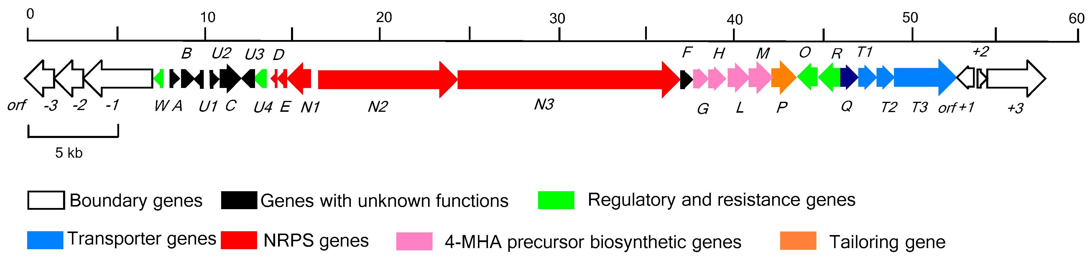
2.1. Identification of the Actinomycin D Biosynthetic Gene Cluster

To identify the gene cluster, we sequenced the S. costaricanus SCSIO ZS0073 genomic DNA using a combination of Hiseq 4000 and PacBio smart technologies [27]. S. costaricanus SCSIO ZS0073 is a mangrove-derived actinomycete which produces antibacterial secondary metabolites such as fungichromin, actinomycin D and actinomycin X [27,28]. We analyzed a 39.8 kb region containing 24 open reading frames (ORFs), suspected of coding for actinomycin D biosynthesis. We elucidated the gene organization as shown in Figure 1 and deduced its biosynthetic pathway (Figure 2) along with assigned gene product functions as noted below in Table 1. The gene cluster has been deposited in Genbank (with the accession number of MK234849). A cosmid library of S. costaricanus SCSIO ZS0073 was constructed using the SuperCos 1 vector system and 2 positive clones (9C2, 9A7) covering the whole gene cluster were screened using targeted gene inactivations.

2.2. Determination of the Actinomycin D Biosynthetic Gene Cluster Boundaries

The upstream boundary of the actinomycin D gene cluster was preliminarily determined as being between orf(−1) and acnW; the downstream boundary was defined between acnT3 and orf(+1). The orf(−1) encodes Type VII secretion system protein EccC. Although orf(−1) carries with it the function of enabling secretion, all the inactivation mutants of orf(−1) failed to show any differences in actinomycin D production levels compared to wild-type (WT) (SI, Figure S7). The upstream boundary region was further delineated by a set of gene inactivations for orf(−1), orf(−2) and orf(−3). None of the inactivation bearing mutants for these genes displayed discernible differences in actinomycin D biosynthesis rates or yields relative to WT producer (SI, Figure S7). Likewise, the downstream boundary was ascertained by bioinformatic analyses and the application of mutants bearing inactivated orf(+1), orf(+2), orf(+3) genes. Although orf(+1) gene encodes an N-acetyltransferase, it is shown by inactivation experiments, the mutant strains produce the same level of actinomycin D compared to wild type strains. Further evaluations proximal to the downstream boundary revealed that genes corresponding to orf(+2) and orf(+3) encode a hypothetical protein and a two-component hybrid sensor and regulator; comparative analyses of ∆orf(+2) and ∆orf(+3) inactivation mutants (vs. WT) revealed that neither of these genes and their respective products play a role in actinomycin D biosynthesis (SI, Figure S7).

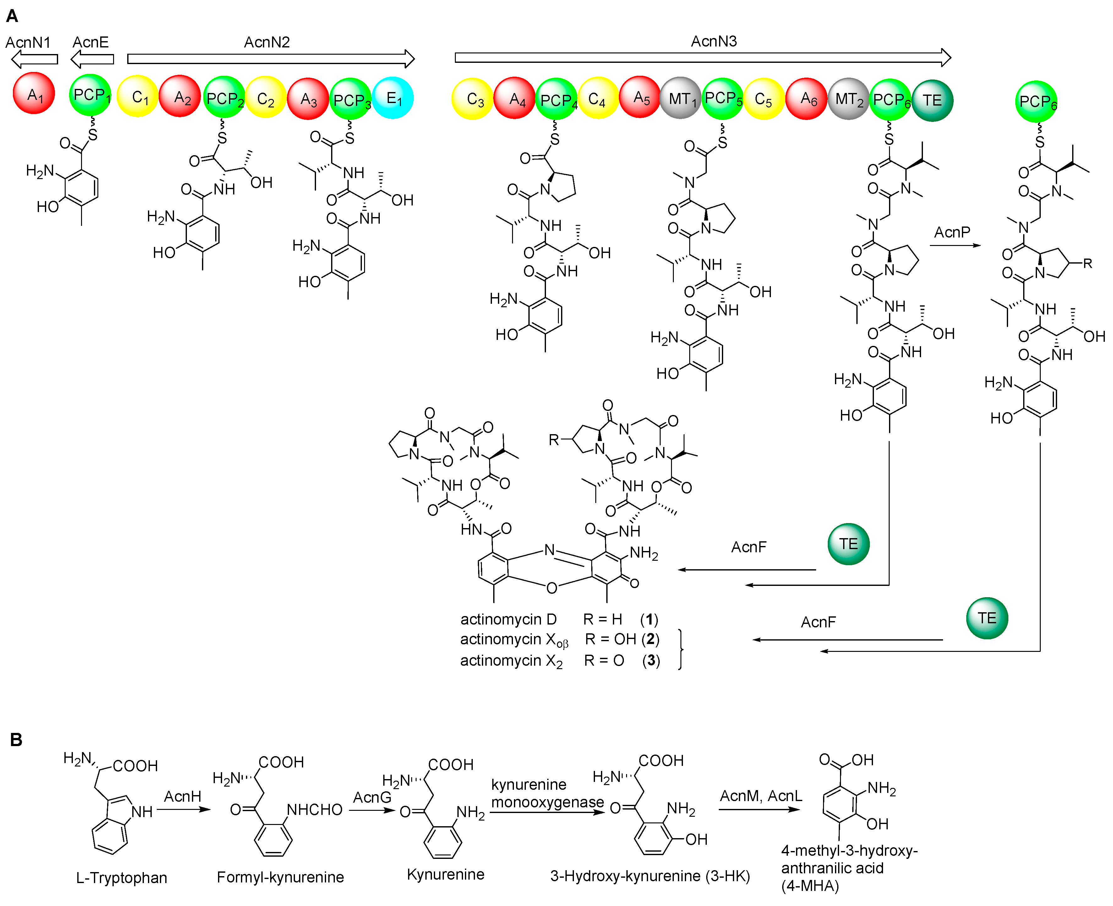
2.3. NRPS Genes for Peptide Chain Assembly in Actinomycin D Biosynthesis

Following the biosynthesis of 4-MHA, this anthranilic acid derivative, along with other amino acids, by several NRPSs ultimately affording the 4-MHA-pentapeptide halves of actinomycin (Figure 2A). The pentapeptide is composed of five different amino acids: l-threonine, d-valine, l-proline, l-sarcosine and methyl-valine. The genes responsible for peptide chain assembly in S. costaricanus SCSIO ZS0073 are acnD, acnE, acnN1, acnN2, acnN3. AcnD was found to contain 66 amino acid residues arranged in a manner that resembles the MbtH protein. MbtH-like proteins are found in many NRPS gene clusters [29] and researchers have demonstrated that MbtH-like proteins participate in some adenylation reactions by tightly binding to NRPS proteins containing adenylation domains [30,31]. MbtH-like proteins, such as gene dptG in S. roseosporus participate in the biosynthesis of the cyclic lipotridecapeptide antibiotic daptomycin [32] and mbtH in Mycobacerium spp. responsible for the siderophore biosynthesis [33]. The NRPS gene acnN1 encodes an adenylation domain protein and acnE encoding 4-MHA carrier protein. Two large multidomain NRPSs acnN1 and acnN3 are responsible for peptide chain assembly and release from the NRPS machinery. Gene deletions of acnD + acnE in combination, acnN1 and acnN3 were found to abolish actinomycin D biosynthesis (Figure 3) in the mutant producers bearing these gene inactivations.

2.4. Biosynthetic Genes of 4-MHA

We identified a set of genes, designated as acnG, acnH, acnL and acnM, that show high functional similarities to enzymes involved in the biosynthesis of 4-MHA, the critical building block of the actinomycin chromophore. These genes are homologous to a set of four counterparts in actinomycin-producing strains S. antibioticus IMRU3720 and S. chrysomallus [17,18]. These genes encode for arylformamidase (acnG), tryptophan-2, 3-dioxygenase (acnH), kynureninase (acnL) and methyltransferase (acnM). The gene acnM shows 88% identities with the 3-hydroxy kynurenine methyltransferase acmL, which is responsible for the methylation of 3-HK [34]. However, the vital step entailing kynurenine conversion to 3-hydroxykynurenine appears to be catalyzed by a kynurenine 3-monooxygenase, the gene for which is not found within the acn gene cluster. Inactivation of acnGHLM in combination, followed by metabolite analysis of the mutant strain revealed that these genes play essential roles in actinomycin D biosynthesis (Figure 3).

2.5. Regulatory and Self-Resistance Genes

We identified six genes acnW, acnU4, acnR, acnT1, acnT2 and acnT3 with apparent regulatory and/or protective functions. AcnR encodes a TetR family transcriptional regulator and usually acts as a transcriptional repressor or activator in many biological processes such as cell-cell communication and metabolite regulation [35,36]. TetR family transcriptional regulators belong to a one-component system and generally possess a two-domain structure composed of an N-terminal HTH DNA-binding motif and a C-terminal ligand regulatory domain. Many of these regulators control the expression of transporters, which help bacteria acclimate to their environment [37,38]. In some cases, members of the TetR family suppress the biosynthesis of antibiotics. For instance, disruption of calR3, a TetR family member in the calcimycin BGC, was found to improve calcimycin titers [39]. Moreover, TrdK, which shows high similarity to TetR family members and is involved in tirandamycin production from marine-derived Streptomyces sp. SCSIO 1666 was found by gene inactivations, to suppress tirandamycin biosynthesis; trdK inactivation led to dramatically improved tirandamycin titers [40]. The inactivation of acnR completely abolished actinomycin D production, consistent with this gene’s essential role as a and important positive regulator of actinomycin D biosynthesis (Figure 3, trace viii). Gene acnW is located downstream of the boundary gene orf(−1) and appears to encode for a hypothetical protein; acnW disruption impaired, but did not abolish, production of actinomycin D. The gene acnU4 also encodes a hypothetical protein and, as with acnW, inactivation of acnU4 correlates to diminished actinomycin D production (Figure 3). On the basis of these findings, AcnW and AcnU4 appear to function as positive regulators of actinomcyin D production although the structural families to which they belong are not yet apparent. AcnO encodes a LmbU-like protein. LmbU is a regulatory gene involved in licomycin biosynthesis in S. lincolnensis 78-11. It always contains a TTA codon close to the N-terminal end of its ORF. The codon is often found in genes involved in the regulation of differentiation or secondary metabolism [41], in neither case is a specific role in actinomycin D assembly known, S. costaricanus SCSIO ZS0073 inactivation mutant strain of ∆acnO was not able to produce even trace amounts of actinomycin D (Figure 3, traces ix). Thus, acnO might serve as a positive regulator in actinomycin D biosynthetic pathway.
Beyond these gene–function correlations, we also identified three putative transporter genes acnT1, acnT2 and acnT3. The acnT1 gene is predicted to encode an ATP-binding cassette (ABC) transporter which uses the energy of ATP to transport molecules through the membrane. Bioinformatics analyses suggest that acnT2 encodes a multidrug ABC transporter permease whereas acnT3 encodes a UvrA-like protein, which is a DNA binding protein involved in the excision repair of DNA. Somewhat surprisingly, inactivations of acnT1 acnT2 and acnT3 all produced mutant strains able to generate actinomycin D with efficiencies rivaling those seen with the WT producer (Figure 3). Consequently, we concluded that these three transporter genes are not necessary in actinomycin D biosynthesis.

2.6. Nonessential Genes of Unknown within the acn Cluster

The BGC for actinomycin D in S. costaricanus SCSIO ZS0073 houses a number of genes not associated with actinomycin D assembly. For instance, close to the upstream boundary reside genes acnA and acnB; all of these encode for hypothetical proteins. Additionally, acnU1, acnU2, acnU3 also encode for proteins whose functions have not been deciphered and whose roles in actinomycin D construction appear nonessential. Inactivations for all these genes have no impact on actinomycin D biosynthesis relative to WT production efficiencies (Figure 4). Bioinformatics have revealed that acnC resides between acnU2 and acnU3 and codes for an acyl-CoA dehydrogenase. This gene’s inactivation also appears to have no impact upon actinomycin D synthesis compared to the WT strain. This too is the case for acnQ. Located downstream of the acnR, acnQ was proposed, based on bioinformatics, to code for a siderophore-interacting protein. Such small molecule siderophore-interacting systems often constitute transport systems by which microbes control intracellular concentrations of specific secondary metabolites. As with the other genes noted however, inactivation of acnQ was found to have no bearing whatsoever on actinomycin D production compared to the WT producer (Figure 4).

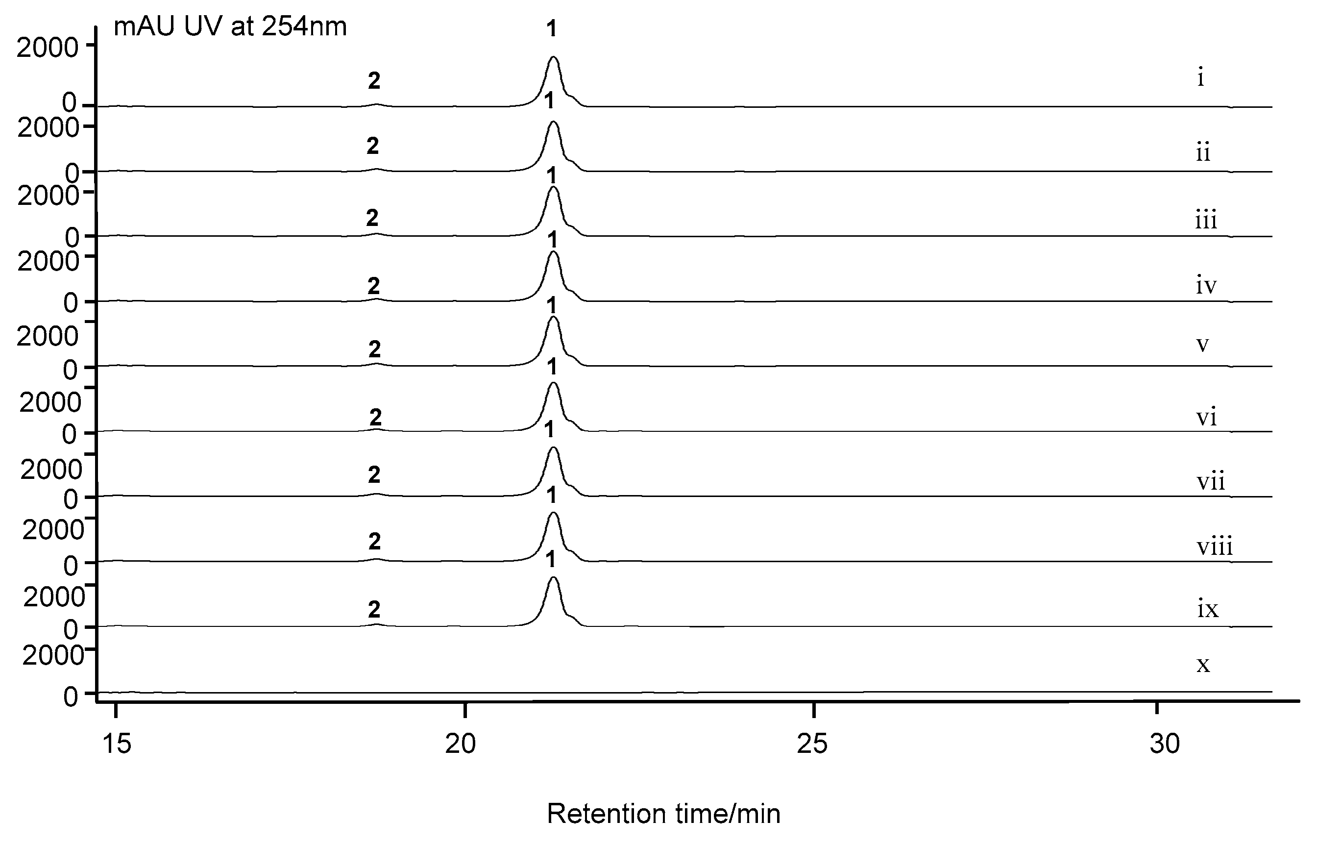
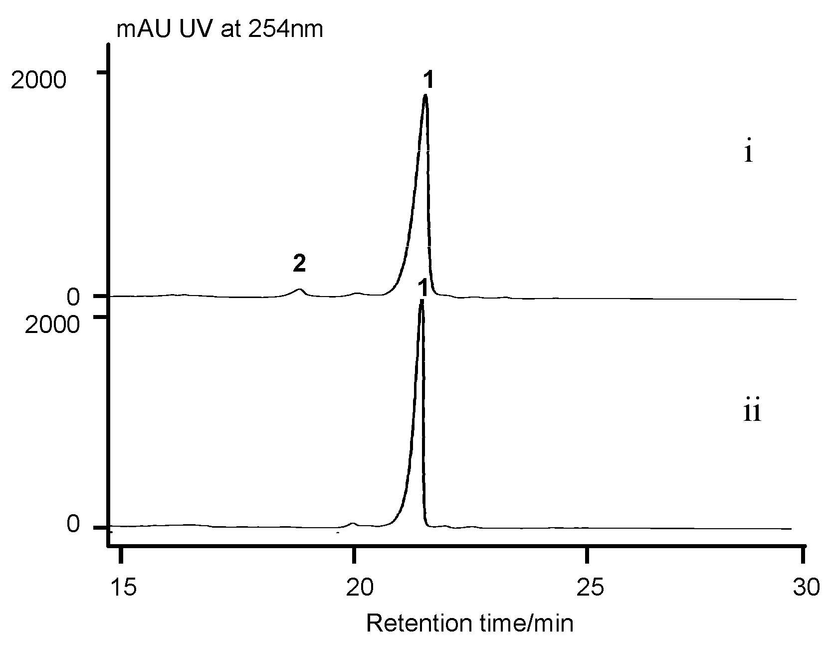
2.7. Cytochrome P450 Gene acnP Is Responsible for the Hydroxylation of Proline in Actinomycin X

X-type actinomycins contain 4-hydroxyproline (actinomycin X) or 4-oxoproline (actinomycin X2) in their β-pentapeptide lactone rings whereas their α-ring contains proline. Previous studies demonstrated the importance of a 4-oxoproline synthase within the actinomycin BGC of S. antibioticus. This enzyme catalyzes proline oxidation in each of the actinomycin halves (prior to dimerization) to form 4-hydroxyproline or 4-oxoproline; condensation of each of the hydroxylated actinomycin halves affords actinomycin X or actinomycin X2 [42]. Within the actinomycin BGC in S. costaricanus SCSIO ZS0073, we identified a cytochrome P450 gene, acnP, that encodes a 436 aa protein. We hypothesized that AcnP is responsible for proline hydroxylation en route to actinomycin X. This speculation was validated by targeted inactivation of acnP; fermentations and metabolite analyses of the ∆acnP mutant revealed the complete abrogation of actinomycin X production and concomitant production of actinomycin D with titers rivaling those seen with the WT producer (Figure 5). These data make clear that AcnP is responsible for the 4-oxoproline found in actinomycin X.
The PHS gene outside the acn cluster is not necessary for actinomycin D biosynthesis. PHS has been presumed to catalyze the oxidative condensation of two 4-MHA-pentapeptide lactone “monomers” in the last step of actinomycin D biosynthesis. Previous studies had, in fact, demonstrated that PHS is involved in a variety of enzymatic condensations of ortho-aminophenols to form phenoxazinones. Within the genome of S. costaricanus SCSIO ZS0073, we identified a phs orf encoding a 627 aa, two copper-containing phenoloxidase separated from the actinomycin biosynthetic cluster by 5.77 Mbp. This phs gene shows sequence similarity (84% identity) to phsA in S. antibioticus IMRU3720. To validate this gene’s involvement in actinomycin D assembly, we generated a ∆phs mutant strain and assessed its biosynthetic capacity relative to the WT producer. Surprisingly, phs inactivation in the S. costaricanus SCSIO ZS0073 strain had no detectable impact upon actinomycin D production relative to the WT producer (Figure 4). This result is consistent with the previous gene inactivation result for phsA in S. antibioticus IMRU3720 [26] and indicated also, that for S. costaricanus SCSIO ZS0073 actinomycin D assembly does not appear to require a PHS.
acnF is essential to actinomycin D biosynthesis. Within the actinomycin cluster, acnF encodes for a hypothetical protein with 212 aa. Gene inactivation for acnF, revealed that acnF is indispensable for actinomycin D construction (Figure 4). S. costaricanus SCSIO ZS0073 inactivation mutant strain of ∆acnF was not able to produce even trace amounts of actinomycin D (Figure 4, traces x), thus showcasing the importance of its gene products to actinomycin D biosynthesis. We proposed acnF might be involved in the dimerization of the 4-MHA-pentapeptide monomer en route to actinomycin (Figure 2). Efforts to identify the exact roles played by acnF products are currently ongoing and will be reported in due course.

3. Materials and Methods

3.1. General Experimental Procedures

Reagents for polymerase chain reactions (PCR) were purchased from Takara Co. (Dalian, China) and Trans Gene Co. (Beijing, China). The plasmid kit and gel extraction kit were from Promega. Unless otherwise indicated, other biochemicals and chemicals were purchased from standard commercial sources and used without further purification. All DNA manipulations were conducted according to the standard procedures or manufactures’ instruction. DNA and aa sequence analyses were performed with the seqmen and editsequence in the Lasergene software package (DNASTAR, Madison, WI, USA). All primers and reagents used in this work were purchased from Sangong Bio-Pharm Technology Co., Ltd., Shanghai, China.

3.2. Bacterial Strains, Plasmids and Culture Conditions

S. costaricanus SCSIO ZS0073, a marine actinomycete isolated from the red sand park of Guangxi (China), was used to identify the actinomycin D gene cluster [27]. The strain and mutants were grown at 30 °C on ISP-2 medium (with 3% sea salt added) for cultivation. E. coli was used for DNA cloning and sequencing. E. coli ET12567/pUZ8002, which is methylation deficient, was employed as the donor cell for conjugal transfer of DNA into S. costaricanus SCSIO ZS0073. All E. coli strains were grown in liquid lysogeny broth (LB) at 37 °C or 30 °C and 200 rpm. When used, antibiotics were added at the following concentrations: chloramphenicol (Chl, 25 μg mL−1), apramycin (Apr, 50 μg mL−1), kanamycin (Kan, 50 μg mL−1), ampicillin (Amp, 100 μg mL−1). The plasmids SuperCos 1 and pIJ773 were used for S. costaricanus SCSIO ZS0073 genomic library construction and the aac(3)IV-oriT resistance gene amplifying, respectively.

3.3. Whole Genome Scanning and Sequence Analysis

S. costaricanus SCSIO ZS0073 genomic DNA was isolated according to the protocol with slight modification [43]. Whole-genome scanning was achieved using both 454 and Solexa technology at Macrogen (Seoul, Korea). Assembly, annotation and bioinformatics analyses allowed us to define the correct contiguous fragments corresponding to the acn cluster. Assignments of ORFs and their functional predictions were accomplished using FramePlot 4.0 (http://nocardia.bih.go.jp/fp4) and Blast (http://blast.ncbi.nlm.nih.gov/) software packages.

3.4. Genomic Library Construction and Screening

Genomic DNA was partially digested with Sau3AI and 30–40 kb fragments were ligated into XbaI/BamHI digested and dephosphorylated SuperCos 1. The resulting ligation mixture was packaged with Gigapack III gold and transduced into E. coli LE392 to generate the genomic library, according to the manufacturer instructions.

3.5. Inactivation of S. costaricanus SCSIO ZS0073 by λ-RED-Mediated PCR-Targeting Mutagenesis

Targeted genes in the S. costaricanus SCSIO ZS0073 biosynthetic gene cluster were inactivated using λ-mediated PCR-targeting methodology [44]. Two cosmids were used to disrupt target genes. An apramycin resistance cassette aac(3)IV-oriT fragment obtained by PCR (digested pIJ773 was used as template), with primer pairs containing 39-nucleotide extensions derived from the 5′- and 3′-ends of the targeted genes, was used to replace an internal region of each of the targeted genes. Each of the mutated genes was verified by PCR with primers designed to be 10–300 bp outside of the disruption region, with verification by restriction enzyme digestion. The constructed mutated cosmids were introduced into non-methylating E. coli ET12567/pUZ8002 for conjugal transfer. For conjugation, harvested S. costaricanus SCSIO ZS0073 spores were suspended in TSB medium and incubated for 3–5 h at 28 °C and 200 rpm after heating for 10 min at 50 °C. The culture was then centrifuged to harvest the germinated spores as the conjugation recipients. At the same time, LB supplied with Kan (50 μg mL−1), Chl (25 μg mL−1) and Apr (100 μg mL−1) was inoculated with E. coli ET12567/pUZ8002 containing each mutated cosmid. After the culture OD600 increased to 0.6–0.8, the cells were harvested, washed three times with LB, resuspended in 500 μL LB medium and mixed with the previously germinated spores. The mixture was plated on modified ISP-4 medium containing MgCl2 (60 mM). The plates ware incubated at 28 °C for 18–20 h, then each plate was covered with sterile deionized water (1 mL), trimethoprim stock solution (Tmp, 30 μL, 50 mg mL−1) and Apr stock solution (25 μL, 50 mg mL−1). Finally, all plates were incubated at 28 °C for an additional 6–7 days until exconjugants appeared. Double-crossover mutants were primarily selected on the basis of KanSAprR phenotypes and the desired double crossover mutants were further verified by PCR with primers listed in Table S3 (SI).

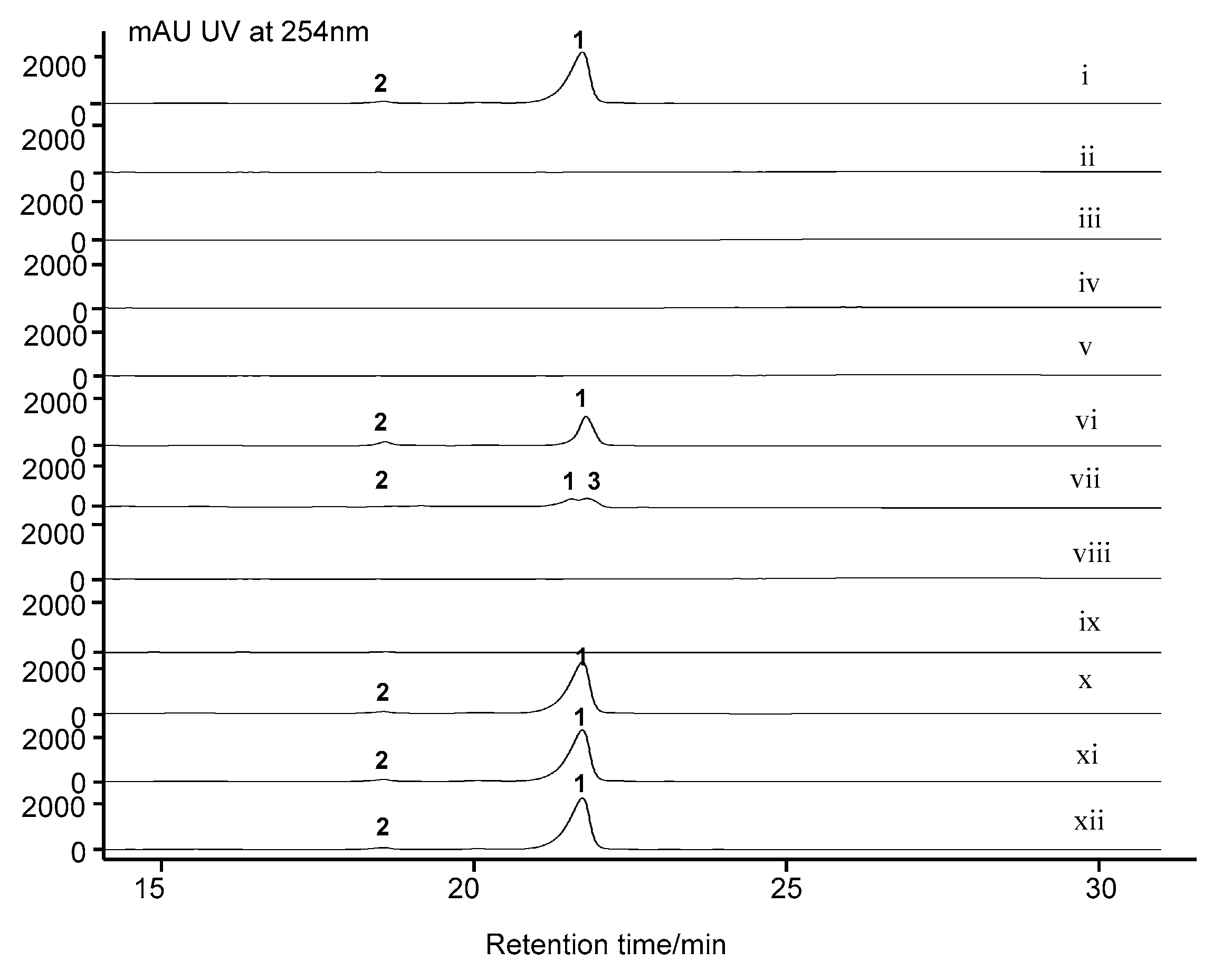
3.6. Fermentation and Analysis of S. costaricanus SCSIO ZS0073 WT and Mutant Strains

S. costaricanus SCSIO ZS0073 WT and mutant strains were inoculated into 250 mL flasks with 50 mL liquid ISP-2 medium and incubated on a rotary shaker at 28 °C, 200 rpm. After 7 days fermentation, each of the 50 mL cultures was added to 100 mL butanone and then vigorously mixed for 30 min. The butanone phase was separated and then evaporated to dryness to yield a residue. The residue was dissolved in 2 mL methanol and centrifuged and the resulting supernatant then subjected to HPLC analysis. Analytical HPLC was performed on an Agilent 1260 HPLC system (Agilent Technologies Inc., USA) equipped with a binary pump and a diode array detector using a Phenomenex Prodigy ODS column (150 × 4.60 mm, 5 μm) with UV detection at 254 nm. The mobile phase was comprised of solvents A and B. Solvent A consisted of 15% CH3CN in water supplemented with 0.1% TFA whereas solvent B consisted of 85% CH3CN in water supplemented with 0.1% TFA. Samples were eluted with a linear gradient from 5% to 90% solvent B in 20 min, followed by 9–100% solvent B for 5 min, then eluted with 100% solvent B for 3 min, at a flow rate of 1 mL/min and UV detection at 254 nm. LC-ESI-MS data were obtained using an amaZon SL ion trap mass spectrometer (Bruker, Billerica, MA, USA). The mobile phase comprises solvents A and B. Solvent A consists of 100% ddH2O supplemented with 0.1% methanoic acid whereas solvent B consists of 100% CH3CN supplemented with 0.1% methanoic acid. Samples were eluted with a linear gradient from 5 % to 90% solvent B in 20 min, followed by 9–100% solvent B for 5 min, then eluted with 100% solvent B for 3 min, at a flow rate of 1 mL/min and UV detection at 254 nm.

4. Conclusions

Actinomycin D is a vital antibiotic in the treatment of Wilms’ tumor, trophoblastic tumors and rhabdonyosarcoma. Here, we have identified and characterized a new actinomycin D BGC (acn) in marine derived producer S. costaricanus SCSIO ZS0073 by carrying out whole genome sequencing and systematic gene disruptions. Relative to the gene cluster for actinomycin of S. chrysomallus and S. antibioticus IMRU3720, the size of the acn cluster in S. costaricanus SCSIO ZS0073 is smaller. In silico analysis, gene inactivation and metabolomics data enabled us to deduce the biosynthetic pathway leading to actinomycin D in S. costaricanus SCSIO ZS0073. We furthermore determined the acn cluster boundaries and elucidated the functions of positive regulatory genes acnWU4RO along with the cytochrome P450 gene acnP which is responsible for installing the 4-oxoproline seen in actinomycin Xoβ. The discovery of this new actinomycin BGC advances initiatives to engineer new actinomycin D analogues for clinical use and to explore the still elusive mechanism of actinomycin 4-MHA-pentapeptide monomer dimerization en route to intact actinomycins.

Supplementary Materials

The following are available online at https://www.mdpi.com/1660-3397/17/4/240/s1, Figures S1–S28. Disruption genes in actinomycin biosynthetic gene cluster in WT S. costaricanus SCSIO ZS0073 via PCR-targeting. Table S1, Structure of actinomycins; Table S2, Strains and plasmids used in this study.

Author Contributions

J.J. designed experiments. M.L. and Y.J. performed the experiments. Y.X., Z.C., J.M., C.S. and J.J. analyzed data. M.L. and J.J. wrote the manuscript. All authors read and approved the final manuscript.

Funding

This study was supported, in part, by the National Natural Science Foundation of China (U1501223, U1706206, 81425022), the Chinese Academy of Sciences (XDA13020302) and Natural Science Foundation of Guangdong Province (2016A030312014).

Acknowledgments

We thank Aijun Sun, Xiaohong Zheng, Yun Zhang, Xuan Ma and Zhihui Xiao, in the analytical facility center of the SCSIO for recording MS and NMR data.

Conflicts of Interest

The authors declare no conflict of interest.

References

  1. Bitzer, J.; Streibel, M.; Langer, H.J.; Grond, S. First Y-type actinomycins from Streptomyces with divergent structure-activity relationships for antibacterial and cytotoxic properties. Org. Biomol. Chem. 2009, 7, 444–450. [Google Scholar] [CrossRef] [PubMed]
  2. Hollstein, U. Actinomycin. Chemistry and mechanism of action. Chem. Rev. 1974, 74, 625–652. [Google Scholar] [CrossRef]
  3. Cai, W.L.; Wang, X.; Elshahawi, S.I.; Ponomareva, L.V.; Liu, X.; McErlean, M.R.; Cui, Z.; Arlinghaus, A.L.; Thorson, J.S.; Van Lanen, S.G. Antibacterial and cytotoxic actinomycins Y6–Y9 and Zp from Streptomyces sp. strain Gö-GS12. J. Nat. Prod. 2016, 79, 2731–2739. [Google Scholar] [CrossRef] [PubMed]
  4. Chen, Y.; Liu, J.; Yuan, B.; Cao, C.; Qin, S.; Cao, X.; Bian, G.; Wang, Z.; Jiang, J. Methylated actinomycin D, a novel actinomycin D analog induces apoptosis in HepG2 cells through Fas- and mitochondria-mediated pathways. Mol. Carcinog. 2013, 52, 983–996. [Google Scholar] [CrossRef] [PubMed]
  5. Dong, M.; Cao, P.; Ma, Y.T.; Luo, J.; Yan, Y.; Li, R.T.; Huang, S.X. A new actinomycin Z analogue with an additional oxygen bridge between chromophore and β-depsipentapeptide from Streptomyces sp. KIB-H714. Nat. Prod. Res. 2018, 2, 1–7. [Google Scholar] [CrossRef]
  6. Jiao, W.H.; Yuan, W.; Li, Z.Y.; Li, J.; Li, L.; Sun, J.B.; Gui, Y.H.; Wang, J.; Ye, B.P.; Lin, H.W. Anti-MRSA actinomycins D1–D4 from the marine sponge-associated Streptomyces sp. LHW52447. Tetrahedron 2018, 74, 5914–5919. [Google Scholar] [CrossRef]
  7. Wang, Q.; Zhang, Y.; Wang, M.; Tan, Y.; Hu, X.; He, H.; Xiao, C.L.; You, X.; Wang, Y.; Gan, M. Neo-actinomycins A and B, natural actinomycins bearing the 5 H-oxazolo [4-b] phenoxazine chromophore, from the marine-derived Streptomyces sp. IMB094. Sci. Rep. 2017, 7, 3591. [Google Scholar] [CrossRef]
  8. Bitzer, J.; Gesheva, V.; Zeeck, A. Actinomycins with altered threonine units in the β-peptidolactone. J. Nat. Prod. 2006, 69, 1153–1157. [Google Scholar] [CrossRef] [PubMed]
  9. Lo, Y.S.; Tseng, W.H.; Chuang, C.Y.; Hou, M.H. The structural basis of actinomycin D-binding induces nucleotide flipping out, a sharp bend and a left-handed twist in CGG triplet repeats. Nucleic Acids Res. 2013, 41, 4284–4294. [Google Scholar] [CrossRef]
  10. Das, T.; Nair, R.R.; Green, R.; Padhee, S.; Howell, M.; Banerjee, J.; Mohapatra, S.S.; Mohapatra, S. Actinomycin D down-regulates SOX2 expression and induces death in breast cancer stem cells. Anticancer Res. 2017, 37, 1655–1663. [Google Scholar] [PubMed]
  11. Choong, M.L.; Yang, H.; Lee, M.A.; Lane, D.P. Specific activation of the p53 pathway by low dose actinomycin D: A new route to p53 based cyclotherapy. Cell Cycle 2009, 8, 2810–2818. [Google Scholar] [CrossRef] [PubMed] [Green Version]
  12. Saijets, S.; Ylipaasto, P.; Vaarala, O.; Hovi, T.; Roivainen, M. Enterovirus infection and activation of human umbilical vein endothelial cells. J. Med. Virol. 2003, 70, 430–439. [Google Scholar] [CrossRef]
  13. Rill, R.L.; Hecker, K.H. Sequence-specific actinomycin D binding to single-stranded DNA inhibits HIV reverse transcriptase and other polymerases. Biochemistry 1996, 35, 3525–3533. [Google Scholar] [CrossRef]
  14. Wang, D.; Wang, C.; Gui, P.; Liu, H.; Khalaf, S.M.H.; Elsayed, E.A.; Wadaan, M.A.M.; Hozzein, W.N.; Zhu, W. Identification, bioactivity and productivity of actinomycins from the marine-derived Streptomyces heliomycini. Front. Microbiol. 2017, 8, 1147. [Google Scholar] [CrossRef] [PubMed]
  15. Chen, C.; Song, F.; Wang, Q.; Abdel-Mageed, W.M.; Guo, H.; Fu, C.; Hou, W.; Dai, H.; Liu, X.; Yang, N.; et al. A marine-derived Streptomyces sp. MS449 produces high yield of actinomycin X2 and actinomycin D with potent anti-tuberculosis activity. Appl. Microbiol. Biotechnol. 2012, 95, 919–927. [Google Scholar] [CrossRef] [PubMed]
  16. Hammer, A.S.; Couto, C.G.; Ayl, R.D.; Shank, K.A. Treatment of tumor-bearing dogs with actinomycin D. J. Vet. Intern. Med. 1994, 8, 236–239. [Google Scholar] [CrossRef] [PubMed]
  17. Keller, U.; Lang, M.; Crnovcic, I.; Pfennig, F.; Schauwecker, F. The actinomycin biosynthetic gene cluster of Streptomyces chrysomallus: A genetic hall of mirrors for synthesis of a molecule with mirror symmetry. J. Bacteriol. 2010, 192, 2583–2595. [Google Scholar] [CrossRef]
  18. Crnovčić, I.; Rückert, C.; Semsary, S.; Lang, M.; Kalinowski, J.; Keller, U. Genetic interrelations in the actinomycin biosynthetic gene clusters of Streptomyces antibioticus IMRU 3720 and Streptomyces chrysomallus ATCC11523, producers of actinomycin X and actinomycin C. Adv. Appl. Bioinform. Chem. 2017, 10, 29. [Google Scholar] [CrossRef]
  19. Wang, X.; Tabudravu, J.; Rateb, M.E.; Annand, K.J.; Qin, Z.; Jaspars, M.; Deng, Z.; Yu, Y.; Deng, H. Identification and characterization of the actinomycin G gene cluster in Streptomyces iakyrus. Mol. Biosyst. 2013, 9, 1286–1289. [Google Scholar] [CrossRef]
  20. Pfennig, F.; Schauwecker, F.; Keller, U. Molecular characterization of the genes of actinomycin synthetase I and of a 4-methyl-3-hydroxyanthranilic acid carrier protein involved in the assembly of the acylpeptide chain of actinomycin in Streptomyces. J. Biol. Chem. 1999, 274, 12508–12516. [Google Scholar] [CrossRef]
  21. Schauwecker, F.; Pfennig, F.; Grammel, N.; Keller, U. Construction and in vitro analysis of a new bi-modular polypeptide synthetase for synthesis of N-methylated acyl peptides. Chem. Biol. 2000, 7, 287–297. [Google Scholar] [CrossRef]
  22. Fawaz, F.; Jones, G.H. Actinomycin synthesis in Streptomyces antibioticus. Purification and properties of a 3-hydroxyanthranilate 4-methyltransferase. J. Biol. Chem. 1988, 4, 4602–4606. [Google Scholar]
  23. Smith, A.W.; Camara-Artigas, A.; Wang, M.; Allen, J.P.; Francisco, W.A. Structure of phenoxazinone synthase from Streptomyces antibioticus reveals a new type 2 copper center. Biochemistry 2006, 45, 4378–4387. [Google Scholar] [CrossRef]
  24. Madu, A.C.; Jones, G.H. Molecular cloning and in vitro expression of a silent phenoxazinone synthase gene from Streptomyces lividans. Gene 1989, 84, 287–294. [Google Scholar] [CrossRef]
  25. Golub, E.E.; Ward, M.A.; Nishimura, J.S. Biosynthesis of the actinomycin chromophore: Incorporation of 3-hydroxy-4-methylanthranilic acid into actinomycins by Streptomyces antibioticus. J. Bacteriol. 1969, 100, 977–984. [Google Scholar] [PubMed]
  26. Jones, G.H. Actinomycin production persists in a strain of Streptomyces antibioticus lacking phenoxazinone synthase. Antimicrob. Agents. Chemother. 2000, 44, 1322–1327. [Google Scholar] [CrossRef]
  27. Song, X.; Jiang, X.; Sun, J.; Zhang, C.; Zhang, Y.; Lu, C.; Ju, J. Antibacterial secondary metabolites produced by mangrove-derived actinomycete Stremptomeces costaricanus SCSIO ZS0073. Nat. Prod. Res. Dev. 2017, 29, 410–414. [Google Scholar]
  28. Jia, Y.; Xie, Y.; Li, Q.; Ma, J.; Huang, H.; Ju, J. Identification of the fungichromin biosynthetic pathway from Streptomyces costaricanus SCSIO ZS0073. Chin. J. Mar. Drug 2017, 36, 1–10. [Google Scholar]
  29. Baltz, R.H. Function of MbtH homologs in nonribosomal peptide biosynthesis and applications in secondary metabolite discovery. J. Ind. Microbiol. Biotechnol. 2011, 38, 1747–1760. [Google Scholar] [CrossRef]
  30. Felnagle, E.A.; Barkei, J.J.; Park, H.; Podevels, A.M.; McMahon, M.D.; Drott, D.W.; Thomas, M.G. MbtH-like proteins as integral components of bacterial nonribosomal peptide synthetases. Biochemistry 2010, 49, 8815–8817. [Google Scholar] [CrossRef]
  31. Imker, H.J.; Krahn, D.; Clerc, J.; Kaiser, M.; Walsh, C.T. N-Acylation during glidobactin biosynthesis by the tridomain nonribosomal peptide synthetase module GlbF. Chem. Biol. 2010, 17, 1077–1083. [Google Scholar] [CrossRef] [PubMed]
  32. Baltz, R.H. Genomics and the ancient origins of the daptomycin biosynthetic gene cluster. J. Antibiot. 2010, 63, 506–511. [Google Scholar] [CrossRef] [PubMed] [Green Version]
  33. Quadri, L.E.; Sello, J.; Keating, T.A.; Weinreb, P.H.; Walsh, C.T. Identification of Mycobacterium tuberculosis gene cluster encoding the biosynthetic enzymes for assembly of the virulence conferring siderophore mycobactin. Chem. Biol. 1998, 5, 631–645. [Google Scholar] [CrossRef]
  34. Crnovčić, I.; Süssmuth, R.; Keller, U. Aromatic C-methyltransferases with antipodal stereoselectivity for structurally diverse phenolic amino acids catalyze the methylation step in the biosynthesis of the actinomycin chromophore. Biochemistry 2010, 49, 9698–9705. [Google Scholar] [CrossRef] [PubMed]
  35. Waters, C.M.; Bassler, B.L. Quorum sensing: Cell-to-cell communication in bacteria. Annu. Rev. Cell Dev. Biol. 2005, 21, 319–346. [Google Scholar] [CrossRef] [PubMed]
  36. Bassler, B.L.; Losick, R. Bacterially speaking. Cell 2006, 125, 237–246. [Google Scholar] [CrossRef] [PubMed]
  37. Ramos, J.L.; Martínez-Bueno, M.; Molina-Henares, A.J.; Terán, W.; Watanabe, K.; Zhang, X.; Gallegos, M.T.; Brennan, R.; Tobes, R. The tetR family of transcriptional repressors. Microbiol. Mol. Biol. Rev. 2005, 69, 326–356. [Google Scholar] [CrossRef]
  38. Nishida, H.; Ohnishi, Y.; Beppu, T.; Horinouchi, S. Evolution of γ-butyrolactone synthases and receptors in Streptomyces. Environ. Microbiol. 2007, 9, 1986–1994. [Google Scholar] [CrossRef] [PubMed]
  39. Gou, L.; Han, T.; Wang, X.; Ge, J.; Liu, W.; Hu, F.; Wang, Z. A novel tetR family transcriptional regulator, CalR3, negatively controls calcimycin biosynthesis in Streptomyces chartreusis NRRL 3882. Front. Microbiol. 2017, 8, 2371. [Google Scholar] [CrossRef]
  40. Mo, X.; Wang, Z.; Wang, B.; Ma, J.; Huang, H.; Tian, X.; Zhang, S.; Zhang, C.; Ju, J. Cloning and characterization of the biosynthetic gene cluster of the bacterial RNA polymerase inhibitor tirandamycin from marine-derived Streptomyces sp. SCSIO 1666. Biochem. Biophys. Res. Commun. 2011, 406, 341–347. [Google Scholar] [CrossRef]
  41. Peschke, U.; Schmidt, H.; Zhang, H.Z.; Pieperserg, W. Molecular characterization of the lincomycin-production gene cluster of Streptomyces lincolnensis 78-11. Mol. Microbiol. 1995, 16, 1137–1156. [Google Scholar] [CrossRef] [PubMed]
  42. Semsary, S.; Crnovčić, I.; Driller, R.; Vater, J.; Loll, B.; Keller, U. Ketonization of proline residues in the peptide chains of actinomycins by a 4-oxoproline synthase. ChemBioChem 2018, 19, 706–715. [Google Scholar] [CrossRef] [PubMed]
  43. Waksman, S.A.; Woodruff, H.B. Bacteriostatic and bactericidal substances produced by a soil actinomyces. Exp. Biol. Med. 1940, 45, 609–614. [Google Scholar] [CrossRef]
  44. Gust, B.; Kieser, T.; Chater, K. PCR targeting system in Streptomyces coelicolor A3 (2). John Innes Centre 2002, 3, 1–39. [Google Scholar]
Figure 1. Gene organization of the actinomycin D cluster in S. costaricanus SCSIO ZS0073. The direction of transcription and the proposed functions of individual ORF are indicated.
Marinedrugs 17 00240 g001
Figure 2. Proposed model for actinomycin D assembly (A) and 4-MHA precursor production (B) in S. costaricanus SCSIO ZS0073. A: adenylation domain; C: condensation; PCP: peptidyl carrier protein; TE: thioesterase.
Marinedrugs 17 00240 g002
Figure 3. HPLC analysis of S. costaricanus strains fermentation extracts. (i) WT producer; (ii) ∆acnDE mutant; (iii) ∆acnN1 mutant; (iv) ∆acnN3 mutant; (v) ∆acnGHLM mutant; (vi) ∆acnW mutant; (vii) ∆acnU4 mutant; (viii) ∆acnR mutant; (ix) ∆acnO mutant; (x) ∆acnT1 mutant; (xi) ∆acnT2 mutant; (xii) ∆acnT3 mutant. 1, actinomycin D; 2, actinomycin X.; 3, actinomycin X2.
Marinedrugs 17 00240 g003
Figure 4. HPLC analyses of S. costaricanus strains fermentation extracts. (i) WT producer; (ii) ∆acnA mutant; (iii) ∆acnB mutant; (iv) ∆acnC mutant; (v) ∆acnU1 mutant; (vi) ∆acnU2 mutant; (vii) ∆acnU3 mutant; (viii) ∆phs mutant; (ix) ∆acnQ mutant; (x) ∆acnF mutant. 1, actinomycin D; 2, actinomycin X.
Marinedrugs 17 00240 g004
Figure 5. HPLC analysis of S. costaricanus strains fermentation extracts. (i) WT producer; (ii) ∆acnP mutant. 1, actinomycin D; 2, actinomycin X.
Marinedrugs 17 00240 g005
Table 1. Deduced orf functions in the acn cluster.
GeneSize aProtein Homolog and OriginID/SM (%)Origin (Protein ID)
orf(−3)492type VII secretion protein EccB96/97SCF88120
Streptomyces sp. LamerLS-31b
orf(−2)464type VII secretion integral membrane protein EccD99/99SCF88128
Streptomyces sp. LamerLS-31b
orf(−1)1289Collagen triple helix repeat-containing protein53/75Clostridium uliginosum
acnW103Hypothetical protein99/100AB905443.1
Streptomyces rochei 7434AN4
acnA121Protein of unknown function93/96SCF88143
Streptomyces sp. LamerLS-31b
acnB415Anti-anti-sigma regulatory factor96/97SCF88151
Streptomyces sp. LamerLS-31b
acnU1121hypothetical protein GA0115258_1155799/100SCF88158
Streptomyces sp. LamerLS-31b
acnU2124hypothetical protein GA0115258_1155895/96SCF88164
Streptomyces sp. LamerLS-31b
acnC392acyl-CoA dehydrogenase91/95AKJ14982
Streptomyces incarnatus
acnU3207hypothetical protein80/91WP_030987180
Streptomyces
acnU4187hypothetical protein82/88WP_030592054
Streptomyces anulatus
acnD66protein mbtH91/95OOQ48080
Streptomyces antibioticus
acnE784-MHA carrier protein73/85ADG27356
Streptomyces anulatus
acnN1467Adenylation domain protein51/65SCD96508
Streptomyces sp. DvalAA-43
acnN22589non-ribosomal peptide synthase73/81WP_057667184
Streptomyces anulatus
acnN34249non-ribosomal peptide synthetase78/85WP_064726364
Streptomyces parvulus
acnF211hypothetical protein84/88OOQ48467
Streptomyces antibioticus
acnG326arylformamidase79/85SCF58504
Streptomyces sp. Cmuel-A718b
acnH284tryptophan 2, 3-dioxygenase84/88OOQ48467
Streptomyces antibioticus
acnL420kynureninase86/90OOQ48077
Streptomyces antibioticus
acnM346methyltransferase88/91WP_030594248
Streptomyces anulatus
acnP386cytochrome P45086/91WP_030594247
Streptomyces anulatus
acnO224LmbU-like protein73/82ADG27350
Streptomyces anulatus
acnR282TetR family transcriptional regulator78/86OOQ48074
Streptomyces antibioticus
acnQ294siderophore-interacting protein78/87WP_030594239
Streptomyces anulatus
acnT1346ABC transporter87/92OOQ48072
Streptomyces antibioticus
acnT2255multidrug ABC transporter permease92/95OOQ48071
Streptomyces antibioticus
acnT3753UvrABC system protein A60/77AMV28079
Gemmata sp. SH-PL17
orf(+1)191GCN5-related N-acetyltransferase81/87UN35851
Streptomyces venezuelae
orf(+2)109hypothetical protein74/83WP_052876109
Streptomyces sp. NRRL F-4335
orf(+3)1130Two-component hybrid sensor and regulator55/68SBO98167
Nonomuraea sp. ATCC 39727
a Size in units of amino acids (aa); ID/SI: identity/similarity; acn: the BGC of actinomycin from S. costaricanus SCSIO ZS0073.

