DNA-Methylation-Based Detection of Urological Cancer in Urine: Overview of Biomarkers and Considerations on Biomarker Design, Source of DNA, and Detection Technologies

Abstract
:1. Introduction
2. Performance Characteristics of DNA Methylation Biomarkers
2.1. Bladder Cancer
2.2. Prostate Cancer
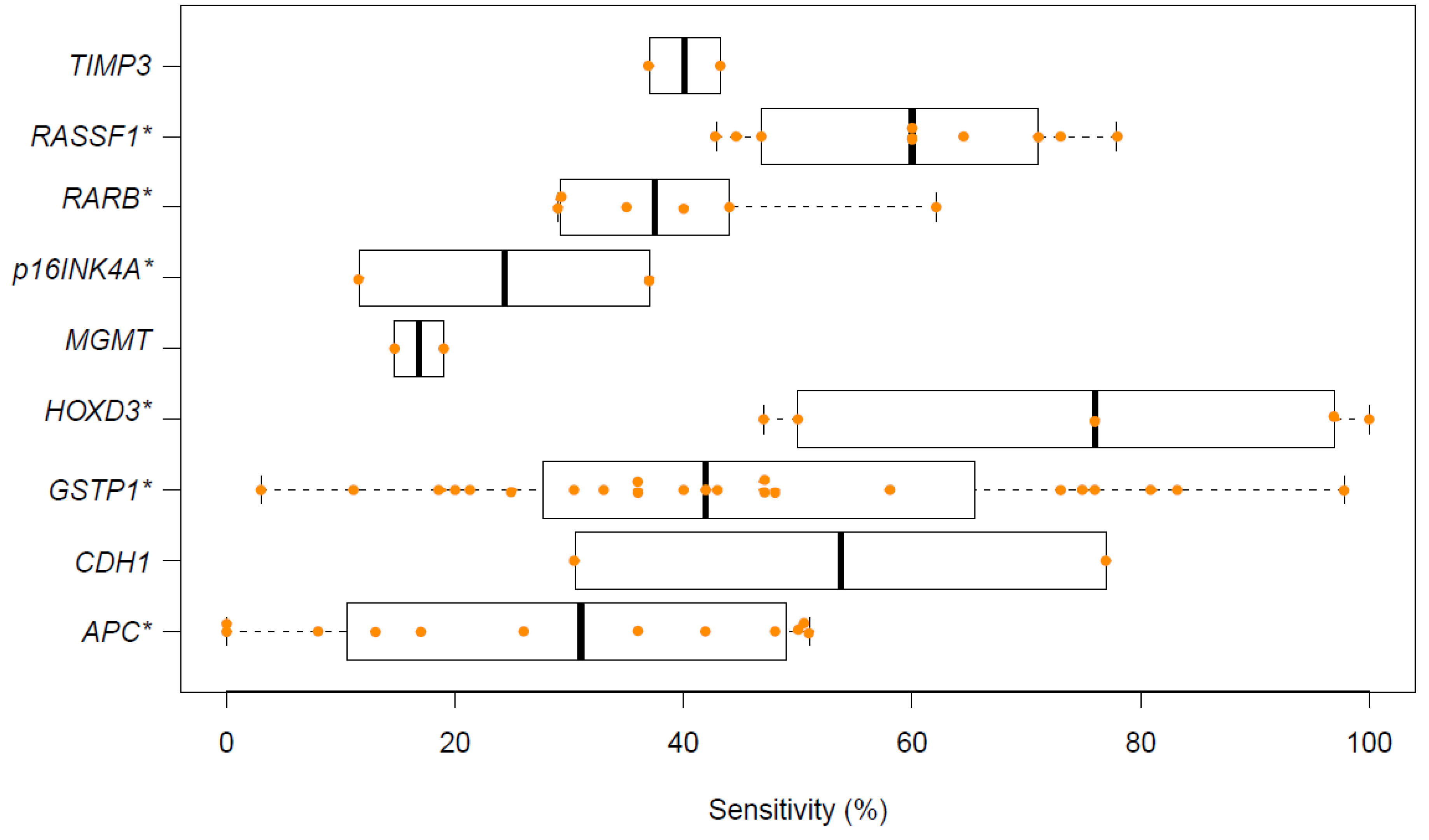
2.3. Renal Cancer
2.4. Upper Urinary Tract Cancer
3. Factors Affecting Biomarker Performance
3.1. Urine Collection and Processing
3.2. Bisulfite Treatment, Detection Technologies, and Sample Scoring
3.3. Genomic Location of Biomarker Assays
3.4. Sensitivity, Specificity, and Control Populations
4. Conclusions
Supplementary Materials
Author Contributions
Funding
Conflicts of Interest
References
- Heitzer, E.; Haque, I.S.; Roberts, C.E.S.; Speicher, M.R. Current and future perspectives of liquid biopsies in genomics-driven oncology. Nat. Rev. Genet. 2019, 20, 71–88. [Google Scholar] [CrossRef]
- Stasik, S.; Salomo, K.; Heberling, U.; Froehner, M.; Sommer, U.; Baretton, G.B.; Ehninger, G.; Wirth, M.P.; Thiede, C.; Fuessel, S. Evaluation of tert promoter mutations in urinary cell-free DNA and sediment DNA for detection of bladder cancer. Clin. Biochem. 2019, 64, 60–63. [Google Scholar] [CrossRef]
- Baylin, S.B.; Jones, P.A. A decade of exploring the cancer epigenome—Biological and translational implications. Nat. Rev. Cancer. 2011, 11, 726–734. [Google Scholar] [CrossRef]
- Trevethan, R. Sensitivity, specificity, and predictive values: Foundations, pliabilities, and pitfalls in research and practice. Front Public Health 2017, 5, 307. [Google Scholar] [CrossRef] [PubMed]
- Laird, P.W. The power and the promise of DNA methylation markers. Nat. Rev. Cancer 2003, 3, 253–266. [Google Scholar] [CrossRef]
- Cairns, P. Gene methylation and early detection of genitourinary cancer: The road ahead. Nat. Rev. Cancer 2007, 7, 531–543. [Google Scholar] [CrossRef] [PubMed]
- Kandimalla, R.; van Tilborg, A.A.; Zwarthoff, E.C. DNA methylation-based biomarkers in bladder cancer. Nat. Rev. Urol. 2013, 10, 327–335. [Google Scholar] [CrossRef] [PubMed]
- Koch, A.; Joosten, S.C.; Feng, Z.; de Ruijter, T.C.; Draht, M.X.; Melotte, V.; Smits, K.M.; Veeck, J.; Herman, J.G.; Van Neste, L.; et al. Analysis of DNA methylation in cancer: Location revisited. Nat. Rev. Clin. Oncol. 2018, 15, 459–466. [Google Scholar] [CrossRef] [PubMed]
- Bosschieter, J.; Lutz, C.; Segerink, L.I.; Vis, A.N.; Zwarthoff, E.C.; van Moorselaar, R.J.A.; van Rhijn, B.W.; Heymans, M.W.; Jansma, E.P.; Steenbergen, R.D.; et al. The diagnostic accuracy of methylation markers in urine for the detection of bladder cancer: A systematic review. Epigenomics 2018, 10, 673–687. [Google Scholar] [CrossRef] [PubMed]
- Chihara, Y.; Kanai, Y.; Fujimoto, H.; Sugano, K.; Kawashima, K.; Liang, G.; Jones, P.A.; Fujimoto, K.; Kuniyasu, H.; Hirao, Y. Diagnostic markers of urothelial cancer based on DNA methylation analysis. BMC Cancer 2013, 13, 275. [Google Scholar] [CrossRef]
- Wang, Y.; Yu, Y.; Ye, R.; Zhang, D.; Li, Q.; An, D.; Fang, L.; Lin, Y.; Hou, Y.; Xu, A.; et al. An epigenetic biomarker combination of pcdh17 and pou4f2 detects bladder cancer accurately by methylation analyses of urine sediment DNA in han chinese. Oncotarget 2016, 7, 2754–2764. [Google Scholar] [CrossRef]
- Yeh, C.M.; Chen, P.C.; Hsieh, H.Y.; Jou, Y.C.; Lin, C.T.; Tsai, M.H.; Huang, W.Y.; Wang, Y.T.; Lin, R.I.; Chen, S.S.; et al. Methylomics analysis identifies znf671 as an epigenetically repressed novel tumor suppressor and a potential non-invasive biomarker for the detection of urothelial carcinoma. Oncotarget 2015, 6, 29555–29572. [Google Scholar] [CrossRef]
- Yegin, Z.; Gunes, S.; Buyukalpelli, R. Hypermethylation of twist1 and nid2 in tumor tissues and voided urine in urinary bladder cancer patients. DNA Cell Biol. 2013, 32, 386–392. [Google Scholar] [CrossRef]
- Andersson, E.; Dahmcke, C.M.; Steven, K.; Larsen, L.K.; Guldberg, P. Filtration device for on-site collection, storage and shipment of cells from urine and its application to DNA-based detection of bladder cancer. PLoS ONE 2015, 10, e0131889. [Google Scholar] [CrossRef]
- Renard, I.; Joniau, S.; van Cleynenbreugel, B.; Collette, C.; Naome, C.; Vlassenbroeck, I.; Nicolas, H.; de Leval, J.; Straub, J.; Van Criekinge, W.; et al. Identification and validation of the methylated twist1 and nid2 genes through real-time methylation-specific polymerase chain reaction assays for the noninvasive detection of primary bladder cancer in urine samples. Eur. Urol. 2010, 58, 96–104. [Google Scholar] [CrossRef]
- Costa, V.L.; Henrique, R.; Danielsen, S.A.; Duarte-Pereira, S.; Eknaes, M.; Skotheim, R.I.; Rodrigues, A.; Magalhaes, J.S.; Oliveira, J.; Lothe, R.A.; et al. Three epigenetic biomarkers, gdf15, tmeff2, and vim, accurately predict bladder cancer from DNA-based analyses of urine samples. Clin. Cancer Res. 2010, 16, 5842–5851. [Google Scholar] [CrossRef]
- Yu, J.; Zhu, T.; Wang, Z.; Zhang, H.; Qian, Z.; Xu, H.; Gao, B.; Wang, W.; Gu, L.; Meng, J.; et al. A novel set of DNA methylation markers in urine sediments for sensitive/specific detection of bladder cancer. Clin. Cancer Res. 2007, 13, 7296–7304. [Google Scholar] [CrossRef]
- Sun, J.; Chen, Z.; Zhu, T.; Yu, J.; Ma, K.; Zhang, H.; He, Y.; Luo, X.; Zhu, J. Hypermethylated sfrp1, but none of other nine genes “informative” for western countries, is valuable for bladder cancer detection in mainland china. J. Cancer Res. Clin. Oncol. 2009, 135, 1717–1727. [Google Scholar] [CrossRef]
- Pietrusinski, M.; Kepczynski, L.; Jedrzejczyk, A.; Borkowska, E.; Traczyk-Borszynska, M.; Constantinou, M.; Kauzewski, B.; Borowiec, M. Detection of bladder cancer in urine sediments by a hypermethylation panel of selected tumor suppressor genes. Cancer Biomark. 2017, 18, 47–59. [Google Scholar] [CrossRef]
- Roperch, J.P.; Grandchamp, B.; Desgrandchamps, F.; Mongiat-Artus, P.; Ravery, V.; Ouzaid, I.; Roupret, M.; Phe, V.; Ciofu, C.; Tubach, F.; et al. Promoter hypermethylation of hs3st2, septin9 and slit2 combined with fgfr3 mutations as a sensitive/specific urinary assay for diagnosis and surveillance in patients with low or high-risk non-muscle-invasive bladder cancer. BMC Cancer 2016, 16, 704. [Google Scholar] [CrossRef]
- Chan, M.W.; Chan, L.W.; Tang, N.L.; Tong, J.H.; Lo, K.W.; Lee, T.L.; Cheung, H.Y.; Wong, W.S.; Chan, P.S.; Lai, F.M.; et al. Hypermethylation of multiple genes in tumor tissues and voided urine in urinary bladder cancer patients. Clin. Cancer Res. 2002, 8, 464–470. [Google Scholar] [PubMed]
- Wu, Y.; Jiang, G.; Zhang, N.; Liu, S.; Lin, X.; Perschon, C.; Zheng, S.L.; Ding, Q.; Wang, X.; Na, R.; et al. Hoxa9, pcdh17, pou4f2, and onecut2 as a urinary biomarker combination for the detection of bladder cancer in chinese patients with hematuria. Eur. Urol. Focus 2018. [Google Scholar] [CrossRef] [PubMed]
- Dahmcke, C.M.; Steven, K.E.; Larsen, L.K.; Poulsen, A.L.; Abdul-Al, A.; Dahl, C.; Guldberg, P. A prospective blinded evaluation of urine-DNA testing for detection of urothelial bladder carcinoma in patients with gross hematuria. Eur. Urol. 2016, 70, 916–919. [Google Scholar] [CrossRef] [PubMed]
- Roupret, M.; Hupertan, V.; Yates, D.R.; Comperat, E.; Catto, J.W.; Meuth, M.; Lackmichi, A.; Ricci, S.; Lacave, R.; Gattegno, B.; et al. A comparison of the performance of microsatellite and methylation urine analysis for predicting the recurrence of urothelial cell carcinoma, and definition of a set of markers by bayesian network analysis. BJU Int. 2008, 101, 1448–1453. [Google Scholar] [CrossRef]
- Reinert, T.; Borre, M.; Christiansen, A.; Hermann, G.G.; Orntoft, T.F.; Dyrskjot, L. Diagnosis of bladder cancer recurrence based on urinary levels of eomes, hoxa9, pou4f2, twist1, vim, and znf154 hypermethylation. PLoS ONE 2012, 7, e46297. [Google Scholar] [CrossRef] [PubMed]
- Fernandez, C.A.; Millholland, J.M.; Zwarthoff, E.C.; Feldman, A.S.; Karnes, R.J.; Shuber, A.P. A noninvasive multi-analyte diagnostic assay: Combining protein and DNA markers to stratify bladder cancer patients. Res. Rep. Urol. 2012, 4, 17–26. [Google Scholar] [CrossRef]
- Zuiverloon, T.C.; Beukers, W.; van der Keur, K.A.; Munoz, J.R.; Bangma, C.H.; Lingsma, H.F.; Eijkemans, M.J.; Schouten, J.P.; Zwarthoff, E.C. A methylation assay for the detection of non-muscle-invasive bladder cancer (nmibc) recurrences in voided urine. BJU Int. 2012, 109, 941–948. [Google Scholar] [CrossRef]
- van der Heijden, A.G.; Mengual, L.; Ingelmo-Torres, M.; Lozano, J.J.; van Rijt-van de Westerlo, C.C.M.; Baixauli, M.; Geavlete, B.; Moldoveanud, C.; Ene, C.; Dinney, C.P.; et al. Urine cell-based DNA methylation classifier for monitoring bladder cancer. Clin. Epigenetics 2018, 10, 71. [Google Scholar] [CrossRef]
- Shindo, T.; Shimizu, T.; Nojima, M.; Niinuma, T.; Maruyama, R.; Kitajima, H.; Kai, M.; Itoh, N.; Suzuki, H.; Masumori, N. Evaluation of urinary DNA methylation as a marker for recurrent bladder cancer: A 2-center prospective study. Urology 2018, 113, 71–78. [Google Scholar] [CrossRef]
- Kandimalla, R.; Masius, R.; Beukers, W.; Bangma, C.H.; Orntoft, T.F.; Dyrskjot, L.; van Leeuwen, N.; Lingsma, H.; van Tilborg, A.A.; Zwarthoff, E.C. A 3-plex methylation assay combined with the fgfr3 mutation assay sensitively detects recurrent bladder cancer in voided urine. Clin. Cancer Res. 2013, 19, 4760–4769. [Google Scholar] [CrossRef]
- Zuiverloon, T.C.; Beukers, W.; van der Keur, K.A.; Nieuweboer, A.J.; Reinert, T.; Dyrskjot, L.; Orntoft, T.F.; Zwarthoff, E.C. Combinations of urinary biomarkers for surveillance of patients with incident nonmuscle invasive bladder cancer: The european fp7 uromol project. J. Urol. 2013, 189, 1945–1951. [Google Scholar] [CrossRef] [PubMed]
- Su, S.F.; de Castro Abreu, A.L.; Chihara, Y.; Tsai, Y.; Andreu-Vieyra, C.; Daneshmand, S.; Skinner, E.C.; Jones, P.A.; Siegmund, K.D.; Liang, G. A panel of three markers hyper- and hypomethylated in urine sediments accurately predicts bladder cancer recurrence. Clin. Cancer Res. 2014, 20, 1978–1989. [Google Scholar] [CrossRef] [PubMed]
- Moreira-Barbosa, C.; Barros-Silva, D.; Costa-Pinheiro, P.; Torres-Ferreira, J.; Constancio, V.; Freitas, R.; Oliveira, J.; Antunes, L.; Henrique, R.; Jeronimo, C. Comparing diagnostic and prognostic performance of two-gene promoter methylation panels in tissue biopsies and urines of prostate cancer patients. Clin. Epigenetics 2018, 10, 132. [Google Scholar] [CrossRef] [PubMed]
- Olkhov-Mitsel, E.; Zdravic, D.; Kron, K.; van der Kwast, T.; Fleshner, N.; Bapat, B. Novel multiplex methylight protocol for detection of DNA methylation in patient tissues and bodily fluids. Sci. Rep. 2014, 4, 4432. [Google Scholar] [CrossRef]
- Rogers, C.G.; Gonzalgo, M.L.; Yan, G.; Bastian, P.J.; Chan, D.Y.; Nelson, W.G.; Pavlovich, C.P. High concordance of gene methylation in post-digital rectal examination and post-biopsy urine samples for prostate cancer detection. J. Urol. 2006, 176, 2280–2284. [Google Scholar] [CrossRef] [PubMed]
- Brikun, I.; Nusskern, D.; Decatus, A.; Harvey, E.; Li, L.; Freije, D. A panel of DNA methylation markers for the detection of prostate cancer from fv and dre urine DNA. Clin. Epigenetics 2018, 10, 91. [Google Scholar] [CrossRef] [PubMed]
- Torres-Ferreira, J.; Ramalho-Carvalho, J.; Gomez, A.; Menezes, F.D.; Freitas, R.; Oliveira, J.; Antunes, L.; Bento, M.J.; Esteller, M.; Henrique, R.; et al. Mir-193b promoter methylation accurately detects prostate cancer in urine sediments and mir-34b/c or mir-129-2 promoter methylation define subsets of clinically aggressive tumors. Mol. Cancer 2017, 16, 26. [Google Scholar] [CrossRef] [PubMed]
- Hoque, M.O.; Begum, S.; Topaloglu, O.; Jeronimo, C.; Mambo, E.; Westra, W.H.; Califano, J.A.; Sidransky, D. Quantitative detection of promoter hypermethylation of multiple genes in the tumor, urine, and serum DNA of patients with renal cancer. Cancer Res. 2004, 64, 5511–5517. [Google Scholar] [CrossRef] [PubMed]
- Costa, V.L.; Henrique, R.; Danielsen, S.A.; Eknaes, M.; Patricio, P.; Morais, A.; Oliveira, J.; Lothe, R.A.; Teixeira, M.R.; Lind, G.E.; et al. Tcf21 and pcdh17 methylation: An innovative panel of biomarkers for a simultaneous detection of urological cancers. Epigenetics 2011, 6, 1120–1130. [Google Scholar] [CrossRef]
- Xin, J.; Xu, R.; Lin, S.; Xin, M.; Cai, W.; Zhou, J.; Fu, C.; Zhen, G.; Lai, J.; Li, Y.; et al. Clinical potential of tcf21 methylation in the diagnosis of renal cell carcinoma. Oncol. Lett. 2016, 12, 1265–1270. [Google Scholar] [CrossRef]
- Battagli, C.; Uzzo, R.G.; Dulaimi, E.; De Caceres, I.I.; Krassenstein, R.; Al-Saleem, T.; Greenberg, R.E.; Cairns, P. Promoter hypermethylation of tumor suppressor genes in urine from kidney cancer patients. Cancer Res. 2003, 63, 8695–8699. [Google Scholar] [PubMed]
- Dulaimi, E.; Uzzo, R.G.; Greenberg, R.E.; Al-Saleem, T.; Cairns, P. Detection of bladder cancer in urine by a tumor suppressor gene hypermethylation panel. Clin. Cancer Res. 2004, 10, 1887–1893. [Google Scholar] [CrossRef] [PubMed]
- Friedrich, M.G.; Weisenberger, D.J.; Cheng, J.C.; Chandrasoma, S.; Siegmund, K.D.; Gonzalgo, M.L.; Toma, M.I.; Huland, H.; Yoo, C.; Tsai, Y.C.; et al. Detection of methylated apoptosis-associated genes in urine sediments of bladder cancer patients. Clin. Cancer Res. 2004, 10, 7457–7465. [Google Scholar] [CrossRef] [PubMed]
- Macgregor-Ramiasa, M.; McNicholas, K.; Ostrikov, K.; Li, J.; Michael, M.; Gleadle, J.M.; Vasilev, K. A platform for selective immuno-capture of cancer cells from urine. Biosens. Bioelectron. 2017, 96, 373–380. [Google Scholar] [CrossRef] [PubMed]
- Birkhahn, M.; Mitra, A.P.; Williams, A.J.; Barr, N.J.; Skinner, E.C.; Stein, J.P.; Skinner, D.G.; Tai, Y.C.; Datar, R.H.; Cote, R.J. A novel precision-engineered microfiltration device for capture and characterisation of bladder cancer cells in urine. Eur. J. Cancer 2013, 49, 3159–3168. [Google Scholar] [CrossRef] [Green Version]
- Andersson, E.; Steven, K.; Guldberg, P. Size-based enrichment of exfoliated tumor cells in urine increases the sensitivity for DNA-based detection of bladder cancer. PLoS ONE 2014, 9, e94023. [Google Scholar] [CrossRef]
- Bosschieter, J.; Bach, S.; Bijnsdorp, I.V.; Segerink, L.I.; Rurup, W.F.; van Splunter, A.P.; Bahce, I.; Novianti, P.W.; Kazemier, G.; van Moorselaar, R.J.A.; et al. A protocol for urine collection and storage prior to DNA methylation analysis. PLoS ONE 2018, 13, e0200906. [Google Scholar] [CrossRef]
- Lin, S.Y.; Linehan, J.A.; Wilson, T.G.; Hoon, D.S.B. Emerging utility of urinary cell-free nucleic acid biomarkers for prostate, bladder, and renal cancers. Eur. Urol. Focus 2017, 3, 265–272. [Google Scholar] [CrossRef]
- Larsen, L.K.; Jakobsen, J.S.; Abdul-Al, A.; Guldberg, P. Noninvasive detection of high grade prostate cancer by DNA methylation analysis of urine cells captured by microfiltration. J. Urol. 2018, 200, 749–757. [Google Scholar] [CrossRef]
- Dahl, C.; Guldberg, P. DNA methylation analysis techniques. Biogerontology 2003, 4, 233–250. [Google Scholar] [CrossRef]
- Grunau, C.; Clark, S.J.; Rosenthal, A. Bisulfite genomic sequencing: Systematic investigation of critical experimental parameters. Nucleic Acids Res. 2001, 29, E65. [Google Scholar] [CrossRef]
- Russo, I.J.; Ju, Y.; Gordon, N.S.; Zeegers, M.P.; Cheng, K.K.; James, N.D.; Bryan, R.T.; Ward, D.G. Toward personalised liquid biopsies for urothelial carcinoma: Characterisation of ddpcr and urinary cfdna for the detection of the tert 228 g>a/t mutation. Bladder Cancer 2018, 4, 41–48. [Google Scholar] [CrossRef]
- Worm Orntoft, M.B.; Jensen, S.O.; Hansen, T.B.; Bramsen, J.B.; Andersen, C.L. Comparative analysis of 12 different kits for bisulfite conversion of circulating cell-free DNA. Epigenetics 2017, 12, 626–636. [Google Scholar] [CrossRef]
- Pharo, H.D.; Honne, H.; Vedeld, H.M.; Dahl, C.; Andresen, K.; Liestol, K.; Jeanmougin, M.; Guldberg, P.; Lind, G.E. Experimental factors affecting the robustness of DNA methylation analysis. Sci. Rep. 2016, 6, 33936. [Google Scholar] [CrossRef]
- Tost, J.; Gut, I.G. DNA methylation analysis by pyrosequencing. Nat. Protoc. 2007, 2, 2265–2275. [Google Scholar] [CrossRef]
- Eads, C.A.; Danenberg, K.D.; Kawakami, K.; Saltz, L.B.; Blake, C.; Shibata, D.; Danenberg, P.V.; Laird, P.W. Methylight: A high-throughput assay to measure DNA methylation. Nucleic Acids Res. 2000, 28, E32. [Google Scholar] [CrossRef]
- Gonzalgo, M.L.; Jones, P.A. Rapid quantitation of methylation differences at specific sites using methylation-sensitive single nucleotide primer extension (ms-snupe). Nucleic Acids Res. 1997, 25, 2529–2531. [Google Scholar] [CrossRef]
- Wojdacz, T.K.; Dobrovic, A. Methylation-sensitive high resolution melting (ms-hrm): A new approach for sensitive and high-throughput assessment of methylation. Nucleic Acids Res. 2007, 35, e41. [Google Scholar] [CrossRef]
- Nygren, A.O.; Ameziane, N.; Duarte, H.M.; Vijzelaar, R.N.; Waisfisz, Q.; Hess, C.J.; Schouten, J.P.; Errami, A. Methylation-specific mlpa (ms-mlpa): Simultaneous detection of cpg methylation and copy number changes of up to 40 sequences. Nucleic Acids Res. 2005, 33, e128. [Google Scholar] [CrossRef]
- consortium, B. Quantitative comparison of DNA methylation assays for biomarker development and clinical applications. Nat. Biotechnol. 2016, 34, 726–737. [Google Scholar] [CrossRef] [Green Version]
- Armbruster, D.A.; Pry, T. Limit of blank, limit of detection and limit of quantitation. Clin. Biochem. Rev. 2008, 29 (Suppl. 1), S49–S52. [Google Scholar]
- Deng, G.; Peng, E.; Gum, J.; Terdiman, J.; Sleisenger, M.; Kim, Y.S. Methylation of hmlh1 promoter correlates with the gene silencing with a region-specific manner in colorectal cancer. Br. J. Cancer 2002, 86, 574–579. [Google Scholar] [CrossRef]
- Fantony, J.J.; Longo, T.A.; Gopalakrishna, A.; Owusu, R.; Lance, R.S.; Foo, W.C.; Inman, B.A.; Abern, M.R. Urinary nid2 and twist1 methylation to augment conventional urine cytology for the detection of bladder cancer. Cancer Biomark. 2017, 18, 381–387. [Google Scholar] [CrossRef]
- Sathyanarayana, U.G.; Maruyama, R.; Padar, A.; Suzuki, M.; Bondaruk, J.; Sagalowsky, A.; Minna, J.D.; Frenkel, E.P.; Grossman, H.B.; Czerniak, B.; et al. Molecular detection of noninvasive and invasive bladder tumor tissues and exfoliated cells by aberrant promoter methylation of laminin-5 encoding genes. Cancer Res. 2004, 64, 1425–1430. [Google Scholar] [CrossRef]
- Urakami, S.; Shiina, H.; Enokida, H.; Kawakami, T.; Kawamoto, K.; Hirata, H.; Tanaka, Y.; Kikuno, N.; Nakagawa, M.; Igawa, M.; et al. Combination analysis of hypermethylated wnt-antagonist family genes as a novel epigenetic biomarker panel for bladder cancer detection. Clin. Cancer Res. 2006, 12, 2109–2116. [Google Scholar] [CrossRef]
- Hoque, M.O.; Begum, S.; Topaloglu, O.; Chatterjee, A.; Rosenbaum, E.; Van Criekinge, W.; Westra, W.H.; Schoenberg, M.; Zahurak, M.; Goodman, S.N.; et al. Quantitation of promoter methylation of multiple genes in urine DNA and bladder cancer detection. J. Natl. Cancer Inst. 2006, 98, 996–1004. [Google Scholar] [CrossRef]
- Yates, D.R.; Rehman, I.; Meuth, M.; Cross, S.S.; Hamdy, F.C.; Catto, J.W. Methylational urinalysis: A prospective study of bladder cancer patients and age stratified benign controls. Oncogene 2006, 25, 1984–1988. [Google Scholar] [CrossRef]
- Bayramov, B.; Gunes, S.; Buyukalpelli, R.; Aydin, O.; Henkel, R. Promoter methylation analysis of cdh1 and p14arf genes in patients with urothelial bladder cancer. Oncol. Targets 2018, 11, 4189–4196. [Google Scholar] [CrossRef]
- Guo, R.Q.; Xiong, G.Y.; Yang, K.W.; Zhang, L.; He, S.M.; Gong, Y.Q.; He, Q.; Li, X.Y.; Wang, Z.C.; Bao, Z.Q.; et al. Detection of urothelial carcinoma, upper tract urothelial carcinoma, bladder carcinoma, and urothelial carcinoma with gross hematuria using selected urine-DNA methylation biomarkers: A prospective, single-center study. Urol. Oncol. 2018, 36, 342.e315–342.e323. [Google Scholar] [CrossRef]
- Mijnes, J.; Veeck, J.; Gaisa, N.T.; Burghardt, E.; de Ruijter, T.C.; Gostek, S.; Dahl, E.; Pfister, D.; Schmid, S.C.; Knuchel, R.; et al. Promoter methylation of DNA damage repair (ddr) genes in human tumor entities: Rbbp8/ctip is almost exclusively methylated in bladder cancer. Clin. Epigenetics 2018, 10, 15. [Google Scholar] [CrossRef]
- Fantony, J.J.; Abern, M.R.; Gopalakrishna, A.; Owusu, R.; Jack Tay, K.; Lance, R.S.; Inman, B.A. Multi-institutional external validation of urinary twist1 and nid2 methylation as a diagnostic test for bladder cancer. Urol. Oncol. 2015, 33, 387.e381–387.e386. [Google Scholar] [CrossRef]
- Maldonado, L.; Brait, M.; Michailidi, C.; Munari, E.; Driscoll, T.; Schultz, L.; Bivalacqua, T.; Schoenberg, M.; Sidransky, D.; Netto, G.J.; et al. An epigenetic marker panel for recurrence risk prediction of low grade papillary urothelial cell carcinoma (lgpucc) and its potential use for surveillance after transurethral resection using urine. Oncotarget 2014, 5, 5218–5233. [Google Scholar] [CrossRef]
- Hayashi, M.; Bernert, H.; Kagohara, L.T.; Maldonado, L.; Brait, M.; Schoenberg, M.; Bivalacqua, T.; Netto, G.J.; Koch, W.; Sidransky, D.; et al. Epigenetic inactivation of vgf associated with urothelial cell carcinoma and its potential as a non-invasive biomarker using urine. Oncotarget 2014, 5, 3350–3361. [Google Scholar] [CrossRef]
- Abern, M.R.; Owusu, R.; Inman, B.A. Clinical performance and utility of a DNA methylation urine test for bladder cancer. Urol. Oncol. 2014, 32, 51.e21–51.e26. [Google Scholar] [CrossRef]
- Beukers, W.; Kandimalla, R.; van Houwelingen, D.; Kovacic, H.; Chin, J.F.; Lingsma, H.F.; Dyrskjot, L.; Zwarthoff, E.C. The use of molecular analyses in voided urine for the assessment of patients with hematuria. PLoS ONE 2013, 8, e77657. [Google Scholar] [CrossRef]
- Garcia-Baquero, R.; Puerta, P.; Beltran, M.; Alvarez, M.; Sacristan, R.; Alvarez-Ossorio, J.L.; Sanchez-Carbayo, M. Methylation of a novel panel of tumor suppressor genes in urine moves forward noninvasive diagnosis and prognosis of bladder cancer: A 2-center prospective study. J. Urol. 2013, 190, 723–730. [Google Scholar] [CrossRef]
- Scher, M.B.; Elbaum, M.B.; Mogilevkin, Y.; Hilbert, D.W.; Mydlo, J.H.; Sidi, A.A.; Adelson, M.E.; Mordechai, E.; Trama, J.P. Detecting DNA methylation of the bcl2, cdkn2a and nid2 genes in urine using a nested methylation specific polymerase chain reaction assay to predict bladder cancer. J. Urol. 2012, 188, 2101–2107. [Google Scholar] [CrossRef]
- Karnes, R.J.; Fernandez, C.A.; Shuber, A.P. A noninvasive multianalyte urine-based diagnostic assay for urothelial cancer of the bladder in the evaluation of hematuria. Mayo Clin. Proc. 2012, 87, 835–842. [Google Scholar] [CrossRef]
- Zhao, Y.; Guo, S.; Sun, J.; Huang, Z.; Zhu, T.; Zhang, H.; Gu, J.; He, Y.; Wang, W.; Ma, K.; et al. Methylcap-seq reveals novel DNA methylation markers for the diagnosis and recurrence prediction of bladder cancer in a chinese population. PLoS ONE 2012, 7, e35175. [Google Scholar] [CrossRef]
- Eissa, S.; Zohny, S.F.; Shehata, H.H.; Hegazy, M.G.; Salem, A.M.; Esmat, M. Urinary retinoic acid receptor-beta2 gene promoter methylation and hyaluronidase activity as noninvasive tests for diagnosis of bladder cancer. Clin. Biochem. 2012, 45, 402–407. [Google Scholar] [CrossRef]
- Reinert, T.; Modin, C.; Castano, F.M.; Lamy, P.; Wojdacz, T.K.; Hansen, L.L.; Wiuf, C.; Borre, M.; Dyrskjot, L.; Orntoft, T.F. Comprehensive genome methylation analysis in bladder cancer: Identification and validation of novel methylated genes and application of these as urinary tumor markers. Clin. Cancer Res. 2011, 17, 5582–5592. [Google Scholar] [CrossRef]
- Eissa, S.; Swellam, M.; El-Khouly, I.M.; Kassim, S.K.; Shehata, H.; Mansour, A.; Esmat, M.; Nossier, A.I.; Hamdy, M.A.; Awad, N.M.; et al. Aberrant methylation of rarbeta2 and apc genes in voided urine as molecular markers for early detection of bilharzial and nonbilharzial bladder cancer. Cancer Epidemiol. Biomark. Prev. 2011, 20, 1657–1664. [Google Scholar] [CrossRef] [PubMed]
- Chen, P.C.; Tsai, M.H.; Yip, S.K.; Jou, Y.C.; Ng, C.F.; Chen, Y.; Wang, X.; Huang, W.; Tung, C.L.; Chen, G.C.; et al. Distinct DNA methylation epigenotypes in bladder cancer from different chinese sub-populations and its implication in cancer detection using voided urine. BMC Med. Genom. 2011, 4, 45. [Google Scholar] [CrossRef] [PubMed]
- Chung, W.; Bondaruk, J.; Jelinek, J.; Lotan, Y.; Liang, S.; Czerniak, B.; Issa, J.P. Detection of bladder cancer using novel DNA methylation biomarkers in urine sediments. Cancer Epidemiol. Biomark. Prev. 2011, 20, 1483–1491. [Google Scholar] [CrossRef]
- Serizawa, R.R.; Ralfkiaer, U.; Steven, K.; Lam, G.W.; Schmiedel, S.; Schuz, J.; Hansen, A.B.; Horn, T.; Guldberg, P. Integrated genetic and epigenetic analysis of bladder cancer reveals an additive diagnostic value of fgfr3 mutations and hypermethylation events. Int. J. Cancer 2011, 129, 78–87. [Google Scholar] [CrossRef] [PubMed]
- Vinci, S.; Giannarini, G.; Selli, C.; Kuncova, J.; Villari, D.; Valent, F.; Orlando, C. Quantitative methylation analysis of bcl2, htert, and dapk promoters in urine sediment for the detection of non-muscle-invasive urothelial carcinoma of the bladder: A prospective, two-center validation study. Urol. Oncol. 2011, 29, 150–156. [Google Scholar] [CrossRef]
- Lin, H.H.; Ke, H.L.; Huang, S.P.; Wu, W.J.; Chen, Y.K.; Chang, L.L. Increase sensitivity in detecting superficial, low grade bladder cancer by combination analysis of hypermethylation of e-cadherin, p16, p14, rassf1a genes in urine. Urol. Oncol. 2010, 28, 597–602. [Google Scholar] [CrossRef]
- Pu, R.T.; Laitala, L.E.; Clark, D.P. Methylation profiling of urothelial carcinoma in bladder biopsy and urine. Acta Cytol. 2006, 50, 499–506. [Google Scholar] [CrossRef]
- Padrao, N.A.; Monteiro-Reis, S.; Torres-Ferreira, J.; Antunes, L.; Leca, L.; Montezuma, D.; Ramalho-Carvalho, J.; Dias, P.C.; Monteiro, P.; Oliveira, J.; et al. Microrna promoter methylation: A new tool for accurate detection of urothelial carcinoma. Br. J. Cancer 2017, 116, 634–639. [Google Scholar] [CrossRef]
- Shimizu, T.; Suzuki, H.; Nojima, M.; Kitamura, H.; Yamamoto, E.; Maruyama, R.; Ashida, M.; Hatahira, T.; Kai, M.; Masumori, N.; et al. Methylation of a panel of microrna genes is a novel biomarker for detection of bladder cancer. Eur. Urol. 2013, 63, 1091–1100. [Google Scholar] [CrossRef]
- van Kessel, K.E.; Van Neste, L.; Lurkin, I.; Zwarthoff, E.C.; Van Criekinge, W. Evaluation of an epigenetic profile for the detection of bladder cancer in patients with hematuria. J. Urol. 2016, 195, 601–607. [Google Scholar] [CrossRef]
- Chan, M.W.; Chan, L.W.; Tang, N.L.; Lo, K.W.; Tong, J.H.; Chan, A.W.; Cheung, H.Y.; Wong, W.S.; Chan, P.S.; Lai, F.M.; et al. Frequent hypermethylation of promoter region of rassf1a in tumor tissues and voided urine of urinary bladder cancer patients. Int. J. Cancer 2003, 104, 611–616. [Google Scholar] [CrossRef]
- Zhao, F.; Olkhov-Mitsel, E.; Kamdar, S.; Jeyapala, R.; Garcia, J.; Hurst, R.; Hanna, M.Y.; Mills, R.; Tuzova, A.V.; O’Reilly, E.; et al. A urine-based DNA methylation assay, procure, to identify clinically significant prostate cancer. Clin. Epigenetics 2018, 10, 147. [Google Scholar] [CrossRef]
- Yao, L.; Ren, S.; Zhang, M.; Du, F.; Zhu, Y.; Yu, H.; Zhang, C.; Li, X.; Yang, C.; Liu, H.; et al. Identification of specific DNA methylation sites on the y-chromosome as biomarker in prostate cancer. Oncotarget 2015, 6, 40611–40621. [Google Scholar] [CrossRef]
- Haluskova, J.; Lachvac, L.; Nagy, V. The investigation of gstp1, apc and rassf1 gene promoter hypermethylation in urine DNA of prostate-diseased patients. Bratislava Lek Listy 2015, 116, 79–82. [Google Scholar] [CrossRef]
- Zhang, L.; Zhang, Q.; Li, L.; Wang, Z.; Ying, J.; Fan, Y.; He, Q.; Lv, T.; Han, W.; Li, J.; et al. Dlec1, a 3p tumor suppressor, represses nf-kappab signaling and is methylated in prostate cancer. J. Mol. Med. (Berl.) 2015, 93, 691–701. [Google Scholar] [CrossRef]
- Daniunaite, K.; Jarmalaite, S.; Kalinauskaite, N.; Petroska, D.; Laurinavicius, A.; Lazutka, J.R.; Jankevicius, F. Prognostic value of rassf1 promoter methylation in prostate cancer. J. Urol. 2014, 192, 1849–1855. [Google Scholar] [CrossRef]
- Dimitriadis, E.; Kalogeropoulos, T.; Velaeti, S.; Sotiriou, S.; Vassiliou, E.; Fasoulis, L.; Klapsas, V.; Synesiou, M.; Apostolaki, A.; Trangas, T.; et al. Study of genetic and epigenetic alterations in urine samples as diagnostic markers for prostate cancer. Anticancer Res. 2013, 33, 191–197. [Google Scholar]
- Daniunaite, K.; Berezniakovas, A.; Jankevicius, F.; Laurinavicius, A.; Lazutka, J.R.; Jarmalaite, S. Frequent methylation of rassf1 and rarb in urine sediments from patients with early stage prostate cancer. Medicina (Kaunas) 2011, 47, 147–153. [Google Scholar]
- Prior, C.; Guillen-Grima, F.; Robles, J.E.; Rosell, D.; Fernandez-Montero, J.M.; Agirre, X.; Catena, R.; Calvo, A. Use of a combination of biomarkers in serum and urine to improve detection of prostate cancer. World J. Urol. 2010, 28, 681–686. [Google Scholar] [CrossRef]
- Payne, S.R.; Serth, J.; Schostak, M.; Kamradt, J.; Strauss, A.; Thelen, P.; Model, F.; Day, J.K.; Liebenberg, V.; Morotti, A.; et al. DNA methylation biomarkers of prostate cancer: Confirmation of candidates and evidence urine is the most sensitive body fluid for non-invasive detection. Prostate 2009, 69, 1257–1269. [Google Scholar] [CrossRef] [PubMed]
- Woodson, K.; O’Reilly, K.J.; Hanson, J.C.; Nelson, D.; Walk, E.L.; Tangrea, J.A. The usefulness of the detection of gstp1 methylation in urine as a biomarker in the diagnosis of prostate cancer. J. Urol. 2008, 179, 508–511. [Google Scholar] [CrossRef]
- Roupret, M.; Hupertan, V.; Yates, D.R.; Catto, J.W.; Rehman, I.; Meuth, M.; Ricci, S.; Lacave, R.; Cancel-Tassin, G.; de la Taille, A.; et al. Molecular detection of localized prostate cancer using quantitative methylation-specific pcr on urinary cells obtained following prostate massage. Clin. Cancer Res. 2007, 13, 1720–1725. [Google Scholar] [CrossRef] [PubMed]
- Hoque, M.O.; Topaloglu, O.; Begum, S.; Henrique, R.; Rosenbaum, E.; Van Criekinge, W.; Westra, W.H.; Sidransky, D. Quantitative methylation-specific polymerase chain reaction gene patterns in urine sediment distinguish prostate cancer patients from control subjects. J. Clin. Oncol. 2005, 23, 6569–6575. [Google Scholar] [CrossRef] [PubMed]
- Goessl, C.; Muller, M.; Heicappell, R.; Krause, H.; Straub, B.; Schrader, M.; Miller, K. DNA-based detection of prostate cancer in urine after prostatic massage. Urology 2001, 58, 335–338. [Google Scholar] [CrossRef]
- Baden, J.; Green, G.; Painter, J.; Curtin, K.; Markiewicz, J.; Jones, J.; Astacio, T.; Canning, S.; Quijano, J.; Guinto, W.; et al. Multicenter evaluation of an investigational prostate cancer methylation assay. J. Urol. 2009, 182, 1186–1193. [Google Scholar] [CrossRef] [PubMed]
- Vener, T.; Derecho, C.; Baden, J.; Wang, H.; Rajpurohit, Y.; Skelton, J.; Mehrotra, J.; Varde, S.; Chowdary, D.; Stallings, W.; et al. Development of a multiplexed urine assay for prostate cancer diagnosis. Clin. Chem. 2008, 54, 874–882. [Google Scholar] [CrossRef]
- Gonzalgo, M.L.; Pavlovich, C.P.; Lee, S.M.; Nelson, W.G. Prostate cancer detection by gstp1 methylation analysis of postbiopsy urine specimens. Clin. Cancer Res. 2003, 9, 2673–2677. [Google Scholar]
- Jeronimo, C.; Usadel, H.; Henrique, R.; Silva, C.; Oliveira, J.; Lopes, C.; Sidransky, D. Quantitative gstp1 hypermethylation in bodily fluids of patients with prostate cancer. Urology 2002, 60, 1131–1135. [Google Scholar] [CrossRef]
- Goessl, C.; Muller, M.; Heicappell, R.; Krause, H.; Miller, K. DNA-based detection of prostate cancer in blood, urine, and ejaculates. Ann. N. Y. Acad. Sci. 2001, 945, 51–58. [Google Scholar] [CrossRef]
- Dumache, R.; Popescu, S.; Minciu, R.; Negru, S.; Puiu, M. Molecular detection of prostate cancer by methylation-specific polymerase chain reaction from urine specimens. J. Med. Biochem. 2013, 32, 233–237. [Google Scholar] [CrossRef]


| Biomarker | Sample Processing | Cases (n) | Controls (n) | Pathology | Control Population | Method | Sens. (%) | Spec. (%) | Ref. | Year |
|---|---|---|---|---|---|---|---|---|---|---|
| SOX1, TJP2, MYOD, HOXA9_1, HOXA9_2, VAMP8, CASP8, SPP1, IFNG, CAPG, HLADPA1, RIPK3 (Positive when six or more markers are present) | Sedimentation | 73 | 18 | Ta-T4, Grade 1–3 | Healthy | Pyrosequencing | 100 | 100 | [10] | 2013 |
| POU4F2, PCDH17, GDF15 | Sedimentation | 72 | 92 | Ta-T4, LG, HG | Mixed urologic diseases and healthy | qMSP | 97 | 75 | [11] | 2016 |
| ZNF671, SFRP1, IRF8 | Sedimentation | 26 | 19 | Ta-T4, LG, HG | Noncancer, not specified | qMSP | 96 | 84 | [12] | 2015 |
| POU4F2, EOMES | Sedimentation | 72 | 92 | Ta-T4, LG, HG | Mixed urologic diseases and healthy | qMSP | 96 | 88 | [11] | 2016 |
| TWIST1, NID2 | Sedimentation | 24 | 15 | Ta-T3, LG, HG | Mixed urologic diseases, and healthy | MSP | 96 | 93 | [13] | 2013 |
| BCL2, EOMES, VIM, SALL3, CCNA1, HOXA9, POU4F2 | Filtration (8 µm) | 33 | 26 | Ta-T2, LH, HG, PUNLMP | Mixed urologic diseases, negative findings | qMSP | 94 | [14] | 2015 | |
| TWIST1, NID2 | Sedimentation | 35 | 57 | Ta-T2, LG, HG | Mixed urologic diseases | qMSP | 94 | 91 | [15] | 2010 |
| VIM, TMEFF2, GDF15 | Sedimentation | 51 | 20 | Not Specified | Healthy | qMSP | 94 | 100 | [16] | 2010 |
| VIM, TMEFF2, GDF15, HSPA2 | Sedimentation | 51 | 20 | Not Specified | Healthy | qMSP | 94 | 100 | [16] | 2010 |
| SALL3, CFTR, ABCC6, HPR1, RASSF1A, MT1A, ALX4, CDH13, RPRM, MINT1, BRCA1 | Sedimentation | 132 | 23 | Stage 0a-IV | Mixed urologic diseases | MSP | 92 | 87 | [17] | 2007 |
| SALL3, CFTR, MT1A, HPP1, ABCC6, RASSF1A, CDH13, RPRM, MINT1, BRCA1, SFRP1 | Sedimentation | 82 | 15 | Stage pTa-IV | Mixed urologic diseases | MSP | 92 | 73 | [18] | 2009 |
| POU4F2, PCDH17 | Sedimentation | 58 | 90 | Ta-T4, LG, HG | Mixed urologic diseases and healthy | qMSP | 91 | 93 | [11] | 2016 |
| POU4F2, PCDH17, GDF15 | Sedimentation | 58 | 90 | Ta-T4, LG, HG | Mixed urologic diseases and healthy | qMSP | 91 | 88 | [11] | 2016 |
| p14ARF, p16INK4A, RASSF1A, DAPK, APC | Sedimentation | 113 | ≥T1, PUNLMP, Grade 1–3 | Healthy | MSP | 91 | [19] | 2017 | ||
| SEPTIN9, SLIT2 | Filtration (11 µm) | 167 | 105 | Ta-T1 (NMIBC), LG, HG | Patients with negative cystoscopy (hematuria) | qMSP | 91 | 71 | [20] | 2016 |
| RARβ, DAPK, CDH1, p16 | Sedimentation | 22 | 17 | NMIBC-MIBC, Grade 1–3 | Healthy | MSP | 91 | 76 | [21] | 2002 |
| HOXA9, PCDH17, POU4F2, ONECUT2 | Sedimentation | Not specified | Not specified | Ta-T4, LG, HG, PUNLMP | Mixed urologic diseases, hematuria | qMSP | 91 | 73 | [22] | 2018 |
| HS3ST2, SEPTIN9, SLIT2 | Filtration (11 µm) | 167 | 105 | Ta-T1 (NMIBC), LG, HG | Patients with negative cystoscopy (hematuria) | qMSP | 90 | 75 | [20] | 2016 |
| HS3ST2, SLIT2 | Filtration (11 µm) | 167 | 105 | Ta-T1 (NMIBC), LG, HG | Patients with negative cystoscopy (hematuria) | qMSP | 90 | 34 | [20] | 2016 |
| HS3ST2, SEPTIN9 | Filtration (11 µm) | 167 | 105 | Ta-T1 (NMIBC), LG, HG | Patients with negative cystoscopy (hematuria) | qMSP | 90 | 72 | [20] | 2016 |
| SALL3, CFTR, MT1A, HPP1, ABCC6, RASSF1A, CDH13, RPRM, MINT1, BRCA1 | Sedimentation | 82 | 15 | Stage Ta-IV | Mixed urologic diseases | MSP | 90 | 80 | [18] | 2009 |
| ONECUT2, VIM, SALL3, CCNA1, BCL2, EOMES | Filtration (8 µm) | 99 | 376 | Ta-T4, LG, HG, PUNLMP | Macroscopic hematuria, no malignancy | qMSP | 90 | 89 | [23] | 2016 |
| Biomarker | Sample Processing | Cases (n) | Controls (n) | Pathology | Control Population | Method | Sens. (%) | Spec. (%) | Ref. | Year |
|---|---|---|---|---|---|---|---|---|---|---|
| APC | Sedimentation | 15 | 25 | Ta-T1 | No recurrence | qMSP | 27 | 80 | [24] | 2008 |
| BCL2 | Sedimentation | 15 | 25 | Ta-T1 | No recurrence | qMSP | 13 | 96 | 2008 | |
| DAPK | Sedimentation | 15 | 25 | Ta-T1 | No recurrence | qMSP | 0 | 96 | 2008 | |
| CDH1 | Sedimentation | 15 | 25 | Ta-T1 | No recurrence | qMSP | 7 | 84 | 2008 | |
| EDNRB | Sedimentation | 15 | 25 | Ta-T1 | No recurrence | qMSP | 20 | 80 | 2008 | |
| EOMES | Sedimentation | 139 | 67 | Ta-T1, Grade 1–3 | Mixed urologic diseases | qMSP | 94 | 55 | [25] | 2012 |
| HOXA9 | Sedimentation | 139 | 67 | Ta-T1, Grade 1–3 | Mixed urologic diseases | qMSP | 93 | 55 | 2012 | |
| IGFBP | Sedimentation | 15 | 25 | Ta-T1 | No recurrence | qMSP | 20 | 84 | [24] | 2008 |
| MGMT | Sedimentation | 15 | 25 | Ta-T1 | No recurrence | qMSP | 20 | 92 | 2008 | |
| NID2 | 48 | 275 | Ta-T3, Grade 1–3 | No recurrence | MSP | 46 | 90 | [26] | 2012 | |
| POU4F2 | Sedimentation | 139 | 67 | Ta-T1, Grade 1–3 | Mixed urologic diseases | qMSP | 88 | 64 | [25] | 2012 |
| RASSF1A | Sedimentation | 15 | 25 | Ta-T1 | No recurrence | qMSP | 50 | 32 | [24] | 2008 |
| TERT | Sedimentation | 15 | 25 | Ta-T1 | No recurrence | qMSP | 13 | 100 | 2008 | |
| TNFRSF25 | Sedimentation | 15 | 25 | Ta-T1 | No recurrence | qMSP | 40 | 56 | 2008 | |
| TWIST1 | Sedimentation | 139 | 67 | Ta-T1, Grade 1–3 | Mixed urologic diseases | qMSP | 90 | 43 | [25] | 2012 |
| TWIST1 | 48 | 275 | Ta-T3, Grade 1–3 | No recurrence | MSP | 75 | 69 | [26] | 2012 | |
| VIM | Sedimentation | 139 | 67 | Ta-T1, Grade 1–3 | Mixed urologic diseases | qMSP | 90 | 59 | [25] | 2012 |
| WIF1 | Sedimentation | 15 | 25 | Ta-T1 | No recurrence | qMSP | 20 | 76 | [24] | 2008 |
| ZNF154 | Sedimentation | 139 | 67 | Ta-T1, Grade 1–3 | Mixed urologic diseases | qMSP | 94 | 67 | [25] | 2012 |
| APC_a, TERT_a, TERT_b, EDNRB | Sedimentation | 65 | 29 | Ta-T4, Grade 0–3 | No recurrence | MS-MLPA | 72 | 55 | [27] | 2012 |
| APC_a, TERT_a, TERT_b, EDNRB | Sedimentation | 49 | 60 | Ta-T1, Grade 1–3 | No recurrence | MS-MLPA | 63 | 58 | 2012 | |
| APC_a, TERT_a, TERT_b, EDNRB | Sedimentation | 68 | 91 | Ta-T1, Grade 1–3 | No BC | MS-MLPA | 2012 | |||
| CFTR, SALL3, TWIST1 | Sedimentation | 173 | 285 | Ta-T1 | Ta-T1 | Pyrosequencing | 90 | 31 | [28] | 2018 |
| HS3ST2, SLIT2, SEPTIN9 | Filtration (11 µm) | 72 | 86 | Ta-T4, LG, HG | Ta-T4, LG, HG | qMSP | [20] | 2016 | ||
| miR-9-3, miR124-2, miR-124-3, miR-137 | Sedimentation | 25 | 107 | Ta-T1 | No recurrence | Pyrosequencing | 62 | 74 | [29] | 2018 |
| OTX1, ONECUT2, OSR1 | Sedimentation | 95 | 40 | NMIBC, Grade 1–3, (recurrence) | No recurrence | SNaPshot | 74 | Fixed = 90% | [30] | 2013 |
| Panel consisting of 41 sequences | Sedimentation | 136 | ≥Ta, LG, HG | Mixed urologic diseases, and healthy | MS-MLPA | [31] | 2013 | |||
| RASSF1A, ECAD, APC, DAPK, MGMT, BCL2, TERT, EDNRB, WIF1, TNFRSF25. IGFBP | Sedimentation | 15 | 25 | Ta-T1 | No recurrence | qMSP | 86 | 8 | [24] | 2008 |
| SOX1, IRAK3, L1-MET (L1-MET hypomethylated) | Sedimentation | 29 | 54 | Ta-T1, LG, HG | Ta-T1, LG, HG | Pyrosequencing | 86 | 89 | [32] | 2014 |
| SOX1, IRAK3, L1-MET (L1-MET hypomethylated) | Sedimentation | 134 | 25 | Ta-T1, LG, HG | Ta-T1, LG, HG | Pyrosequencing | 80 | 97 | 2014 |
| Biomarkers | Urine Collection | Sample Processing | Cancer (n) | Controls (n) | Pathology | Control Population | Method | Sens. (%) | Spec. (%) | Ref. | Year |
|---|---|---|---|---|---|---|---|---|---|---|---|
| GSTP1, RARβ2, APC, miR-34b/c + miR-193b | Morning | Sedimentation | 87 | 32 | T2-T3b | Asymptomatic donors | qMSP | 100 | 75 | [33] | 2018 |
| TGFB2, HOXD3, APC | Post DRE | Sedimentation | 10 | 5 | Organ confined | Cancer free (not further specified) | qMSP | 100 | 60 | [34] | 2014 |
| EDNRB, APC, GSTP1 | Post DRE/biopsy | Sedimentation | 12 | 5 | GS 6–7 | Biopsy Negative | MSP | 100 | 40 | [35] | 2006 |
| miR-34b/c + miR-193b | Morning | Sedimentation | 87 | 32 | T2-T3b | Asymptomatic donors | qMSP | 95 | 84 | [33] | 2018 |
| GSTP1, RARβ2, APC | Morning | Sedimentation | 87 | 32 | T2-T3b | Asymptomatic donors | qMSP | 94 | 84 | 2018 | |
| ≥6 positive of 19 markers | First Void | Sedimentation | 32 | 35 | GS 6–10 | Negative biopsy results | Nested qMSP | 94 | 71 | [36] | 2018 |
| miR-34b/c + miR-193b | No DRE | Sedimentation | 95 | 46 | GS ≥ 6 | No urological malignancy, healthy | qMSP | 91 | 98 | [37] | 2017 |
| Biomarker | Sample Processing | Cancer (n) | Controls (n) | Pathology | Control Population | Method | Sens. (%) | Spec. (%) | Ref. | Year |
|---|---|---|---|---|---|---|---|---|---|---|
| APC | Sedimentation | 26 | 91 | Not specified | Various conditions, malignant and nonmalignant | qMSP | 38 | 96 | [38] | 2004 |
| ARF | Sedimentation | 26 | 91 | Not specified | Various conditions, malignant and nonmalignant | qMSP | 31 | 100 | 2004 | |
| CDH1 | Sedimentation | 26 | 91 | Not specified | Various conditions, malignant and nonmalignant | qMSP | 38 | 95 | 2004 | |
| GDF15 | Sedimentation | 19 | 20 | Not specified | Healthy | qMSP | 5 | 100 | [16] | 2010 |
| GSTP1 | Sedimentation | 26 | 91 | Not specified | Various conditions, malignant and nonmalignant | qMSP | 15 | 100 | [38] | 2004 |
| HSPA2 | Sedimentation | 19 | 20 | Not specified | Healthy | qMSP | 11 | 100 | [16] | 2010 |
| MGMT | Sedimentation | 26 | 91 | Not specified | Various conditions, malignant and nonmalignant | qMSP | 8 | 100 | [38] | 2004 |
| p16 | Sedimentation | 26 | 91 | Not specified | Various conditions, malignant and nonmalignant | qMSP | 35 | 100 | 2004 | |
| PCDH17 | Sedimentation | 50 | 48 | Not specified | Healthy | qMSP | 20 | 100 | [39] | 2011 |
| RARB2 | Sedimentation | 26 | 91 | Not specified | Various conditions, malignant and nonmalignant | qMSP | 31 | 91 | [38] | 2004 |
| RASSF1A | Sedimentation | 26 | 91 | Not specified | Various conditions, malignant and nonmalignant | qMSP | 65 | 89 | 2004 | |
| TCF21 | Sedimentation | 33 | 15 | Grades I–IV | Healthy | Pyrosequencing | 79 | 100 | [40] | 2016 |
| TCF21 | Sedimentation | 50 | 48 | Not specified | Healthy | qMSP | 28 | 100 | [39] | 2011 |
| TIMP3 | Sedimentation | 26 | 91 | Not specified | Various conditions, malignant and nonmalignant | qMSP | 46 | 91 | [38] | 2004 |
| TMEFF2 | Sedimentation | 19 | 20 | Not specified | Healthy | qMSP | 11 | 100 | [16] | 2010 |
| VIM | Sedimentation | 19 | 20 | Not specified | Healthy | qMSP | 5 | 100 | 2010 | |
| PCDH17, TCF21 | Sedimentation | 50 | 48 | Not specified | Healthy | qMSP | 32 | 100 | [39] | 2011 |
| APC, ARF, CDH1, GSTP1, MGMT, P16, RAR-β2, RASSF1A, TIMP3 | Sedimentation | 26 | 91 | Not specified | Various conditions, malignant and nonmalignant | qMSP | 88 | [38] | 2004 | |
| VHL, p16/cdkn2a, p14ARF, APC, RASSF1A, Timp-3 | Sedimentation | 50 | 24 | T1–T3 | Healthy, renal stones, benign renal cysts | MSP | 88 | 100 | [41] | 2004 |
| Biomarkers | Sample Processing | Cancer (n) | Controls (n) | Pathology | Control Population | Method | Sens. (%) | Spec. (%) | Ref | Year |
|---|---|---|---|---|---|---|---|---|---|---|
| ABCC6 | Sedimentation | 98 | 113 | Not specified | Benign urologic conditions | MSP | 44 | 54 | [42] | 2018 |
| BRCA1 | Sedimentation | 98 | 113 | Not specified | Benign urologic conditions | MSP | 26 | 58 | 2018 | |
| CDH1 | Sedimentation | 98 | 113 | Not specified | Benign urologic conditions | MSP | 28 | 98 | 2018 | |
| GDF15 | Sedimentation | 98 | 113 | Not specified | Benign urologic conditions | MSP | 30 | 90 | 2018 | |
| HSPA2 | Sedimentation | 98 | 113 | Not specified | Benign urologic conditions | MSP | 83 | 36 | 2018 | |
| RASSF1A | Sedimentation | 98 | 113 | Not specified | Benign urologic conditions | MSP | 48 | 73 | 2018 | |
| SALL3 | Sedimentation | 98 | 113 | Not specified | Benign urologic conditions | MSP | 23 | 80 | 2018 | |
| THBS1 | Sedimentation | 98 | 113 | Not specified | Benign urologic conditions | MSP | 74 | 25 | 2018 | |
| TMEFF2 | Sedimentation | 98 | 113 | Not specified | Benign urologic conditions | MSP | 67 | 40 | 2018 | |
| VIM | Sedimentation | 98 | 113 | Not specified | Benign urologic conditions | MSP | 73 | 61 | 2018 | |
| VIM | Sedimentation | 22 | 20 | Not specified | Healthy | qMSP | 82 | 100 | [43] | 2014 |
| CDH1, VIM | Sedimentation | 98 | 113 | Not specified | Benign urologic conditions | MSP | 82 | 60 | [42] | 2018 |
| CDH1, VIM, RASSF1A | Sedimentation | 98 | 113 | Not specified | Benign urologic conditions | MSP | 82 | 60 | 2018 | |
| CDH1, VIM, RASSF1A, HSPA2 | Sedimentation | 98 | 113 | Not specified | Benign urologic conditions | MSP | 85 | 59 | 2018 | |
| CDH1, VIM, RASSF1A, HSPA2, GDF15 | Sedimentation | 98 | 113 | Not specified | Benign urologic conditions | MSP | 82 | 65 | 2018 | |
| CDH1, VIM, RASSF1A, HSPA2, GDF15, TMEFF2 | Sedimentation | 98 | 113 | Not specified | Benign urologic conditions | MSP | 82 | 68 | 2018 | |
| VIM, GDF15 | Sedimentation | 22 | 20 | Papillary, invasive, LG, HG | Healthy, renal cell carcinoma, prostate carcinoma | qMSP | 91 | 100 | [43] | 2014 |
| VIM, GDF15, TMEFF2 | Sedimentation | 22 | 20 | Papillary, invasive, LG, HG | Healthy, renal cell carcinoma, prostate carcinoma | qMSP | 91 | 100 | 2014 |
© 2019 by the authors. Licensee MDPI, Basel, Switzerland. This article is an open access article distributed under the terms and conditions of the Creative Commons Attribution (CC BY) license (http://creativecommons.org/licenses/by/4.0/).
Share and Cite
Larsen, L.K.; Lind, G.E.; Guldberg, P.; Dahl, C. DNA-Methylation-Based Detection of Urological Cancer in Urine: Overview of Biomarkers and Considerations on Biomarker Design, Source of DNA, and Detection Technologies. Int. J. Mol. Sci. 2019, 20, 2657. https://doi.org/10.3390/ijms20112657
Larsen LK, Lind GE, Guldberg P, Dahl C. DNA-Methylation-Based Detection of Urological Cancer in Urine: Overview of Biomarkers and Considerations on Biomarker Design, Source of DNA, and Detection Technologies. International Journal of Molecular Sciences. 2019; 20(11):2657. https://doi.org/10.3390/ijms20112657
Chicago/Turabian StyleLarsen, Louise Katrine, Guro Elisabeth Lind, Per Guldberg, and Christina Dahl. 2019. "DNA-Methylation-Based Detection of Urological Cancer in Urine: Overview of Biomarkers and Considerations on Biomarker Design, Source of DNA, and Detection Technologies" International Journal of Molecular Sciences 20, no. 11: 2657. https://doi.org/10.3390/ijms20112657