Individual and Combined Impact of Oxygen and Oxygen Transporter Supplementation during Kidney Machine Preservation in a Porcine Preclinical Kidney Transplantation Model

 , , and
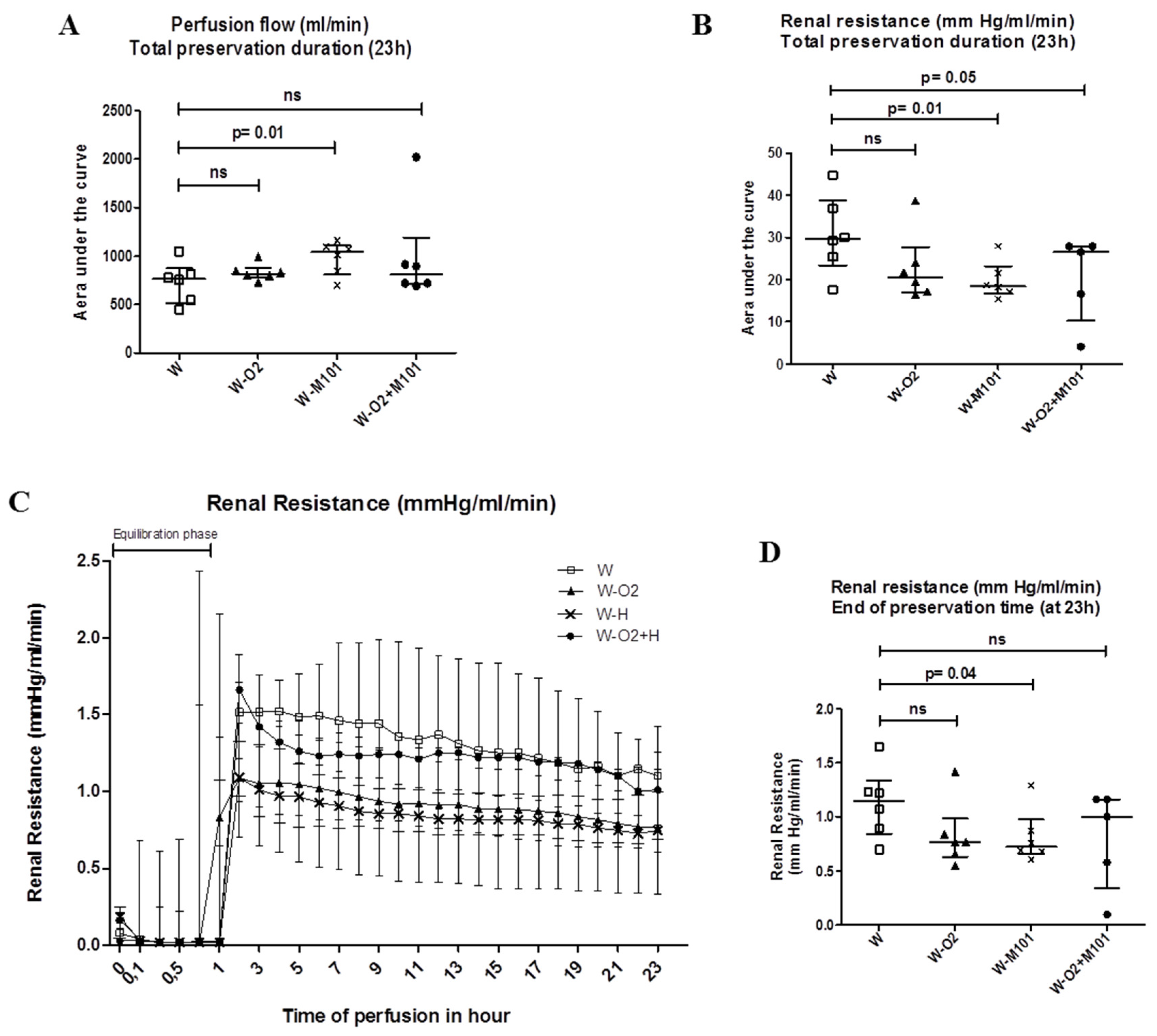
Abstract
:1. Introduction
2. Results
2.1. Ex Vivo Perfusion Parameters Evaluation
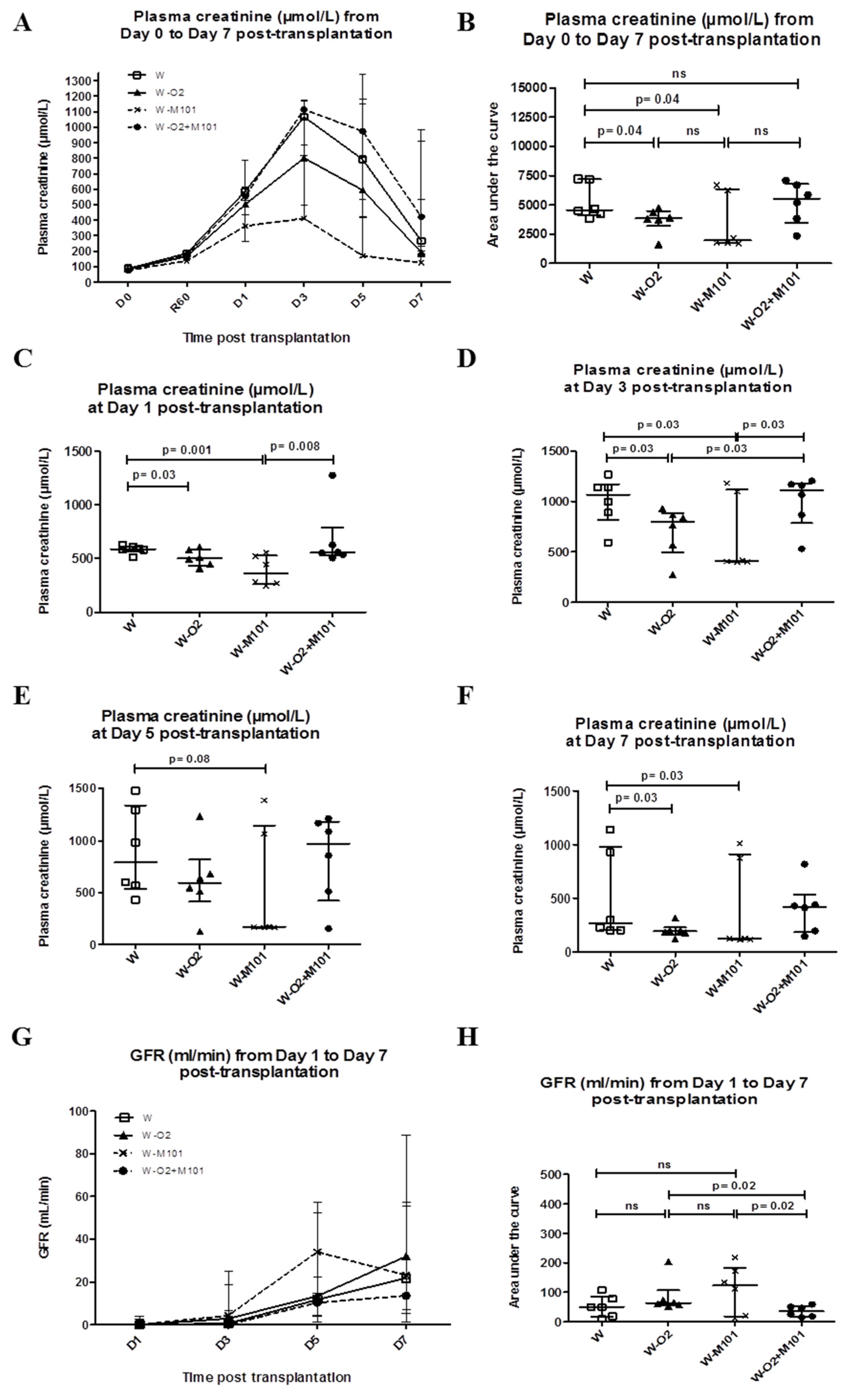
2.2. Kidney Function Recovery from Day 0 to Day 7 Post-Transplantation
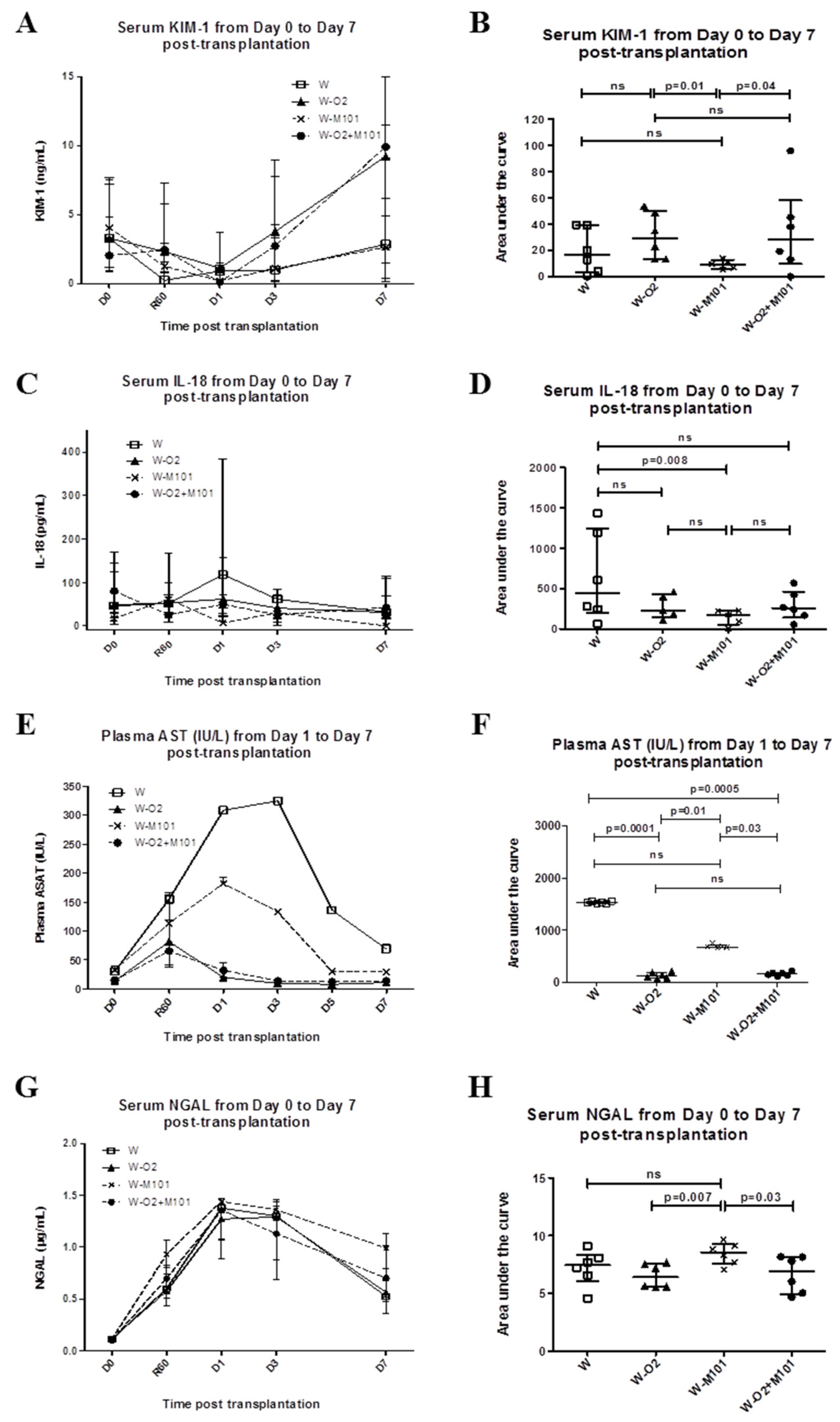
2.3. Kidney Injury Biomarker Evaluation from Day 0 to Day 7 Post-Transplantation
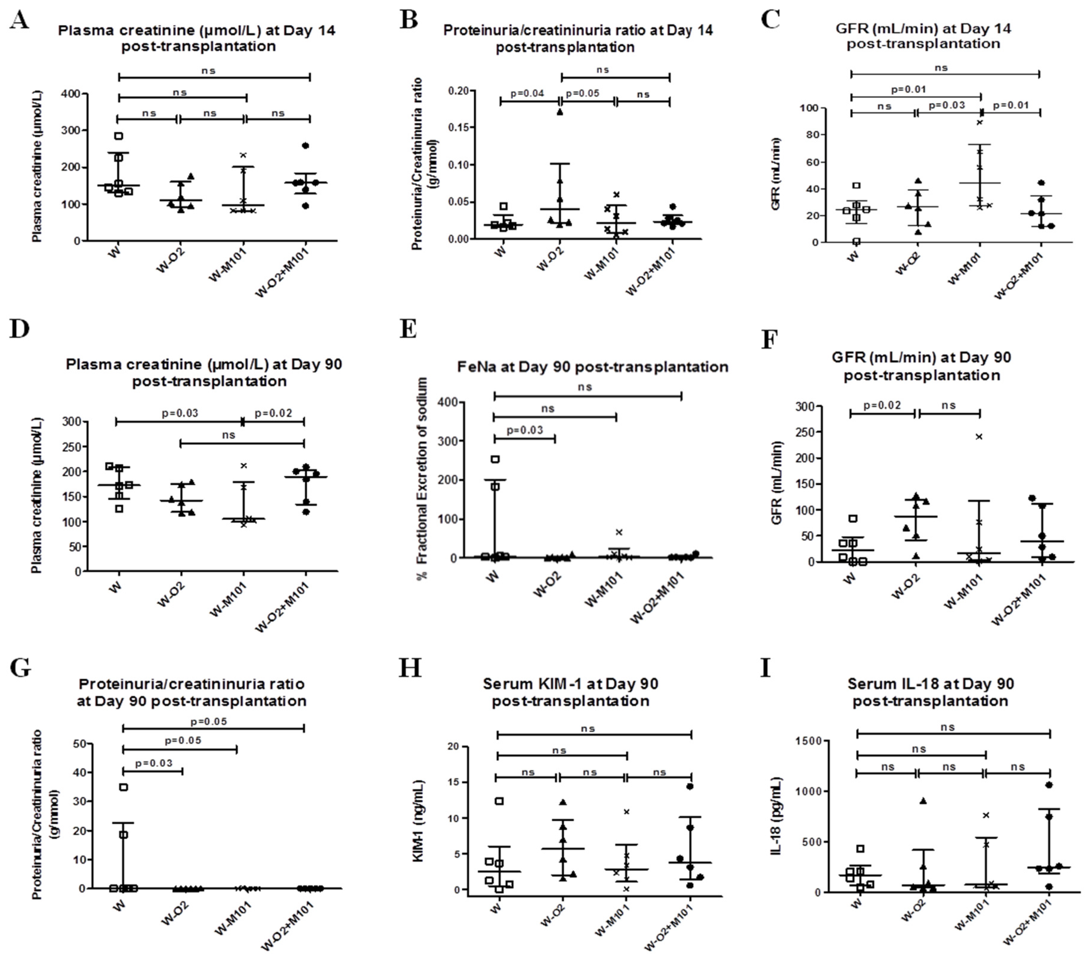
2.4. Kidney Function and Injury Biomarker Evaluation at Day 14 Post-Transplantation
2.5. Kidney Function Evaluation at Day 90 Post-Transplantation
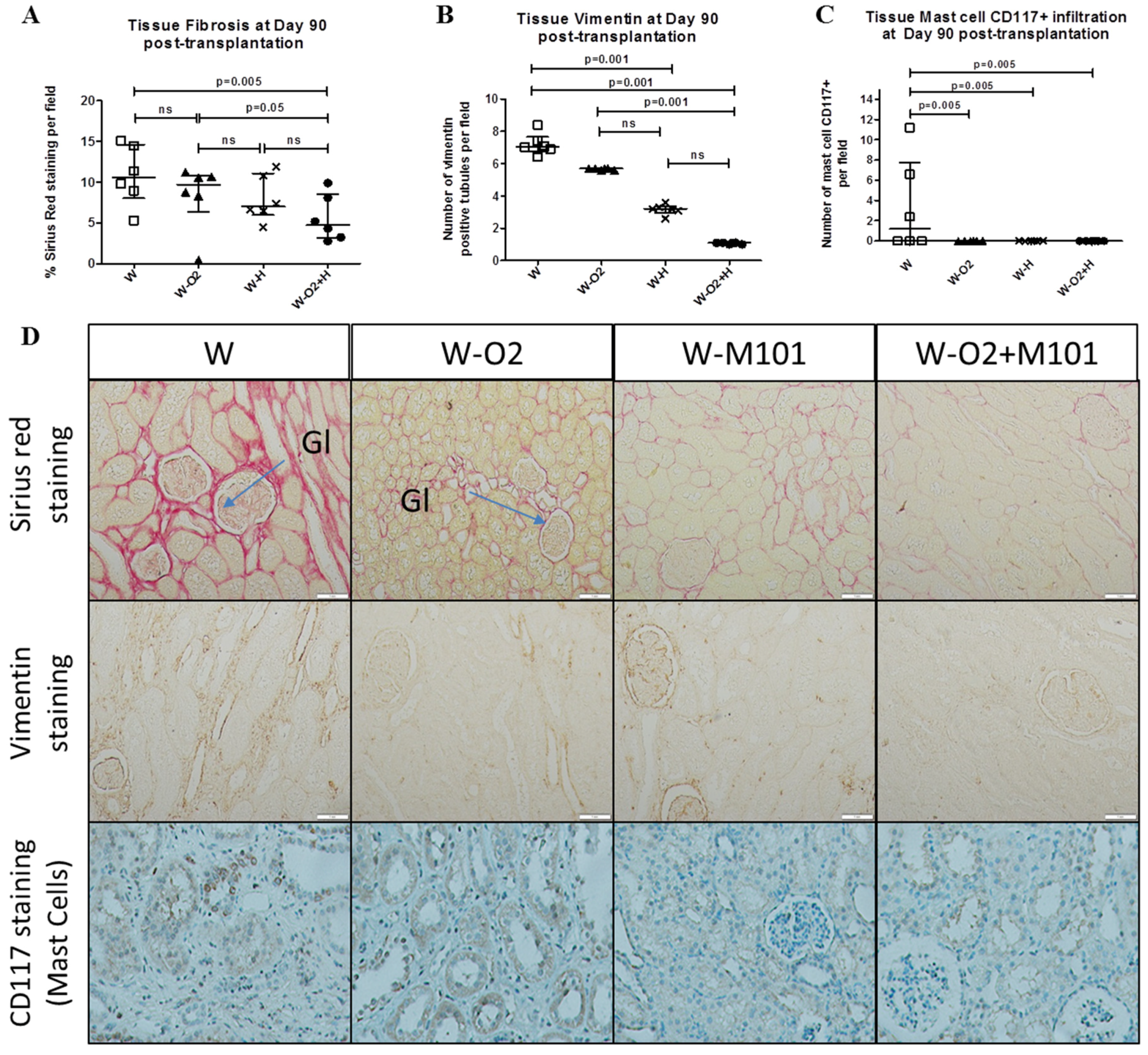
2.6. Histological Evaluation at Day 90 Post-Transplantation
3. Discussion
4. Materials and Methods
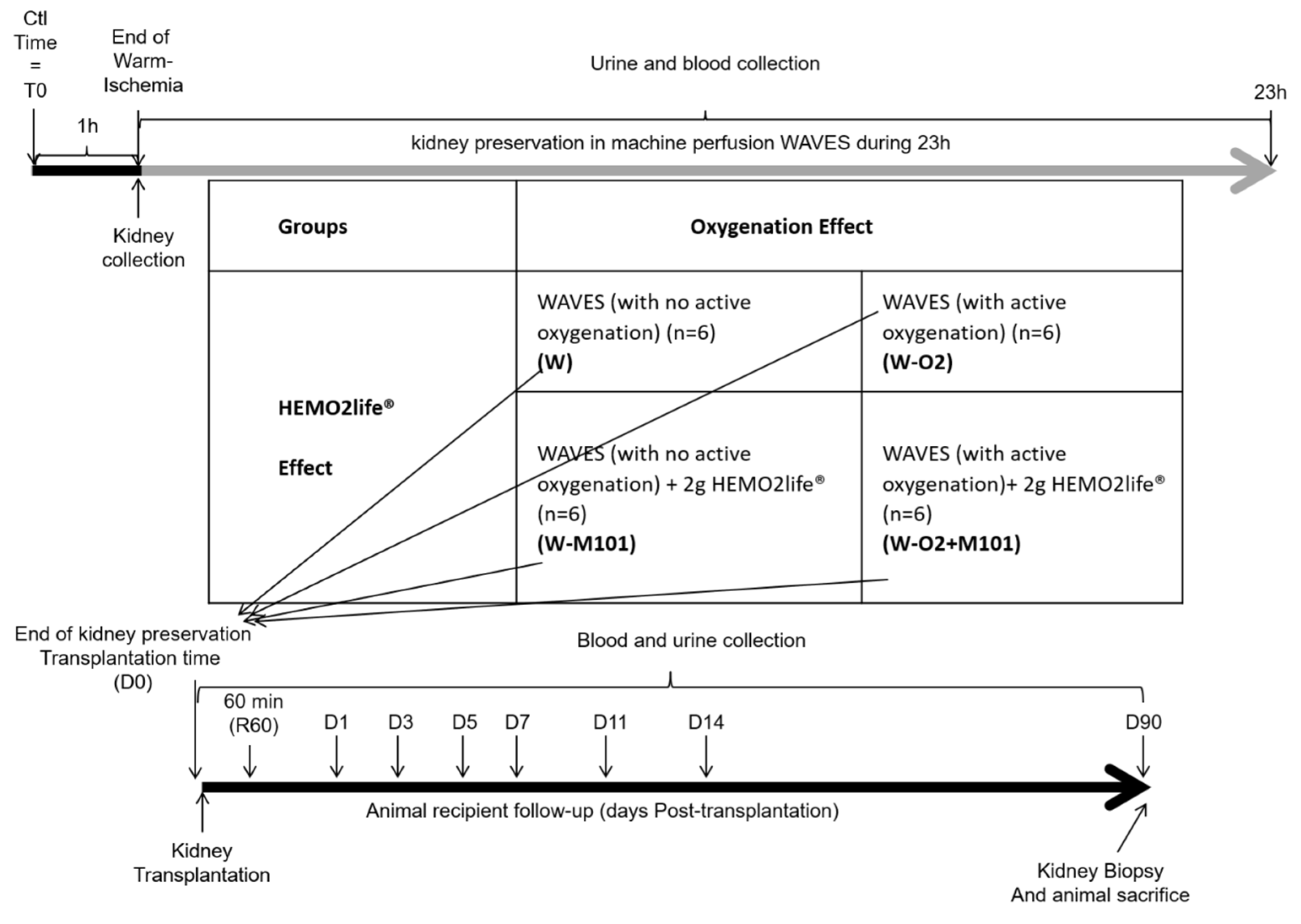
4.1. Experimental Model and Design of Experiments
4.2. Ex Vivo Perfusion Parameters
4.3. Kidney Function and Biomarkers Evaluation
4.4. Renal Histochemical and Immunohistochemical Evaluation at 3 Months Post-Transplantation
4.5. Statistical Analysis
5. Conclusions
Author Contributions
Funding
Acknowledgments
Conflicts of Interest
Abbreviations
| AST | Aspartate transaminase |
| AUC | Area under the curve |
| DCD | Donation after circulatory death |
| DGF | Delayed graft function |
| ECD | Expanded criteria donors |
| FeNa | Fractional excretion of sodium |
| GFR | Glomerular filtration rate |
| IL-18 | Interleukine-18 |
| IR | Ischemia-reperfusion |
| IRI | Ischemia-reperfusion injuries |
| KIM-1 | Kidney injury molecule-1 |
| MP | Machine perfusion |
| NGAL | Neutrophil gelatinase-associated lipocalin |
| pmh | Per million habitants |
| SOD | Superoxide dismutase |
Appendix A

References
- Oberbauer, R. Progression of Interstitial Fibrosis in Kidney Transplantation. Clin. J. Am. Soc. Nephrol. 2016, 11, 2110–2112. [Google Scholar] [CrossRef] [PubMed]
- Martínez Arcos, L.; Fabuel Alcañiz, J.J.; Gómez Dos Santos, V.; Burgos Revilla, F.J. Functional Results of Renal Preservation in Hypothermic Pulsatile Machine Perfusion Versus Cold Preservation: Systematic Review and Meta-Analysis of Clinical Trials. Transplant. Proc. 2018, 50, 24–32. [Google Scholar] [CrossRef]
- Thuillier, R.; Allain, G.; Celhay, O.; Hebrard, W.; Barrou, B.; Badet, L.; Leuvenink, H.; Hauet, T. Benefits of active oxygenation during hypothermic machine perfusion of kidneys in a preclinical model of deceased after cardiac death donors. J. Surg. Res. 2013, 184, 1174–1181. [Google Scholar] [CrossRef] [PubMed]
- ’t Hart, N.A.; van der Plaats, A.; Faber, A.; Leuvenink, H.G.D.; Olinga, P.; Wiersema-Buist, J.; Verkerke, G.J.; Rakhorst, G.; Ploeg, R.J. Oxygenation during hypothermic rat liver preservation: An in vitro slice study to demonstrate beneficial or toxic oxygenation effects. Liver Transpl. 2005, 11, 1403–1411. [Google Scholar] [CrossRef] [Green Version]
- Rousselot, M.; Delpy, E.; Drieu La Rochelle, C.; Lagente, V.; Pirow, R.; Rees, J.-F.; Hagege, A.; Le Guen, D.; Hourdez, S.; Zal, F. Arenicola marina extracellular hemoglobin: A new promising blood substitute. Biotechnol. J. 2006, 1, 333–345. [Google Scholar] [CrossRef]
- Thuillier, R.; Dutheil, D.; Trieu, M.T.N.; Mallet, V.; Allain, G.; Rousselot, M.; Denizot, M.; Goujon, J.-M.; Zal, F.; Hauet, T. Supplementation with a New Therapeutic Oxygen Carrier Reduces Chronic Fibrosis and Organ Dysfunction in Kidney Static Preservation: A New O2 Therapeutic Molecule Improves Static Kidney Preservation. Am. J. Transplant. 2011, 11, 1845–1860. [Google Scholar] [CrossRef]
- Le Meur, Y.; Morellon, E.; Essig, M.; Thierry, A.; Buchler, M.; Drouin, S.; Deruelle, C.; Badet, L.; Pesteil, F.; Delpech, P.O.; et al. Evaluation of a Marine OXYgen Carrier (Hemo2Life®) for Organ Preservation: First Use in Kidney Transplantation in Humans. Am. J. Transplant. 2018, 18, 905. [Google Scholar]
- Hoyer, D.P.; Gallinat, A.; Swoboda, S.; Wohlschlaeger, J.; Rauen, U.; Paul, A.; Minor, T. Influence of oxygen concentration during hypothermic machine perfusion on porcine kidneys from donation after circulatory death. Transplantation 2014, 98, 944–950. [Google Scholar] [CrossRef] [PubMed]
- Doucet, C.; Milin, S.; Favreau, F.; Desurmont, T.; Manguy, E.; Hebrard, W.; Yamamoto, Y.; Mauco, G.; Eugene, M.; Papadopoulos, V.; et al. A p38 mitogen-activated protein kinase inhibitor protects against renal damage in a non-heart-beating donor model. Am. J. Physiol. Ren. Physiol. 2008, 295, F179–F191. [Google Scholar] [CrossRef]
- Vaziri, N.; Thuillier, R.; Favreau, F.D.; Eugene, M.; Milin, S.; Chatauret, N.P.; Hauet, T.; Barrou, B. Analysis of machine perfusion benefits in kidney grafts: A preclinical study. J. Transl. Med. 2011, 9, 15. [Google Scholar] [CrossRef]
- Kaminski, J.; Hannaert, P.; Kasil, A.; Thuillier, R.; Leize, E.; Delpy, E.; Steichen, C.; Goujon, J.M.; Zal, F.; Hauet, T. Efficacy of the natural oxygen transporter HEMO2life® in cold preservation in a preclinical porcine model of donation after cardiac death. Transpl. Int. 2019. [Google Scholar] [CrossRef]
- Jochmans, I.; Moers, C.; Smits, J.M.; Leuvenink, H.G.D.; Treckmann, J.; Paul, A.; Rahmel, A.; Squifflet, J.-P.; van Heurn, E.; Monbaliu, D.; et al. The Prognostic Value of Renal Resistance During Hypothermic Machine Perfusion of Deceased Donor Kidneys: Renal Resistance of Machine-Perfused Kidney Grafts. Am. J. Transplant. 2011, 11, 2214–2220. [Google Scholar] [CrossRef]
- De Vries, E.E.; Hoogland, E.R.P.; Winkens, B.; Snoeijs, M.G.; van Heurn, L.W.E. Renovascular Resistance of Machine-Perfused DCD Kidneys Is Associated with Primary Nonfunction. Am. J. Transplant. 2011, 11, 2685–2691. [Google Scholar] [CrossRef] [Green Version]
- Thuillier, R.; Allain, G.; Giraud, S.; Saintyves, T.; Delpech, P.O.; Couturier, P.; Billault, C.; Marchand, E.; Vaahtera, L.; Parkkinen, J.; et al. Cyclodextrin curcumin formulation improves outcome in a preclinical pig model of marginal kidney transplantation. Am. J. Transplant. 2014, 14, 1073–1083. [Google Scholar] [CrossRef]
- Sabbisetti, V.S.; Waikar, S.S.; Antoine, D.J.; Smiles, A.; Wang, C.; Ravisankar, A.; Ito, K.; Sharma, S.; Ramadesikan, S.; Lee, M.; et al. Blood Kidney Injury Molecule-1 Is a Biomarker of Acute and Chronic Kidney Injury and Predicts Progression to ESRD in Type I Diabetes. J. Am. Soc. Nephrol. 2014, 25, 2177. [Google Scholar] [CrossRef]
- Sung, W.-C.; Yu, H.-P.; Tsai, Y.-F.; Chung, P.C.-H.; Lin, C.-C.; Lee, W.-C. The ratio of plasma interleukin-18 is a sensitive biomarker for acute kidney injury after liver transplantation. Transplant. Proc. 2014, 46, 816–817. [Google Scholar] [CrossRef] [PubMed]
- Jochmans, I.; Lerut, E.; van Pelt, J.; Monbaliu, D.; Pirenne, J. Circulating AST, H-FABP, and NGAL are early and accurate biomarkers of graft injury and dysfunction in a preclinical model of kidney transplantation. Ann. Surg. 2011, 254, 784–791. [Google Scholar] [CrossRef]
- Serwin, N.M.; Wiśniewska, M.; Jesionowska, A.; Skwirczyńska, E.; Marcinowska, Z.; Dołęgowska, B. Serum levels of 12 renal function and injury markers in patients with glomerulonephritis. Pol. Arch. Med. Wewn. 2016, 126, 483–493. [Google Scholar] [CrossRef]
- Gandhi, R.; Yi, J.; Ha, J.; Shi, H.; Ismail, O.; Nathoo, S.; Bonventre, J.V.; Zhang, X.; Gunaratnam, L. Accelerated receptor shedding inhibits kidney injury molecule-1 (KIM-1)-mediated efferocytosis. Am. J. Physiol. Ren. Physiol. 2014, 307, F205. [Google Scholar] [CrossRef] [PubMed]
- Bonventre, J.V. Kidney injury molecule-1: a translational journey. Trans. Am. Clin. Climatol. Assoc. 2014, 125, 293–299. [Google Scholar]
- Peters, S.M.; Rauen, U.; Tijsen, M.J.; Bindels, R.J.; van Os, C.H.; de Groot, H.; Wetzels, J.F. Cold preservation of isolated rabbit proximal tubules induces radical-mediated cell injury. Transplantation 1998, 65, 625–632. [Google Scholar] [CrossRef] [PubMed]
- Liang, D.; Liu, H.-F.; Yao, C.-W.; Liu, H.-Y.; Huang-Fu, C.-M.; Chen, X.-W.; Du, S.-H.; Chen, X.-W. Effects of interleukin 18 on injury and activation of human proximal tubular epithelial cells. Nephrology 2007, 12, 53–61. [Google Scholar] [CrossRef]
- Lombi, F.; Muryan, A.; Canzonieri, R.; Trimarchi, H. Biomarkers in acute kidney injury: Evidence or paradigm? Nefrol. Engl. Ed. 2016, 36, 339–346. [Google Scholar] [CrossRef] [Green Version]
- Patel, S.K.; Pankewycz, O.G.; Nader, N.D.; Zachariah, M.; Kohli, R.; Laftavi, M.R. Prognostic Utility of Hypothermic Machine Perfusion in Deceased Donor Renal Transplantation. Transplant. Proc. 2012, 44, 2207–2212. [Google Scholar] [CrossRef]
- Chapman, J.R.; O’Connell, P.J.; Nankivell, B.J. Chronic Renal Allograft Dysfunction. J. Am. Soc. Nephrol. 2005, 16, 3015–3026. [Google Scholar] [CrossRef] [PubMed] [Green Version]
- Li, M.; Luan, F.; Zhao, Y.; Hao, H.; Zhou, Y.; Han, W.; Fu, X. Epithelial-mesenchymal transition: An emerging target in tissue fibrosis. Exp. Biol. Med. 2016, 241, 1. [Google Scholar] [CrossRef] [PubMed]
- Summers, S.A.; Gan, P.-Y.; Dewage, L.; Ma, F.T.; Ooi, J.D.; O’Sullivan, K.M.; Nikolic-Paterson, D.J.; Kitching, A.R.; Holdsworth, S.R. Mast cell activation and degranulation promotes renal fibrosis in experimental unilateral ureteric obstruction. Kidney Int. 2012, 82, 676–685. [Google Scholar] [CrossRef] [PubMed] [Green Version]
- Patel, K.; Smith, T.B.; Neil, D.A.; Thakker, A.; Tsuchiya, Y.; Higgs, E.B.; Hodges, N.J.; Ready, A.R.; Nath, J.; Ludwig, C. The effects of oxygenation on ex vivo kidneys undergoing Hypothermic Machine Perfusion. Transplantation 2018, 103, 314–322. [Google Scholar] [CrossRef] [PubMed]
- Abramowicz, D.; Oberbauer, R.; Heemann, U.; Viklicky, O.; Peruzzi, L.; Mariat, C.; Crespo, M.; Budde, K.; Oniscu, G.C. Recent advances in kidney transplantation: a viewpoint from the Descartes advisory board. Nephrol. Dial. Transplant. 2018, 33, 1699–1707. [Google Scholar] [CrossRef] [PubMed]
- O’Callaghan, J.; Pall, K.; Pengel, L. Supplemental oxygen during hypothermic kidney preservation: A systematic review. Transplant. Rev. 2017, 31, 172–179. [Google Scholar] [CrossRef]
- Morito, N.; Obara, H.; Matsuno, N.; Enosawa, S.; Furukawa, H. Oxygen consumption during hypothermic and subnormothermic machine perfusions of porcine liver grafts after cardiac death. J. Artif. Organs 2018, 21, 450–457. [Google Scholar] [CrossRef] [PubMed]


© 2019 by the authors. Licensee MDPI, Basel, Switzerland. This article is an open access article distributed under the terms and conditions of the Creative Commons Attribution (CC BY) license (http://creativecommons.org/licenses/by/4.0/).
Share and Cite
Kasil, A.; Giraud, S.; Couturier, P.; Amiri, A.; Danion, J.; Donatini, G.; Matillon, X.; Hauet, T.; Badet, L. Individual and Combined Impact of Oxygen and Oxygen Transporter Supplementation during Kidney Machine Preservation in a Porcine Preclinical Kidney Transplantation Model. Int. J. Mol. Sci. 2019, 20, 1992. https://doi.org/10.3390/ijms20081992
Kasil A, Giraud S, Couturier P, Amiri A, Danion J, Donatini G, Matillon X, Hauet T, Badet L. Individual and Combined Impact of Oxygen and Oxygen Transporter Supplementation during Kidney Machine Preservation in a Porcine Preclinical Kidney Transplantation Model. International Journal of Molecular Sciences. 2019; 20(8):1992. https://doi.org/10.3390/ijms20081992
Chicago/Turabian StyleKasil, Abdelsalam, Sebastien Giraud, Pierre Couturier, Akbar Amiri, Jerome Danion, Gianluca Donatini, Xavier Matillon, Thierry Hauet, and Lionel Badet. 2019. "Individual and Combined Impact of Oxygen and Oxygen Transporter Supplementation during Kidney Machine Preservation in a Porcine Preclinical Kidney Transplantation Model" International Journal of Molecular Sciences 20, no. 8: 1992. https://doi.org/10.3390/ijms20081992