Article

Celf1 Is Required for Formation of Endoderm-Derived Organs in Zebrafish

Gene Regulation Research, Nara Institute Science and Technology, 8916-5 Takayama, Nara 630-0101, Japan
*
Author to whom correspondence should be addressed.
Int. J. Mol. Sci. 2013, 14(9), 18009-18023; https://doi.org/10.3390/ijms140918009
Submission received: 25 June 2013 / Revised: 21 August 2013 / Accepted: 26 August 2013 / Published: 3 September 2013
(This article belongs to the Special Issue Post-Transcriptional Gene Regulation by Ribonucleoprotein Complexes)

Abstract

:
We recently reported that an RNA binding protein called Cugbp Elav-like family member 1 (Celf1) regulates somite symmetry and left-right patterning in zebrafish. In this report, we show additional roles of Celf1 in zebrafish organogenesis. When celf1 is knocked down by using an antisense morpholino oligonucleotides (MO), liver buds fail to form, and pancreas buds do not form a cluster, suggesting earlier defects in endoderm organogenesis. As expected, we found failures in endoderm cell growth and migration during gastrulation in embryos injected with celf1-MOs. RNA immunoprecipitation revealed that Celf1 binds to gata5 and cdc42 mRNAs which are known to be involved in cell growth and migration, respectively. Our results therefore suggest that Celf1 regulates proper organogenesis of endoderm-derived tissues by regulating the expression of such targets.

Graphical Abstract

1. Introduction

Cugbp Elav-like family member 1 (Celf1), which is a member of the Celf family of RNA binding proteins, regulates gene expression at multiple post-transcriptional levels including alternative splicing and mRNA decay, and fine-tunes the amounts of proteins that are synthesized from its target mRNAs [13]. Celf1 target mRNAs have been identified by several approaches such as the yeast three hybrid system [4], systematic evolution of ligands by exponential enrichment (SELEX) [5], RNA immunoprecipitation followed by microarray (RIP-Chip), and cross-linking immunoprecipitation followed by sequencing (CLIP-Seq) [610]. Celf1 binds to hundreds of short-lived mRNAs, which are involved in cell growth, migration and death [7]. On the basis of these analyses, UG-rich or UGU repeats are identified as a Celf1 binding sequences [3,7].
It has been reported that Celf1 is involved in the regulation of somite segmentation, muscle formation, and spermatogenesis in vertebrate development [6,11,12]. In addition, we recently showed that celf1 is required for somite symmetry and cardiac laterality [13]. In the course of our investigation of other laterality organs such as liver and pancreas, we find an unexpected role for celf1 in zebrafish organogenesis; celf1 is essential for the formation of endoderm-derived organs.

2. Results and Discussion

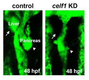
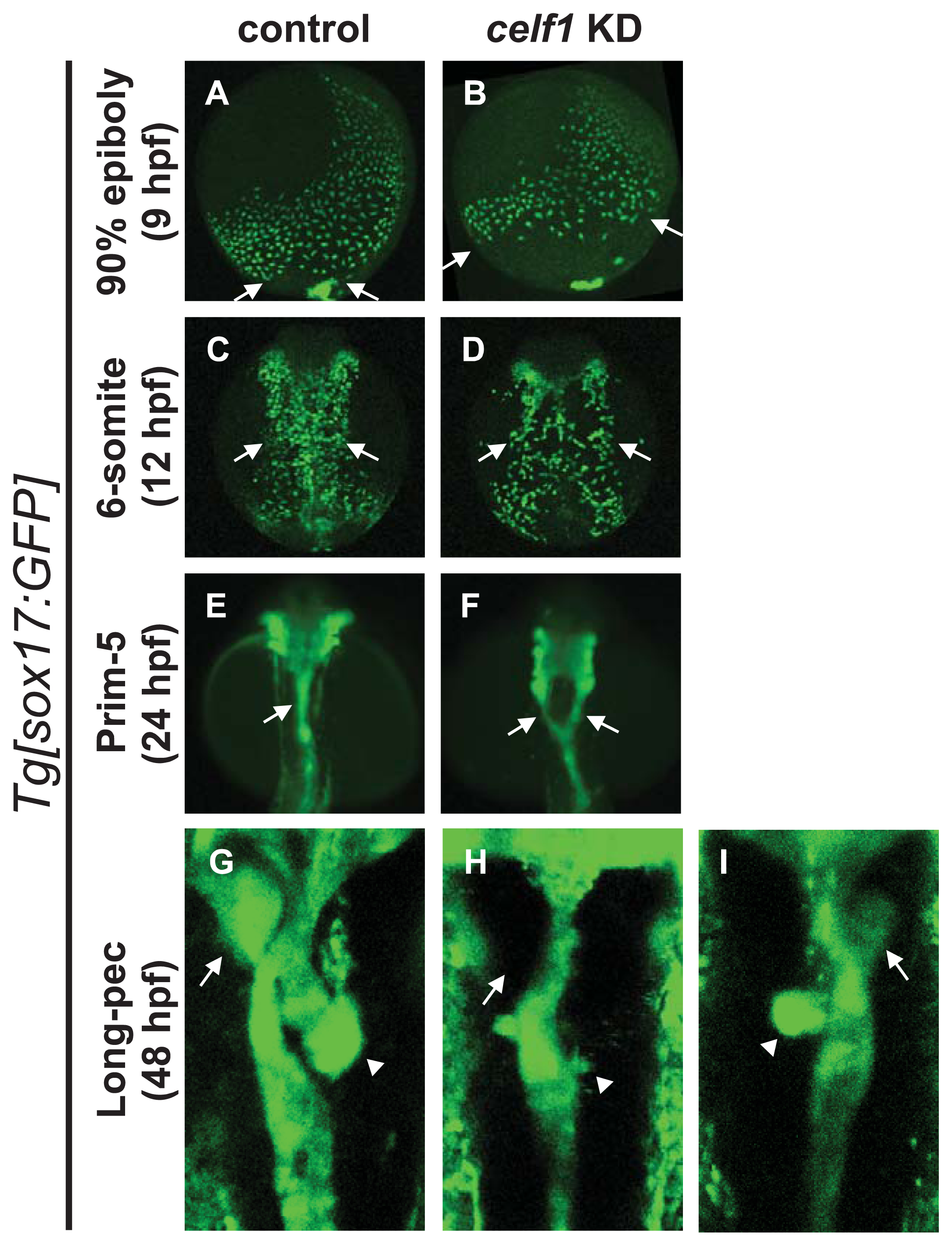
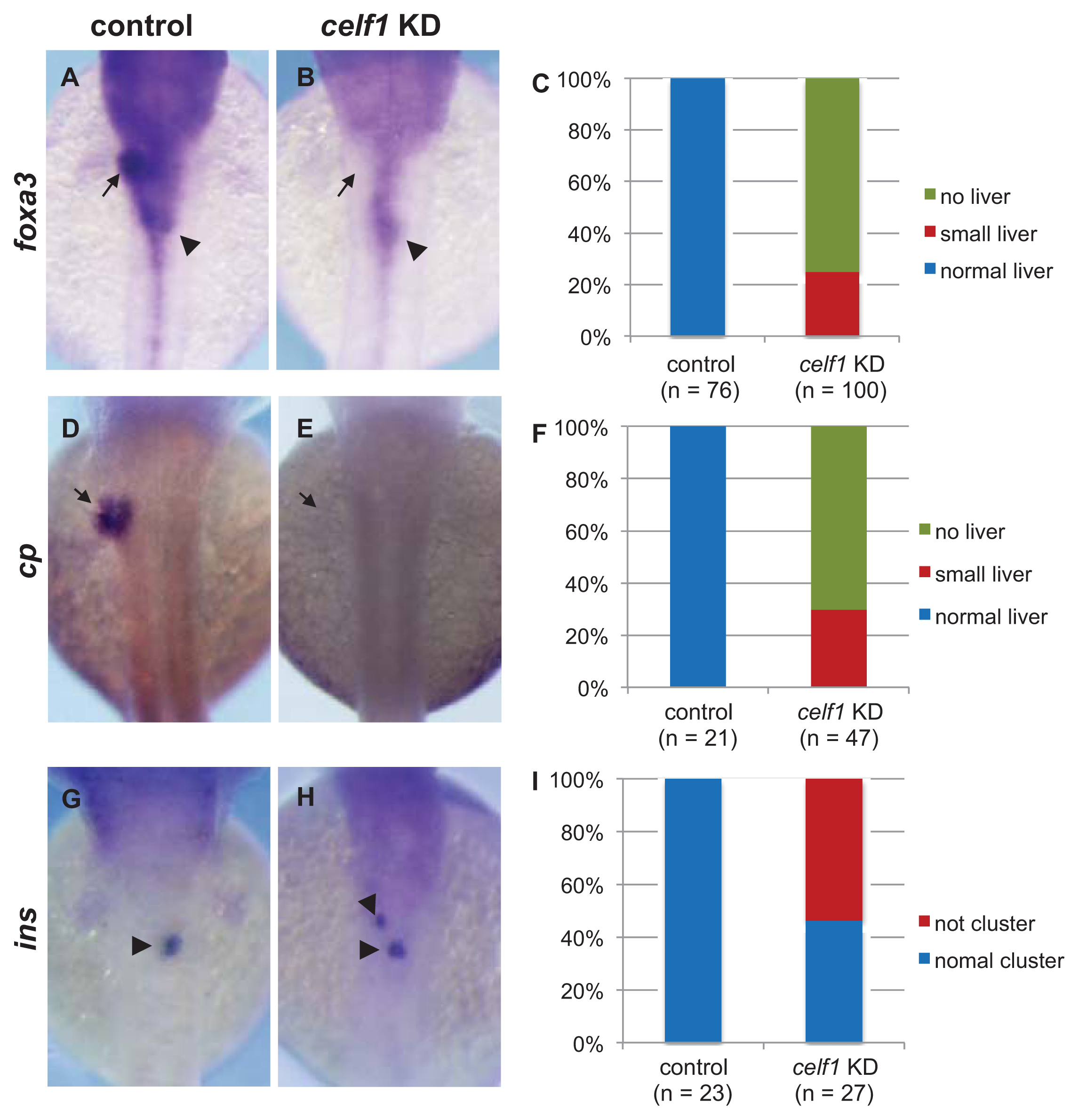
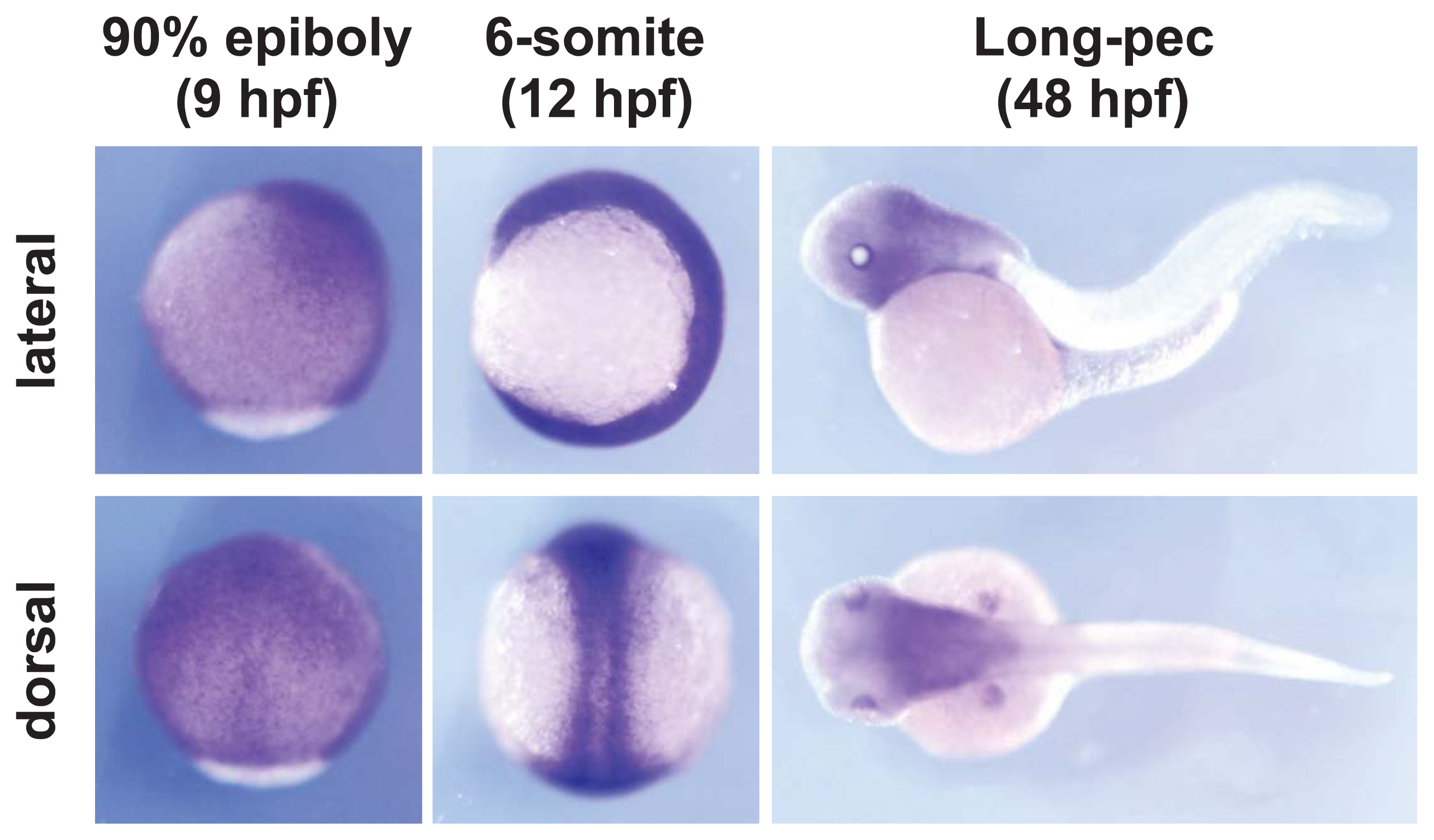
2.1. Celf1 Is Involved in Formation of Endoderm-Derived Organs

Since cardiac laterality was altered in celf1 knockdown (KD) embryos [13], we reasoned that the lateralities of endoderm-derived organs such as liver, pancreas and gut are also altered in celf1 KD embryos. To test this, we injected celf1 morpholinos (celf1-MOs) into transgenic line Tg [sox17:GFP] [14], whose GFP is expressed in endoderm cells and dorsal forerunner cells, and observed the formation of endoderm-derived organs in celf1 KD embryos. In control embryos at 48 h postfertilization (hpf), liver and pancreas were formed on the left and right sides of the gut tube, and this tube was bent because the placement of these organs and looping are regulated by left-right patterning (Figure 1G) [1517]. However, in the celf1 KD embryos, signs of the liver bud, pancreas buds were low, and the gut tube tended to be straight (Figure 1H). Although we could observe left-right defects in the gut tube in the celf1 KD embryos (Figure 1I), other defects were unexpected. These results therefore suggest that celf1 has an additional role(s) in the formation of endoderm-derived organs in zebrafish.
To investigate the role of celf1 in the formation of endoderm-derived organs, we analyzed the expression of markers for general endoderm derivatives (forkhead box A3, foxa3), liver fate (ceruloplasmin, cp), and pancreas differentiation (preproinsulin, ins). Consistent with the results seen in the Tg [sox17:GFP] embryos, liver buds became smaller or absent in the celf1 KD embryos (Figure 2A–F). Although β-cells in the pancreas formed a cluster by 48 hpf in the control embryos, two or three populations of β-cells were visible in the celf1 KD embryos (Figure 2G–I). These results suggest that celf1 is essential for the formation of endoderm-derived organs.

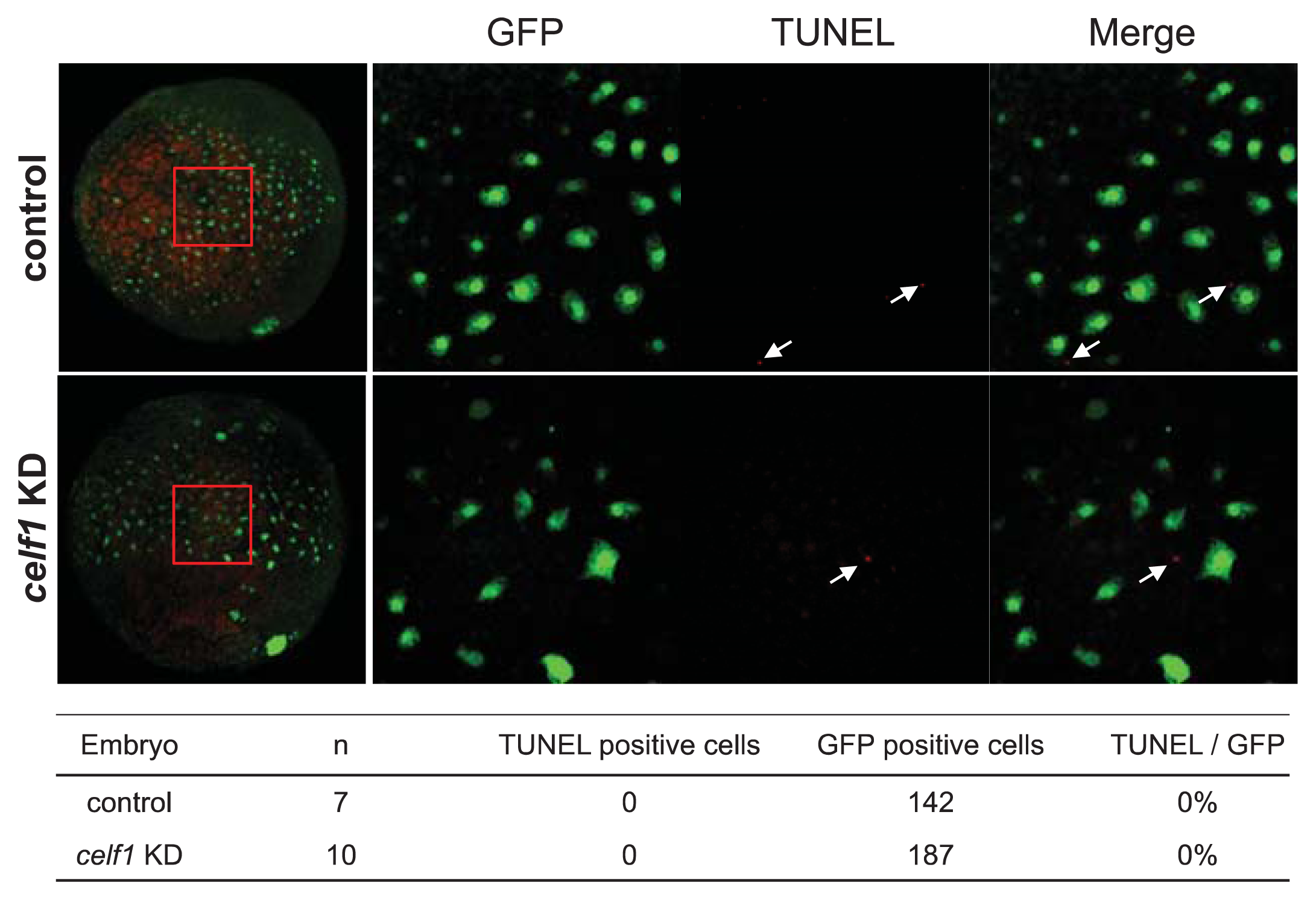
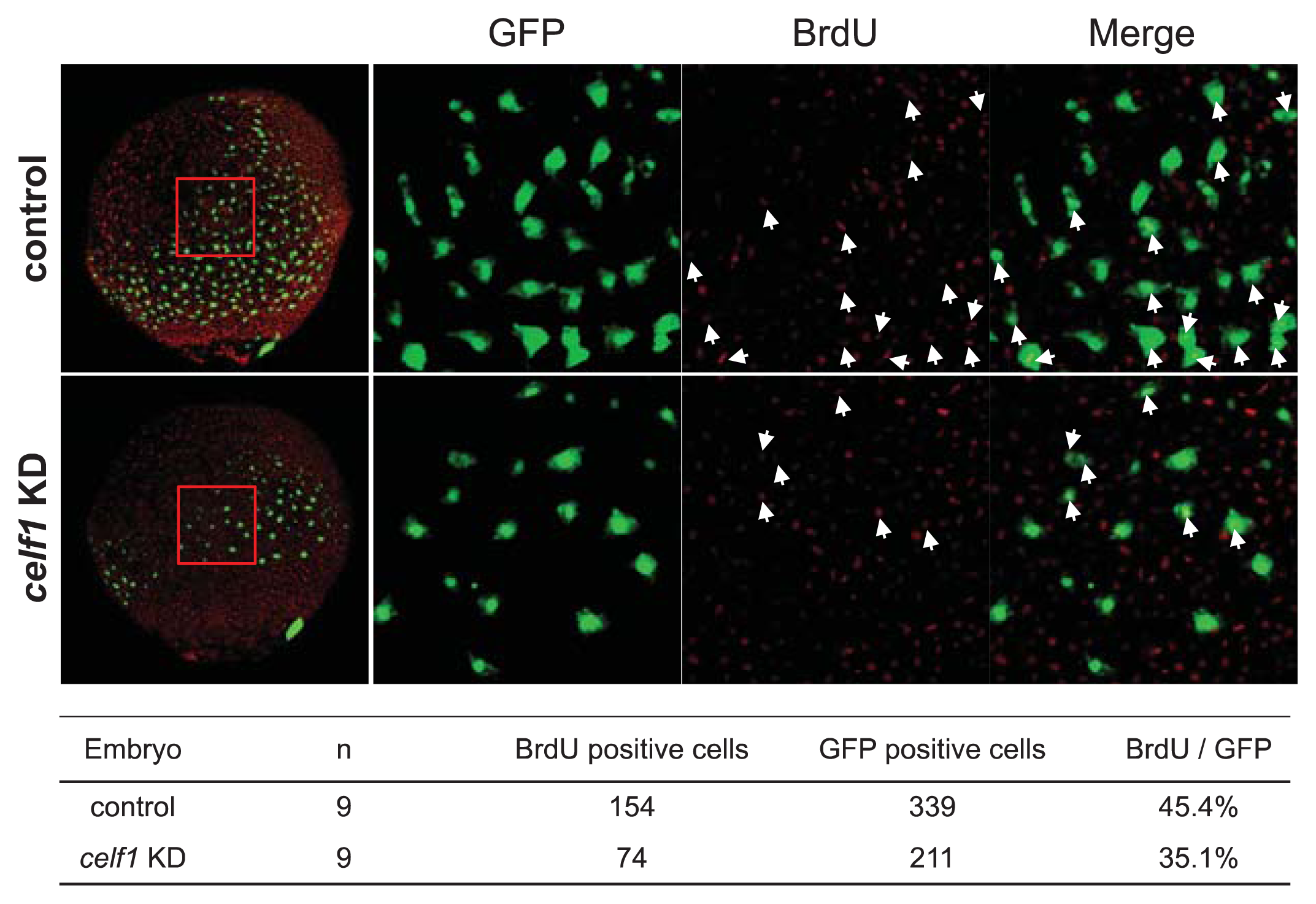
2.2. Celf1 Controls Endoderm Cell Growth during Gastrulation

As a maternal factor, celf1 is broadly expressed in zebrafish embryos by gastrulation stages, whereas celf1 expression is not detected in endoderm derivatives (Figure A1). Instead, celf1 expression is restricted to specific regions such as eyes and pectoral fins at later stages [13,18,19]. We therefore reasoned that celf1 regulates endoderm formation during gastrulation and secondarily affects the formation of endoderm-derived organs in later embryos. To test the possibility, we observed the behavior of endoderm cells during gastrulation by using Tg [sox17:GFP] embryos. GFP-expressing endoderm cells were distributed around the blastoderm margin in a salt-and-pepper pattern in control embryos at 6 hpf and they then migrated dorsally and proliferated (Movie 1). Although endoderm cells appeared normally in celf1 KD embryos at 6 hpf, dorsal migration of endoderm cells became slow and the number of the cells seemed to be low in comparison with the controls (Figure 1A,B, and Movies 1 and 2).
To confirm whether the endoderm cell number is reduced in the celf1 KD embryos, we analyzed the expression of an endoderm specification marker (sox32) and counted the number of sox32-expressing endoderm cells. The number of cells in the celf1 KD embryos was normal at 6 hpf but became significantly lower at 9 hpf (Figure 3). These results suggest that, in celf1 KD embryos, endoderm specification occurs normally, but proliferation and/or death of endoderm cells are altered. We thus tested whether celf1 regulates endoderm cell death, growth or both during gastrulation. Fragmented GFP signals, which are a sign of dead cells [20], were not observed both in the control and celf1 KD embryos during the dorsal migration of the endoderm cells (Table 1). This result was supported by the data from TUNEL assays (Figure A2). In contrast, the number of cell divisions became significantly lower in the celf1 KD embryos (p < 0.05, Table 1 and Movies 1 and 2). In agreement with this, BrdU incorporation of endoderm cells in celf1 KD embryos significantly reduced as compared to that of control embryos (p < 0.05, Figure A3). These results suggest that celf1 regulates endoderm proliferation during gastrulation.

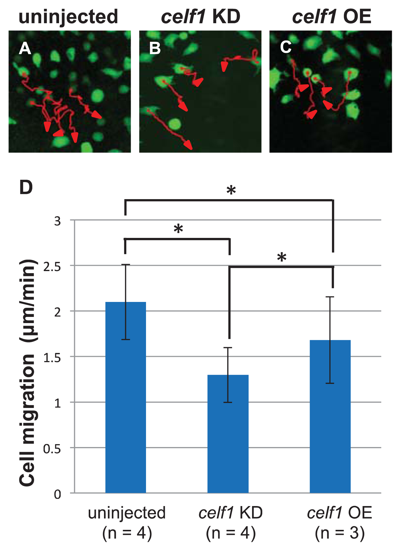
2.3. Celf1 Regulates Endoderm Cell Migration during Gastrulation

Since time lapse observations suggest that cell migration is also limited in celf1 KD embryos, we next investigated endoderm cell movements during gastrulation by tracing the trajectory of each GFP-expressing endoderm cell. In the celf1 KD embryos, many endoderm cells migrated toward the midline, but the speed of the migration became slower than that of the uninjected control, leading to a defect in endoderm cell assembly around the midline (Figure 4). Consistent with the defect in endoderm migration during gastrulation, distribution of endoderm cells in the celf1 KD embryos at 12 hpf became wider in comparison with that of the control (Figure 1C,D). Knockdown of celf1 then resulted in a failure to fuse the anterior gut tube, leading to formation of a Y-shaped tube (Figure 1E,F). With the data taken all together, our results suggest that celf1 regulates the growth and migration of endoderm cells during gastrulation to generate endoderm-derived organs properly.

2.4. Celf1 Binds to gata5 and cdc42 mRNAs In Vivo

Celf1 binds to UG-rich elements or UGU repeats within the 3′ untranslated regions (3′ UTR) of mRNAs and regulates gene expression at multiple post-transcriptional levels [13]. Since celf1 is essential for the growth and migration of endoderm cells, we looked for possible targets by undertaking a search for genes involved in endoderm proliferation and migration. Because either UG rich sequences or UGU repeats are existed in 3′ UTR of many mRNAs that encode cell cycle and migration regulators [5] and because flanking U-rich or UA-rich elements to UG/UGU sequences also affects the binding affinity of Celf1 [13], we thought that a sequence containing U/UA-rich elements and at least four UGU repeats within 35 bp would be a strong candidate for the Celf1-binding site. We thus selected gata5 (gata-binding protein 5) and cdc42 as potential targets of Celf1 for the following reasons (Figure 5A). gata5 is known to control endoderm proliferation in zebrafish [21,22]. Both five UGU repeats and U-rich elements are present in 3′ UTR of gata5 mRNA (Figure 5A). Rho family G proteins (Rho, Rac, Cdc42) regulate the convergence extension (CE) movements of mesoendoderm [23], but Rac and Cdc42 (but not Rho) control primordial midgut cells in the fly [24]. Among Rho family G proteins, only cdc42 mRNA carries a putative Celf1 binding site that is composed of seven UGU repeats and U-rich sequences (Figure 5A). In addition, Gata5 and/or Cdc42 are identified as putative Celf1 targets in mouse muscle cells [6] and human T cells [8]. To test whether Celf1 binds to gata5 and cdc42 mRNAs in vivo, we performed RIP assays by using Celf1 antiserum. Although Celf1 did not bind to cyclinA1 (ccna1) mRNA (negative control) [13], Celf1 associated with gata5 and cdc42 mRNAs (Figure 5B). To investigate whether Celf1 affects the expression of gata5 and cdc42, we performed qPCR analyses in control and celf1 KD embryos. Knockdown of celf1 resulted in a 23% and 39% increase of the amounts of gata5 and cdc42 mRNAs relative to control, respectively (Figure 5C). These results suggest that Celf1 controls the formation of endoderm-derived organs through modulating protein expression from such targets during zebrafish development.

2.5. Celf1 Targets

Because loss-of-function of gata5 resulted in the reduction of endoderm proliferation [21,22], we expected that Celf1 stabilize gata5 mRNA. However, we got opposite results from qPCR analyses: Celf1 may destabilize gata5 mRNA (Figure 5C). In addition, overexpression of celf1 did not affect endoderm proliferation (Figure A4). Although Celf1 controls the levels of gata5 mRNA, our and previous observations suggest that endoderm proliferation is regulated by complicated mechanisms, to which several factors contribute. To control the CE movements during gastrulation, Cdc42 is activated by non-canonical Wnt signaling [23]. Consistent with the fact that both loss- and gain-of-functions of the signaling showed CE defects [23], knockdown and overexpression of celf1 resulted in slower migration of endoderm cells relative to uninjected control samples (Figure 4). Thus, cdc42 is a strong candidate of the target to regulate endoderm migration.
However, we could not conclude yet whether these interactions are sufficient for controlling endoderm proliferation and migration. As reported previously [13], blocking the interaction of Celf1 with either gata5 or cdc42 mRNA using specific target protector morpholinos will be required. Because UG-rich elements or UGU repeats are present in numerous mRNAs, it is also possible that Celf1 coordinates protein expression from several targets to generate endoderm-derived organs properly. Therefore, systematic analyses including cross-linking RNA immunoprecipitation followed by microarray or sequencing will be important for understanding all of the roles of Celf1 in endoderm formation.

2.6. Roles of Celf1 in Generation of Endoderm-Derived Organs

Our data suggest that Celf1 regulates endoderm formation during gastrulation. However, in celf1 KD embryos at later stages, we could find several failures such as defective convergence of the gut tube, loss of the liver, and malformation of the pancreas (Figures 1 and 2). Although it is possible that these failures are secondary defects of endoderm formation at an earlier stage, one possibility is that Celf1 also contributes to generating endoderm-derived organs in later stages. Since celf1 is not expressed in endoderm-derived organs in later stages (Figure A1), it is suggested that celf1 non-cell autonomously affects the formation of endoderm-derived organs. It would be of great interest to prove the stage and cell type specific roles of Celf1 in zebrafish embryos.

3. Experimental Section

3.1. Zebrafish and Whole-Mount In Situ Hybridization

Wild-type and Tg [sox17:GFP] [14] zebrafish were used in this study. Whole-mount in situ hybridization was performed as described previously [20,25]. cDNA fragments of celf1, cp, foxa3, ins, and sox32 were used as templates for the antisense probes.

3.2. Morpholino and mRNA Injection

Antisense MO oligonucleotides named celf1_long-MO, celf1_short-MO, and control-MO were obtained from Gene Tools. celf1_long-MO and celf1_short-MO were designed to target the AUG initiation codon of these mRNAs.
The MO sequences were as follows:
  • control-MO: 5′-CCTCTTACCTCAGTTACAATTTATA-3′;
  • celf1_long-MO: 5′-GCTTCAGCTTCGATACTATCCATCC-3′ [13];
  • celf1_short-MO: 5′-GTGGTCCAGAGACCCATTCATCTTC-3′ [13].
To knock down celf1, we co-injected 2.5 ng celf1_long-MO and 2.5 ng celf1_short-MO (5 ng celf1-MOs) into one-cell-stage zebrafish embryos. As a control, we injected 5 ng control-MO. We previously evaluated the specificity and efficacy of celf1_long-MO and celf1_short-MO [13].
pCS2-celf1 (long form) and pCS2-monomeric red fluorescent protein (mRFP) were used in this study. celf1 and mRFP mRNAs were synthesized using SP mMassage mMachine System (Ambion, Carlsbad, CA, USA). To overexpress celf1, we injected 150 pg celf1 mRNA into one-cell-stage embryos. As a control, 150 pg mRFP mRNA was injected.

3.3. TUNEL and Immunofluorescence Analyses

Tg [sox17:GFP] embryos at 90% epiboly stage (9 hpf) were fixed with 4% paraformaldehyde (PFA). Dead cells within the embryos were detected using In Situ Cell Death Detection Kit, POD (Roche, Mannheim, Germany), and the signals were amplified using an Alexa Fluor 647-Tyramide Signal Amplification Kit (Invitrogen, Carlsbad, CA, USA) following the manufacturer’s instructions. To visualize the location of GFP-positive endoderm cells within the embryos after TUNEL, immunofluorescence analyses were performed as described [20]. Anti-GFP (Chicken antibodies, IgY fraction) (aves, Tigard, OR, USA) and CF488A goat anti-chicken IgY (Biotium, Hayward, CA, USA) were used.

3.4. BrdU Labeling and Detection

About 1 nL of 25 mM BrdU (Sigma, St. Louis, MO, USA) was injected into the yolk of Tg [sox17:GFP] embryos at shield stage (6 hpf). After 3 h incubation, embryos were fixed with 4% PFA. After immunofluorescence analyses for GFP, embryos were treated with 5 μg/mL of protenase K (Roche, Mannheim, Germany) for 5 min, washed with PBSDT (1% DMSO, 0.1% TritonX100 in PBS) and fixed with 4% PFA. Re-fixed embryos were treated with 2 N HCl for 20 min, treated with 0.1 M Boric acid for 10 min, washed with PBSDT and blocked with 2% FBS in PBSDT (blocking buffer) for at least 30 min. The embryos were incubated with rat anti-BrdU antibody (AbD Serotec, Oxford, UK) in blocking buffer for at least 16 h at 4 °C. After extensive washing with PBSDT, embryos were incubated with CF647 donkey anti-rat IgG (Biotium, Hayward, CA, USA) in blocking buffer for 16 h at 4 °C. Embryos were extensively washed with PBSDT and fixed with 4% PFA.

3.5. Imaging of Fluorescence Signals

Embryos were embedded in 1% low-melt agarose. Time-lapse image acquisition was performed with an LSM710 confocal microscope and Zen software (Zeiss, Oberkochen, Germany). Immunefluorescence signals in fixed embryos or GFP signals in live embryos were visualized and photographed using an SZX12 stereo microscope (Olympus, Tokyo, Japan), LSM710 or LSM-Duo confocal microscopes (Zeiss, Oberkochen, Germany).

3.6. RNA Immunoprecipitation (RIP) Assay

In accordance with the manufacturer’s protocol of the RIP-Assay Kit (MBL), complexes which consist of mRNAs and Celf1 were isolated from uninjected embryos at 9–10 hpf by using rabbit anti-Celf1 antiserum (a kind gift from Dr. Kunio Inoue). Normal rabbit serum (Thermo Scientific, Waltham, MA, USA) was used as a control. In vivo interaction between Celf1 and several mRNAs was tested by RT-PCR with gene-specific primers. Signal intensity was quantified using Image J software (NIH: Bethesda, MD, USA).
The sequences of gene-specific primers were as follows:
  • gata5-F: 5′-TGGTGTGCTCTGTCTCAAGC-3′;
  • gata5-R: 5′-GCTTTCCTAAGCACCGTCTG-3′;
  • cdc42-F: 5′-GACAGTAGCCCTGTAAATGGTTG-3′;
  • cdc42-R: 5′-GTTAGAAAGTTCCCTGCTTGAGAG-3′;
  • ccna1-F: 5′-CTCCCACAATCCACCAGTTT-3′;
  • ccna1-R: 5′-AATCACAGCCAGAGAGTAACCAG-3′.

3.7. Quantitative PCR (qPCR)

Embryos injected with control-MO or celf1-MOs were grown to 10 hpf. Total RNAs of embryos were isolated using Sepasol RNA I (Nacalai Tesque, Kyoto, Japan). First-strand cDNAs were synthesized from total RNA with SuperScript II (Invitrogen, Carlsbad, CA, USA) and oligo-dT primers (Invitrogen, Carlsbad, CA, USA). Quantitative real-time PCR using gene-specific primers (see above) was performed in LightCycler 480 system II (Roche, Mannheim, Germany) using KAPA SYBR FAST qPCR Kit Master Mix (KAPABIOSYSTEMS, Boston, MA, USA).

3.8. Statistics

An ANOVA followed by Scheffe’s test was used for comparisons of three or four groups, and a Student’s t test for comparisons of two groups. Results were presented as the mean ± SD, and considered significant when p < 0.05.

4. Conclusions

RNA-binding proteins control gene expression at post-transcriptional levels by binding to numerous and diverse mRNAs. A RNA-binding protein named Celf1 was characterized in organisms ranging from humans to flies. In the present study, we used zebrafish as a model system and revealed a novel role of Celf1 during early vertebrate development. We provided the evidence that Celf1 is involved in endoderm proliferation and migration, and we proposed a possible mechanism in which Celf1-dependent regulation of gata5 and cdc42 is required for proper formation of endoderm-derived organs during zebrafish development.

Appendix

Figure A1. celf1 expression in zebrafish embryos. Lateral (upper panels) and dorsal views (lower panels). We could observe dotted signals at 9 hpf (left panels), reminiscent for the distribution of endoderm cells in this stage (see also Figure 1A). Although endoderm cells distributed in a salt and pepper pattern at 12 hpf as shown in Figure 1C, the salt and pepper distribution of celf1 could not be seen in embryos at 12 hpf. At 48 hpf, celf1 was expressed in pectoral fins, heads and eyes, but not in endoderm-derived organs. Because mouse Celf1 and chick Celf1 are expressed in livers [2630], we think that the expression patterns of celf1 are specific for zebrafish.
Ijms 14 18009f6
Figure A2. Cell death in zebrafish embryos at 90% epiboly stage. TUNEL assays were performed in Tg [sox17:GFP] embryos at 9 hpf. Green and red signals indicated GFP-expressing endoderm cells and TUNEL positive cells, respectively. Arrows marked TUNEL positive cells. Right panels were higher magnification images highlighting endoderm cells with the embryo (left panel). TUNEL signals were detected within the embryos, but we could not find TUNEL positive endoderm cells in control and celf1 KD embryos.
Ijms 14 18009f7
Figure A3. Cell proliferation in zebrafish embryos during gastrulation. BrdU staining was performed in Tg [sox17:GFP] embryos at 9 hpf. Green and red signals indicated GFP-expressing endoderm cells and BrdU positive cells, respectively. Right panels were higher magnification images highlighting endoderm cells with the embryo (left panel). The number of BrdU positive endoderm cells (arrows, double positive cells) significantly decreased in celf1 KD embryos (*p < 0.05).
Ijms 14 18009f8
Figure A4. Effect of celf1 overexpression on endoderm proliferation. (AD) sox32 expression in endoderm cells at 6 (A,B) and 9 hpf (C,D) in mRFP OE (A,C) and celf1 OE embryos (B,D). Anterior to the top. (E) Overexpression of celf1 did not affect endoderm proliferation. Asterisks indicate statistically significant differences (* p < 0.05).
Ijms 14 18009f9

Supplementary Files

  • Supplementary File 1:

    Supplementary (ZIP, 27778 KB)

  • Acknowledgments

    We thank Maiko Yokouchi, and Hiroko Shigesato for their technical assistance; Tatsuro Matta for help with statistical analysis; and Kunio Inoue for sharing the anti-Celf1 antiserum. This work was supported by Grants-in-Aid for Scientific Research [23111517 and 24681043 to T.M., 17017027 to Y.B. and T.M.] from the Ministry of Education, Culture, Sports, Science and Technology (MEXT), Japan and by the Mochida Memorial Foundation for Medical and Pharmaceutical Research, Japan.

    Conflicts of Interest

    The authors declare no conflict of interest.

    References

    1. Barreau, C.; Paillard, L.; Mereau, A.; Osborne, H.B. Mammalian CELF/Bruno-Like RNA-Binding proteins: Molecular characteristics and biological functions. Biochimie 2006, 88, 515–525. [Google Scholar]
    2. Vlasova, I.A.; Tahoe, N.M.; Fan, D.; Larsson, O.; Rattenbacher, B.; Sternjohn, J.R.; Vasdewani, J.; Karypis, G.; Reilly, C.S.; Bitterman, P.B.; et al. Conserved GU-Rich elements mediate mRNA decay by binding to CUG-Binding protein 1. Mol. Cell 2008, 29, 263–270. [Google Scholar]
    3. Vlasova-St Louis, I.; Dickson, A.M.; Bohjanen, P.R.; Wilusz, C.J. CELFish ways to modulate mRNA decay. Biochim. Biophys. Acta 2013, 1829, 695–707. [Google Scholar]
    4. Takahashi, N.; Sasagawa, N.; Suzuki, K.; Ishiura, S. The CUG-Binding protein binds specifically to UG dinucleotide repeats in a yeast three-hybrid system. Biochem. Biophys. Res. Commun 2000, 277, 518–523. [Google Scholar]
    5. Marquis, J.; Paillard, L.; Audic, Y.; Cosson, B.; Danos, O.; le Bec, C.; Osborne, H.B. CUG-BP1/CELF1 requires UGU-Rich sequences for high-affinity binding. Biochem. J 2006, 400, 291–301. [Google Scholar]
    6. Lee, J.E.; Lee, J.Y.; Wilusz, J.; Tian, B.; Wilusz, C.J. Systematic analysis of Cis-Elements in unstable mRNAs demonstrates that CUGBP1 is a key regulator of mRNA decay in muscle cells. PLoS One 2010, 5, e11201. [Google Scholar]
    7. Rattenbacher, B.; Beisang, D.; Wiesner, D.L.; Jeschke, J.C.; von Hohenberg, M.; St Louis-Vlasova, I.A.; Bohjanen, P.R. Analysis of CUGBP1 targets identifies GU-Repeat sequences that mediate rapid mRNA decay. Mol. Cell Biol 2010, 30, 3970–3980. [Google Scholar]
    8. Beisang, D.; Rattenbacher, B.; Vlasova-St Louis, I.A.; Bohjanen, P.R. Regulation of CUG-Binding protein 1 (CUGBP1) binding to target transcripts upon T cell activation. J. Biol. Chem 2012, 287, 950–960. [Google Scholar]
    9. Graindorge, A.; le Tonqueze, O.; Thuret, R.; Pollet, N.; Osborne, H.B.; Audic, Y. Identification of CUG-BP1/EDEN-BP target mRNAs in xenopus tropicalis. Nucleic Acids Res 2008, 36, 1861–1870. [Google Scholar]
    10. Masuda, A.; Andersen, H.S.; Doktor, T.K.; Okamoto, T.; Ito, M.; Andresen, B.S.; Ohno, K. CUGBP1 and MBNL1 preferentially bind to 3′ UTRs and facilitate mRNA decay. Sci. Rep 2012, 2, 209. [Google Scholar]
    11. Gautier-Courteille, C.; le Clainche, C.; Barreau, C.; Audic, Y.; Graindorge, A.; Maniey, D.; Osborne, H.B.; Paillard, L. EDEN-BP-dependent post-transcriptional regulation of gene expression in Xenopus somitic segmentation. Development 2004, 131, 6107–6117. [Google Scholar]
    12. Kress, C.; Gautier-Courteille, C.; Osborne, H.B.; Babinet, C.; Paillard, L. Inactivation of CUG-BP1/CELF1 causes growth, viability, and spermatogenesis defects in mice. Mol. Cell Biol 2007, 27, 1146–1157. [Google Scholar]
    13. Matsui, T.; Sasaki, A.; Akazawa, N.; Otani, H.; Bessho, Y. Celf1 regulation of dmrt2a is required for Somite symmetry and left-right patterning during zebrafish development. Development 2012, 139, 3553–3560. [Google Scholar]
    14. Mizoguchi, T.; Verkade, H.; Heath, J.K.; Kuroiwa, A.; Kikuchi, Y. Sdf1/Cxcr4 signaling controls the dorsal migration of endodermal cells during zebrafish gastrulation. Development 2008, 135, 2521–2529. [Google Scholar]
    15. Field, H.A.; Ober, E.A.; Roeser, T.; Stainier, D.Y. Formation of the digestive system in zebrafish. I. liver morphogenesis. Dev. Biol 2003, 253, 279–290. [Google Scholar]
    16. Field, H.A.; Dong, P.D.; Beis, D.; Stainier, D.Y. Formation of the digestive system in zebrafish. II. Pancreas morphogenesis. Dev. Biol 2003, 261, 197–208. [Google Scholar]
    17. Matsui, T.; Bessho, Y. Left-Right asymmetry in zebrafish. Cell. Mol. Life Sci 2012, 69, 3069–3077. [Google Scholar]
    18. ZFIN. Zebrafish celf1 Data for this Paper Were Retrieved from the Zebrafish Information Network (ZFIN); University of Oregon, Eugene: OR, USA. Available online: http://zfin.Org/ (accessed on 24 June 2013).
    19. Hashimoto, Y.; Suzuki, H.; Kageyama, Y.; Yasuda, K.; Inoue, K. Bruno-like protein is localized to zebrafish germ plasm during the early cleavage stages. Gene Expression Patterns 2006, 6, 201–205. [Google Scholar]
    20. Matsui, T.; Thitamadee, S.; Murata, T.; Kakinuma, H.; Nabetani, T.; Hirabayashi, Y.; Hirate, Y.; Okamoto, H.; Bessho, Y. Canopy1, a positive feedback regulator of FGF signaling, controls progenitor cell clustering during Kupffer’s vesicle organogenesis. Proc. Natl. Acad. Sci. USA 2011, 108, 9881–9886. [Google Scholar]
    21. Reiter, J.F.; Alexander, J.; Rodaway, A.; Yelon, D.; Patient, R.; Holder, N.; Stainier, D.Y. Gata5 is required for the development of the heart and endoderm in zebrafish. Genes Dev 1999, 13, 2983–2995. [Google Scholar]
    22. Li, N.; Wei, C.; Olena, A.F.; Patton, J.G. Regulation of endoderm formation and left-right asymmetry by mir-92 during early zebrafish development. Development 2011, 138, 1817–1826. [Google Scholar]
    23. Roszko, I.; Sawada, A.; Solnica-Krezel, L. Regulation of convergence and extension movements during vertebrate gastrulation by the Wnt/PCP pathway. Semin. Cell Dev. Biol 2009, 20, 986–997. [Google Scholar]
    24. Martin-Bermudo, M.D.; Alvarez-Garcia, I.; Brown, N.H. Migration of the drosophila primordial midgut cells requires coordination of diverse PS integrin functions. Development 1999, 126, 5161–5169. [Google Scholar]
    25. Matsui, T.; Raya, A.; Kawakami, Y.; Callol-Massot, C.; Capdevila, J.; Rodriguez-Esteban, C.; Izpisua Belmonte, J.C. Noncanonical Wnt signaling regulates midline convergence of organ primordia during zebrafish development. Genes Dev 2005, 19, 164–175. [Google Scholar]
    26. Blech-Hermoni, Y.; Stillwagon, S.J.; Ladd, A.N. Diversity and conservation of CELF1 and CELF2 RNA and protein expression patterns during embryonic development. Dev. Dyn 2013, 242, 767–777. [Google Scholar]
    27. Ladd, A.N.; Charlet, N.; Cooper, T.A. The CELF family of RNA binding proteins is implicated in cell-specific and developmentally regulated alternative splicing. Mol. Cell Biol 2001, 21, 1285–1296. [Google Scholar]
    28. Ladd, A.N.; Nguyen, N.H.; Malhotra, K.; Cooper, T.A. CELF6, a member of the CELF family of RNA-binding proteins, regulates muscle-specific splicing enhancer-dependent alternative splicing. J. Biol. Chem 2004, 279, 17756–17764. [Google Scholar]
    29. Ladd, A.N.; Stenberg, M.G.; Swanson, M.S.; Cooper, T.A. Dynamic balance between activation and repression regulates Pre-mRNA alternative splicing during heart development. Dev. Dyn 2005, 233, 783–793. [Google Scholar]
    30. Brimacombe, K.R.; Ladd, A.N. Cloning and embryonic expression patterns of the chicken CELF family. Dev. Dyn 2007, 236, 2216–2224. [Google Scholar]
    Figure 1. Knockdown of celf1 leads to defects in endoderm-derived organs. (AD) Lateral views of Tg [sox17:GFP] transgenic embryos at 9 hpf (A,B). Dorsal views of the mid-trunk region of Tg [sox17:GFP] transgenic embryos at 12 hpf (C,D). Anterior to the top. The migration of GFP-expressing endoderm cells to the dorsal midline was delayed in celf1 KD embryos. Arrows in panels A, B and C, D point at the caudal and lateral edges of endoderm cells, respectively. Panels A and B are frames of supplementary movies 1 and 2, respectively. (E,F) Dorsal views of the pharyngeal and foregut regions of Tg [sox17:GFP] transgenic embryos at 24 hpf. Anterior to the top. celf1 KD embryos showed a splitting of the anterior gut (arrow). (GI) Dorsal views of the mid-trunk region of Tg [sox17:GFP] transgenic embryos at 48 hpf. Anterior to the top. Signs of liver buds (arrows) and pancreas buds (arrowheads) were lost or lower. celf1 KD embryos resulted in defects of endoderm-derived organs (H,I) and left-right patterning (I).
    Figure 1. Knockdown of celf1 leads to defects in endoderm-derived organs. (AD) Lateral views of Tg [sox17:GFP] transgenic embryos at 9 hpf (A,B). Dorsal views of the mid-trunk region of Tg [sox17:GFP] transgenic embryos at 12 hpf (C,D). Anterior to the top. The migration of GFP-expressing endoderm cells to the dorsal midline was delayed in celf1 KD embryos. Arrows in panels A, B and C, D point at the caudal and lateral edges of endoderm cells, respectively. Panels A and B are frames of supplementary movies 1 and 2, respectively. (E,F) Dorsal views of the pharyngeal and foregut regions of Tg [sox17:GFP] transgenic embryos at 24 hpf. Anterior to the top. celf1 KD embryos showed a splitting of the anterior gut (arrow). (GI) Dorsal views of the mid-trunk region of Tg [sox17:GFP] transgenic embryos at 48 hpf. Anterior to the top. Signs of liver buds (arrows) and pancreas buds (arrowheads) were lost or lower. celf1 KD embryos resulted in defects of endoderm-derived organs (H,I) and left-right patterning (I).
    Ijms 14 18009f1
    Figure 2. Malformations of endoderm-derived organs in celf1 KD embryos. The expression of foxa3 (gut and its associated organs), cp (liver), and ins (β-cells in pancreas) was examined in celf1 KD embryos at 48 hpf. Expression of foxa3 was specifically lost at the anterior part of the foregut in celf1 KD embryos (AC). Moreover, the liver buds did not form (arrows, DF), and β-cells in the pancreas bud did not form a cluster (arrowheads, GI) in the celf1 KD embryos.
    Figure 2. Malformations of endoderm-derived organs in celf1 KD embryos. The expression of foxa3 (gut and its associated organs), cp (liver), and ins (β-cells in pancreas) was examined in celf1 KD embryos at 48 hpf. Expression of foxa3 was specifically lost at the anterior part of the foregut in celf1 KD embryos (AC). Moreover, the liver buds did not form (arrows, DF), and β-cells in the pancreas bud did not form a cluster (arrowheads, GI) in the celf1 KD embryos.
    Ijms 14 18009f2
    Figure 3. celf1 controls endoderm cell growth during gastrulation. (AD) sox32 expression in endoderm cells at 6.5 and 9 hpf in control and celf1 KD embryos. Anterior to the top. (E) The number of endoderm cells was the same at 6.5 hpf but reduced at 9 hpf. Asterisks indicate statistically significant differences (* p < 0.05).
    Figure 3. celf1 controls endoderm cell growth during gastrulation. (AD) sox32 expression in endoderm cells at 6.5 and 9 hpf in control and celf1 KD embryos. Anterior to the top. (E) The number of endoderm cells was the same at 6.5 hpf but reduced at 9 hpf. Asterisks indicate statistically significant differences (* p < 0.05).
    Ijms 14 18009f3
    Figure 4. celf1 regulates migration of endoderm cells during gastrulation. (AC) The migration tracks individual endoderm cells in a period of 80 min in uninjected (A), celf1 KD (B) or celf1 overexpressing (OE) embryos (C). The cell position was determined every two minutes. Each red arrow indicates the trajectory of the cell migration. (D) Migration of endoderm cells (n = 58, 59 or 50) in uninjected (n = 4), celf1 KD (n = 4) or celf1 OE embryos (n = 3). The speed of cell migration in celf1 KD embryos became slower than that of the uninjected control. Statistically significant differences (* p < 0.05) could be seen in control vs. celf1 KD embryos.
    Figure 4. celf1 regulates migration of endoderm cells during gastrulation. (AC) The migration tracks individual endoderm cells in a period of 80 min in uninjected (A), celf1 KD (B) or celf1 overexpressing (OE) embryos (C). The cell position was determined every two minutes. Each red arrow indicates the trajectory of the cell migration. (D) Migration of endoderm cells (n = 58, 59 or 50) in uninjected (n = 4), celf1 KD (n = 4) or celf1 OE embryos (n = 3). The speed of cell migration in celf1 KD embryos became slower than that of the uninjected control. Statistically significant differences (* p < 0.05) could be seen in control vs. celf1 KD embryos.
    Ijms 14 18009f4
    Figure 5. Celf1 binds to several mRNAs which encodes regulators of cell growth and migration in zebrafish embryos. (A) Putative Celf1 binding sequences within 3′ UTR of candidate mRNAs. Underlines mark UGU repeats. (B) Binding of Celf1 with specific RNA was tested with RIP assays. Celf1 associated with gata5 and cdc42 mRNAs but not with ccna1 mRNA (negative control). Signal intensities ware quantified by the Image J software. The signal intensity of each RIP sample by normal serum was stated as a basal level (1.0). (C) Effect of Celf1 on expression of gata5 and cdc42 in zebrafish embryos. Total RNAs extracted from embryos injected with control-MO or celf1-MOs at 10 hpf were subjected to qPCR for gata5, cdc42 and ccna1. The samples were normalized to ccna1 as a reference. Asterisks indicate statistically significant differences (* p < 0.05).
    Figure 5. Celf1 binds to several mRNAs which encodes regulators of cell growth and migration in zebrafish embryos. (A) Putative Celf1 binding sequences within 3′ UTR of candidate mRNAs. Underlines mark UGU repeats. (B) Binding of Celf1 with specific RNA was tested with RIP assays. Celf1 associated with gata5 and cdc42 mRNAs but not with ccna1 mRNA (negative control). Signal intensities ware quantified by the Image J software. The signal intensity of each RIP sample by normal serum was stated as a basal level (1.0). (C) Effect of Celf1 on expression of gata5 and cdc42 in zebrafish embryos. Total RNAs extracted from embryos injected with control-MO or celf1-MOs at 10 hpf were subjected to qPCR for gata5, cdc42 and ccna1. The samples were normalized to ccna1 as a reference. Asterisks indicate statistically significant differences (* p < 0.05).
    Ijms 14 18009f5
    Table 1. Frequency of cell death and division.
    Table 1. Frequency of cell death and division.
    EmbryoCell death (times/h)Cell division (times/h)
    control (n = 3)0.11 ± 0.1627.77 ± 3.72
    celf1 KD (n = 4)0 ± 017.00 ± 3.32 *
    Statistically significant difference (* p < 0.05) could be seen in control vs. celf1 KD embryos.

    Share and Cite

    MDPI and ACS Style

    Tahara, N.; Bessho, Y.; Matsui, T. Celf1 Is Required for Formation of Endoderm-Derived Organs in Zebrafish. Int. J. Mol. Sci. 2013, 14, 18009-18023. https://doi.org/10.3390/ijms140918009

    AMA Style

    Tahara N, Bessho Y, Matsui T. Celf1 Is Required for Formation of Endoderm-Derived Organs in Zebrafish. International Journal of Molecular Sciences. 2013; 14(9):18009-18023. https://doi.org/10.3390/ijms140918009

    Chicago/Turabian Style

    Tahara, Naoyuki, Yasumasa Bessho, and Takaaki Matsui. 2013. "Celf1 Is Required for Formation of Endoderm-Derived Organs in Zebrafish" International Journal of Molecular Sciences 14, no. 9: 18009-18023. https://doi.org/10.3390/ijms140918009

    Article Metrics

    Back to TopTop