Review

The Role of Altered Nucleotide Excision Repair and UVB-Induced DNA Damage in Melanomagenesis

Centre for Information Based Medicine, Hunter Medical Research Institute, and School of Biomedical Sciences & Pharmacy, Faculty of Health, University of Newcastle, Newcastle, NSW 2289, Australia
*
Author to whom correspondence should be addressed.
Int. J. Mol. Sci. 2013, 14(1), 1132-1151; https://doi.org/10.3390/ijms14011132
Submission received: 31 October 2012 / Revised: 29 November 2012 / Accepted: 26 December 2012 / Published: 9 January 2013
(This article belongs to the Special Issue UV-Induced Cell Death 2012)

Abstract

:
UVB radiation is the most mutagenic component of the UV spectrum that reaches the earth’s surface and causes the development of DNA damage in the form of cyclobutane pyrimidine dimers and 6-4 photoproducts. UV radiation usually results in cellular death, but if left unchecked, it can affect DNA integrity, cell and tissue homeostasis and cause mutations in oncogenes and tumour-suppressor genes. These mutations, if unrepaired, can lead to abnormal cell growth, increasing the risk of cancer development. Epidemiological data strongly associates UV exposure as a major factor in melanoma development, but the exact biological mechanisms involved in this process are yet to be fully elucidated. The nucleotide excision repair (NER) pathway is responsible for the repair of UV-induced lesions. Patients with the genetic disorder Xeroderma Pigmentosum have a mutation in one of eight NER genes associated with the XP complementation groups XP-A to XP-G and XP variant (XP-V). XP is characterized by diminished repair capacity, as well as a 1000-fold increase in the incidence of skin cancers, including melanoma. This has suggested a significant role for NER in melanoma development as a result of UVB exposure. This review discusses the current research surrounding UVB radiation and NER capacity and how further investigation of NER could elucidate the role of NER in avoiding UV-induced cellular death resulting in melanomagenesis.

1. Introduction

It has been long known that solar radiation is genotoxic, with UV radiation being the most mutagenic component [1]. UV-irradiation usually results in cellular death, but if left unchecked, it can affect DNA integrity, cell and tissue homeostasis and cause mutations in oncogenes and tumour-suppressor genes. These mutations, if unrepaired, can lead to abnormal cell growth, increasing the risk of cancer development [2,3]. Epidemiological data strongly associates sunlight exposure as a major factor in melanoma development [46], but the exact biological mechanisms involved in this process are yet to be fully elucidated.
The UV spectrum is composed of UVC (100–280 nm), UVB (280–315 nm) and UVA (315–400 nm) (Figure 1). UVC has the shortest wavelength and is the most toxic, however, no UVC penetrates the ozone layer and reaches the earth’s surface [7]. The UV that does reach the surface of the earth is composed of approximately 95% UVA and 5% UVB, as much of UVB is also absorbed by the ozone layer [8]. Despite this, UVB remains the most mutagenic component of terrestrial UV, due to the fact that it is directly absorbed by DNA. While it is less abundant than UVA, it is absorbed by DNA more efficiently, with a tenfold difference between the two wavelengths. This results in UVB inducing more significant damage at much lower doses than UVA. It is currently debated whether UVA directly damages DNA or causes indirect oxidative damage. Therefore, it has been generally assumed that UVB is the predominant carcinogen in sunlight for melanoma development [1,9,10].
The main photochemical reaction between UVB and DNA is absorption by pyrimidine bases, leading to the formation of two individual DNA photoproducts, cyclobutane pyrimidine dimers and 6-4 photoproducts (Figure 2). Photoproducts form at dipyrimidine sites (CC, CT, TT, TC), upon absorption of UVB. This predominantly occurs at sites containing a thymine, with TC and TT being more photoreactive. The resulting photoproduct creates a bulky lesion that distorts the DNA helix, creating adducts that can halt transcription and DNA replication (Figure 2) [11,12].
6-4 Photoproducts (6-4 PPs) are formed when UV reacts with the carbonyl group and double carbon bond in adjacent pyrimidines. This leads to an excited state and unstable oxetane, which is spontaneously rearranged to form a 6-4 PP [1]. These lesions are not believed to play a significant role in UV carcinogenesis, as they are removed with high efficiency [13]. Cyclobutane pyrimidine dimers (CPDs) are formed when UV induces the formation of additional covalent bonds between pyrimidine bases by reacting with double carbon bonds. The major type of CPD (TT) is formed approximately ten-times higher than that of the corresponding TT 6-4 PP, making CPDs the major photoproduct of UVB radiation. While the helix distortion created by 6-4 PPs is greater, CPDs are removed slower and are more mutagenic, being responsible for 80% of UVB mutations [1,14,15].
If unrepaired, UVB-induced DNA photoproducts can lead to mutations, most frequently C>T or CC>TT transitions. These are commonly referred to as UV fingerprint mutations, as they are the most specific mutation caused by UV, being rarely found in internal organs [16]. The exact mechanisms of how photoproducts lead to these UV fingerprint mutations are still unclear; however, it has been hypothesised that two pathways may exist. Mutations may be introduced when damaged DNA is replicated and translesion polymerase incorrectly replicates the pyrimidine base. Even though TT photoproducts are the most common, translesion polymerases, such as DNA polymerase η, can correctly replicate these bases, leading to a low mutation frequency [17,18]. The other pathway involves the deamination of 5-methylcytosine within CPDs leading to C>T transitions. Studies have shown that dipyrimidine sites containing 5-methylcytosine are 15-times more predisposed to CPD formation after UVB [19,20] and that deamination of these cytosines occurs at a significant rate [21]. Therefore, it is hypothesised that CPDs containing cytosine are susceptible to spontaneous deamination, which results in a C>T or CC>TT transition.

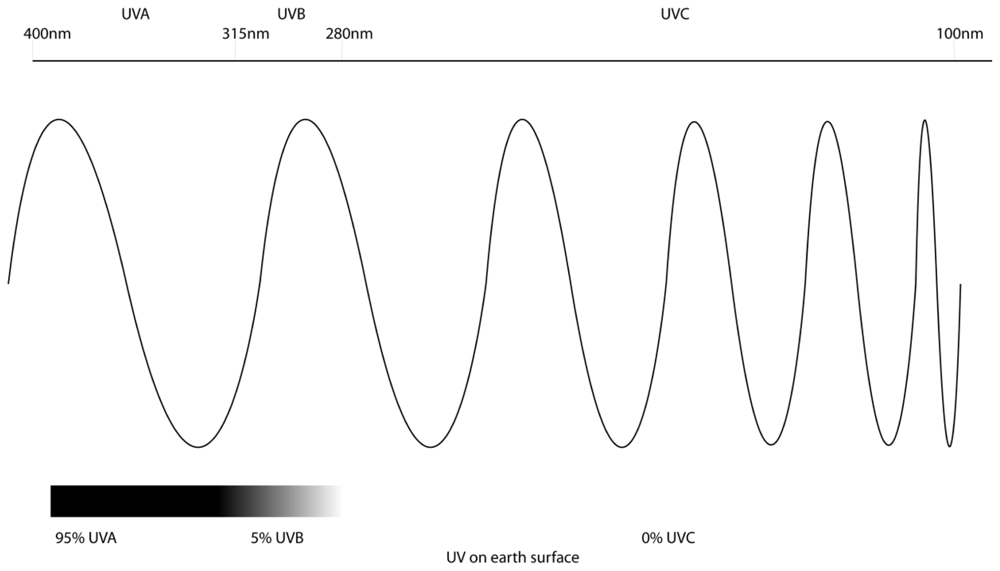
2. Ultraviolet Radiation and Melanoma

One hallmark feature of melanoma is development as a consequence of chronic UV exposure (reviewed in [22]). Both the incidence and mortality rates for melanoma are increasing rapidly worldwide. Incidence rates in Australia are amongst the highest in the world [23] and account for around 10% of all cancer cases in Australia [24]. It is the most common cancer in young Australians aged between 15 and 54 years [24]. Due to its aggressive nature and extreme resistance to chemotherapy treatments, there are very few effective treatments once melanoma has metastasised [25]. Therefore, survival for invasive melanoma is only 15%, making it a major clinical problem [26,27]. It is generally accepted that the two main factors in melanoma development are genetics and sun exposure [28]. However, while epidemiological data supports the undeniable causative link between melanoma and ultraviolet (UV) radiation, the exact molecular mechanisms are not completely understood [2,29].

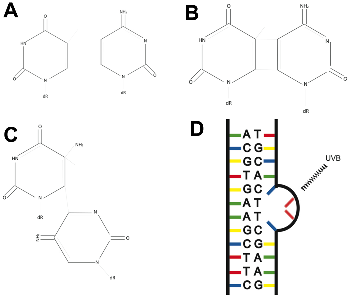
3. Fingerprint Mutations Suggest a Role for UVB in Melanoma

UV fingerprint mutations are found across the genome of melanoma. Recently, a study by Pleasance et al.[30] sequenced the entire genome of a metastatic melanoma cell line, and a study by Berger et al.[31] sequenced the genome of 25 metastatic melanomas to compile a catalogue of somatic mutations. Additional studies have also sequenced the melanoma exome (protein coding regions of the genome) of up to 147 melanoma tumours [3234]. All these studies identified that the vast majority of mutations present were C>T or CC>TT transitions. This mutational signature is indicative of UV-induced DNA damage, with C>T and CC>TT transitions being UV fingerprint mutations. Further investigation revealed that the majority of these substitutions occurred at dipyrimidines, sites at which photoproducts develop. The frequency of both these substitutions were elevated at CpG sites, which can be methylated to 5-methylcytosine, making them susceptible to CPD formation. The mutation spectrum in melanoma unequivocally demonstrates that the majority of somatic mutations are a result of UV-induced DNA damage; however, the role these mutations play in melanoma development is yet to be elucidated. Despite this, the most common somatic mutation found in melanoma, BRAF V600E (A>T), can be targeted with new therapies [35], but is not a UV fingerprint (C>T) mutation.
In addition to being found across the genome, UVB fingerprint mutations have also been found in key genes in all types of skin cancer. Interestingly, the most common mutations found in melanomas, in BRAF and NRAS, do not occur as a result of UV-irradiation. Basal cell carcinomas (BCCs) and squamous cell carcrinomas (SCCs) occur as a result of UV-exposure at a much higher frequency than melanomas, but rarely metastasize. This may be due in part to UV-induced mutations occurring in different genes in these skin cancer types. Mutations in the sonic hedgehog signalling genes PTCH or SMO are thought to be initiating mutations in >70% of BCCs [36]. SCCs, however, have characteristic large chromosomal aberrations and loss of heterozygosity (LOH), which underpins genomic instability [37,38]. Both BCCs and SCCs have high frequencies of UV-fingerprint mutations, particularly in p53[37,39,40] and N-ras[38].
p53 is one of the most commonly mutated genes in cancer, which leads to alterations in cell cycle control and apoptosis [41]. While p53 mutations are present in all types of skin cancer, they are less common in melanoma than in BCCs and SCCs [39]; p53 has been shown to harbour UV signature C>T and CC>TT transitions in approximately 50% of all skin cancers [37,42], but has a higher rate of A>T in melanoma compared to other skin cancers [39], further implicating UV damage. The p53 mutation hot spots are distinctly different in melanoma compared to BCCs and SCCs, which leads to the possibility that the pathways leading to a given tumour needs specific p53 mutations [39].
Cyclin-dependent kinase inhibitor 2A (CDKN2A) is one of the best known genetic factors in melanoma development and codes for two proteins controlling cell proliferation: p16Ink4A and p14ARF [43]. A CDKN2A knockout mouse model has shown that melanoma develops after a single neonatal UV dose, suggesting that alterations in the CDKN2A may play an important role in the UV-induced development of melanoma [44]. A systemic review of studies on mutations in melanoma by Hocker et al.[45] showed that CDNK2A had statistically higher rates of UVB fingerprint mutations than other genes, such as BRAF and NRAS in melanoma, as well as compared to other non-skin cancers.
PTEN is a tumour suppressor gene that carries mutations in many human cancers and may also play a role in UV-induced DNA damage repair [46]. Studies have shown a mutation rate of approximately 30%–40% in melanoma cell lines [47,48]. A study examining mutations in PTEN in melanomas from patients with the skin cancer predisposing disorder Xeroderma Pigmentosum showed that of the melanomas analysed, 56% had mutations in PTEN, and of those, 91% were UVB fingerprint (C>T) mutations [49]. Additionally, 46% had more than one UVB mutation. This data provides further evidence for a role of UVB in the induction of melanoma. The evidence of UVB fingerprint mutations both across the melanoma genome and in specific genes that may be important in melanoma development or progression, including p53, PTEN and CDNK2A, highlights an important role for UVB in melanoma, strengthening the link between UV and melanoma.
The two most recent studies that have sequenced the melanoma exome by Krauthammer et al.[33] and Hodis et al.[32] both identified a novel mutation in the gene RAC1. This gene had a high rate of repeated mutation in 9.2% and 5% of melanomas, respectively, and carried a strong UV signature. RAC1 is a member of the Rho family of small GTPases and plays important roles in cell proliferation, cytoskeletal rearrangement, cell migration and adhesion [5052]. The resulting amino acid change leads to stabilization of the active form of the protein, resulting in enhanced downstream signalling. Expression of the mutated form of RAC1 in melanocytes leads to enhanced MAP kinase signalling, as well as increased proliferation and migration, altering the melanocytes phenotype [33]. The data also indicated that this mutation is likely to occur during early mutagenesis, providing a possible link between this mutation, UV exposure and avoidance of cellular death.

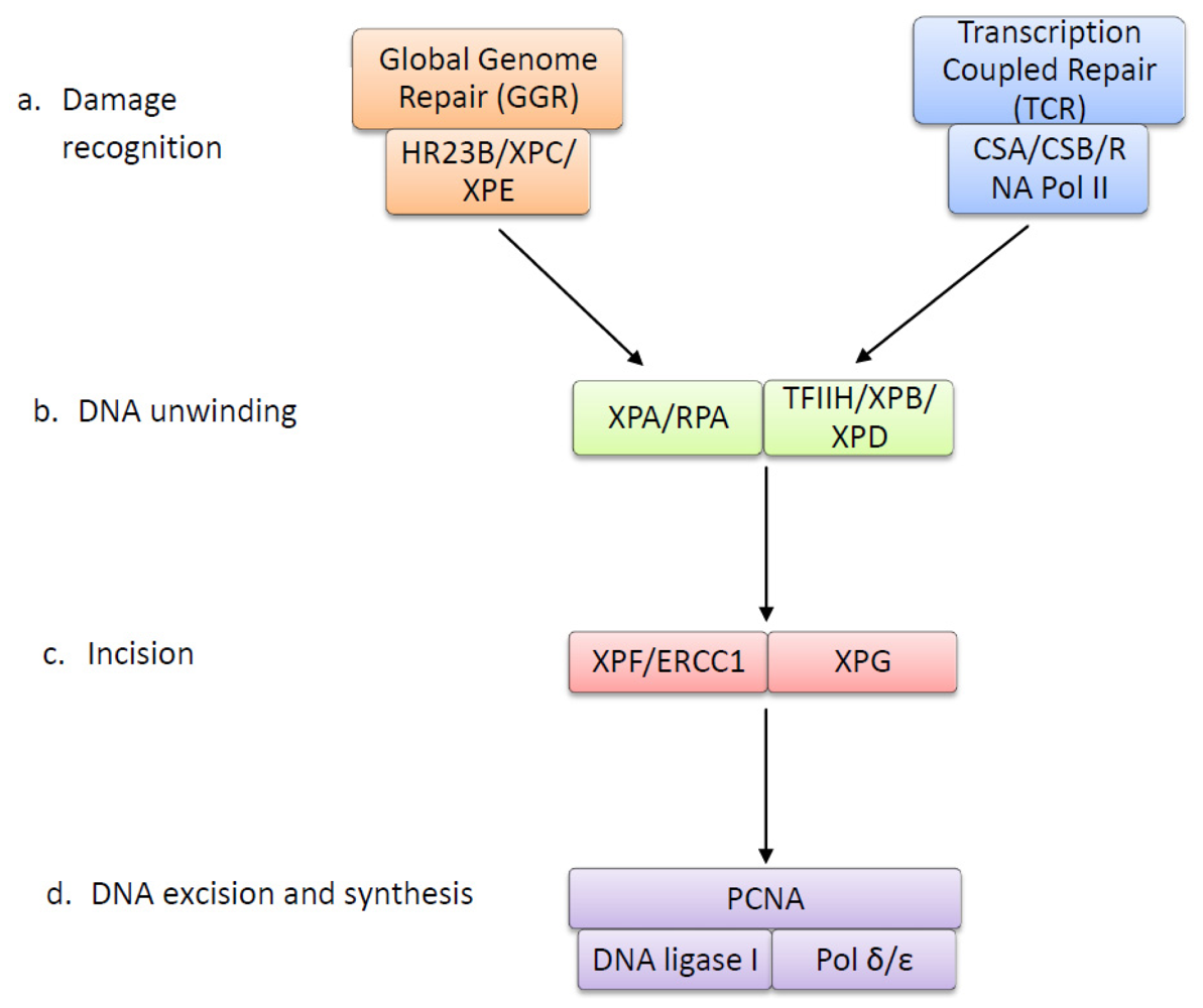
4. The Nucleotide Excision Repair Pathway Removes UV-Induced Lesions

UV photoproducts alter the double helix structure of DNA by creating bulky adducts that halt transcription and trigger apoptosis (Reviewed in [53]), but if unrepaired, can result in mutations. In cells, DNA repair mechanisms are present that allow for damaged DNA to be removed and repaired, preserving the integrity of the genome. The nucleotide excision repair (NER) pathway is the system that is responsible for repairing bulky DNA damage, including UV-induced photoproducts, in particular CPDs and 6-4 PPs [54]. The NER pathway is a highly conserved system comprising around 30 proteins and functions in four main steps; damage recognition, DNA unwinding, excision and DNA synthesis [55]. The NER pathway is illustrated in Figure 3, and Table 1 summarises the function of each NER protein.
There are two sub-pathways in NER damage recognition, global genome repair (GGR) and transcription coupled repair (TCR). TCR repairs lesions that block transcription in actively transcribed genes. TCR functions with a higher priority than GGR and may work in concert with homologous recombination to efficiently repair damage [56]. It serves to rapidly recover transcriptional activity after DNA damage to prevent DNA-damage-induced apoptosis [57]. Induction of TCR is tightly linked to RNA polymerase II, which is stalled at the site of UV lesions and signals for repair with proteins CSA and CSB (reviewed in [58]). GGR is a slower repair process that functions across the entire genome, repairing silent, transcribed and non-transcribed regions. It is suggested to be the preferred pathway for removing 6-4 PPs and can remove these faster and more efficiently than CPDs [59]. In GGR, lesions are recognized by DNA-binding protein complexes XPC and XPE, which recognize and bind to UV lesions, signalling for repair. The main purpose of GGR is to remove lesions that may induce mutations during DNA replication, when translesion polymerases may introduce errors, thereby preventing carcinogenesis [60].
Following damage recognition, these pathways converge on a common repair pathway in which the first step is the unwinding of DNA around the damage by DNA helicases. The multi-protein complex TFIIH contains the helicases XPB and XPD and is recruited to the site of damage, unwinding the DNA around the site of the UV lesion to form a DNA bubble (reviewed in [61]). This allows for proteins XPA and RPA to bind to the DNA, XPA to the site of damage and RPA to the undamaged strand. These proteins allow for the binding of endonucleases XPG and XPF-ERCC1. XPG stabilizes the DNA bubble and cleaves the DNA 3′ to the UV-lesion. XPF-ERCC1 then cleaves the DNA 5′ to the lesion, and an oligonucleotide fragment containing the damage is removed. PCNA and DNA polymerase δ or ɛ then synthesise new DNA complementary to the undamaged strand. DNA ligase seals the nicks, completing the repair process [62].
The NER pathway has been linked to the tumour suppressor p53, particularly in the activation of the GGR pathway. P53 is a transcription factor that plays an important role in cell-cycle control, apoptosis and DNA damage response, with a critical role in protection against carcinogenesis [41]. One way that p53 may regulate DNA damage response is through its downstream target Gadd45, which plays a role in DNA repair, as Gadd45-deficient cells have reduced UV-induced DNA damage repair [84]. However, recent studies suggest that p53 may directly affect the expression of GGR genes XPC and DDB2 (XPE). It has been shown that cells with mutations in p53 have deficiencies in the removal of both CPDs and 6-4 PPs, however this is limited to genomic DNA in non-transcribed regions, suggesting that it is GGR that requires functional p53 and that TCR removal of CPDs is still proficient [85,86]. One study has since shown that expression of XPC is UV-inducible in p53 wild-type cells; however, no induction is observed in p53 deficient cells. Further analysis of the gene showed a possible p53 response element, suggesting that p53 may directly control the expression of the XPC gene [87]. Another study has demonstrated that expression of DDB2 (XPE) is also dependent on p53. It was observed that DDB2 mRNA levels was dependent on basal p53 expression and increased after DNA damage in a p53-dependent manner [88]. A subsequent study analysing the DDB2 gene has shown that it also contains a region that binds p53 to control transcription [89]. Together, these results show that p53 directly controls DNA damage repair after UV damage through the expression of GGR genes XPC and DDB2 (XPE) and that wild-type p53 expression is important for efficient GGR repair.

5. Xeroderma Pigmentosum: A Link between Nucleotide Excision Repair and Melanoma

The exact molecular mechanisms of the link between UV and melanoma are not completely understood, but a possible link is evident in the genetic disorder Xeroderma Pigmentosum (XP) in which the dysfunction of the NER pathway directly results in UV-induced skin cancers. XP is a rare autosomal recessive disease characterized by defective NER, extreme sun sensitivity, pigmentation abnormalities and an increased incidence of skin cancers, including melanoma [90]. Another genetic disorder, Cockayne syndrome (CS), is linked to the TCR component of the NER pathway and is caused by a mutation in CSA or CSB. However, unlike XP, there is no increase in cancer incidence, and all symptoms are neurological and developmental. Furthermore, no cases of cancer have been reported in CS patients [91]. Studies show that without functional TCR, the apoptotic response after UV is enhanced, with a significant induction of p53 and its target genes [92]. This suggests that after damage, the persistent blockage of RNA polymerase II tips the equilibrium of cell survival towards apoptosis, protecting from carcinogenesis [93]. As there is no increased incidence of cancer in CS patients as a result of TCR deficiency, it has been assumed that TCR would not play a role in melanoma development, as any disruption to TCR would lead to an enhanced apoptotic response, eliminating cancerous cells.
XP is a heterogeneous disease resulting from different mutations in one of seven NER genes or one DNA polymerase gene, resulting in eight different complementation groups, XP-A to XP-G and XP-V. Patients within each group will exhibit a large variability in symptoms and NER capacity depending on the nature of the mutation [94]. Groups XP-A and XP-C are the most prevalent (90% of cases) and are caused by mutation in the genes XPA and XPC, respectively. Only a few patients from other groups have been reported, most likely due to the biological importance of the enzymes that are mutated [95]. Most patients have a defect in both GGR and TCR, with the exception of patients from the XP-C and XP-E groups, who are only GGR-deficient. Patients from groups XP-A and XP-G are almost completely repair deficient, highlighting the critical role of XPA and XPG in the NER pathway. XP-B patients are very rare, due to the important roles of XPB outside the NER pathway, and XP-D and XP-F patients exhibit low levels of repair [96].
Mutation in the NER pathway and the subsequent reduced repair capacity results in XP patients having a high incidence of skin cancers compared to the normal population. This includes a 1000-fold increase in the incidence of melanoma [97,98]. There is a stronger association with cancer development in the complementation groups XP-C and XP-E, who have mutations in the GGR pathway [99]. XP-C is the most common group and results from a mutation in the XPC gene, leading to a repair capacity of 10%–20%, while XP-E patients, with a mutation in the DDB2 subunit of the XPE complex, show milder symptoms and have a repair capacity over 50%. Data from CS patients suggests that TCR capacity is the main determinant of cell survival after UV, supported by the fact that GGR deficient, but TCR proficient, XP-C and XP-E cells are less sensitive to UV-induced cell death. Therefore, as GGR is important in protecting from mutagenesis, it would be expected that low GGR capacity in XP-C and XP-E cells would to lead to an increase in mutation rates. This would then lead to an increased cancer predisposition, without cancerous cells being removed by UV-induced apoptosis [59,94,96,100]. Interestingly, most rodent cells are deficient in the repair of UV-induced CPDs in non-transcribed DNA and, hence, exhibit a defect in GGR [101]. Therefore, since rodents are used as surrogates for humans in environmental oncogenic studies, how they differ from humans with respect to GGR should be taken into consideration [102].
A relationship between UV mutagenesis and cancer development in XP patients is also seen when examining mutations. UV fingerprint mutations have been found in many genes in cancers from XP patients, including p53, CDKN2A and Ras oncogenes, many of which are also present in cutaneous melanomas that develop from sun exposure in the normal population [103105].
In summary, hyper-mutability from UV-induced DNA damage and a higher incidence of melanoma in XP patients provides evidence of a relationship between reduced NER, UV-induced DNA damage and skin cancer development. Complementation groups XP-C and XP-E have impaired GGR and are more strongly associated with cancer development. This suggests that NER, in particular GGR, could play an important role in melanoma development as a result of UV exposure. If NER was compromised in melanocytes, then UV lesions would go unrepaired, leading to the accumulation of mutations and promotion of the transformation of melanoma. This could be a potential mechanism that links melanoma development to UV damage. Despite this possible connection between NER and melanoma and the recognized importance of DNA repair in melanomagenesis [106], very few studies have been performed to investigate the relationship between NER and UV damage in melanoma.

6. Research into NER and Melanoma

One area of increasing research is the investigation of single nucleotide polymorphisms (SNPs) in NER genes in predisposition to developing melanoma. Genetic variation that exists in DNA repair genes could lead to differences in repair capacity within the population, therefore predisposing to melanoma development. Several studies have investigated SNPs in NER genes, including XPC, XPG, XPD and XPF in case-control studies. The results of these studies are inconsistent and all studies have found different associations or no associations at all, often contradicting each other. Replication of results in any of these studies has not been achieved [107109].
Recently, a Xiphophorus fish model of melanoma has been used by Fernandez et al.[110] to determine whether NER capacity plays a role in melanoma development. The Xiphophorus model develops melanoma, in part by overexpression and constitutive activation of fish paralogs of human EGFR and CDNK2 (reviewed in [111]), but also after UVB exposure. This study used a hybrid model that results in reduced 6-4 PP removal to investigate the role of NER capacity. DNA was extracted from fish immediately and 24 hours after UVB exposure, and levels of 6-4 PPs were measured. It was reported that approximately 62% of 6-4 PPs were repaired after 24 h, with no significant difference in NER capacity between fish that developed melanoma and those that did not. It was also found that fish with 13.2% and 88.5% NER capacity both developed melanoma. From this data, the authors concluded that melanomagenesis is not associated with NER capacity. Some of the limitations of this study were the different DNA repair processes in Xiphophorus fish, the absence of quantitation of both 6-4PPs and CPDs, and the melanomas that develop in the Xiphophorus fish in this model have different histology to human melanomas [28]. Therefore, it cannot be concluded from this study that NER capacity does not play a role in the development of melanoma.
To date, there has only been one study that has compared UV-induced NER capacity in human melanocytes and melanoma cells. Gaddameedhi et al.[112] compared the repair of 6-4 PPs and CPDs between the two cell types to determine if NER plays a role in melanoma development. It was reported that there was a time-dependent reduction in photoproducts with 50%–80% of 6-4 PPs and 40%–80% CPDs being removed in both cell types. From this, it was concluded that there was no difference in the repair capacity of melanocytes and melanoma cells; therefore, melanoma cells retain normal NER function, which does not contribute to melanomagenesis. While it was concluded that there was no reduction in NER capacity, both cell lines showed high variation in repair capacity, ranging approximately 40%–80%. In addition, statistical analysis was not provided to prove that this reduction was not significant. The study was also not specific to measuring GGR or TCR, only the overall removal of photoproducts. Previous data has suggested that melanoma shows low levels of GGR, which would not have been identifiable in this study.
Melanocytes may initially have a lower NER capacity when compared with other cell types [113]. The repair of oxidative damage and photoproducts in melanocytes, normal and XP fibroblasts were compared, using host cell reactivation and in vitro repair synthesis assays. The ability of melanocytes to reactivate a UV damaged luciferase gene was lower than normal fibroblasts, but not as low as XP fibroblasts. Similarly melanocytes had a lower capacity of DNA damage-induced repair synthesis after UV irradiation compared to normal fibroblasts. It was also tested to see if the low repair was due to melanin in the melanocytes interfering with excision repair or expression of repair genes. Melanin was added to fibroblast lysates and showed a reduced repair capacity proportional to the amount of melanin. Additionally, it was found that melanin co-purified with DNA, and when separated after electrophoresis, the DNA became sensitive again to repair. This evidence suggests that melanin in melanocytes binds to DNA interfering with damage recognition and incision by NER enzymes, resulting in reduced repair. The results in this study may explain the low percentage of CPD and 6-4 PP removal in both melanocytes and melanoma cells reported by Gaddameedhi et al.[112]. Moreover, if melanocytes originally have a low repair capacity, then the lack of difference between melanocytes and melanoma cells cannot rule out NER playing a role melanoma development. Having a low NER capacity may predispose melanocytes to the accumulation of UV-induced DNA mutations that could lead to avoidance of cell death and melanomagenesis.
There are two hallmark features of melanoma; firstly, its development as a result of chronic UV exposure and, secondly, the limited efficacy of chemotherapy drugs, especially the DNA-damaging agent cisplatin [114]. Cisplatin induces intra- and inter-strand cross links in DNA that cause helix distorting lesions similar to UV damage. In addition to repairing UV-induced photoproducts, NER also removes cisplatin lesions [54]. A study by Bowden et al.[115] investigated the level of NER gene expression after cisplatin-induced DNA damage in melanoma and melanocytes. Results showed a higher basal expression of NER genes in melanoma; however, when compared to melanocytes, there was a significant lack of induction of GGR genes XPC, DDB1 and DDB2. In contrast, the expression of TCR genes CSA and CSB was low in both melanoma and melanocytes, suggesting that TCR does not play a role in the response to cisplatin. The findings of this study indicate that a lack of NER induction, especially GGR, may play a role in the resistance of melanoma to cisplatin. Subsequent studies identified that the NER component ERCC1 could play a key role in melanomagenesis and resistance to cisplatin [116,117]. In addition, the DNA damage caused by cisplatin is similar to that caused by UVB, with both causing helix distorting lesions. As a result, it could be hypothesized that melanoma would also have low expression of NER in response to UV damage similar to that seen in cisplatin. This could suggest a potential role for NER, particularly GGR, in the development of melanoma as a result of UV.
Furthermore, the genome of the melanoma cell lines sequenced by Pleasance et al.[30] and Berger et al.[31], which showed a mutational signature indicating DNA damage by UV exposure, also displayed an uneven distribution of mutations across the genome. There was a lower prevalence of UV mutations on the transcribed strand, as well as within exons compared to introns. This suggests that the TCR component of NER, which is responsible for removing damage from transcribed genes, is still functional in melanoma. Reduced GGR activity could also be responsible for the significant amount of UV mutations across the rest of the genome, as GGR removes UV lesions from the entire genome. These results are similar to those suggested by Bowden et al.[115]; that melanoma has low expression of GGR genes, but sufficient TCR expression and that low levels of the GGR component of NER may play a role in the development of melanoma. The idea that TCR does not play a role in melanoma development can be supported by the fact that Cockayne syndrome patients, who lack TCR, do not show an increased incidence of melanoma that is seen in XP patients.
If GGR was compromised in melanocytes, then UV-induced DNA damage across the genome would go unrepaired and lead to mutations when DNA is replicated. Additionally, cells would not undergo apoptosis to prevent cancer development, due to a functional TCR. This could then potentially lead to cell transformation and melanomagenesis, highlighting a potential role for NER in melanoma development.

7. Conclusions

The data suggests that the NER pathway may play a role in avoidance of UV-induced cell death and the subsequent development of melanoma. In particular the GGR pathway of NER may be important, as it functions to repair damage that can lead to mutations during DNA replication, and XP patients with defective GGR have a stronger association with cancer development. This idea has been supported by studies that found low GGR expression in melanoma cells treated with cisplatin and the genome of a melanoma line showing a high frequency of UV mutations in non-transcribed regions. Furthermore, melanocytes may potentially have low NER levels, which could predispose them to UV mutagenesis and transformation.
Despite this possible connection between NER and melanoma, little research in this area has been done, and the studies that have been performed are often contradictory. Given the scarce research into NER in melanoma, more investigation is needed to fully elucidate this relationship.

Acknowledgments

The authors would like to acknowledge the financial support from the Australian National Health & Medical Research Council, Cancer Australia, Cure Cancer Australia Foundation, Hunter Medical Research Institute and the Rotary Club of Cardiff (Australia).
  • Conflict of InterestThe authors declare no conflict of interest.

References

  1. Ravanat, J.L.; Douki, T.; Cadet, J. Direct and indirect effects of UV radiation on DNA and its components. J. Photochem. Photobiol. B 2001, 63, 88–102. [Google Scholar]
  2. Marrot, L.; Meunier, J.-R. Skin DNA photodamage and its biological consequences. J. Am. Acad. Dermatol 2008, 58, S139–S148. [Google Scholar]
  3. Brenner, M.; Degitz, K.; Besch, R.; Berking, C. Differential expression of melanoma-associated growth factors in keratinocytes and fibroblasts by ultraviolet A and ultraviolet B radiation. Br. J. Dermatol 2005, 153, 733–739. [Google Scholar]
  4. Garland, C.F.; Garland, F.C.; Gorham, E.D. Epidemiologic evidence for different roles of ultraviolet A and B radiation in melanoma mortality rates. Ann. Epidemiol 2003, 13, 395–404. [Google Scholar]
  5. Tran, T.T.; Schulman, J.; Fisher, D.E. UV and pigmentation: Molecular mechanisms and social controversies. Pigment Cell Melanoma Res 2008, 21, 509–516. [Google Scholar]
  6. Tucker, M.A.; Goldstein, A.M. Melanoma etiology: Where are we? Oncogene 2003, 22, 3042–3052. [Google Scholar]
  7. Besaratinia, A.; Bates, S.E.; Synold, T.W.; Pfeifer, G.P. Similar mutagenicity of photoactivated porphyrins and ultraviolet A radiation in mouse embryonic fibroblasts: Involvement of oxidative DNA lesions in mutagenesis. Biochemistry 2004, 43, 15557–15566. [Google Scholar]
  8. Brozyna, A.; Zbytek, B.; Granese, J.; Carlson, A.; Ross, J.; Slominski, A. Mechanism of UV-related carcinogenesis and its contribution to nevi/melanoma. Expert Rev. Dermatol 2007, 2, 451–469. [Google Scholar]
  9. Slominski, A.; Pawelek, J. Animals under the sun: Effects of ultraviolet radiation on mammalian skin. Clin. Dermatol 1998, 16, 503–515. [Google Scholar]
  10. Bennett, D.C. Ultraviolet wavebands and melanoma initiation. Pigment Cell Melanoma Res 2008, 21, 520–524. [Google Scholar]
  11. Hussein, M.R. Ultraviolet radiation and skin cancer: Molecular mechanisms. J. Cutan. Pathol 2005, 32, 191–205. [Google Scholar]
  12. Pfeifer, G.P. Formation and processing of UV photoproducts: Effects of DNA sequence and chromatin environment. Photochem. Photobiol 1997, 65, 270–283. [Google Scholar]
  13. Courdavault, S.; Baudouin, C.; Charveron, M.; Canguilhem, B.; Favier, A.; Cadet, J.; Douki, T. Repair of the three main types of bipyrimidine DNA photoproducts in human keratinocytes exposed to UVB and UVA radiations. DNA Repair 2005, 4, 836–844. [Google Scholar]
  14. Kim, J.-K.; Patel, D.; Choi, B.-S. Contrasting structural impacts induced by cis-syn cyclobutane dimer and (6-4) adduct in DNA duplex decamers: Implication in mutagenesis and repair activity. Photochem. Photobiol 1995, 62, 44–50. [Google Scholar]
  15. You, Y.H.; Lee, D.H.; Yoon, J.H.; Nakajima, S.; Yasui, A.; Pfeifer, G.P. Cyclobutane pyrimidine dimers are responsible for the vast majority of mutations induced by UVB irradiation in mammalian cells. J. Biol. Chem 2001, 276, 44688–44694. [Google Scholar]
  16. Brash, D.E. Roles of the transcription factor p53 in keratinocyte carcinomas. Br. J. Dermatol 2006, 154, S8–S10. [Google Scholar]
  17. Johnson, R.E.; Prakash, S.; Prakash, L. Efficient bypass of a thymine-thymine dimer by yeast DNA polymerase, Poleta. Science 1999, 283, 1001–1004. [Google Scholar]
  18. Masutani, C.; Kusumoto, R.; Yamada, A.; Dohmae, N.; Yokoi, M.; Yuasa, M.; Araki, M.; Iwai, S.; Takio, K.; Hanaoka, F. The XPV (xeroderma pigmentosum variant) gene encodes human DNA polymerase η. Nature 1999, 399, 700–704. [Google Scholar]
  19. Drouin, R.; Therrien, J.-P. UVB-induced cyclobutane pyrimidine dimer frequency correlates with skin cancer mutational hotspots in p53. Photochem. Photobiol 1997, 66, 719–726. [Google Scholar]
  20. Tommasi, S.; Denissenko, M.F.; Pfeifer, G.P. Sunlight induces pyrimidine dimers preferentially at 5-methylcytosine bases. Cancer Res 1997, 57, 4727–4730. [Google Scholar]
  21. Tu, Y.; Dammann, R.; Pfeifer, G.P. Sequence and time-dependent deamination of cytosine bases in UVB-induced cyclobutane pyrimidine dimers in vivo. J. Mol. Biol 1998, 284, 297–311. [Google Scholar]
  22. Garibyan, L.; Fisher, D.E. How sunlight causes melanoma. Curr. Oncol. Rep 2010, 12, 319–326. [Google Scholar]
  23. World Health Organisation (WHO), The Global Burden of Disease: 2004 Update; WHO: Geneva, Switzerland, 2008.
  24. AIHW, Cancer in Australia: An overview, 2008; Cancer series no. 46; Cat. no. CAN 42; AIHW: Canberra, Australia, 2008.
  25. Soengas, M.S.; Lowe, S.W. Apoptosis and melanoma chemoresistance. Oncogene 2003, 22, 3138–3151. [Google Scholar]
  26. Fayolle, C.; Pourchet, J.; de Fromentel, C.C.; Puisieux, A.; Dore, J.F.; Voeltzel, T. Gadd45a activation protects melanoma cells from ultraviolet B-induced apoptosis. J. Investig. Dermatol 2008, 128, 196–202. [Google Scholar]
  27. Tracey, E.; Kerr, T.; Dobrovic, A.; Currow, D. Cancer in New South Wales: Incidence and Mortality Report 2008; Cancer Institute NSW: Sydney, Australia, 2008. [Google Scholar]
  28. Jhappan, C.; Noonan, F.P.; Merlino, G. Ultraviolet radiation and cutaneous malignant melanoma. Oncogene 2003, 22, 3099–3112. [Google Scholar]
  29. De Gruijl, F.R. Skin cancer and solar UV radiation. Eur. J. Cancer 1999, 35, 2003–2009. [Google Scholar]
  30. Pleasance, E.D.; Cheetham, R.K.; Stephens, P.J.; McBride, D.J.; Humphray, S.J.; Greenman, C.D.; Varela, I.; Lin, M.L.; Ordonez, G.R.; Bignell, G.R.; et al. A comprehensive catalogue of somatic mutations from a human cancer genome. Nature 2010, 463, 191–196. [Google Scholar]
  31. Berger, M.F.; Hodis, E.; Heffernan, T.P.; Deribe, Y.L.; Lawrence, M.S.; Protopopov, A.; Ivanova, E.; Watson, I.R.; Nickerson, E.; Ghosh, P.; et al. Melanoma genome sequencing reveals frequent PREX2 mutations. Nature 2012, 485, 502–506. [Google Scholar]
  32. Hodis, E.; Watson, I.R.; Kryukov, G.V.; Arold, S.T.; Imielinski, M.; Theurillat, J.-P.; Nickerson, E.; Auclair, D.; Li, L.; Place, C.; et al. A landscape of driver mutations in melanoma. Cell 2012, 150, 251–263. [Google Scholar]
  33. Krauthammer, M.; Kong, Y.; Ha, B.H.; Evans, P.; Bacchiocchi, A.; McCusker, J.P.; Cheng, E.; Davis, M.J.; Goh, G.; Choi, M.; et al. Exome sequencing identifies recurrent somatic RAC1 mutations in melanoma. Nat. Genet 2012, 44, 1006–1014. [Google Scholar]
  34. Stark, M.S.; Woods, S.L.; Gartside, M.G.; Bonazzi, V.F.; Dutton-Regester, K.; Aoude, L.G.; Chow, D.; Sereduk, C.; Niemi, N.M.; Tang, N.; et al. Frequent somatic mutations in MAP3K5 and MAP3K9 in metastatic melanoma identified by exome sequencing. Nat. Genet 2012, 44, 165–169. [Google Scholar]
  35. Flaherty, K.T.; Puzanov, I.; Kim, K.B.; Ribas, A.; McArthur, G.A.; Sosman, J.A.; O’Dwyer, P.J.; Lee, R.J.; Grippo, J.F.; Nolop, K.; et al. Inhibition of mutated, activated BRAF in metastatic melanoma. N. Engl. J. Med 2010, 363, 809–819. [Google Scholar]
  36. Gailani, M.R.; Stahle-Backdahl, M.; Leffell, D.J.; Glynn, M.; Zaphiropoulos, P.G.; Pressman, C.; Unden, A.B.; Dean, M.; Brash, D.E.; Bale, A.E.; et al. The role of the human homologue of Drosophila patched in sporadic basal cell carcinomas. Nat. Genet 1996, 14, 78–81. [Google Scholar]
  37. Boukamp, P. Non-melanoma skin cancer: What drives tumor development and progression? Carcinogenesis 2005, 26, 1657–1667. [Google Scholar]
  38. Popp, S.; Waltering, S.; Herbst, C.; Moll, I.; Boukamp, P. UV-B type mutations and chromosomal imbalances indicate common pathways for the development of Merkel and skin squamous cell carcinomas. Int. J. Cancer 2002, 99, 352–360. [Google Scholar]
  39. Giglia-Mari, G.; Sarasin, A. TP53 mutations in human skin cancers. Human Mutat 2003, 21, 217–228. [Google Scholar]
  40. Ziegler, A.; Jonason, A.; Leffell, D.; Simon, J.; Sharma, H.; Kimmelman, J.; Remington, L.; Jacks, T.; Brash, D. Sunburn and p53 in the onset of skin cancer. Nature 1994, 372, 773–776. [Google Scholar]
  41. Balint, E.; Vousden, K.H. Activation and activities of the p53 tumour suppressor protein. Br. J. Cancer 2001, 85, 1813–1823. [Google Scholar]
  42. Zerp, S.F.; van Elsas, A.; Poltenburg, L.T.C.; Schrier, P.I. p53 mutations in human cutaneous melanoma correlate with sun exposure, but are not always involved in melanomagenesis. Br. J. Cancer 1999, 79, 921. [Google Scholar]
  43. Piepkorn, M. Melanoma genetics: An update with focus on the CDKN2A(p16)/ARF tumor suppressors. J. Am. Acad. Dermatol. 2000, 42, 705–722, quiz 723–706. [Google Scholar]
  44. Recio, J.A.; Noonan, F.P.; Takayama, H.; Anver, M.R.; Duray, P.; Rush, W.L.; Lindner, G.; de Fabo, E.C.; DePinho, R.A.; Merlino, G.; et al. Ink4a/arf deficiency promotes ultraviolet radiation-induced melanomagenesis. Cancer Res 2002, 62, 6724–6730. [Google Scholar]
  45. Hocker, T.; Tsao, H. Ultraviolet radiation and melanoma: A systematic review and analysis of reported sequence variants. Human Mutat 2007, 28, 578–588. [Google Scholar]
  46. Ming, M.; Feng, L.; Shea, C.R.; Soltani, K.; Zhao, B.; Han, W.; Smart, R.C.; Trempus, C.S.; He, Y.-Y. PTEN positively regulates UVB-induced DNA damage repair. Cancer Res 2011, 71, 5287–5295. [Google Scholar]
  47. Guldberg, P.; thor Straten, P.; Birck, A.; Ahrenkiel, V.; Kirkin, A.F.; Zeuthen, J. Disruption of the MMAC1/PTEN gene by deletion or mutation is a frequent event in malignant melanoma. Cancer Res 1997, 57, 3660–3663. [Google Scholar]
  48. Tsao, H.; Zhang, X.; Benoit, E.; Haluska, F.G. Identification of PTEN/MMAC1 alterations in uncultured melanomas and melanoma cell lines. Oncogene 1998, 16, 3397–3402. [Google Scholar]
  49. Wang, Y.; Digiovanna, J.J.; Stern, J.B.; Hornyak, T.J.; Raffeld, M.; Khan, S.G.; Oh, K.S.; Hollander, M.C.; Dennis, P.A.; Kraemer, K.H.; et al. Evidence of ultraviolet type mutations in xeroderma pigmentosum melanomas. Proc. Nat. Acad. Sci. USA 2009, 106, 6279–6284. [Google Scholar]
  50. Hakoshima, T.; Shimizu, T.; Maesaki, R. Structural basis of the Rho GTPase signaling. J. Biochem 2003, 134, 327–331. [Google Scholar]
  51. Jaffe, A.B.; Hall, A. RHO GTPASES: Biochemistry and Biology. Annu. Rev. Cell Dev. Biol 2005, 21, 247–269. [Google Scholar]
  52. Wennerberg, K.; Rossman, K.L.; Der, C.J. The Ras superfamily at a glance. J. Cell Sci 2005, 118, 843–846. [Google Scholar]
  53. Batista, L.F.Z.; Kaina, B.; Meneghini, R.; Menck, C.F.M. How DNA lesions are turned into powerful killing structures: Insights from UV-induced apoptosis. Mutat. Res 2009, 681, 197–208. [Google Scholar]
  54. Friedberg, E.C. How nucleotide excision repair protects against cancer. Nat. Rev. Cancer 2001, 1, 22–33. [Google Scholar]
  55. Marini, F.; Nardo, T.; Giannattasio, M.; Minuzzo, M.; Stefanini, M.; Plevani, P.; Falconi, M.M. DNA nucleotide excision repair-dependent signaling to checkpoint activation. Proc. Natl. Acad. Sci. USA 2006, 103, 17325–17330. [Google Scholar]
  56. Aboussekhra, A.; Al-Sharif, I.S. Homologous recombination is involved in transcription-coupled repair of UV damage in Saccharomyces cerevisiae. EMBO J 2005, 24, 1999–2010. [Google Scholar]
  57. Van Oosten, M.; Rebel, H.; Friedberg, E.C.; van Steeg, H.; van der Horst, G.T.J.; van Kranen, H.J.; Westerman, A.; van Zeeland, A.A.; Mullenders, L.H.F.; de Gruijl, F.R. Differential role of transcription-coupled repair in UVB-induced G2 arrest and apoptosis in mouse epidermis. Proc. Natl. Acad. Sci. USA 2000, 97, 11268–11273. [Google Scholar]
  58. Fousteri, M.; Mullenders, L.H. Transcription-coupled nucleotide excision repair in mammalian cells: Molecular mechanisms and biological effects. Cell Res 2008, 18, 73–84. [Google Scholar]
  59. Van Hoffen, A.; Venema, J.; Meschini, R.; van Zeeland, A.A.; Mullenders, L.H. Transcription-coupled repair removes both cyclobutane pyrimidine dimers and 6-4 photoproducts with equal efficiency and in a sequential way from transcribed DNA in xeroderma pigmentosum group C fibroblasts. EMBO J 1995, 14, 360–367. [Google Scholar]
  60. Sugasawa, K. UV-induced ubiquitylation of XPC complex, the UV-DDB-ubiquitin ligase complex and DNA repair. J. Mol. Histol 2006, 37, 189–202. [Google Scholar]
  61. Compe, E.; Egly, J.-M. TFIIH: When transcription met DNA repair. Nat. Rev. Mol. Cell Biol 2012, 13, 343–354. [Google Scholar]
  62. Rastogi, R.P.; Richa; Kumar, A.; Tyagi, M.B.; Sinha, R.P. Molecular mechanisms of ultraviolet radiation-induced DNA damage and repair. J. Nucleic Acids 2010, 2010, 1–32. [Google Scholar]
  63. Sugasawa, K.; Ng, J.M.; Masutani, C.; Iwai, S.; van der Spek, P.J.; Eker, A.P.; Hanaoka, F.; Bootsma, D.; Hoeijmakers, J.H. Xeroderma pigmentosum group C protein complex is the initiator of global genome nucleotide excision repair. Mol. Cell 1998, 2, 223–232. [Google Scholar]
  64. Fitch, M.E.; Nakajima, S.; Yasui, A.; Ford, J.M. In vivo recruitment of XPC to UV-induced cyclobutane pyrimidine dimers by the DDB2 gene product. J. Biol. Chem 2003, 278, 46906–46910. [Google Scholar]
  65. Scrima, A.; Konickova, R.; Czyzewski, B.K.; Kawasaki, Y.; Jeffrey, P.D.; Groisman, R.; Nakatani, Y.; Iwai, S.; Pavletich, N.P.; Thomä, N.H. Structural Basis of UV DNA-Damage Recognition by the DDB1/DDB2 Complex. Cell 2008, 135, 1213–1223. [Google Scholar]
  66. Li, J.; Bhat, A.; Xiao, W. Regulation of nucleotide excision repair through ubiquitination. Acta Biochim. Biophys. Sin 2011, 43, 919–929. [Google Scholar]
  67. Xie, Z.; Liu, S.; Zhang, Y.; Wang, Z. Roles of Rad23 protein in yeast nucleotide excision repair. Nucleic Acids Res 2004, 32, 5981–5990. [Google Scholar]
  68. Ng, J.M.Y.; Vermeulen, W.; van der Horst, G.T.J.; Bergink, S.; Sugasawa, K.; Vrieling, H.; Hoeijmakers, J.H.J. A novel regulation mechanism of DNA repair by damage-induced and RAD23-dependent stabilization of xeroderma pigmentosum group C protein. Genes Dev 2003, 17, 1630–1645. [Google Scholar]
  69. Svejstrup, J.Q. Rescue of arrested RNA polymerase II complexes. J. Cell Sci 2003, 116, 447–451. [Google Scholar]
  70. Bregman, D.B.; Halaban, R.; van Gool, A.J.; Henning, K.A.; Friedberg, E.C.; Warren, S.L. UV-induced ubiquitination of RNA polymerase II: A novel modification deficient in Cockayne syndrome cells. Proc. Natl. Acad. Sci. USA 1996, 93, 11586–11590. [Google Scholar]
  71. Ratner, J.N.; Balasubramanian, B.; Corden, J.; Warren, S.L.; Bregman, D.B. Ultraviolet radiation-induced ubiquitination and proteasomal degradation of the large subunit of RNA Polymerase II. J. Biol. Chem 1998, 273, 5184–5189. [Google Scholar]
  72. Bradsher, J.; Coin, F.; Egly, J.M. Distinct roles for the helicases of TFIIH in transcript initiation and promoter escape. J. Biol. Chem 2000, 275, 2532–2538. [Google Scholar]
  73. Oksenych, V.; Coin, F. The long unwinding road: XPB and XPD helicases in damaged DNA opening. Cell Cycle 2010, 9, 90–96. [Google Scholar]
  74. Volker, M.; Mone, M.J.; Karmakar, P.; van Hoffen, A.; Schul, W.; Vermeulen, W.; Hoeijmakers, J.H.; van Driel, R.; van Zeeland, A.A.; Mullenders, L.H. Sequential assembly of the nucleotide excision repair factors in vivo. Mol. Cell 2001, 8, 213–224. [Google Scholar]
  75. He, Z.; Henricksen, L.A.; Wold, M.S.; Ingles, C.J. RPA involvement in the damage-recognition and incision steps of nucleotide excision repair. Nature 1995, 374, 566–569. [Google Scholar]
  76. Matsuda, T.; Saijo, M.; Kuraoka, I.; Kobayashi, T.; Nakatsu, Y.; Nagai, A.; Enjoji, T.; Masutani, C.; Sugasawa, K.; Hanaoka, F.; et al. DNA repair protein XPA binds replication protein A (RPA). J. Biol. Chem 1995, 270, 4152–4157. [Google Scholar]
  77. Clarkson, S.G. The XPG story. Biochimie 2003, 85, 1113–1121. [Google Scholar]
  78. Gaillard, P.H.; Wood, R.D. Activity of individual ERCC1 and XPF subunits in DNA nucleotide excision repair. Nucleic Acids Res 2001, 29, 872–879. [Google Scholar]
  79. Sijbers, A.M.; de Laat, W.L.; Ariza, R.R.; Biggerstaff, M.; Wei, Y.-F.; Moggs, J.G.; Carter, K.C.; Shell, B.K.; Evans, E.; de Jong, M.C.; et al. Xeroderma pigmentosum group F caused by a defect in a structure-specific DNA repair endonuclease. Cell 1996, 86, 811–822. [Google Scholar]
  80. Tsodikov, O.V.; Ivanov, D.; Orelli, B.; Staresincic, L.; Shoshani, I.; Oberman, R.; Scharer, O.D.; Wagner, G.; Ellenberger, T. Structural basis for the recruitment of ERCC1-XPF to nucleotide excision repair complexes by XPA. EMBO J 2007, 26, 4768–4776. [Google Scholar]
  81. Popanda, O.; Thielmann, H.W. The function of DNA polymerases in DNA repair synthesis of ultraviolet-irradiated human fibroblasts. Biochim. Biophys. Acta 1992, 1129, 155–160. [Google Scholar]
  82. Shivji, K.K.; Kenny, M.K.; Wood, R.D. Proliferating cell nuclear antigen is required for DNA excision repair. Cell 1992, 69, 367–374. [Google Scholar]
  83. Nocentini, S. Rejoining kinetics of DNA single- and double-strand breaks in normal and DNA ligase-deficient cells after exposure to ultraviolet C and gamma radiation: An evaluation of ligating activities involved in different DNA repair processes. Radiat. Res 1999, 151, 423–432. [Google Scholar]
  84. Smith, M.L.; Ford, J.M.; Hollander, M.C.; Bortnick, R.A.; Amundson, S.A.; Seo, Y.R.; Deng, C.-X.; Hanawalt, P.C.; Fornace, A.J. p53-Mediated DNA repair responses to UV radiation: Studies of mouse cells lacking p53, p21 and/orgadd45 genes. Mol. Cell. Biol 2000, 20, 3705–3714. [Google Scholar]
  85. Ford, J.M.; Hanawalt, P.C. Expression of wild-type p53 is required for efficient global genomic nucleotide excision repair in UV-irradiated human fibroblasts. J. Biol. Chem 1997, 272, 28073–28080. [Google Scholar]
  86. Mathonnet, G.; Leger, C.; Desnoyers, J.; Drouin, R.; Therrien, J.-P.; Drobetsky, E.A. UV wavelength-dependent regulation of transcription-coupled nucleotide excision repair in p53-deficient human cells. Proc. Natl. Acad. Sci 2003, 100, 7219–7224. [Google Scholar]
  87. Adimoolam, S.; Ford, J.M. p53 and DNA damage-inducible expression of the xeroderma pigmentosum group C gene. Proc. Natl. Acad. Sci. USA 2002, 99, 12985–12990. [Google Scholar]
  88. Hwang, B.J.; Ford, J.M.; Hanawalt, P.C.; Chu, G. Expression of the p48 xeroderma pigmentosum gene is p53-dependent and is involved in global genomic repair. Proc. Natl. Acad. Sci. USA 1999, 96, 424–428. [Google Scholar]
  89. Tan, T.; Chu, G. p53 binds and activates the xeroderma pigmentosum DDB2 gene in humans, but not mice. Mol. Cell. Biol 2002, 22, 3247–3254. [Google Scholar]
  90. Nouspikel, T. DNA repair in mammalian cells: Nucleotide excision repair: Variations on versatility. Cell. Mol. Life Sci 2009, 66, 994–1009. [Google Scholar]
  91. Nance, M.A.; Berry, S.A. Cockayne syndrome: Review of 140 cases. Am. J. Med. Genet 1992, 42, 68–84. [Google Scholar]
  92. Frontini, M.; Proietti-de-Santis, L. Cockayne syndrome B protein (CSB): Linking p53, HIF-1 and p300 to robustness, lifespan, cancer and cell fate decisions. Cell Cycle 2009, 8, 693–696. [Google Scholar]
  93. Hoeijmakers, J.H.J. DNA damage, aging and cancer. N. Engl. J. Med 2009, 361, 1475–1485. [Google Scholar]
  94. Chavanne, F.; Broughton, B.C.; Pietra, D.; Nardo, T.; Browitt, A.; Lehmann, A.R.; Stefanini, M. Mutations in the XPC gene in families with xeroderma pigmentosum and consequences at the cell, protein and transcript levels. Cancer Res 2000, 60, 1974–1982. [Google Scholar]
  95. Lehmann, A.; McGibbon, D.; Stefanini, M. Xeroderma pigmentosum. Orphanet J. Rare Dis 2011, 6, 70. [Google Scholar]
  96. Stary, A.; Sarasin, A. The genetics of the hereditary xeroderma pigmentosum syndrome. Biochimie 2002, 84, 49–60. [Google Scholar]
  97. Bradford, P.T.; Goldstein, A.M.; Tamura, D.; Khan, S.G.; Ueda, T.; Boyle, J.; Oh, K.-S.; Imoto, K.; Inui, H.; Moriwaki, S.-I.; et al. Cancer and neurologic degeneration in xeroderma pigmentosum: Long term follow-up characterises the role of DNA repair. J. Med. Genet 2011, 48, 168–176. [Google Scholar]
  98. Kraemer, K.H.; Lee, M.M.; Scotto, J. Xeroderma pigmentosum: Cutaneous, ocular and neurologic abnormalities in 830 published cases. Arch. Dermatol 1987, 123, 241–250. [Google Scholar]
  99. Cleaver, J.E.; Thompson, L.H.; Richardson, A.S.; States, J.C. A summary of mutations in the UV-sensitive disorders: Xeroderma pigmentosum, Cockayne syndrome and trichothiodystrophy. Human Mutation 1999, 14, 9–22. [Google Scholar]
  100. Queille, S.; Drougard, C.; Sarasin, A.; Daya-Grosjean, L. Effects of XPD mutations on ultraviolet-induced apoptosis in relation to skin cancer-proneness in repair-deficient syndromes. J. Invest. Dermatol 2001, 117, 1162–1170. [Google Scholar]
  101. Van der Wees, C.; Jansen, J.; Vrieling, H.; van der Laarse, A.; van Zeeland, A.; Mullenders, L.H. Nucleotide excision repair in differentiated cells. Mutat. Res 2007, 614, 16–23. [Google Scholar]
  102. Hanawalt, P.C.; Ford, J.M.; Lloyd, D.R. Functional characterization of global genomic DNA repair and its implications for cancer. Mutat. Res 2003, 544, 107–114. [Google Scholar]
  103. Daya-Grosjean, L.; Robert, C.; Drougard, C.; Suarez, H.; Sarasin, A. High mutation frequency in ras genes of skin tumors isolated from DNA repair deficient xeroderma pigmentosum patients. Cancer Res 1993, 53, 1625–1629. [Google Scholar]
  104. Dumaz, N.; Drougard, C.; Sarasin, A.; Daya-Grosjean, L. Specific UV-induced mutation spectrum in the p53 gene of skin tumors from DNA-repair-deficient xeroderma pigmentosum patients. Proc. Natl. Acad. Sci 1993, 90, 10529–10533. [Google Scholar]
  105. Soufir, N.; Daya-Grosjean, L.; de La Salmoniere, P.; Moles, J.-P.; Dubertret, L.; Sarasin, A.; Basset-Seguin, N. Association between INK4a-ARF and p53 mutations in skin carcinomas of xeroderma pigmentosum patients. J. Nat. Cancer Inst 2000, 92, 1841–1847. [Google Scholar]
  106. Law, M.H.; Macgregor, S.; Hayward, N.K. Melanoma genetics: Recent findings take us beyond well-traveled pathways. J. Invest. Dermatol 2012, 132, 1763–1774. [Google Scholar]
  107. Blankenburg, S.; Konig, I.R.; Moessner, R.; Laspe, P.; Thoms, K.M.; Krueger, U.; Khan, S.G.; Westphal, G.; Berking, C.; Volkenandt, M.; et al. Assessment of 3 xeroderma pigmentosum group C gene polymorphisms and risk of cutaneous melanoma: A case-control study. Carcinogenesis 2005, 26, 1085–1090. [Google Scholar]
  108. Blankenburg, S.; Konig, I.R.; Moessner, R.; Laspe, P.; Thoms, K.M.; Krueger, U.; Khan, S.G.; Westphal, G.; Volkenandt, M.; Neumann, C.; et al. No association between three xeroderma pigmentosum group C and one group G gene polymorphisms and risk of cutaneous melanoma. Eur. J. Human Genet 2005, 13, 253–255. [Google Scholar]
  109. Povey, J.E.; Darakhshan, F.; Robertson, K.; Bisset, Y.; Mekky, M.; Rees, J.; Doherty, V.; Kavanagh, G.; Anderson, N.; Campbell, H.; et al. DNA repair gene polymorphisms and genetic predisposition to cutaneous melanoma. Carcinogenesis 2007, 28, 1087–1093. [Google Scholar]
  110. Fernandez, A.A.; Garcia, R.; Paniker, L.; Trono, D.; Mitchell, D.L. An experimental population study of nucleotide excision repair as a risk factor for UVB-induced melanoma. Photochem. Photobiol 2011, 87, 335–341. [Google Scholar]
  111. Patton, E.E.; Mitchell, D.L.; Nairn, R.S. Genetic and environmental melanoma models in fish. Pigment Cell Melanoma Res 2010, 23, 314–337. [Google Scholar]
  112. Gaddameedhi, S.; Kemp, M.G.; Reardon, J.T.; Shields, J.M.; Smith-Roe, S.L.; Kaufmann, W.K.; Sancar, A. Similar nucleotide excision repair capacity in melanocytes and melanoma cells. Cancer Res 2010, 70, 4922–4930. [Google Scholar]
  113. Wang, H.-T.; Choi, B.; Tang, M.-s. Melanocytes are deficient in repair of oxidative DNA damage and UV-induced photoproducts. Proc. Natl. Acad. Sci. USA 2010, 107, 12180–12185. [Google Scholar]
  114. Bradbury, P.A.; Middleton, M.R. DNA repair pathways in drug resistance in melanoma. Anti-Cancer Drugs 2004, 15, 421–426. [Google Scholar]
  115. Bowden, N.A.; Ashton, K.A.; Avery-Kiejda, K.A.; Zhang, X.D.; Hersey, P.; Scott, R.J. Nucleotide excision repair gene expression after Cisplatin treatment in melanoma. Cancer Res 2010, 70, 7918–7926. [Google Scholar]
  116. Li, W.; Melton, D.W. Cisplatin regulates the MAPK kinase pathway to induce increased expression of DNA repair gene ERCC1 and increase melanoma chemoresistance. Oncogene 2012, 31, 2412–2422. [Google Scholar]
  117. Song, L.; Ritchie, A.-M.; McNeil, E.M.; Li, W.; Melton, D.W. Identification of DNA repair gene Ercc1 as a novel target in melanoma. Pigment Cell Melanoma Res 2011, 24, 966–971. [Google Scholar]
Figure 1. Ultraviolet radiation spectrum: Ultraviolet radiation is composed of three wavelengths. UVA has the largest wavelength (315–400 nm), making it the least energetic, but UV at the earth’s surface is composed of 95% UVA. UVB (280–315 nm) composes approximately 5% of the UV on earth surface. UVC is the smallest wavelength (100–280 nm) and also the most energetic and toxic. UVC is absorbed by the ozone layer, and none reaches the surface of the earth.
Ijms 14 01132f1
Figure 2. Structure of DNA photoproducts caused by UVB radiation: Cyclobutane pyrimidine dimers (CPD) and 6-4 Photoproducts (6-4 PPs) are the most common UV induced mutations. (A) Chemical structures of cytosine (C) and thymine (T); (B) The chemical structure of a TC CPD. UVB radiation reacts with the double carbon bonds in the adjacent thymine bases and creates additional covalent bonds; (C) The structure of TC 6-4 PP. A single covalent bond forms between a double carbon bond and a carbonyl group in adjacent pyrimidines, linking the two bases; (D) The additional bonds in both of these lesions result in a bulky adduct that affects the double helix structure of DNA, which can halt transcription and DNA replication (Image adapted from [2]).
Ijms 14 01132f2
Figure 3. The nucleotide excision repair pathway: There are two primary damage recognition paths in nucleotide excision repair. Global genome repair (GGR) functions across the entire genome, including non-coding and non-transcribed genes. Transcription coupled repair (TCR) acts with higher priority to repair actively transcribed genes. In GGR, the XPE complex and XPC sense UV lesions and act to recruit repair proteins. In TCR, RNA polymerase II is stalled at the UV lesion and CSA and CSB recruit repair proteins. Following damage recognition, both pathways converge on a single repair path in which the TFIIH complex unwinds the DNA around the damage site, by means of helicases XPB and XPD. XPA binds to the site of DNA damage, while RPA binds to the undamaged DNA and allows for binding of endonucleases XPF-ERCC1 and XPG, respectively, which cleave and excise the damaged strand. Following this, DNA polymerase and PCNA synthesize new DNA to replace the damage. The process is complete when DNA ligase seals the nicks between the old and new DNA.
Ijms 14 01132f3
Table 1. Components of the nucleotide excision repair pathway and their functions.
ProteinFunction
XPCDNA-binding protein that recognizes UV lesions in global genome repair and recruits subsequent repair proteins. Can easily detect 6-4 photoproducts (6-4 PPs), but requires XPE to recognize and bind to CPDs [63,64].
XPEComposed of subunits DDB1 and DDB2. Detects and binds to both CPDs and 6-4 PPs via DDB2 subunit. Forms ubiquitin ligase complex, which polyubiquitinates XPC to increase affinity to UV damage, allowing recognition of CPDs [65,66].
HR23BForms a complex with XPC and prevents it from being degraded after polyubiquitination [67,68].
RNA Polymerase IIStalled RNA polymerase II at the site of a UV lesion is the damage recognition step in transcription couples repair [69].
CSA (ERCC8)Together with CSB, displaces stalled RNA polymerase and acts to recruit repair proteins [70].
CSB (ERCC6)Together with CSA, displaces stalled RNA polymerase and acts to recruit repair proteins [70]. May ubiquitinate RNA polymerase II to enhance this process [71].
TFIIH ComplexTen subunit protein ring complex, including XPB and XPD, that unwinds DNA around UV lesion to form denaturation bubble [72].
XPB (ERCC3)DNA helicase subunit of TFIIH that unwinds the DNA damage site in the 3′ to 5′ direction [73].
XPD (ERCC2)DNA helicase subunit of TFIIH that unwinds the DNA damage site in the 5′ to 3′ direction [73].
XPADNA damage verification by binding to the DNA damage that is marked by XPE and XPC and unwound by TFIIH. Allows for binding of XPF-ERCC1 complex [74].
RPASingle-stranded DNA binding protein that binds to the undamaged strand opposite UV lesion and allows for binding of XPG [75,76].
XPG (ERCC5)Endonuclease that makes the first incision 3′ to UV lesion [77].
XPF (ERCC4)Forms an endonuclease complex with ERCC1 that makes the incision 5′ to UV lesion [78]. The XPF subunit of the XPF-ERCC1 complex contains the nuclease activity [79].
ERCC1Forms an endonuclease complex with XPF that makes the incision 5′ to UV lesion [78]. ERCC1 subunit is required for binding to DNA [80].
DNA Polymerase δ/ɛSynthesises a new strand of DNA to replace the excised DNA containing the UV lesion [81].
PCNARequired for DNA synthesis by acting with DNA polymerase δ/ɛ to form short repair patches [82].
DNA LigaseSeals the nicks between the newly synthesised DNA strands [83].

