Article

Transcriptional Profiling and Molecular Characterization of the yccT Mutant Link: A Novel STY1099 Protein with the Peroxide Stress Response and Cell Division of Salmonella enterica Serovar Enteritidis

1
Department of Veterinary and Biomedical Sciences, University of Minnesota, Saint Paul, MN 55108, USA
2
University of Minnesota Informatics Institute, University of Minnesota, Minneapolis, MN 55455, USA
3
Department of Animal Science, University of Minnesota, Saint Paul, MN 55108, USA
*
Author to whom correspondence should be addressed.
Biology 2019, 8(4), 86; https://doi.org/10.3390/biology8040086
Submission received: 12 September 2019 / Revised: 7 November 2019 / Accepted: 8 November 2019 / Published: 13 November 2019
(This article belongs to the Section Microbiology)

Abstract

:
Uncharacterized protein STY1099, encoded by the yccT gene, was previously identified as the most altered (i.e., upregulated) protein among the ZnO nanoparticle (NP) stimulon of Salmonella enterica serovar Enteritidis. Here we combined various stress response-related assays with functional genetics, global transcriptomic and proteomic analyses to characterize the yccT gene and its STY1099 product. Exposure of S. enterica Enteritidis to H2O2 (i.e., hydrogen peroxide) resulted in a significant (p < 0.0001) upregulation of the yccT gene, whereas exposure to paraquat (i.e., superoxide) did not alter the expression of the yccT gene. The ∆yccT mutant of S. enterica Enteritidis exposed to 0.75 mM H2O2, showed significantly reduced (p < 0.05) viability compared to the wild type strain. Further, comparative transcriptome analyses supported by Co-immunoprecipitation (Co-IP) assay revealed that STY1099 protein plays a role in redox homeostasis during the peroxide stress assault via involvement in the processes of respiratory nitrate reductase, oxidoreductase activities, cellular uptake and stress response. In addition, we found that the STY1099 protein has the monopolar subcellular location and that it interacts with key cell division proteins, MinD, and FtsH, as well as with a rod shape-determining protein MerB.

1. Introduction

Non-typhoidal Salmonella (NTS) are zoonotic pathogens of global health importance [1]. The lifestyle of NTS includes frequent multi-host transmission events and short- or relatively long-term survival in the external environment outside animal hosts [2]. To respond to these changing conditions, NTS has acquired a variety of adaptive stress response mechanisms that ensure long-term survival of the pathogen in harsh environments [3,4]. Response to oxidative stress is considered a critical adaptive mechanism for NTS, both within the host [5] and outside the primary habitat of this pathogen [6].
Generally, Salmonella enterica possesses two distinct oxidative stress-response systems: (i) a peroxide stress-response system and (ii) a superoxide stress-response system [7]. OxyR, a 34-kDa protein composed of an N-terminal DNA-binding motif and a C-terminal regulatory domain, is the major transcriptional regulator for the expression of the peroxide stress-response genes [8]. OxyR controls the expression of genes encoding enzymes that degrade peroxide; catalase (KatG) and alkyl hydroperoxide-NADPH oxidoreductase (AhpCF), proteins involved in DNA protection (DpS), redox balance (GorA, GrxA and TrxC), as well as repressors of iron transport (Fur) [9]. In addition to direct transcriptional control, OxyR controls numerous genes indirectly, via the synthesis of the small regulatory RNA (oxyS) by affecting mRNA stability and translational efficiency [10].
A two-regulatory component system, superoxide radical response (SoxRS) is essential for superoxide stress response [11]. Exposure of NTS to a superoxide-generating agent (e.g., paraquat) leads to synthesis of SoxR, a 17-kDa protein of the MerR family of transcriptional activators, which further initiates expression of the soxS gene [12]. Upregulation of the soxS results in the activation of the soxRS regulon, which includes an efflux system (AcrAB), the manganese-containing superoxide dismutase (SodA), a DNA repair system (endonuclease IV, Nfo), iron uptake (Fur) and electron transport (FldA and FldB).
The oxidative stress regulon commonly overlaps with other stress regulons, such as osmotically inducible genes (chaA and nrdHIEF), heat stress-response genes (dnaJ, dnaK, htpX, clpB, hslO, hslU and hslV), further indicating a strong pleiotropic effect of the oxidative stress response on bacterial physiology [13]. Exploring the antimicrobial effect of zinc oxide nanoparticles (ZnO NPs), specifically their ability to prevent S. enterica serovar Enteritidis from forming biofilms, we discovered a group of stress response proteins that showed significant upregulation with exposure to ZnO NPs [14]. This group of stress-response proteins consisted primarily of chaperones, proteases governing cell wall, membrane and envelope biogenesis (HtrA, DegP, ClpC, TolB, AotJ, GroEL, and CspC), as well as a hypothetical protein (STY1099). Among this ZnO response stimulon, STY1099 exhibited the most significant upregulation (16-fold), indicating a possible role in protecting S. Enteritidis from reactive oxygen species generated by ZnO NPs.
In this study, we employed functional genetics and proteomics as well as global transcriptomic approaches to characterize the yccT gene that encodes STY1099 protein, a novel prokaryotic stress response protein, using S. enterica subs. enterica serovar Enteritidis as a model organism.

2. Materials and Methods

2.1. Bacterial Strains, Plasmids and Growth Conditions

Escherichia coli DH5α was used as host for the recombinant plasmid pTre99A, while Salmonella enterica subs. enterica serovar Enteritidis ATCC 13076 strain served as the wild type. Plasmid pKD3 was used as a template for amplification of the Cm resistance cassette and the pTre99A was used as an expression vector. Plasmids pKD46 and pCP20 were used during the Red Lambda procedure. Growth media was supplemented with ampicillin (100 μg/mL), chloramphenicol (30 μg/mL) and arabinose 10 mM (Sigma Chemical Co., St. Louis, MO, USA) for maintenance of plasmids and selection of bacterial strains, as required.

2.2. Gene Expression Assay

Overnight cultures of the wild type S. enterica Enteritidis strain were diluted 1/100 in 100 mL of Luria–Bertani (LB) medium and grown at 37 °C with constant shaking at 190 rpm to optical density at 600 nm of 0.5. This mid-exponential growth phase culture was exposed to 3 mM of H2O2. Cultures were additionally incubated for 60 min and harvested by centrifugation. Total RNAs were extracted using the RNeasy Mini kit (Qiagen), following the manufacturer’s instructions. Synthesis of complementary DNA (cDNA) was carried out using iScriptTM Reverse Transcription (Bio-Rad Laboratories, Inc., Hercules, CA, USA). Quantitative PCR was performed using Power SYBR green master mix kit (Applied Biosystems, Foster, CA, USA). The rtcR gene was selected as an internal reference control and the data were reported as the fold change relative to levels in the susceptible strains using the comparative CT method [14].

2.3. Construction of ∆yccT, ∆napB, ∆gutM and ∆ahpCF Salmonella enterica Enteritidis Strains

Construction of chromosomal deletions was performed using the Red Lambda recombination system as previously described [15]. Briefly, the chloramphenicol resistance cassette, cat, flanked by Flp recognition sites, was amplified using the pKD3 plasmid as the DNA template. All primers used for the construction of mutants and for the complementation study are listed in Table 1. Amplified cat cassettes were used to transform the wild type strain harboring the Red recombination plasmid, pKD46. Introduction of desirable mutations was verified by PCR and DNA Sanger sequencing. The deletions were transferred to strains by P22 transduction. To excise the cat cassette, a temperature sensitive Flp recombinase-expressing vector, pCP20, was introduced via electroporation. The pCP20 plasmid was further cured by growing the mutants at an elevated temperature (42 °C). All mutants were verified with PCR and sequencing and used for later functional analysis.

2.4. Oxidative Stress Killing Assay

The oxidative killing assay was performed using overnight cultures of the wild type S. enterica Enteritidis ATCC 13076 and its ∆yccT, ∆napB, ∆gutM and ∆ahpCF mutant strains grown in LB at 37 °C with constant shaking at 180 rpm. Seed cultures were diluted 1/100 in 100 mL freshly prepared LB and grown to optical density at 600 nm of 0.4. Each wild type and mutant culture (∆yccT, ∆napB, ∆gutM and ∆ahpCF) was exposed at mid-exponential growth phase to H2O2 in a final concentration of 3 mM. Oxidative stress was maintained for 60 min under the same temperature and shaking conditions. Viable cell counts were carried out by 10-fold dilutions in 0.9% NaCl at time zero (before H2O2 exposure) and then at every 15 min of incubation. Aliquots of 0.1 mL were plated on LB agar in triplicate and then incubated at 37 °C for 24 h.

2.5. Complementation Study

A DNA fragment containing the yccT gene, plus 150 bp upstream and downstream was amplified using the genomic DNA of S. enterica Enteritidis ATTC 13076 as template. Plasmid pTrc99A was digested with SmaI and SalI restriction enzymes, and the 975 bp product containing an integral yccT gene was cloned into pTrc99A. The recombinant plasmid pTrc99A-yccT was introduced into ∆yccT S. enterica Enteritidis ATCC 13076 strain, and transformants were selected on LB agar containing ampicillin. As control for the complemented ∆yccT S. enterica Enteritidis ATCC 13076 strain, the wild type S. enterica Enteritidis ATTC 13076 and ∆yccT S. enterica Enteritidis ATCC 13076 strains were transformed with an empty pTrc99A vector.

2.6. Preparation of Samples for RNA Extraction

Untreated and 30 min treated cultures (10 mL volume) of the wild type and its three mutants were centrifuged at 4000 rpm for 5 min. The resultant cell pellets were washed two times followed by RNA extraction using the RNeasy Mini kit (Qiagen) following the manufacturer’s instructions. Sample quality was assessed using capillary electrophoresis (e.g., Agilent BioAnalyzer 2100), generating an RNA integrity number (RIN). A RIN of eight or greater was required to pass initial QC.

2.7. RNA-Seq Analysis
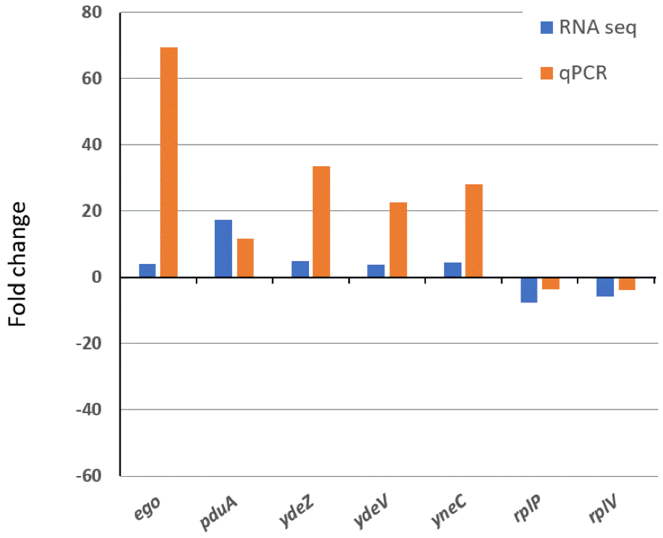
After quality control, RNA samples were converted to Illumina sequencing libraries using Illumina’s TruSeq Stranded Total RNA Library Prep Human/Mouse/Rat Sample Preparation Kit (Cat. # 20020597) at the University of Minnesota Genomics Center. Approximately 500 nanograms of total RNA was rRNA depleted using sequence-specific Ribozero capture probes. The mRNA was then fragmented and reverse transcribed into cDNA. The cDNA fragments were blunt-ended and ligated to indexed (barcoded) adaptors and amplified using 15 cycles of PCR. Final library size distribution was validated using capillary electrophoresis and quantified using fluorimetry (PicoGreen). Indexed libraries were then normalized and pooled in an equimolar fashion. Truseq libraries were hybridized to a single read flow cell and individual fragments were clonally amplified by bridge amplification on the Illumina cBot. Once clustering was complete, the flow cell was loaded on the HiSeq 2500 and sequenced using Illumina’s SBS chemistry. Base call (bcl) files for each cycle of sequencing were generated by Illumina Real Time Analysis (RTA) software. Primary analysis and index de-multiplexing were performed using Illumina’s bcl2fastq v2.20.0.422. The end result of the bcl2fastq workflow resulted in creation of de-multiplexed FASTQ files (i.e., raw sequence data). Quality control on raw sequence data for each sample was performed with FastQC. Read mapping was performed via Hisat2 (v2.0.2) using the Salmonella enterica Enteritidis genome P125109 as reference. Gene quantification was done via Feature Counts for raw read counts. Differentially expressed genes were identified using the edgeR (negative binomial) feature in CLCGWB (Qiagen, Valencia, CA, USA), followed by filtration based on a minimum 2× absolute fold change and false discovery rate (FDR) corrected p < 0.05.

2.8. Real-Time PCR

The RNA Seq data were validated by quantitative real-time PCR (qRT-PCR). Synthesis of cDNA was carried out using iScriptTM Reverse Transcription (Bio-Rad Laboratories, Inc. Hercules, CA, USA). qRT-PCR was performed on a MiniOpticonTM Real-Time PCR Detection System (Bio-Rad Laboratories, Hercules, CA, USA) with iQTM SYBR® Green Supermix kit (Bio-Rad Laboratories, Hercules, CA, USA). Gene rtcR, encoding for transcriptional regulator protein RtcR, was selected as an internal reference gene. The expression of rtcR in the wild type and its isogenic yccT mutant was not affected by H2O2 treatment or yccT mutation. Primers of seven genes (ego, pduA, ydeZ, ydeV, yneC, rplP and rplV) were designed to generate internal fragments ranging from 91 bp to 129 bp in size (Table S1).

2.9. Subcellular Localization of STY1099

Subcellular localization of the STY1099-fused with green fluorescent protein (GFP) was carried out in the transformed S. enterica Enteritidis ATCC 13076 wild type strain by inverted TiE deconvolution microscope. Briefly, the yccT-egfp fusion gene was amplified using the genome of S. enterica serovar Enteritidis ATCC 13076 and egfp plasmid (AddGene, Watertown, MA, USA) as the DNA template. The primers for yccT PCR amplification were 5′–GGAATTCCATATGAAAACCGGCGCGCTAGCCACCTT–3′ and 5′–TCCTCGCCCTTGCTCACCATGGATCCAGAGGGCGGCTGTTTTTCCGC–3′and the primers for egfp PCR amplification were 5′–GCGGAAAAACAGCCGCCCTCTGGATCCATGGTGAGCAAGGGCGAGGA–3′ and 5′–GGAATTCCATATGAAAACCGGCGCGCTAGCCACCTT–3′. Fusion of yccT and egfp was carried using amplified yccT and egfp as DNA templets and primers 5′–GGAATTCCATATGAAAACCGGCGCGCTAGCCACCTT–3′ and 5′–GGAATTCCATATGAAAACCGGCGCGCTAGCCACCTT–3′. The amplified yccT-egfp gene was digested with NdeI and XholI and ligated into the pET42b vector. An egfp gene intended for non-fusion was amplified with 5′–GGAATTCCATATGGTGAGCAAGGGCGAGGA–3′ and 5′–CCGCTCGAGTTACTTGTACAGCTCGTCCA–3′. The amplified egfp gene was digested with NdeI and XhoIl and followed ligation into the pET4b vector. After plasmid isolation and sequence verification, the pET42b::yccT-egfp and pET42::egfp plasmids were used to transform S. enterica Enteritidis ATCC 13076 wild type. Concentration of 0.5 mM of isopropyl ß-D-1-thiogalactopyranoside (IPTG) was used to induce expression of the yccTEGFP and EGFP, respectively. Transformed S. enterica Enteritidis cells were spotted on slides with ProLong® Diamond Antifade Mountant (4′6-diamidino-2-phenylindole) and imaged. Images were acquired in a Nikon TiE microscope equipped with a confocal A1R scan head. All images were captured with a 100× Apo TIRF 1.49 NA objective. GFP fluorescence was excited with a 488 nm laser and fluorescence collected through a 500–550 nm emission filter. The confocal aperture was set to 16.6 µm (0.3 AU) and the voxel size was 31 × 31 × 100 nm (XYZ). All acquisition parameters were kept constant between experimental and control samples. The images were processed with 3D automatic deconvolution (Nikon Elements software, 20 iterations).

2.10. Co-Immunoprecipitation of STY1099

Co-immunoprecipitation (Co-IP) assay was carried out at 4 °C unless otherwise indicated, using E. coli 21 and S. enterica Enteritidis ATCC 13076 strains. Briefly, the fusion yccT-flag gene was amplified using the genome of S. enterica Enteritidis ATCC 13076 as the DNA template. The primers for PCR amplification were 5′–GGA ATTCCATATGAAAACCGGCGCGCTAGCCACCTT–3′ and 5′–CCGCTCGAGTCACTTGT CGTCATCGTCTTTGTAGTCAGAGGGCGGCTGTTTTTCCGCCCATT–3′. The amplified yccT-flag gene was digested with NdeI and XholI and ligated into the pET42b vector. Two plasmids, pET42::yccT and pET42b empty vector, were used to transform E. coli, respectively. E. coli pET42b was used as negative controls. Expression of yccT was induced using 0.5 mM of IPTG. The expression of yccT was confirmed by performing Western blot. To pull down the STY1099-flag tagged protein, the lysates (E. coli served as negative control, or mixture of E. coli and S. enterica Enteritidis) were incubated with anti-FLAG M2 agarose (Sigma) at 4 °C overnight on an end-over-end rotator. After centrifugation the beads were washed with lysis buffer three times followed by proteins elution. The samples of eluted proteins were loaded onto a 10% SDS-PAGE gel. The protein bands were visualized by Coomassie Brilliant Blue R-250 (Thermo Fisher Scientific, Waltham, MA, USA) and excised for protein identification. After in-gel trypsin digestion the peptide mixture was analyzed by capillary liquid chromatography-mass spectrometry (LC-MS) on an Eksigent 1D plus LC with a MicroAS autosampler (Dublin, Ireland) online with an Orbitrap Velos MS system (Thermo Fisher Scientific, Waltham, MA, USA) as previously described [16].

2.11. Experimental Replications and Bioinformatics

Data from the gene expression, oxidative killing assays, global transcriptomics and RT-qPCR represent the average of three biological replicates. Kinetic data were analyzed by CoStat version 6.4 software (Co-Hort Software, Monterey, CA, USA) using the homogeneity of linear regression slopes method to test for significant (p < 0.05) differences. Gene expression data were analyzed by t-tests. Nucleotide sequence translation was carried out using EMBOSS Transeq [17] (the European Molecular Biology Laboratory—European Bioinformatics Institute; Hinxton, Cambridge, United Kingdom). The neighbor-joining algorithm [18] was used to generate the phylogenetic tree. The protein sequences were aligned using Clustal Omega [19] and colored using the “Percentage Identity” color-scheme in Jalview [20]. The STY1099 protein binding site and tertiary structure predictions were determined using RaptorX algorithms [21]. The gene ontology (GO) analysis was conducted using the Database for Annotation, Visualization and Integrated Discovery (DAVID) [22]. Signal peptide was determined using the SignalP 5.0 software version [23].

3. Results

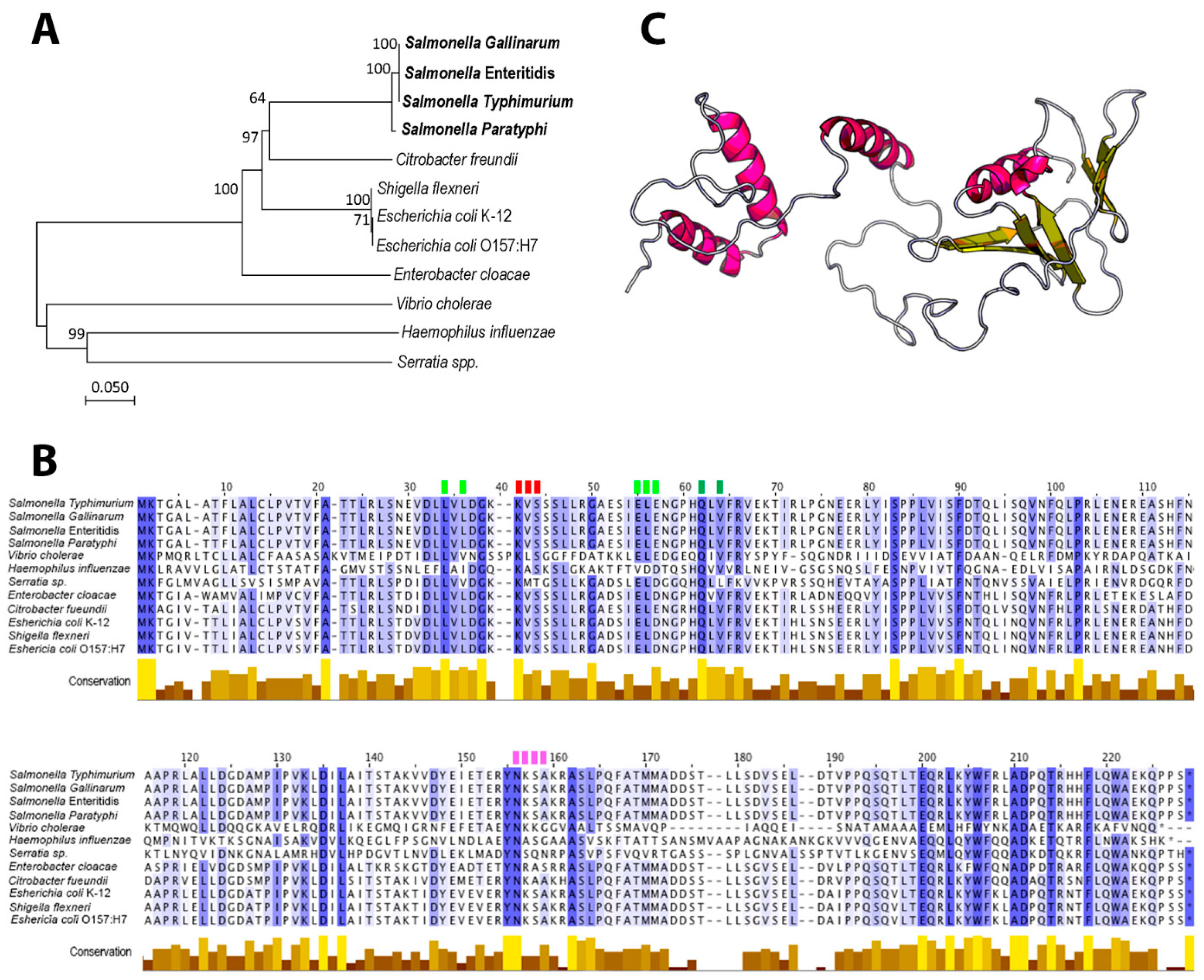
3.1. Phylogeny, Conserved Domains and Structural Characteristics of STY1099 Protein

Phylogeny of the yccT gene sequence greatly resembled taxonomic relationships of the tested species (Figure 1A). Four different Salmonella serovars formed a distinct cluster, while Shigella flexneri, a close relative of the Escherichia coli species, clustered together with E. coli (Figure 1A). Vibrio cholerae (Vibrionaceae), Haemophilus influenzae (Pasteurellaceae) and Serratia spp. (Enterobacteriaceae) were joined to the main tree basally, indicating a distant phylogeny of their yccT sequences (Figure 1A).
The overall average pairwise distance among the three families, Enterobacteriaceae, Vibrionaceae and Pasteurellaceae, was 0.437, indicating any two bacterial strains from this group would diverge, on average, 43.7% from each other based on the yccT gene sequence. Alignment of the STY1099 protein sequence revealed numerous conserved domains, including amino acid positions, 1, 2, 21, 34, 38, 42, 62, 83, 90, 103, 135, 137, 155, 156, 162, 200, 203, 206, 210, 211, 214 and 218 (Figure 1B). The STY1099 protein sequences, from this diverged group of bacteria, exhibited common binding sites with high pocket multiplicity (PM) values for SO4 (PM 20), Zn+ (PM 20), Na+ (PM 9) and HO (PM 9). Interestingly, the first amino acid binding site for each ligand, SO4 (L42), Na+ (N156), Zn (Q62) and HO (L34) was completely conserved among the tested species (Figure 1B), suggesting a ubiquitous physiological role of this protein. Based on the S. enterica Enteritidis protein sequence the STY1099 protein contains two primary domains; domain 1 (p = 6.07 × 10⁻³; from 1 to 158 aa) and domain 2 (p = 4.14 × 10⁻²; from 159 to 220 aa), forming four α-helixes and three antiparallel β-pleated sheets (Figure 1C). Also, the same protein sequence, with a high likelihood (0.9497), contains a Sec/SPI “standard” secretory signal peptide with a cleavage site located between positions 20 and 21 and VFA/TT amino acid sequences (probability 0.9324).

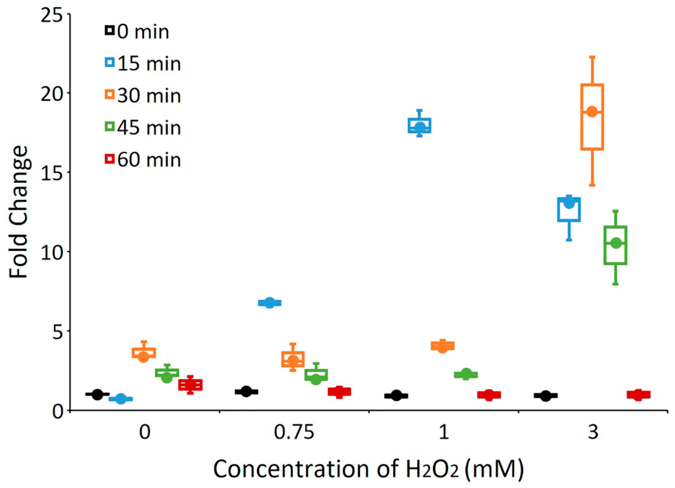
3.2. The yccT Gene Is Highly Inducible upon Exposure to Hydrogen Peroxide, But Not to Parquet

In our previous study we carried out a STRING analysis to identify an interactome of protein STY1099 [23]. This analysis with a high confidence score (confidence score, 0.70) showed that the STY1099 interacts with several proteins in the proteome of S. enterica Enteritidis including NapD (an assembly protein for periplasmic nitrate reductase) and GutM (a DNA-binding transcriptional activator). To determine whether or not reactive oxygen species caused upregulation of STY1099 and its interactome (NapD and GutM proteins), we tested the expression of genes, yccT (STY1099), gutM (GutM), napD (NapD) and two reference genes oxyR and sodA, using peroxide (H2O2) and superoxide (paraquat) as oxidative agents. No significant changes in expression of yccT, napD and gutM were observed during treatment of S. enterica Enteritidis with paraquat (Figure S1). In sharp contrast, exposure of S. enterica Enteritidis to H2O2 resulted in a significant upregulation of the yccT gene. Figure 2 shows the expression dynamics of yccT, gutM and napD alongside oxyR and sodA in the presence of 3 mM H2O2, and in controls over 60 min.
Most notably, the expression of yccT exhibited a profound upregulation with the H2O2 treatment. Significant upregulation (p < 0.0001) of yccT was observed at 15 min (11.68-fold), 30 min (18.71-fold) and 45 min (9.95-fold) of the peroxide treatment. Induction of yccT during peroxide treatment was greater than that of oxyR, which is a regulatory hallmark of the peroxide stress response in S. enterica. Interestingly, another positive control, sodA, showed a massive change in expression in the presence of H2O2, resulting in three- and six-times higher expression compared to those of yccT and oxyR, respectively, during 30 and 45 min treatments (Figure 2). A reason of the massive expression of sodA most likely lies in the fact that this gene encodes an enzyme, SodA, which catalyzes an oxidative agent, whereas oxyR encodes a protein that regulates expression of other oxidative stress response genes. In other words, sodA must be expressed at much higher levels compared to that of oxyR, as these two proteins have quite different modes of action combating an oxidative assault. Based on the expression level, the yccT gene is positioned between these two oxidative stress response genes, oxyR and sodA. This may indicate that yccT has a different mode of action during oxidative stress response of S. enterica compared to those of oxyR and sodA. Both yccT and oxyR exhibited the same pattern of gene expression, a significant increase at 15 min, followed by highest expression at 30 min and a return to basal levels after 60 min (Figure 2). Two other genes, napD and gutM, did not show significant induction during peroxide treatment.
To evaluate the sensitivity of yccT to peroxide stress, we treated S. enterica Enteritidis with three different concentrations (0.75 mM, 1 mM and 3 mM) of H2O2 over the same period of time. Data show yccT significantly upregulated (p < 0.005) at concentrations of 0.75- and 1-mM after 15 min treatment followed by a sharp decrease in the gene expression (Figure 3). The efficiency of RT-qPCR primers are presented in Table S1 and the expression data for the reference gene rtcR are provided in Table S2.
Most notably, the expression of yccT exhibited a profound upregulation with the H2O2 treatment. Significant upregulation (p < 0.0001) of yccT was observed at 15 min (11.68-fold), 30 min (18.71-fold) and 45 min (9.95-fold) of the peroxide treatment. Induction of yccT during peroxide treatment was greater than that of oxyR, which is a regulatory hallmark of the peroxide stress response in S. enterica. Both yccT and oxyR exhibited the same pattern of gene expression, a significant increase at 15 min, followed by highest expression at 30 min and a returned to basal levels after 60 min (Figure 2). Two other genes, napD and gutM, did not show significant induction during peroxide treatment.

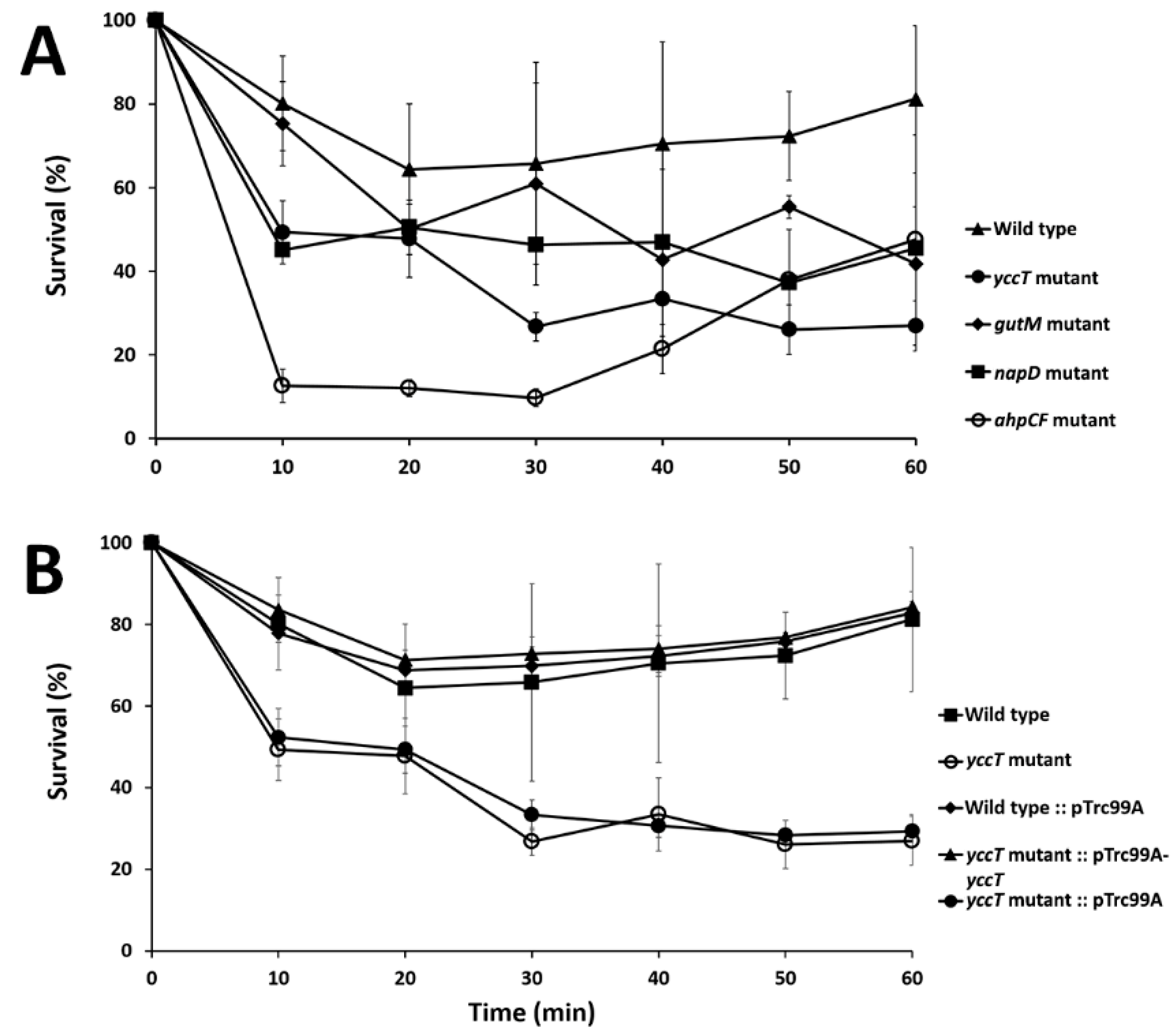
3.3. The ∆yccT Mutant Exhibits Increased Sensitivity to Hydrogen Peroxide

After demonstrating yccT induction by peroxide, we investigated whether yccT plays a role in hydrogen peroxide tolerance of S. enterica Enteritidis. Two additional genes, gutM and napD, that encode interactome proteins of STY1099 were also included in oxidative killing assays. As a reference strain with known susceptibility to oxidative stress, the ahpCF mutant strain of S. enterica Enteritidis was included in this assay too. Sensitivity of the ∆yccT, ∆gutM, ∆napD and ∆ahpCF mutants to oxidative stress was determined by exposing exponentially grown cultures of these mutants and their wild type strain to 0.75 mM of H2O2 over 60 min. Concentrations of H2O2 higher than 0.75 mM affected the wild type strain too, so for the oxidative killing assay concentration of 0.75 mM was selected. Homogeneity of linear regression slope analysis revealed a statistically significant difference (p < 0.05) in the viability between the wild type and yccT mutant, suggesting that the yccT gene plays a role in oxidative tolerance of S. enterica Enteritidis (Figure 4A).
Two other mutants, ∆gutM and ∆napD, showed reduced viability, albeit less pronounced compared to the ∆yccT mutant. The ∆ahpCF mutant showed the highest level of susceptibility to H2O2 during the first 40 min of assay. After this period, the ∆ahpCF mutant showed an increased survivability, which resulted in a higher survival rate compared to that of the ∆yccT mutant (Figure 4A). During hydrogen peroxide treatment, the ∆yccT mutant complemented with the pTre99A-yccT vector had increased viability during the peroxide treatment compared to the ∆yccT mutant (Figure 4B).

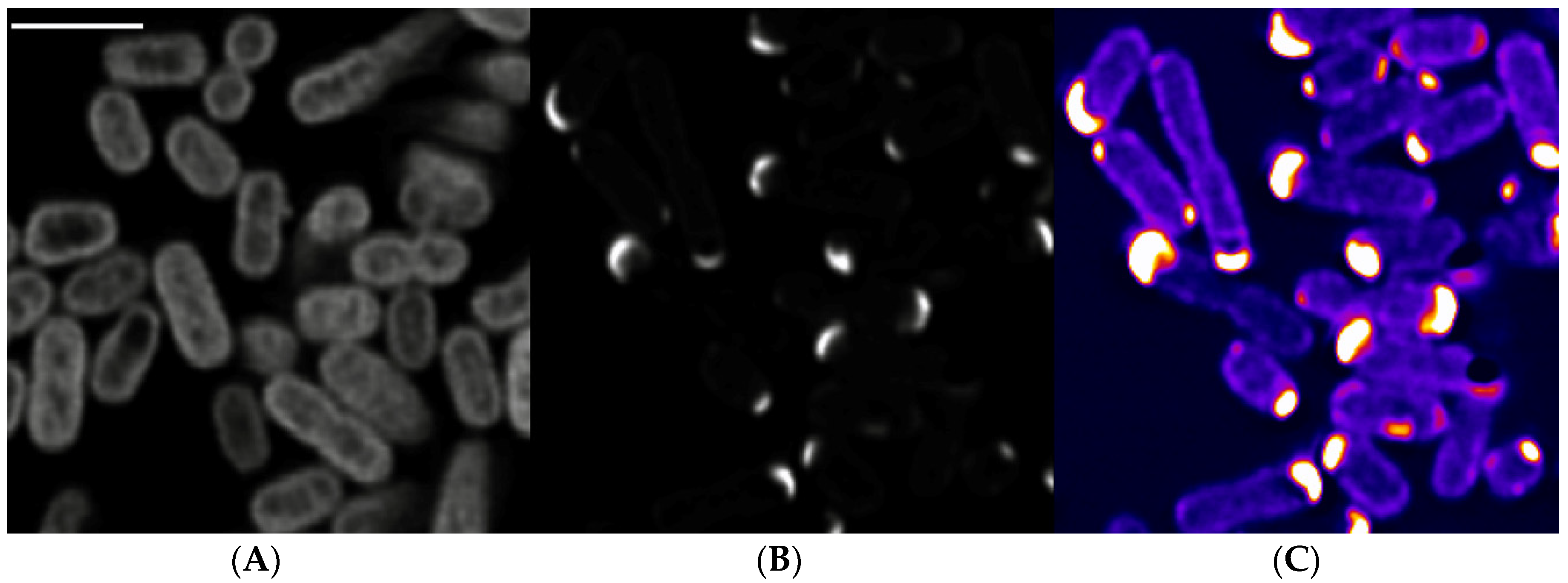
3.4. Subcellular Localization of STY1099

As STY1099 protein contains a conserved domain of unknown function 2057 (DUF2057), which occurs only in prokaryotes, we aimed to determine subcellular location of this prokaryotic protein. STY1099 was fused with green fluorescent protein (GFP) and expressed in S. enterica Enteritidis cells by a lac IPTG (isopropyl ß-D-1-thiogalactopyranoside) inducible promoter. Inverted TiE microscopy coupled with confocal scanning option showed strong and consistent fluorescence signal in transformed S. enterica Enteritidis cells, clearly indicating monopolar location of STY1099 protein (Figure 5).
These data suggest that STY1099 protein has a specific function in bacterial cells that may correspond to cell motility (flagella), cell division or other cellular function that requires monopolar location of the studied protein.

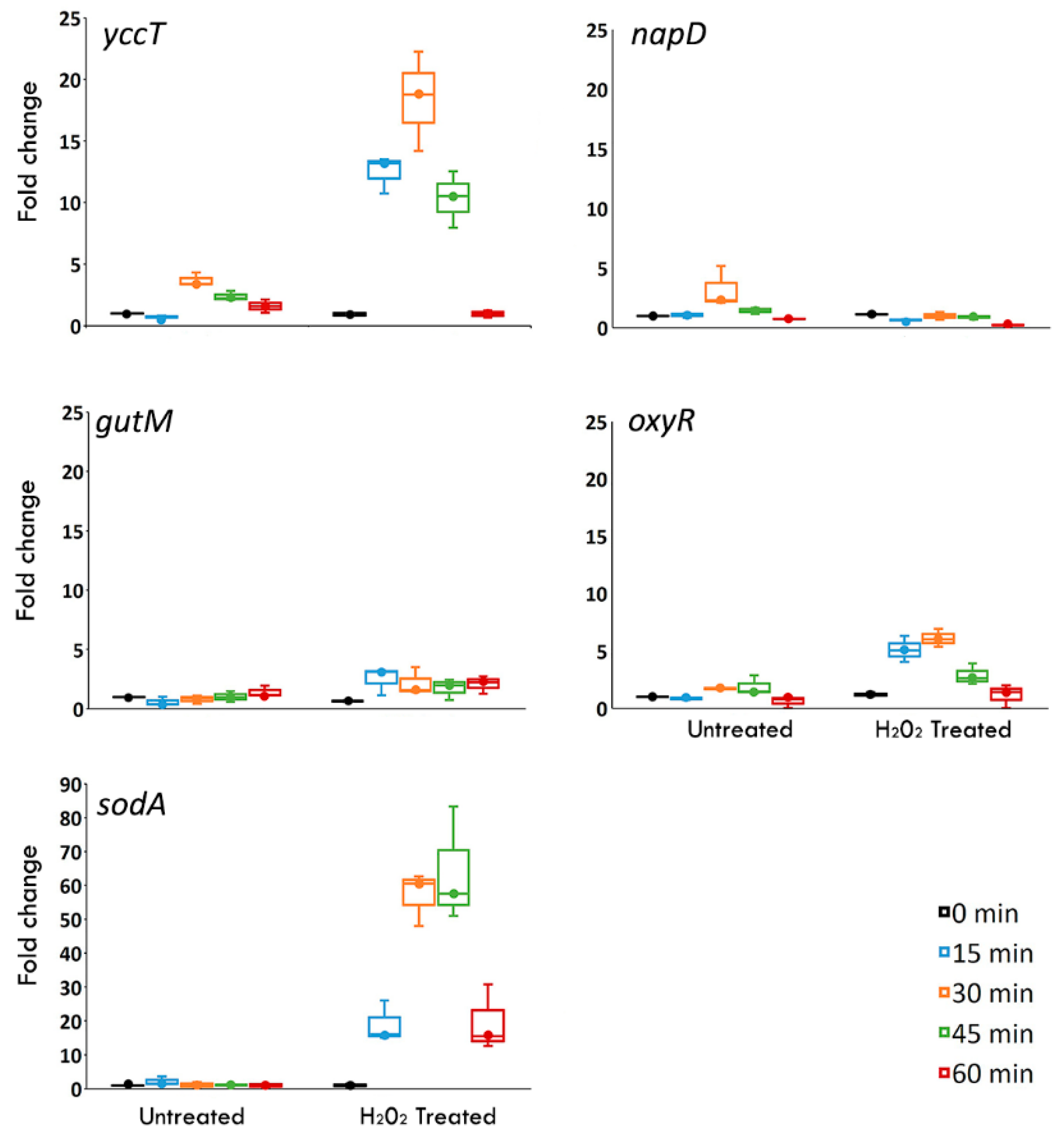
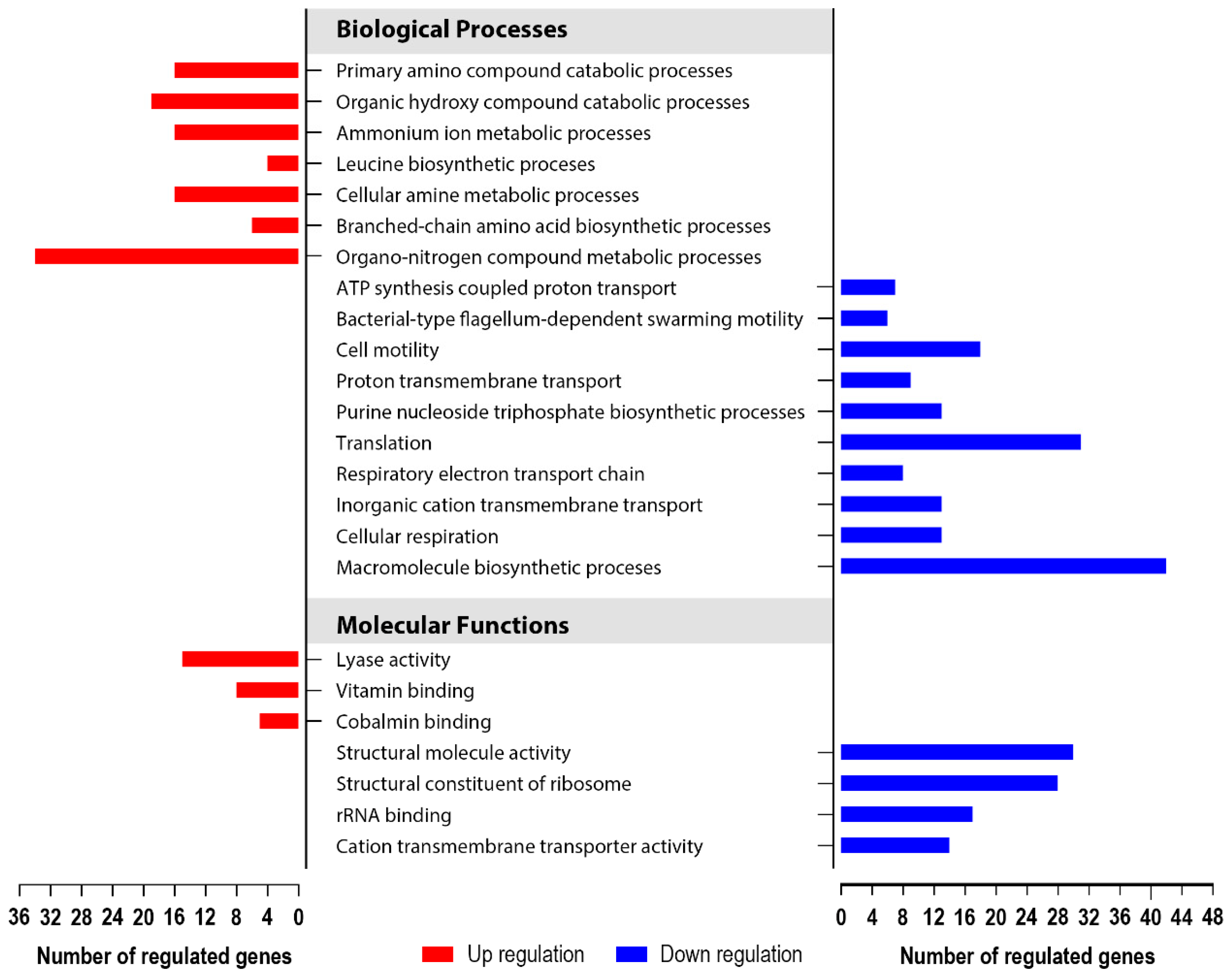
3.5. Global Transcriptomics of the S. enterica Enteritidis ∆yccT Mutant Strain Reveal the STY1099 Regulon

To determine the effect of yccT deletion on the oxidative stimulon of S. enterica Enteritidis, and specifically to identify proteins affected by the STY1099 protein, we carried out global transcriptome (RNAseq) analyses using the wild type and the ∆yccT mutant strains in combination with the oxidative stress assay. Alterations in gene expression were studied by comparing the transcriptomes with a minimum 2× absolute fold change and false discovery rate (FDR) corrected p < 0.05 used as cutoffs for significance.
In total, 2051 genes were significantly differently expressed (DE) by H2O2 in the wild type strain, and 1712 DE genes were observed in the ∆yccT mutant (Table S3). Of the 1712 DE genes, 1453 were shared between the comparisons, while 259 were unique to the yccT mutant and 598 unique to the wild type treatment group. There were 22 genes exhibiting statistically significant changes in expression between the wild type and yccT mutant during the H2O2 treatment (Table 2). Out of the 22 genes, 13 genes (narGHIJK, hycDF, dmsA3, rrmJ, SEN0541, SEN1163, SEN1249 and SEN3184) were unique to the oxidative response, and nine genes (citEFT, kdgT, nirC, yccT, SEN0167, SEN0271 and SEN0992) of the oxidative stimulon were shared with the core group of the yccT affected genes (Tables S3 and S4). When exposed to oxidative stress, the ∆yccT mutant showed significant upregulation (p < 0.05) of the citrate metabolism (citEFT), oxidoreductase activity/electron transport (hycDF, dmsA3, SEN1249/3184), rRNA processing/stress response (SEN0992, rrmJ) and gluconate transmembrane transport (kdgT) (Table 2). The ∆yccT mutant significantly downregulated (p < 0.05) nitrate metabolism (narJHIGK and nirC) (Table 2). Also, affected by oxidative stress in the ∆yccT mutant was a group of genes that encodes proteins of unknown function (SEN0167, SEN0271, SEN1163 and SEN0541).
To investigate how the yccT gene deletion specifically affected the physiology of growing cells, we compared the transcriptomes of exponentially growing culture of the yccT mutant to that of the wild type strain. Global transcriptomic analyses identified 352 (343 unique genes) differentially expressed genes in the no treatment controls; 137 genes significantly upregulated and 215 genes downregulated (Table S5).
The most pronounced changes in biological processes occurred via downregulation of genes involved in macromolecular biosynthetic processes, translation, cell motility and inorganic cation transmembrane transport (Figure 6). Among this large group of 215 genes, the most profound gene expression alterations were exhibited by a group of genes associated with electron transport via the process of nitrate reduction, including napA (−22.95; FDR p = 0.0), napB (−34.04; FDR p = 7.92 × 10⁻²²), napC (−31.42; FDR p = 1.88 × 10⁻¹⁹), napD (−16.35; FDR p = 8.76 × 10⁻¹⁷), napF (−17.48; FDR p = 1.87 × 10⁻¹⁸), napG (−29.60; FDR p = 5.58 × 10⁻²⁵) and napH (−27.25; FDR p = 6.36 × 10⁻²¹).
Upregulated biological processes of the ∆yccT mutant included organo-nitrogen compound metabolic processes, organic hydroxyl and primary amino compound catabolic processes, cellular amine metabolic and branched-chain amino acid biosynthetic processes (Figure 6). This group of upregulated genes had less pronounced expression changes, compared to the downregulated genes. Largest upregulation was observed in genes involved in propanediol metabolism, including pduA (17.25; FDR p = 8.72 × 10−36), pudB (12.69; FDR p = 7.20 × 10⁻³¹), pudD (10.70; FDR p = 1.48 × 10⁻³²), and pudC (9.77; FDR p = 3.43 × 10⁻²⁷).
To validate the RNA-seq data, the expression of seven randomly chosen genes was validated by quantitative real-time PCR (qRT-PCR). The qRT-PCR analysis confirmed the same pattern of expression, including five upregulated genes (e.g., ego, pduA, ydeZ, ydeV and yneC) and two downregulated genes (e.g., rplP and rplV) (Figure 7).

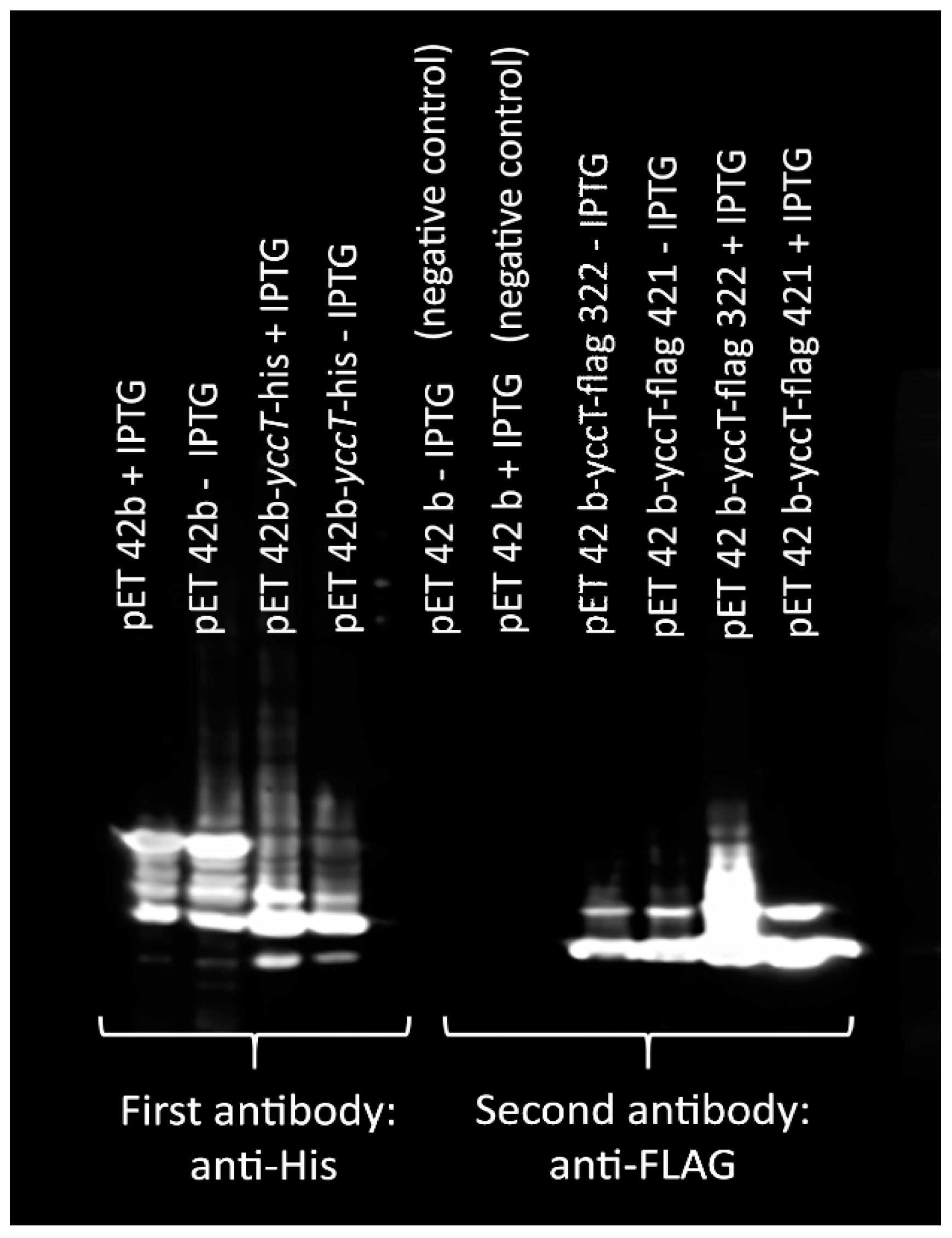
3.6. Determination of STY1099 Interactome

To identify proteins that are associated with the STY1099 protein, we carried out a Co-IP assay. Using STY1099-flag protein as a bait and proteome of S. enterica Enteritidis as a pray, we identified 36 proteins that were associated with the STY1099 protein (Table 3). Validation of the Co-IP assay were performed by Western blot, and SDS-PAGE analyses (Figure 8, Figure S2).
This group of STY1099-associated proteins consisted of a group of cell division and shape proteins, including septum site-determining protein MinD, cell division protein FtsH and rod shape-determining protein MreB, indicating that the STY1099 protein may play a role in cell division and determination of cell shape. Besides association with this functionally very specific group of proteins, the STY1099 protein showed interaction with a group of proteins involved in oxidoreductase activity. Proteins involved in oxidoreductase activities perform various biological processes, including lipid metabolism (e.g., multifunctional fatty acid oxidation complex, FadB), amino acid metabolism (e.g., aminoacyl-histidine dipeptidase, PepD), carbohydrate metabolism (e.g., bifunctional acetaldehyde-CoA/alcohol dehydrogenase, AdhE), and electron transport (e.g., cytochrome d terminal oxidase subunit 1) (Table 3). The STY1099 protein interacted with a group of ribosomal and DNA binding proteins involved in transcription and translation as well as several stress response proteins associated with heat stress (e.g., DnaK protein), DNA damage (e.g., DNA-binding ATP-dependent proteases La), osmotic stress (e.g., two-component response regulator OmpR) and extracytoplasmic stress (e.g., ATP-dependent protease ATP-binding subunit ClpX) (Table 3). The STY1099 protein interacted also with two proteins, maltose/maltodextrin transporter ATP-binding protein and oligopeptide ABC transporter ATP-binding protein OppF, involved in cellular uptake (Table 3).

4. Discussion

Our previous study identified a protein of unknown function STY1099 (i.e., encoded by yccT gene) as the most abundant among a small group of proteins associated with the ZnO NPs stimulon [24]. It is important to emphasize that ZnO NPs affect cells via two distinct pathways: Zn toxicity and oxidative stress [25,26]. The Zn toxicity-mediated pathway is primarily based on inhibition of the major enzymes involved in the tricarboxylic acid cycle [25], glycolysis [27] and the capsule biosynthesis [28]. It has been postulated that lethality of ZnO NPs is also associated with the production of increased levels of reactive oxygen species, mainly hydroxyl (OH) radicals [26], which further interfere with phospholipids, lipopolysaccharides and lipoproteins, leading to extensive cell wall disorganization of Gram-negative bacteria [29]. To distinguish this dual effect of ZnO NPs and, more importantly, to pinpoint a stimulus-causing upregulation of STY1099 in S. enterica Enteritidis exposed to ZnO NPs, we measured expression of the yccT gene during oxidative treatment. Our data show that yccT is significantly overexpressed upon exposure to H2O2, but not upon exposure to paraquat, indicating a specific role of yccT in peroxide homeostasis of this zoonotic pathogen.
We demonstrated that under peroxide stress an S. enterica Enteritidis strain lacking yccT showed a significant decrease in viability, compared to that of the wild type, indicating that the STY1099 protein likely plays a role in protecting S. enterica Enteritidis from the peroxide stress assault. A possible polar effect caused by the yccT deletion was ruled out by complementation. Increased viability of the ∆yccT mutant complemented with the pTre99A-yccT vector clearly showed that intolerance to peroxide stress of the ∆yccT mutant is due to the yccT deletion.
Transcriptomic analysis of the ∆yccT mutant and wild type provided insight into the transcriptome of this mutant in non-stressful conditions. Most notably, a majority (61%) of the altered genes became significantly downregulated including genes associated with macromolecular biosynthetic processes and translation being most common in the ∆yccT mutant. Specifically, most significant was downregulation of the napABCDGHF operon, which encodes periplasmic nitrate reductase (Nap) proteins that catalyze the transformation of nitrate to nitrite. In prokaryotic organisms, reduction of nitrate to nitrite occurs via three distinct systems, namely periplasmic nitrate reductase (Nap), respiratory nitrate reductase (Nar) and assimilatory nitrate reductase (Nas) [30]. Unlike other nitrate reductase systems, Nap is functionally diverse. It is demonstrated to be involved in dissimilatory nitrate reduction (both denitrification and nitrate reduction to ammonia) [31], maintenance of cellular oxidation–reduction potential (i.e., redox poise) [30] and nitrate scavenging and pathogenicity [32]. Besides nitrate reduction, the ∆yccT mutant exhibited a significant downregulation of the rpsQCS operon (i.e., from eight- to five-fold), which encodes one of the primary rRNA binding proteins, RpsQ (30S ribosomal protein S17). The RpsQ protein plays a critical role in translational accuracy [33], while RpsC (30S ribosomal protein S3) is involved in mRNA unwinding [34] and RpsS (30S ribosomal protein S19) in ribosomal small subunit assembly [35]. Downregulation of the Nap system and the rpsQCS operon in the ∆yccT mutant indicates that STY1099 impacts primary nitrate metabolism, cellular oxidation–reduction potential and rRNA biology during exponential growth of S. enterica Enteritidis.
Among a group of upregulated genes, the pduABCDE operon exhibited the greatest alteration, ranging from 17-fold for the pduA gene (propanediol utilization protein A) to 6.4-fold for the pduE gene (propanediol dehydratase small subunit). Proteins encoded by the pduABCDE operon catalyze the dehydration of 1,2-propanediol to propionaldehyde, which further serves as the carbon and energy source for bacterial growth [36]. The yccT mutant also showed significant upregulation of the yneABC operon (~ four-fold), which encodes proteins involved in substrate binding, trans-membrane transport, and interconverting aldoses and ketoses [37].
Indeed, our Co-IP assay showed that the STY1099 protein interacts with a group of proteins involved in oxidoreductase activity, electron transport, rRNA biology and cellular uptake, clearly indicating that the STY1099 protein is involved in these cellular processes. Besides these proteins involved in the central metabolic processes, the STY1099 protein interacted with proteins of key importance for cell division (e.g., septum-site determining protein MinD and cell division protein FtsH) and a protein involved in determination of cell shape (e.g., rod shape determining protein MreB). Monopolar location of the STY1099 protein additionally suggests that the STY1099 protein may play a role in cell division and cell shape determination too.
The molecular response of the ∆yccT mutant to peroxide stress included downregulation of nitrate reductase (narJHIGK, and nirC) and upregulation of oxidoreductase activity (hycDF, dmsA3, SEN1249 and SEN3184) and citrate metabolism (citEFT). Interestingly, during exponential growth with no peroxide treatment, the ∆yccT mutant exhibited a profound downregulation of nitrate reductase mediated by Nap, while during peroxide treatment nitrate reductase was altered via the respiratory nitrate reductase (Nar) system. Although both Nap and Nar systems belong to dissimilatory nitrate reductases, the Nar system is strictly involved in redox regulation and energy acquisition, whereas the Nap system is functionally diverse [30]. Both nitrate reductases, Nap and Nar, are membrane associated, whereas Nas is made of soluble proteins located in the cytoplasm [38]. We showed that the STY1099 protein only affects membrane associated nitrate reductase, which is further supported by the fact that this novel protein is associated with cellular membrane (i.e., monopolar localization), but not with cytoplasm. In addition, it has been shown with the high likelihood (0.9324) that the STY1099 protein contains a Sec/SPI signal peptide. This secretory signal peptide, transported by the Sec translocon and cleaved by signal peptidase I, is an integral part of periplasmic proteins, which suggests that the novel STY1099 protein belongs to a group of periplasmic proteins. Downregulation of the Nar system in the ∆yccT mutant during peroxide treatment indicates that STY1099 plays a role in redox homeostasis. Indeed, upregulation of the genes associated with oxidoreductase activity and citrate metabolism provides a second line of evidence that STY1099 is involved in maintenance of cellular redox balance. In addition to this, interactome of STY1099, defined by the Co-IP assay, showed that the STY1099 protein is associated with a group of proteins involved in oxidoreductase activities. These oxidoreductase proteins are involved in various biological processes (e.g., lipid, amino acid and carbohydrate metabolisms as well as electron transport), clearly indicating that their association with the STY1099 protein is based on their role in oxidoreductase activities rather than involvement in some other biological processes.
Fu and colleagues [38], examining response of S. enterica Typhimurium to H2O2 treatment, found that this Salmonella serovar significantly induced several proteins associated with DNA repair machinery, including RecA, RecE and GyrI. The current study showed that the STY1099 protein interacted with DNA-binding ATP-dependent protease La, showing that this novel protein is indirectly associated with DNA repair machinery, an important response of Salmonella enterica to peroxide stress [38]. Besides DNA-binding proteases La, the STY1099 protein interacted with key protease, ClpX, chaperone, DnaK, and the two-component response regulator, OmpR, which controls a variety of membrane-associated transporters that mediate uptake or efflux of small molecules [39], showing its importance in the stress response physiology.

5. Conclusions

We demonstrated that yccT is a highly peroxide-inducible gene, and the ∆yccT mutant exhibits peroxide intolerance compared to its wild type strain. Interestingly, the ∆yccT mutant showed a tolerance to parquet, indicating that this novel protein is specifically involved in the peroxide stress response of S. enterica serovar Enteritidis. Comparative transcriptome analyses supported by Co-IP assay showed that the STY1099 protein is involved in oxidoreductase processes, respiratory nitrate reductase, rRNA biology and stress response. In addition, we showed that the STY1099 protein has monopolar location and that interacts with key cell division proteins MinD and FtsH as well as with a rod shape-determining protein MreB.

Supplementary Materials

The following are available online at https://www.mdpi.com/2079-7737/8/4/86/s1, Figure S1: mRNA expression levels of yccT, gutM, napD, oxyR and sodA in the wild type S. enterica Enteritidis strain during its exponential growth exposed to 4 mM of parquet. Values on the y-axis are relative expression levels (fold change) normalized to wild type during the oxidative treatment. The data correspond to the mean value of three biological replications. Error bars correspond to the standard deviation. Figure S2: SDS-PAGE gel showing. Lanes (1) Vector pET 42b in BL21 + S. enterica Enteritidis; (2) one volume of pET 42 b-yccT-flag in BL21 + ten volumes of S. enterica Enteritidis unbinding cell lysis; (3) one volume pET 42 b-yccT-flag in BL21 + two volumes of S. enterica Enteritidis unbinding cell lysis; (4) pET 42b-yccT-FLAG in BL21 unbinding cell lysis; (5) Marker; (6) pET 42b in BL21 + S. enterica Enteritidis; (7) one volume pET 42 b-yccT-FLAG in BL21 + ten volumes of S. enterica serovar Enteritidis; (8) one volume pET 42 b-yccT-FLAG in BL21 + two volumes of S. enterica Enteritidis; (9) pET 42b-yccT-FLAG in BL21. Table S1: Primers and their efficiency used for the gene expression assay. Table S2: Expression data of the reference gene rtcR in the wild type and its yccT mutant. Table S3: List of genes exhibiting statistically significant changes in expression between the yccT mutant treated and not treated with H2O2.Table S4: List of the genes exhibiting statistically significant changes in expression between the wild type and yccT mutant treated with H2O2. Table S5: List of the genes exhibiting statistically significant changes in expression between the yccT mutant and wild type during exponential growth with no H2O2 treatment.

Author Contributions

Conceptualization, S.V.; methodology, S.V. and X.L.; investigation, X.L., R.A. and S.V., formal analysis, S.V., X.L. and J.E.A.; assistance with the RNA-seq data, K.M.M. and K.M.R.; statistical analysis for the gene expression assay, A.K.J.; writing—original draft, S.V.; funding acquisition, S.V.

Funding

This research was funded by the United States Department of Agriculture, National Institute of Food and Agriculture, Hatch Capacity Grant # 592 and Grant-in-Aid of Research, Artistry and Scholarship, University of Minnesota, USA to S.V.

Acknowledgments

The authors gratefully acknowledge the technical support from the University Imaging Centers at the University of Minnesota, Jim Imlay, Sergei Korshunov and Daniela Vidovic. We also thank Misara Omar and Sahar Alshalchi for their assistance. This project was supported by the Signature Program AES GAR grant. The present study was also supported, in part, by resources provided by the Minnesota Supercomputing Institute.

Conflicts of Interest

The authors declare no conflict of interest.

References

  1. Scallan, E.; Hoekstra, R.M.; Angulo, F.J.; Tauxe, R.V.; Widdowson, M.A.; Roy, S.L.; Jones, J.L.; Griffin, P.M. Foodborne illness acquired in the United States–major pathogens. Emerg. Infect. Dis. 2011, 17, 7–15. [Google Scholar] [CrossRef]
  2. de Moraes, M.H.; Desai, P.; Porwollik, S.; Canals, R.; Perez, D.R.; Chu, W.; McClelland, M.; Teplitski, M. Salmonella persistence in tomatoes requires a distinct set of metabolic functions identified by transposon insertion sequencing. Appl. Environ. Microbiol. 2017, 83, e03028-16. [Google Scholar] [CrossRef]
  3. Parker, W.F.; Mee, B.J. Survival of Salmonella adelaide and fecal coliforms in coarse sands of the swan costal plain, western Australia. Appl. Environ. Microbiol. 1982, 43, 981–986. [Google Scholar] [PubMed]
  4. Mezrioui, N.; Baleux, B.; Trousselier, M. A microcosm study of the survival of Escherichia coli and Salmonella Typhimurium in brackish water. Water Res. 1995, 29, 459–465. [Google Scholar] [CrossRef]
  5. Fang, F.C.; Frawley, E.R.; Tapscott, T.; Vázquez-Torres, A. Bacterial stress responses during host infection. Cell Host Microbe 2016, 20, 133–143. [Google Scholar] [CrossRef] [PubMed]
  6. Mangalappalli-Illathu, A.K.; Vidovic, S.; Korber, D.R. Differential adaptive response and survival of Salmonella enterica serovar Enteritidis planktonic and biofilm cells exposed to benzalkonium chloride. Antimicrob. Agents Chemother. 2008, 52, 3669–3680. [Google Scholar] [CrossRef]
  7. Farr, S.B.; Kogoma, T. Oxidative stress responses in Escherichia coli and Salmonella Typhimurium. Microbiol. Rev. 1991, 55, 561–585. [Google Scholar] [PubMed]
  8. Aslund, F.; Zheng, M.; Beckwith, J.; Storz, G. Regulation of the OxyR transcription factor by hydrogen peroxide and the cellular thiol-disulfide status. Proc. Natl. Acad. Sci. USA 1999, 96, 6161–6165. [Google Scholar] [CrossRef]
  9. Zheng, M.; Wang, X.; Templeton, L.J.; Smulski, D.R.; Larossa, R.A.; Storz, G. DNA microarray-mediated transcriptional profiling of the Escherichia coli response to hydrogen peroxide. J. Bacteriol. 2001, 183, 4562–4570. [Google Scholar] [CrossRef]
  10. Altuvia, S.; Weinstein-Fischer, D.; Zhang, A.; Postow, L.; Storz, G. A small, stable RNA induced by oxidative stress: Role as a pleiotropic regulator and antimutator. Cell 1997, 90, 43–53. [Google Scholar] [CrossRef]
  11. Wu, J.; Weiss, B. Two divergently transcribed genes, soxR and soxS, control a superoxide response regulon in Escherichia Coli. J. Bacteriol. 1991, 173, 2864–2871. [Google Scholar] [CrossRef] [PubMed]
  12. Pomposiello, P.J.; Demple, B. Redox-operated genetic switches: The SoxR and OxyR transcription factors. Trends Biotechnol. 2001, 19, 109–114. [Google Scholar] [CrossRef]
  13. Vidovic, S.; Korber, D. Escherichia coli O157: Insights into the adaptive stress physiology and the influence of stressors on epidemiology and ecology of this human pathogen. Crit. Rev. Microbiol. 2016, 42, 83–93. [Google Scholar] [CrossRef] [PubMed]
  14. Schmittgen, T.D.; Livak, K.J. Analyzing real-time PCR data by the comparative CT method. Nat. Protocol. 2008, 3, 1101–1108. [Google Scholar] [CrossRef] [PubMed]
  15. Datsenko, K.A.; Wanner, B.L. One-step inactivation of chromosomal genes in Escherichia coli K-12 using PCR products. Proc. Natl. Acad. Sci. USA 2000, 97, 6640–6645. [Google Scholar] [CrossRef] [PubMed]
  16. Thu, Y.M.; Riper, S.K.V.; Higgins, L.A.; Zhang, T.; Becker, J.R.; Markowski, T.W.; Nguyen, H.D.; Griffin, T.J.; Bielinsky, A.K. Slx5/Slx8 promotes replication stress tolerance by facilitating mitotic progression. Cell Rep. 2016, 15, 1254–1265. [Google Scholar] [CrossRef] [PubMed]
  17. Kearse, M.; Moir, R.; Wilson, A.; Stones-Havas, S.; Cheung, M.; Sturrock, S.; Buxton, S.; Cooper, A.; Markowitz, S.; Duran, C.; et al. Geneious Basic: An integrated and extendable desktop software platform for the organization and analysis of sequence data. Bioinformatics 2012, 28, 1647–1649. [Google Scholar] [CrossRef] [PubMed]
  18. Saitou, N.; Nei, M. The neighbor-joining method: A new method for reconstructing phylogenetic trees. Mol. Biol. Evol. 1987, 4, 406–425. [Google Scholar] [PubMed]
  19. McWilliam, H.; Li, W.; Uludag, M.; Squizzato, S.; Park, Y.M.; Buso, N.; Cowley, A.P.; Lopez, R. Analysis tool web services from the EMBL-EBI. Nucleic Acids Res. 2013, 41, W597–W600. [Google Scholar] [CrossRef]
  20. Waterhouse, A.M.; Procter, J.B.; Martin, D.M.A.; Clamp, M.; Barton, G.J. Jalview Version 2-A multiple sequence alignment editor and analysis workbench. Bioinformatics 2014, 25, 1189–1191. [Google Scholar] [CrossRef]
  21. Kallberg, M.; Wang, H.; Wang, S.; Peng, J.; Wang, Z.; Lu, H.; Xu, J. Template-based protein structure modeling using the RaptorX web server. Nat. Protocol. 2012, 7, 1511–1522. [Google Scholar] [CrossRef] [PubMed]
  22. Huang, D.W.; Sherman, B.T.; Lempicki, R.A. Systematic and integrative analysis of large gene lists using DAVID Bioinformatics Resources. Nat. Protocol. 2009, 4, 44–57. [Google Scholar] [CrossRef] [PubMed]
  23. Armenteros, J.J.A.; Tsirigos, K.D.; Sonderby, C.K.; Petersen, T.N.; Winther, O.; Brunak, S.; von Heijne, G.; Nielsen, H. SignalP 5.0 improves signal peptide predictions using deep neural networks. Nat. Biotechnol. 2019, 37, 420–423. [Google Scholar] [CrossRef]
  24. Vidovic, S.; Elder, J.; Medihala, P.; Lawrence, J.R.; Predicala, B.; Zhang, H.; Korber, D.R. ZnO nanoparticles impose a panmetabolic toxic effect along with strong necrosis, inducing activation of the envelope stress response in Salmonella enterica serovar Enteritidis. Antimicrob. Agents Chemother. 2015, 59, 3317–3328. [Google Scholar] [CrossRef]
  25. Alhasawi, A.; Auger, C.; Appanna, V.P.; Chahma, M.; Appanna, V.D. Zinc toxicity and ATP production in Pseudomonas fluorescens. J. Appl. Microbiol. 2014, 117, 65–73. [Google Scholar] [CrossRef]
  26. Applerot, G.; Lipovsky, A.; Dror, R.; Perkas, N.; Nitzan, Y.; Lubart, R.; Gedanken, A. Enhanced antimicrobial activity of nanocrystalline ZnO due to increased ROS-mediated cell injury. Adv. Funct. Mater. 2009, 19, 842–852. [Google Scholar] [CrossRef]
  27. Stocks, C.J.; Phan, M.D.; Achard, M.E.S.; Nhu, N.T.K.; Condon, N.D.; Gawthorne, J.A.; Lo, A.W.; Peters, K.M.; McEwan, A.G.; Kapetanovic, R.; et al. Uropathogenic Escherichia coli employs both evasion and resistance to subvert innate immune-mediated zinc toxicity for dissemination. Proc. Natl. Acad. Sci. USA 2019, 116, 6341–6350. [Google Scholar] [CrossRef]
  28. Ong, C.I.Y.; Walker, M.J.; McEwan, A.G. Zinc disrupts central carbon metabolism and capsule biosynthesis in Streptococcus pyogenes. Sci. Rep. 2015, 5, 10799. [Google Scholar] [CrossRef]
  29. Brayner, R.; Ferrari-Iliou, R.; Brivovis, N.; Djediat, S.; Benedetti, M.C.; Fievet, F. Toxicological impact studies based on Escherichia coli bacteria in ultrafine ZnO nanoparticles colloidal medium. Nano Lett. 2006, 6, 866–870. [Google Scholar] [CrossRef]
  30. Sparacino-Watkins, C.; Stolz, J.F.; Basu, P. Nitrate and periplasmic nitrate reductases. Chem. Soc. Rev. 2014, 43, 676–706. [Google Scholar] [CrossRef]
  31. Cruz-Garcia, C.; Murray, A.E.; Klappenbach, J.A.; Stewart, V.; Tiedje, J.M. Respiratory nitrate ammonification by Shewanella oneidensis MR-1. J. Bacteriol. 2007, 189, 656–662. [Google Scholar] [CrossRef] [PubMed] [Green Version]
  32. Vegge, C.S.; Brondsted, L.; Li, Y.P.; Bang, D.D.; Ingmer, H. Energy taxis drives Campylobacter jejuni toward the most favorable conditions for growth. Appl. Environ. Microbiol. 2009, 75, 5308–5314. [Google Scholar] [CrossRef] [PubMed] [Green Version]
  33. Yaguchi, M.; Wittmann, H.G.; Cabezon, T.; DeWilde, M.; Villarroel, R.; Herzog, A.; Bollen, A. Alteration of ribosomal protein S17 by mutation linked to neamine resistance in Escherichia coli. II. Localization of the amino acid replacement in protein S17 from a meaA mutant. J. Mol. Biol. 1976, 104, 617–620. [Google Scholar] [CrossRef]
  34. Takyar, S.; Hickerson, R.P.; Noller, H.F. mRNA helicase activity of the ribosome. Cell 2005, 120, 49–58. [Google Scholar] [CrossRef] [PubMed] [Green Version]
  35. Valle, M.; Zavialov, A.; Sengupta, J.; Rawat, U.; Ehrenberg, M.; Frank, J. Locking and unlocking of ribosomal motions. Cell 2003, 114, 123–134. [Google Scholar] [CrossRef] [Green Version]
  36. Jeter, R.M. Cobalmin-dependent 1,2-propanediol utilization by Salmonella Typhimurium. J. Gen. Microbiol. 1990, 136, 887–896. [Google Scholar] [CrossRef] [Green Version]
  37. Caspi, R.; Billington, R.; Ferrer, L.; Foerster, H.; Fulcher, C.A.; Keseler, I.M.; Kothari, A.; Krummenacker, M.; Latendresse, M.; Mueller, L.A.; et al. The MetaCyc database of metabolic pathways and enzymes and the BioCyc collection of pathway/genome databases. Nucleic Acids Res. 2015, 44, D471–D480. [Google Scholar] [CrossRef]
  38. Fu, J.; Qi, L.; Hua, M.; Liu, Y.; Yu, K.; Liub, Q.; Liu, X. Salmonella proteomics under oxidative stress reveals coordinated regulation of antioxidant defense with the iron metabolism and bacterial virulence. J. Proteom. 2017, 157, 52–58. [Google Scholar] [CrossRef]
  39. Shimada, T.; Takada, H.; Yamamoto, K.; Ishihama, A. Expended roles of two-component response regulator OmpR in Escherichia coli: Genomic SELEX search for novel regulation targets. Genes Cells 2015, 20, 915–931. [Google Scholar] [CrossRef]
Figure 1. Characterization of the yccT gene and its STY1099 protein sequence. (A) Phylogeny of the yccT gene sequence using seven bacterial species from Enterobacteriaceae, Vibrionaceae, and Pasteurellaceae families. (B) Sequence alignment of STY1099 protein from Salmonella enterica, Escherichia coli, Shigella flexneri, Citrobacter freundi, Enterobacter cloacae, Vibrio cholerae, Haemophilus influenzae and Serratia spp. Color gradient depicts highly conserved sequences (dark blue), moderate conserved (light blue) and no conserved sequences (white). Binding sites for SO4 (red), Na+ (pink), Zn (dark green) and OH (light green) have been highlighted by rectangles above the STY1099 sequence. (C) A predicted model of the STY1099 tertiary structure was generated using a publicly available RaptorX platform at www.raptorx.uchicago.edu/StructurePrediction/documentation.
Biology 08 00086 g001
Figure 2. Messenger RNA (mRNA) expression levels of yccT (STY1099 protein), gutM, napD (interactomes of STY1099) and oxyR, sodA (positive controls for oxidative stress) in the wild type S. enterica serovar Enteritidis strain during its exponential growth exposed to 3 mM of H2O2. Values on the y axis are relative expression levels (fold change) normalized to wild type during the oxidative treatment. The data correspond to the mean value of three biological replications. Error bars correspond to the standard deviation.
Biology 08 00086 g002
Figure 3. mRNA expression levels of yccT in the wild type strain exposed to 0.75 mM, 1 mM and 3 mM concentrations of H2O2, respectively, during the exponential growth phase. The data correspond to the mean value of three biological replications.
Biology 08 00086 g003
Figure 4. Hydrogen peroxide killing assay. (A) Evaluation of the effect of H2O2 on the ∆yccT, ∆gutM, ∆napD mutant strains compared to that of their parental wild type strain during their exponential growth phases. (B) Validation of the effect of the yccT gene deletion on the survivability of S. Enteritidis during peroxide treatment.
Biology 08 00086 g004
Figure 5. Subcellular localization of STY1099 protein. Single confocal section of deconvolved three D acquisitions are shown. (A) GFP expressing bacteria. (B,C) STY1099-GFP fusions. Panels (A,B) share the same lookup table, panel C shows the STY1099-GFP fusion with a lookup table intended to show the bacterial cell outline. Scale bar 2 µm.
Biology 08 00086 g005
Figure 6. Gene ontology (GO) enrichment analysis portraying the most important biological processes and molecular functions of S. enterica serovar Enteritidis during the exponential growth phase that were affected by ∆yccT mutation. Included in analysis were genes that were more than two-fold up- or downregulated with FDR (p < 0.05) and high reproducibility across the biological replicates.
Biology 08 00086 g006
Figure 7. Validation of RNA-sequencing (RNA-seq) data by qRT-PCR analysis. Data represent fold changes in expression of selected seven genes in the wild type and the ∆yccT mutant treated with 3 mM H2O2. Genes differently expressed between the wild type and ∆yccT mutant during H2O2 treatment represent the mean value of three biological replications.
Biology 08 00086 g007
Figure 8. Validation of STY1099 expression of the yccT-flag gene. Western blot analysis shows the results for first antibody: anti His (lanes 1–4) and second antibody: anti-FLAG (lanes 5–10). Negative controls lanes 5 and 6.
Biology 08 00086 g008
Table 1. Primers used in this study for gene deletions and complementation.
Primer NameSequence (5′–3′)
Primers for yccT, napD, gutM, ahpC and ahpF Deletion
yccT: ForwardCTT CCT TGC TCT CTG TTT GCC GGT GAC TGT TTT TGC CAC AAC GCT CCG TTT GTA GGC TGG AGC TGC TTC G
yccT: ReverseCCC ATT GCA GAA AAT GAT GGC GTG TCT GCG GGT CGG CCA GTC GGA ACC AAC ATA TGA ATA TCC TCC TTA G
napD: ForwardATG CGC ACT AAC TGG CAG GTC TGT AGC CTG GTC GTG CAG GCC AAA AGT CAT GTA GGC TGG AGC TGC TTC G
napD: ReverseTCA TGG TGT TTC CTC ACC TTG CTC ATC CTG CTG GTG ATA AAC CAG CGA CAC ATA TGA ATA TCC TCC TTA G
gutM: ForwardATG GTT TCC ACT CTG ATT ACC GTC GCC GTT ATC GCC TGG TGT GCG CAA CTT GTA GGC TGG AGC TGC TTC G
gutM: ReverseTTA CCC ATG TTT CAG TTT AAG CGC CAA TGA TAG TGC ATT CTG CGC GAG CGC ATA TGA ATA TCC TCC TTA G
ahpC: ForwardATG TCC TTA ATT AAC ACC AAA ATC AAA CCT TTC AAA AAC CAG GCG TTC AAT GTA GGC TGG AGC TGC TTC G
ahpC: ReverseTTA GAT TTT ACC GAC CAG GTC TAA AGA TGG AGC CAG AGT CGC TTC GCC TTC ATA TGA ATA TCC TCC TTA G
ahpF: ForwardATG CTC GAC ACA AAT ATG AAA ACC CAG CTC AGG GCT TAC CTT GAG AAA CTT GTA GGC TGG AGC TGC TTC G
ahpF: ReverseTTA TGC GAT TTT GGT ACG AAT CAG ATA ATC AAA GGC GCT CAA CGA GGC TTC ATA TGA ATA TCC TCC TTA G
Primers for Conformation of yccT, napD, gutM, ahpC and ahpF gene Deletions
yccT mutant FAAA GTG TAC GAC AAA CCT GAC A
yccT mutant RTGA GCA ACC AAC GCG ATA TGT T
napD mutant FGCT GCC AGG ACA GTT GTG AAC C
napD mutant RCCG TTC CGC AAA AAC GGC ACG G
gutM mutant FTTT ATG CCA GCC CGA AAG CGT C
gutM mutant RGCA TGT GCG TTG GCT TTC CTC G
ahpC mutant FACT TTA GAT GGC TGA CAG GGC GCA
ahpC mutant RCGC GGG TGC GCC CAT GAC TGA AAC
ahpF mutant FGCC GCC TTA CTC TGA CGT GAA ATA
ahpF mutant RTTA ACA GAC CGT TTC AGA GTA TTG
Primers for yccT Mutant Complementation a
yccT-FTCA CCC GGG ATT CGA TTC CGT AGT TAA CCT
yccT-RGCG GTC GAC AGC AAA ACG CCA AAA TAT GTC
a Underline bases represent restriction endonuclease sites: SmaI and SalI.
Table 2. Altered genes during peroxide treatment of the ∆yccT mutant of S. Enteritidis.
Biological Processes and GenesLocus TagProtein DescriptionFold ChangeFDR p-Value 1
Citrate metabolism
citESEN0591Citryl-CoA lyase2.670.0018
citFSEN0590Citrate CoA-transferase4.180.0307
citTSEN0587Citrate carrier2.550.0413
Oxidoreductase activity/electron transport
hycDSEN2692Respiratory-chain NADH dehydrogenase2.350.0274
hycFSEN2690Formate hydrogenlyase complex iron-sulfur2.640.0274
dmsA3SEN1552Dimethyl sulfoxide reductase3.870.0274
SEN1249SEN1249Hydrogenases b-type cytochrome2.120.0448
SEN3184SEN3184Na+-transporting methylmalonyl-CoA/oxaloacetate decarboxylase2.060.0317
rRNA processing/stress response
SEN0992SEN0992Oligogalacturonate-specific porin4.660.0274
rrmJSEN313023S rRNA methyltransferase J2.120.0378
Gluconate transmembrane transport
kdgTSEN01662-keto-3-deoxygluconate permease5.330.0045
Nitrate metabolism
narJSEN1277Nitrate reductase−16.960.0162
narHSEN12764Fe–4S ferredoxin, respiratory nitrate reductase−15.310.0178
narISEN1278Nitrate reductase, gamma subunit−11.240.0178
narGSEN1275Respiratory nitrate reductase, subunit alpha−10.980.0162
narKSEN1274Nitrite extrusion protein−7.480.0162
nirCSEN3303Nitrite transporter−3.580.0274
Unknown
SEN0167SEN0167Hypothetical protein4.750.0274
SEN0271SEN0271Hypothetical protein2.330.0306
yycT2SEN0942STY1099−1525.689.0780 × 10−56
SEN1163SEN1163Phage membrane protein−8.980.0008
SEN0541SEN0541Protein of unknown function DUF1471−2.310.0274
1 False discovery rate (FDR) p < 0.05; 2 Deleted yccT gene that encodes STY1099 protein.
Table 3. Proteins of S. enterica Enteritidis that form an interactom with the STY1099 protein.
Biological Processes and Protein NamesAccession NumberIdentification Probability (%)Molecular WeightIdentified Unique Peptide Sequences
Cell Division and Shape
Septum site-determining protein MinDYP_005216705.110029,509.3ASNQGEPVILDATADAGK; AYADTVDR; IIVVTSGK; IKLVGVIPEDQSVLR; LVGVIPEDQSVLR; TENLFILPASQTR; TTSSAAIATGLAQK
Cell division protein FtsHZP_12129674.110070,784.5QKLESQISTLYGGR; QVVVGLPDVR
Rod shape-determining protein MreBZP_11739057.110036,934.4DGVIADFFVTEK; GMVLTGGGALLR; IKHEIGSA YPGDEVR; IKHEIGSAYPGD EVREIEVR; NLAEG VPR; NYGSLIGEATAER; RNYGSLIGEATAER; VL VCVPVGATQVER
Oxidoreductase Activity/Electron Transport
Biotin carboxylase of acetyl-CoA carboxylaseZP_12119137.197.0 VVEEAPAPGITPELRR; YLENPR
Multifunctional fatty acid oxidation complex subunit alphaZP_15867656.110079,596.2DFSDDEIIAR; KEEDAAVDDLLASVSQTKR; QAI TGDLDWR
Glutamate dehydrogenaseZP_15809056.110045,978.8CAALNLPYGGAK; LFAGAGAR; RANIAVEGAR; TAAYIVACER; VAVQGFGNVGSEAAR
Precorrin-4 C11-methyltransferaseZP_11737843.110028,358.5EQGEELTR; GTLADISDKVR; LQTGDVSLYGSVR
Short chain dehydrogenaseZP_13073001.110027,852.2ADVRDFASVQAAVAR; AKETEGRIDILVNNAG VCR; SLAVEYAQSGIR; TALITGASQGIGEGIAR; TPMAESIAR; VNAICPGYVR
Aminoacyl-histidine dipeptidaseYP_005235990.110052,419.9EAVPAGFACFK; FLAGHAEELDLR; LLNATPNGVIR
Cytochrome d terminal oxidase subunit 1ZP_15859114.199.958,265.5AYELLEQLR; DLGYGLLLKR
Anaerobic glycerol-3-phosphate dehydrogenase subunit AYP_005397674.110057,893.3ACEAAGIR; AEAIDPQQAR; EGATVCGVHVR; H DIATGATGR; HGDRTPGWLSEGR; IAEYADLSI R; IISLPAPLR; INQHVINR
Bifunctional acetaldehyde-CoA/alcohol dehydrogenaseZP_15844816.196.996,199.8AVTNVAELNALVER; AAALAAADAR
Transcription and Translation
30S ribosomal protein S3ZP_15868039.110025,965.5EFADNLDSDFKVR; EGRVPLHTLR; GIKVEVSGR; IVIERPAK; KPELDAK; KVEVSGR; KVVADIAGPAQINIAEVR; LGGAEIAR; LVADSITSQLER; VPLH TLR
DNA-binding transcriptional regulator PhoPZP_15835390.199.725,483.5IQAQYPHDVITTVR; VLVVEDNALLR
30S ribosomal protein S9YP_002638940.110014,790.5AENQYYGTGR; GGGISGQAGAIR; SLEQYFGR
30S ribosomal protein S11NP_462321.110013,812.8ALNAAGFR; CADAVKEYGIK; STPFAAQVAAER
LSU ribosomal protein L6pZP_12153653.110018,841.3APVVVPAGVDVK; DGYADGWAQAGTAR; GA DKQVIGQVAADLR; KLQLVGVGYR; YADEVVR
30S ribosomal protein S4ZP_09771019.199.623,467.7AALELAEQR; LSDYGVQLR; VKAALELAEQR
Threonyl-tRNA synthetaseYP_002226745.110073,887.2ALNAYLQR; IYGTAWADKK; LSASYVGEDNER; PVITLPDGSQR
50S ribosomal protein L5YP_005183280.110020,300.6GLDITITTTAK; ITLNMGVGEAIADKK; LITIAVPR; QGYPIGCK
cAMP-regulatory proteinZP_09770968.110023,595.9QEIGQIVGCSR; VGNLAFLDVTGR
50S ribosomal protein L21NP_457683.195.711,560MYAVFQSGGK; VSEGQTVR
Stress Response
ATP-dependent protease ATP-binding subunit ClpXZP_15869131.110046,159.1KHPQQEFLQVDTSK; LLYCSFCGK; SNILLIGPT GSGK
Two-component response regulator OmpRYP_005215137.199.827,317.4SIDVQISR; SVANAEQMDR
DNA-binding ATP-dependent protease LaYP_002225562.110087,395.7DIHVHVPEGATPK; LGINPDFYEKR
DnaK protein (heat shock protein 70)YP_005211303.199.969,241.2FQDEEVQR; RFQDEEVQR
Carbohydrate Metabolism
Phosphoenolpyruvate synthaseZP_15868324.110087,132.5AAAIVTNR; AVIEELAR; IEDVPQQQR
Dihydrolipoamide succinyltransferaseZP_15861596.110043,825.8APAVEPAAQPALGAR; LLAEHNLEASAIKGTGV GGR; QYGEVFEKR
Phosphoenolpyruvate carboxylaseYP_005214624.110099,020.2GEAASNPEVIAR; GGAPAHAALLSQPPGSLK; H VLLLSR
Carbohydrate Metabolism Continued
S-adenosylmethionine synthetaseZP_09770040.110042,437.9HGGGAFSGKDPSKVDR; KIIVDTYGGMAR; QSPDINQGVDR; TDKAQLLR
Amino Acid Metabolism
Carbamate kinaseZP_11788746.110033,332.7DHLVICNGGGGVPVVEK; KNIELAAR; NHLPER; RGEPLEADIQRK; RIVENDAIR; TLVVALGGNALLK; VTACAEFVSHCR; YIGPIYDEAQAR
Glutamate-1-semialdehyde aminotransferaseYP_002242369.110045,417.4ELIPGGVNSPVR; HTLTCTYNDLTSVR; NAVIEA AER; SKSENLYSAAR
Arginine deiminaseZP_15827530.199.445,571.6AGEEHDIFANTLR; LGPTFAADIR
Lipid Metabolism
3-oxoacyl-(acyl carrier protein) synthase IIZP_11731244.198.641,678.9ASTPLGVGGFGAAR; SVFGDAASR
Cellular Transport
Maltose/maltodextrin transporter ATP-binding proteinZP_14274887.110040,780.7FVAGFIGSPK; MNDIPPAER; MNFLPVK; QQI WLPVESR; RLHQEPGV; TLVAEPR; VAQVGKPL ELYHYPADR; VTATAIEQVQVELPNR
Oligopeptide ABC transporter ATP-binding protein OppFZP_15831204.110037,252.3HAVSCLKVDPL; LYEGETLGVVGESGCGK; VGLLPNLINR

