


Toxins 2022, 14(6), 396; https://doi.org/10.3390/toxins14060396 - 08 Jun 2022
Cited by 2 | Viewed by 2920
Abstract
►
Show Figures
Non-toxic derivatives of the cholera toxin are extensively used in neuroscience, as neuronal tracers to reveal the location of cells in the central nervous system. They are, also, being developed as vaccine components and drug-delivery vehicles. Production of cholera-toxin derivatives is often non-reproducible;
[...] Read more.
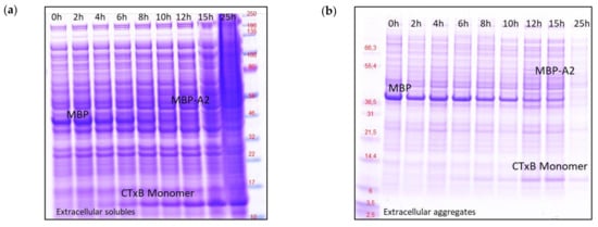
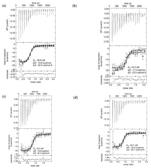
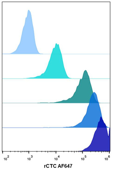
Non-toxic derivatives of the cholera toxin are extensively used in neuroscience, as neuronal tracers to reveal the location of cells in the central nervous system. They are, also, being developed as vaccine components and drug-delivery vehicles. Production of cholera-toxin derivatives is often non-reproducible; the quality and quantity require extensive fine-tuning to produce them in lab-scale settings. In our studies, we seek a resolution to this problem, by expanding the molecular toolbox of the Escherichia coli expression system with suitable production, purification, and offline analytics, to critically assess the quality of a probe or drug delivery, based on a non-toxic derivative of the cholera toxin. We present a re-engineered Cholera Toxin Complex (rCTC), wherein its toxic A1 domain was replaced with Maltose Binding Protein (MBP), as a model for an rCTC-based targeted-delivery vehicle. Here, we were able to improve the rCTC production by 11-fold (168 mg/L vs. 15 mg/L), in comparison to a host/vector combination that has been previously used (BL21(DE3) pTRBAB5-G1S). This 11-fold increase in the rCTC production capability was achieved by (1) substantial vector backbone modifications, (2) using Escherichia coli strains capable of growth-decoupling (V strains), (3) implementing a well-tuned fed-batch production protocol at a 1 L scale, and (4) testing the stability of the purified product. By an in-depth characterization of the production process, we revealed that secretion of rCTC across the E. coli Outer Membrane (OM) is processed by the Type II secretion-system general secretory pathway (gsp-operon) and that cholera toxin B-pentamerization is, likely, the rate-limiting step in complex formation. Upon successful manufacturing, we have validated the biological activity of rCTC, by measuring its binding affinity to its carbohydrate receptor GM1 oligosaccharide (Kd = 40 nM), or binding to Jurkat cells (93 pM) and delivering the cargo (MBP) in a retrograde fashion to the cell.
Full article
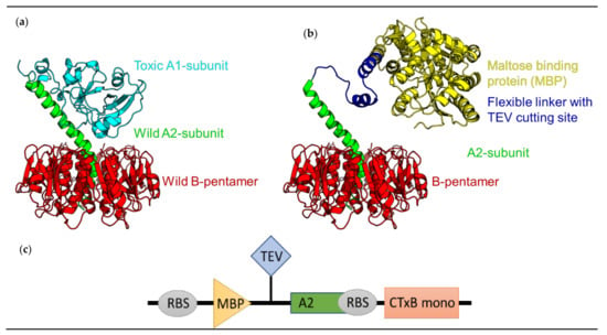
Figure 1
