


Toxins 2023, 15(10), 587; https://doi.org/10.3390/toxins15100587 - 23 Sep 2023
Cited by 1 | Viewed by 1124
Abstract
►
Show Figures
Okadaic acid (OA) and its analogues cause diarrhetic shellfish poisoning (DSP) in humans, and risk assessments of these toxins require toxicity equivalency factors (TEFs), which represent the relative toxicities of analogues. However, no human death by DSP toxin has been reported, and its
[...] Read more.
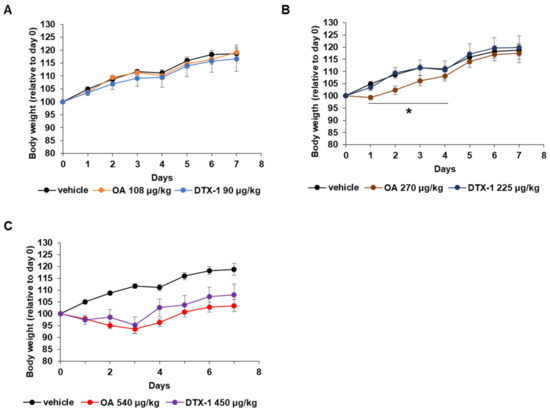
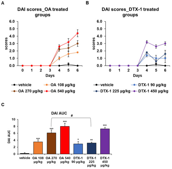
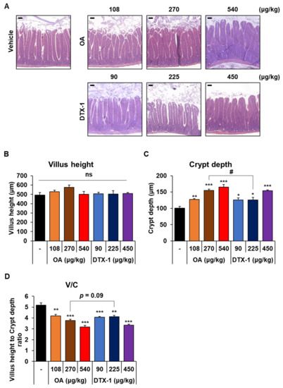
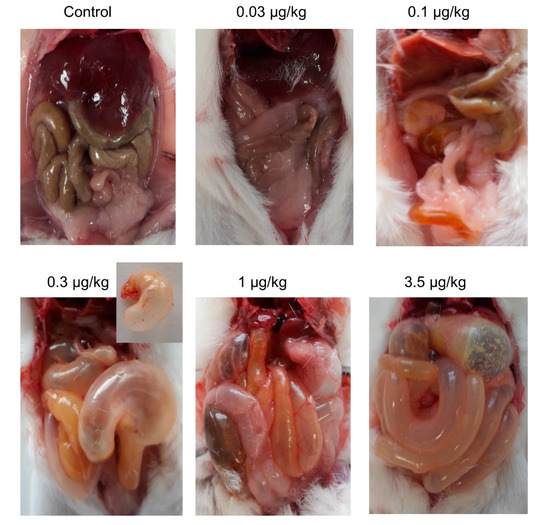
Okadaic acid (OA) and its analogues cause diarrhetic shellfish poisoning (DSP) in humans, and risk assessments of these toxins require toxicity equivalency factors (TEFs), which represent the relative toxicities of analogues. However, no human death by DSP toxin has been reported, and its current TEF value is based on acute lethality. To properly reflect the symptoms of DSP, such as diarrhea without death, the chronic toxicity of DSP toxins at sublethal doses should be considered. In this study, we obtained acute oral LD50 values for OA and dinophysistoxin-1 (DTX-1) (1069 and 897 μg/kg, respectively) to set sublethal doses. Mice were treated with sublethal doses of OA and DTX-1 for 7 days. The mice lost body weight, and the disease activity index and intestinal crypt depths increased. Furthermore, these changes were more severe in OA-treated mice than in the DTX-1-treated mice. Strikingly, ascites was observed, and its severity was greater in mice treated with OA. Our findings suggest that OA is at least as toxic as DTX-1 after repeated oral administration at a low dose. This is the first study to compare repeated oral dosing of DSP toxins. Further sub-chronic and chronic studies are warranted to determine appropriate TEF values for DSP toxins.
Full article
Figure 1
