Advanced Biomaterials for Cells Adhesion, Proliferation and Differentiation
A topical collection in Materials (ISSN 1996-1944). This collection belongs to the section "Biomaterials".
Viewed by 98172
Share This Topical Collection
Editor
 Prof. Dr. Guoping Chen
Prof. Dr. Guoping Chen
E-Mail
Website
Collection Editor
Research Center for Functional Materials, National Institute for Materials Science, 1-1 Namiki, Tsukuba, Ibaraki 3050044, Japan
Interests: scaffolds; porous materials; nanobiomaterials; biomimetic materials; extracellular matrices; composite materials; surface modification; micropatterning; hydrogels; tissue engineering
Topical Collection Information
Dear Colleagues,
Cells adhesion, proliferation and differentiation are involved in various natural phenomena, such as embryogenesis, histogenesis, maintenance of tissue structure, immune response, metastasis, wound healing, as well as tissue integration of biomaterial. Lack of native tissue integration is one of frequent problems associated with the biomaterials surfaces of dental implants. It has prompted a significant body of research regarding the modification of these surfaces. Therefore, there is a constant need to find advanced biomaterials which are very closely related to cell behaviors and particularly to cell adhesion, proliferation and differentiation, that aim to restore a patient’s mobility and alleviate pains.
Properties of biomaterials and scaffolds, such as pore structures, mechanical properties and degradation, play a pivotal role in accomplishment of their functions for tissue repairing or regeneration. Surface characteristics of materials, whether their topography, chemistry or surface energy, play an essential part in cell–material interaction and implant integration.
This Special Issue will focus on the recent progress of biopolymers, biometals, bioceramics, biomimetic materials, nanobiomaterials, scaffolds, porous materials, composite materials, smart biomaterials for cell responses in terms of adhesion, spreading, viability, proliferation and differentiation.
Prof. Dr. Guoping Chen
Guest Editor
Manuscript Submission Information
Manuscripts should be submitted online at www.mdpi.com by registering and logging in to this website. Once you are registered, click here to go to the submission form. Manuscripts can be submitted until the deadline. All submissions that pass pre-check are peer-reviewed. Accepted papers will be published continuously in the journal (as soon as accepted) and will be listed together on the collection website. Research articles, review articles as well as short communications are invited. For planned papers, a title and short abstract (about 100 words) can be sent to the Editorial Office for announcement on this website.
Submitted manuscripts should not have been published previously, nor be under consideration for publication elsewhere (except conference proceedings papers). All manuscripts are thoroughly refereed through a single-blind peer-review process. A guide for authors and other relevant information for submission of manuscripts is available on the Instructions for Authors page. Materials is an international peer-reviewed open access semimonthly journal published by MDPI.
Please visit the Instructions for Authors page before submitting a manuscript.
The Article Processing Charge (APC) for publication in this open access journal is 2600 CHF (Swiss Francs).
Submitted papers should be well formatted and use good English. Authors may use MDPI's
English editing service prior to publication or during author revisions.
Keywords
- Biopolymers
- biometals
- bioceramics
- biomimetic materials
- nanobiomaterials
- scaffolds
- porous materials
- composite materials
- smart biomaterials
- hydrogels
- extracellular matrices
- surface modification
- tissue engineering
- micropatterns
- nanopatterns
- regenerative medicine
- stem cells
- cell adhesion
- cell proliferation
- differentiation
- photothermal therapy
- cell–material interaction
- cell encapsulation
- biocompatibility
- biodegradation
Published Papers (24 papers)
Open AccessReview
Antimicrobial and Antiproliferative Coatings for Stents in Veterinary Medicine—State of the Art and Perspectives
by
Szymon Graczyk, Robert Pasławski, Arkadiusz Grzeczka, Urszula Pasławska, Beata Świeczko-Żurek, Klaudia Malisz, Ketul Popat, Alina Sionkowska, Patrycja Golińska and Mahendra Rai
Cited by 1 | Viewed by 1099
Abstract
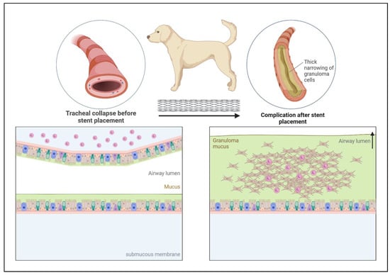
Microbial colonization in veterinary stents poses a significant and concerning issue in veterinary medicine. Over time, these pathogens, particularly bacteria, can colonize the stent surfaces, leading to various complications. Two weeks following the stent insertion procedure, the colonization becomes observable, with the aggressiveness
[...] Read more.
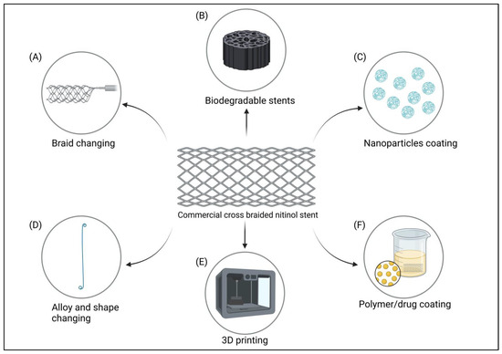
Microbial colonization in veterinary stents poses a significant and concerning issue in veterinary medicine. Over time, these pathogens, particularly bacteria, can colonize the stent surfaces, leading to various complications. Two weeks following the stent insertion procedure, the colonization becomes observable, with the aggressiveness of bacterial growth directly correlating with the duration of stent placement. Such microbial colonization can result in infections and inflammations, compromising the stent’s efficacy and, subsequently, the animal patient’s overall well-being. Managing and mitigating the impact of these pathogens on veterinary stents is a crucial challenge that veterinarians and researchers are actively addressing to ensure the successful treatment and recovery of their animal patients. In addition, irritation of the tissue in the form of an inserted stent can lead to overgrowth of granulation tissue, leading to the closure of the stent lumen, as is most often the case in the trachea. Such serious complications after stent placement require improvements in the procedures used to date. In this review, antibacterial or antibiofilm strategies for several stents used in veterinary medicine have been discussed based on the current literature and the perspectives have been drawn. Various coating strategies such as coating with hydrogel, antibiotic, or other antimicrobial agents have been reviewed.
Full article
►▼
Show Figures
Open AccessArticle
In Vitro Study of the Interaction of Innate Immune Cells with Liquid Silicone Rubber Coated with Zwitterionic Methyl Methacrylate and Thermoplastic Polyurethanes
by
Franziska Woitschach, Marlen Kloss, Karsten Schlodder, Alexander Borck, Niels Grabow, Emil C. Reisinger and Martina Sombetzki
Cited by 8 | Viewed by 1716
Abstract
The biocompatibility of medical devices, such as implants and prostheses, is strongly determined by the host’s immune response to the implanted material. Monocytes and macrophages are main actors of the so-called foreign body reaction. The innate immune system macrophages (M) can be broadly
[...] Read more.
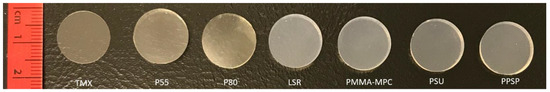
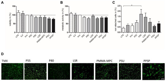
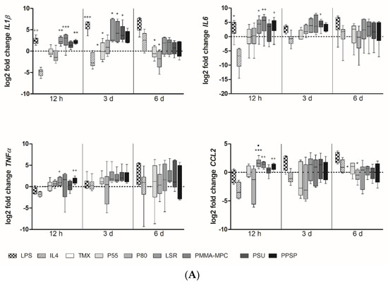
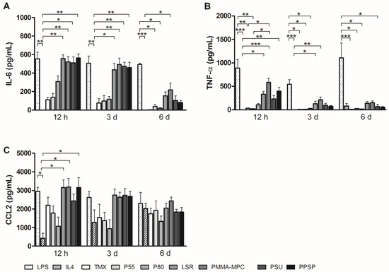
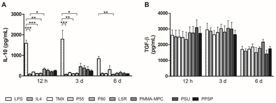
The biocompatibility of medical devices, such as implants and prostheses, is strongly determined by the host’s immune response to the implanted material. Monocytes and macrophages are main actors of the so-called foreign body reaction. The innate immune system macrophages (M) can be broadly classified into the pro-inflammatory M1-type and the anti-inflammatory, pro-healing M2-type. While a transient inflammatory initial state can be helpful during an infection, persistent inflammation interferes with proper healing and subsequent regeneration. The functional orientation of the immune response, mirrored by monocyte polarization, during interaction with different biomaterials has not yet been sufficiently explored. In implant manufacturing, thermoplastic polyurethane (TPU) represents the state-of-the-art material. The constantly growing areas of application and the associated necessary adaptations make the optimization of these materials indispensable. In the present study, modified liquid silicone rubber (LSR) were compared with two of the most commonly used TPUs, in terms of monocyte adhesion and M1/M2 polarization in vitro. Human monocytes isolated from venous blood were evaluated for their ability to adhere to various biomaterials, their gene expression profile, and their cytokine release. Based on the results, the different polymers exhibit different potential to bias monocytes with respect to early pro-inflammatory cytokine production and gene transcription. Furthermore, none of our test materials showed a clear trend towards M1 or M2 polarization. However, we were able to evaluate the inflammatory potential of the materials, with the classic TPUs appearing to be the most unreactive compared to the silicone-based materials.
Full article
►▼
Show Figures
Open AccessArticle
Adhesion and Growth of Neuralized Mouse Embryonic Stem Cells on Parylene-C/SiO2 Substrates
by
Alan F. Murray and Evangelos Delivopoulos
Cited by 1 | Viewed by 2079
Abstract
Neuronal patterning on microfabricated architectures has developed rapidly over the past few years, together with the emergence of soft biocompatible materials and tissue engineering scaffolds. Previously, we introduced a patterning technique based on serum and the biopolymer parylene-C, achieving highly compliant growth of
[...] Read more.
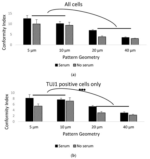
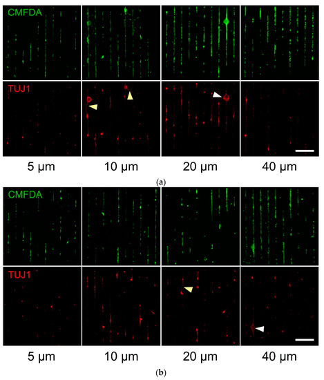
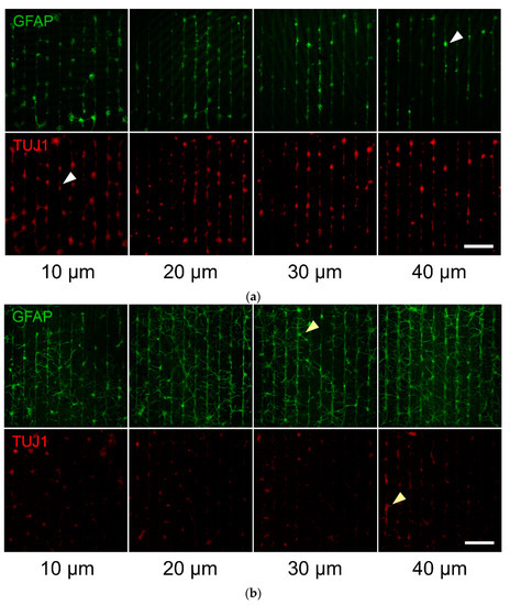
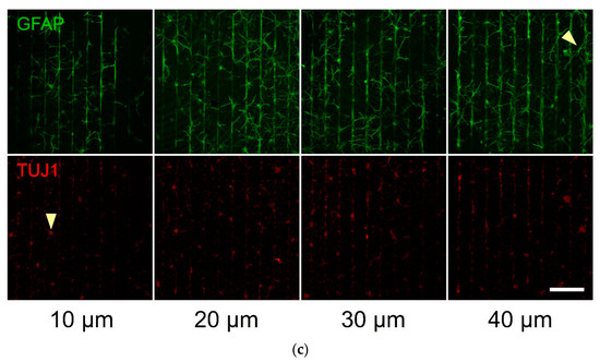
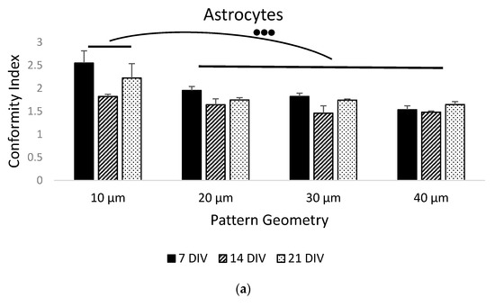
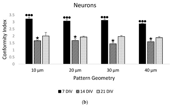
Neuronal patterning on microfabricated architectures has developed rapidly over the past few years, together with the emergence of soft biocompatible materials and tissue engineering scaffolds. Previously, we introduced a patterning technique based on serum and the biopolymer parylene-C, achieving highly compliant growth of primary neurons and astrocytes on different geometries. Here, we expanded this technique and illustrated that neuralized cells derived from mouse embryonic stem cells (mESCs) followed stripes of variable widths with conformity equal to or higher than that of primary neurons and astrocytes. Our results indicate the presence of undifferentiated mESCs, which also conformed to the underlying patterns to a high degree. This is an exciting and unexpected outcome, as molecular mechanisms governing cell and ECM protein interactions are different in stem cells and primary cells. Our study enables further investigations into the development and electrophysiology of differentiating patterned neural stem cells.
Full article
►▼
Show Figures
Open AccessArticle
A2BC-Type Porphyrin SAM on Gold Surface for Bacteria Detection Applications: Synthesis and Surface Functionalization
by
Laurie Neumann, Lea Könemund, Valentina Rohnacher, Annemarie Pucci, Hans-Hermann Johannes and Wolfgang Kowalsky
Cited by 2 | Viewed by 2047
Abstract
Currently used elaborate technologies for the detection of bacteria can be improved in regard to their time consumption, labor intensity, accuracy and reproducibility. Well-known electrical measurement methods might connect highly sensitive sensing systems with biological requirements. The development of modified sensor surfaces with
[...] Read more.
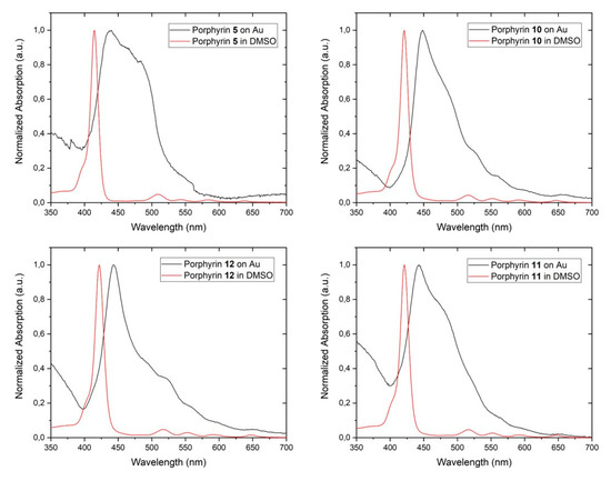
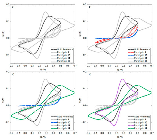
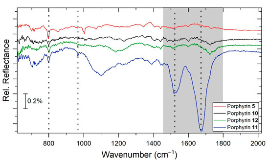
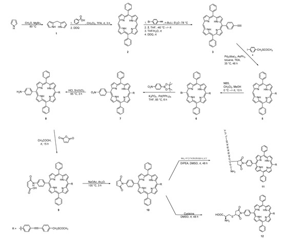
Currently used elaborate technologies for the detection of bacteria can be improved in regard to their time consumption, labor intensity, accuracy and reproducibility. Well-known electrical measurement methods might connect highly sensitive sensing systems with biological requirements. The development of modified sensor surfaces with self-assembled monolayers (SAMs) from functionalized porphyrin for bacteria trapping can lead to a highly sensitive sensor for bacteria detection. Different A
2BC-type porphyrin structures were synthesized and examined regarding their optical behavior. We achieved the synthesis of a porphyrin for SAM formation on a gold surface as electrode material. Two possible bio linkers were attached on the opposite
meso-position of the porphyrin, which allows the porphyrin to react as a linker on the surface for bacteria trapping. Different porphyrin structures were attached to a gold surface, the SAM formation and the respective coverage was investigated.
Full article
►▼
Show Figures
Open AccessArticle
Fibroblast-Like-Synoviocytes Mediate Secretion of Pro-Inflammatory Cytokines via ERK and JNK MAPKs in Ti-Particle-Induced Osteolysis
by
Ashish Ranjan Sharma, Supriya Jagga, Chiranjib Chakraborty and Sang-Soo Lee
Cited by 10 | Viewed by 2654
Abstract
Biomaterials are designed to replace and augment living tissues in order to provide functional support to skeletal deformities. However, wear debris produced from the interfaces of metal implants initiates inflammatory bone loss, causing periprosthetic osteolysis. Lately, fibroblast-like synoviocytes (FLS) have been shown to
[...] Read more.
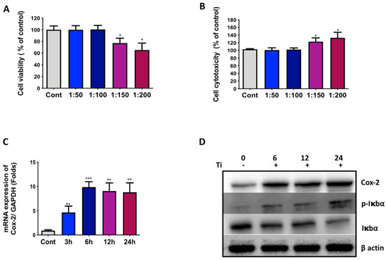
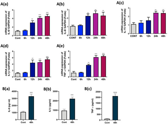
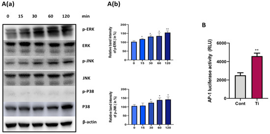
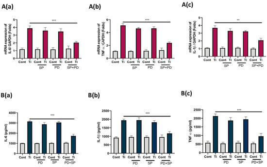
Biomaterials are designed to replace and augment living tissues in order to provide functional support to skeletal deformities. However, wear debris produced from the interfaces of metal implants initiates inflammatory bone loss, causing periprosthetic osteolysis. Lately, fibroblast-like synoviocytes (FLS) have been shown to play a role in wear-debris-induced osteolysis. Thus, here we have tried to understand the underlying mechanism of FLS involvement in wear-debris-induced osteolysis. Our results demonstrate that the effects of Ti particle (1:100 cell-to-Ti particle ratio) on FLS can induce Cox-2 expression and activate NFkB signaling. Moreover, the mRNA expression of pro-inflammatory cytokines such as IL-6, IL-8, IL-11, IL-1β, and TNFα was found to be elevated. However, among these pro-inflammatory cytokines, the mRNA and protein levels of only IL-6, IL-1β, and TNFα were found to be significantly higher. Ti particles activated extracellular signal-regulated kinase (ERK) and c-Jun N-terminal kinase (JNK) mitogen-activated protein kinases (MAPKs) as an early response in FLS. Co-inhibition of ERK and JNK signaling pathways by their specific inhibitors (PD9805 and SP600125, respectively) resulted in the suppression of mRNA and protein levels of IL-6, IL-1β, and TNFα in FLS. Taken together, targeting ERK and JNK MAPKs in FLS might provide a therapeutic option for reducing the secretion of bone-resorbing pro-inflammatory cytokines, thus preventing periprosthetic osteolysis.
Full article
►▼
Show Figures
Open AccessArticle
RGD-Dendrimer-Poly(L-lactic) Acid Nanopatterned Substrates for the Early Chondrogenesis of Human Mesenchymal Stromal Cells Derived from Osteoarthritic and Healthy Donors
by
Cristina Rodríguez-Pereira, Anna Lagunas, Ignasi Casanellas, Yolanda Vida, Ezequiel Pérez-Inestrosa, José A. Andrades, José Becerra, Josep Samitier, Francisco J. Blanco and Joana Magalhães
Cited by 2 | Viewed by 5485
Abstract
Aiming to address a stable chondrogenesis derived from mesenchymal stromal cells (MSCs) to be applied in cartilage repair strategies at the onset of osteoarthritis (OA), we analyzed the effect of arginine–glycine–aspartate (RGD) density on cell condensation that occurs during the initial phase of
[...] Read more.
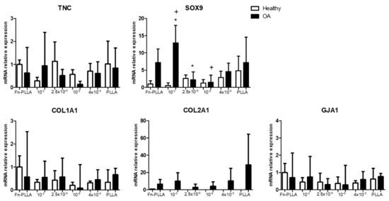
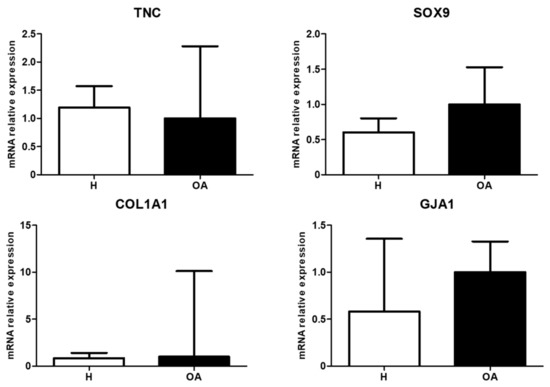
Aiming to address a stable chondrogenesis derived from mesenchymal stromal cells (MSCs) to be applied in cartilage repair strategies at the onset of osteoarthritis (OA), we analyzed the effect of arginine–glycine–aspartate (RGD) density on cell condensation that occurs during the initial phase of chondrogenesis. For this, we seeded MSC-derived from OA and healthy (H) donors in RGD-dendrimer-poly(L-lactic) acid (PLLA) nanopatterned substrates (RGD concentrations of 4 × 10
−9, 10
−8, 2.5 × 10
−8, and 10
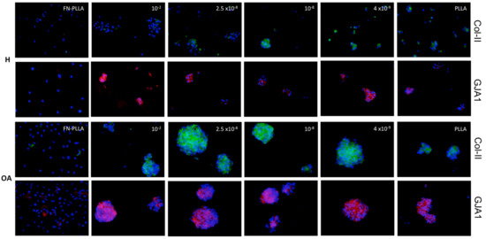
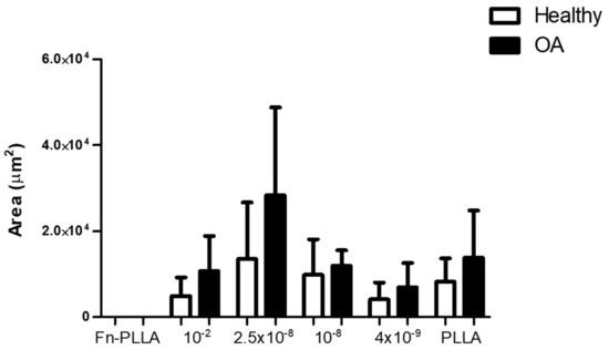
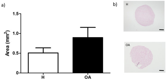
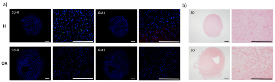
−2 w/w), during three days and compared to a cell pellet conventional three-dimensional culture system. Molecular gene expression (collagens type-I and II–COL1A1 and COL2A1, tenascin-TNC, sex determining region Y-box9-SOX9, and gap junction protein alpha 1–GJA1) was determined as well as the cell aggregates and pellet size, collagen type-II and connexin 43 proteins synthesis. This study showed that RGD-tailored first generation dendrimer (RGD-Cys-D1) PLLA nanopatterned substrates supported the formation of pre-chondrogenic condensates from OA- and H-derived human bone marrow-MSCs with enhanced chondrogenesis regarding the cell pellet conventional system (presence of collagen type-II and connexin 43, both at the gene and protein level). A RGD-density dependent trend was observed for aggregates size, in concordance with previous studies. Moreover, the nanopatterns’ had a higher effect on OA-derived MSC morphology, leading to the formation of bigger and more compact aggregates with improved expression of early chondrogenic markers.
Full article
►▼
Show Figures
Open AccessArticle
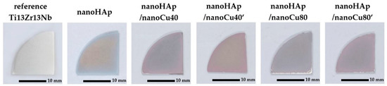
Properties of Nanohydroxyapatite Coatings Doped with Nanocopper, Obtained by Electrophoretic Deposition on Ti13Zr13Nb Alloy
by
Michał Bartmański, Łukasz Pawłowski, Gabriel Strugała, Aleksandra Mielewczyk-Gryń and Andrzej Zieliński
Cited by 29 | Viewed by 2868
Abstract
Nowadays, hydroxyapatite coatings are the most common surface modification of long-term implants. These coatings are characterized by high thickness and poor adhesion to the metallic substrate. The present research is aimed at characterizing the properties of nanohydroxyapatite (nanoHAp) with the addition of copper
[...] Read more.
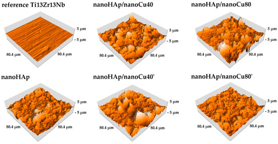
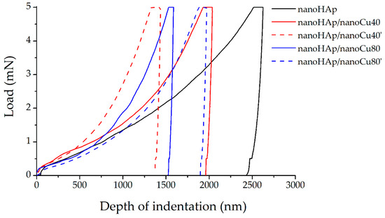
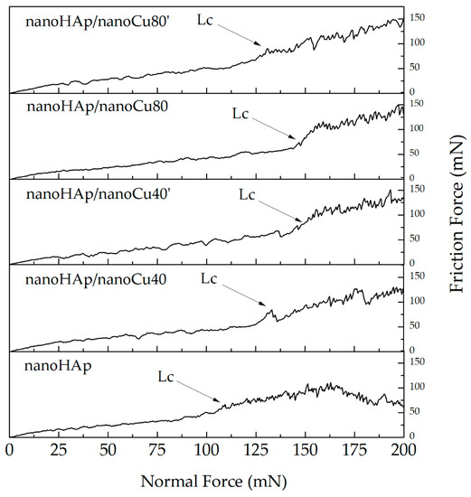
Nowadays, hydroxyapatite coatings are the most common surface modification of long-term implants. These coatings are characterized by high thickness and poor adhesion to the metallic substrate. The present research is aimed at characterizing the properties of nanohydroxyapatite (nanoHAp) with the addition of copper nanoparticle (nanoCu) coatings deposited on the Ti13Zr13Nb alloy by an electrophoresis process. The deposition of coatings was carried out for various amounts of nanoCu powder and various average particle sizes. Microstructure, topography, phase, and chemical composition were examined with scanning electron microscopy, atomic force microscopy, and X-ray diffraction. Corrosion properties were determined by potentiodynamic polarization technique in simulated body fluid. Nanomechanical properties were determined based on nanoindentation and scratch tests. The wettability of coatings was defined by the contact angle. It was proven that nanoHAp coatings containing nanocopper, compared to nanoHAp coatings without nanometals, demonstrated smaller number of cracks, lower thickness, and higher nanomechanical properties. The influence of the content and the average size of nanoCu on the quality of the coatings was observed. All coatings exhibited hydrophilic properties. The deposition of nanohydroxyapatite coatings doped with nanocopper may be a promising way to improve the antibacterial properties and mechanical stability of coatings.
Full article
►▼
Show Figures
Open AccessArticle
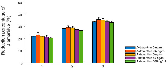
The Influence of Astaxanthin on the Proliferation of Adipose-derived Mesenchymal Stem Cells in Gelatin-Methacryloyl (GelMA) Hydrogels
by
Bo Young Choi, Elna Paul Chalisserry, Myoung Hwan Kim, Hyun Wook Kang, Il-Whan Choi and Seung Yun Nam
Cited by 10 | Viewed by 4756
Abstract
Recently, astaxanthin, a red lipophilic pigment belonging to the xanthophyllic family of carotenoids, has shown the feasibility of its uses in tissue engineering and regenerative medicine, due to its excellent antioxidant activities and its abilities to enhance the self-renewal potency of stem cells.
[...] Read more.
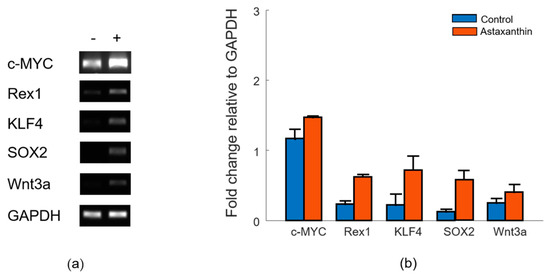
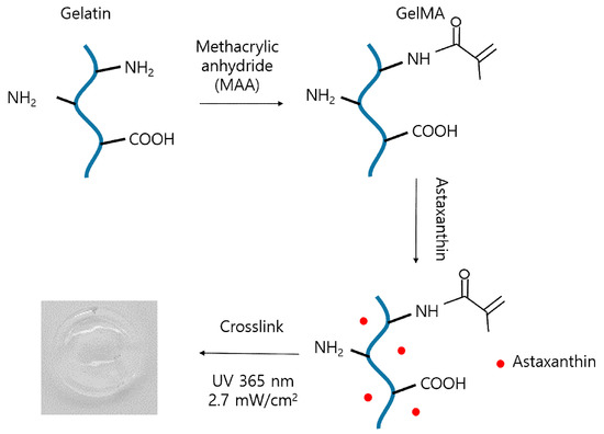
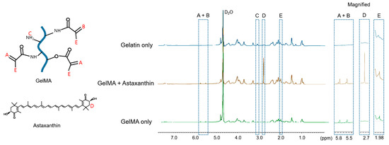
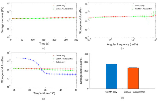
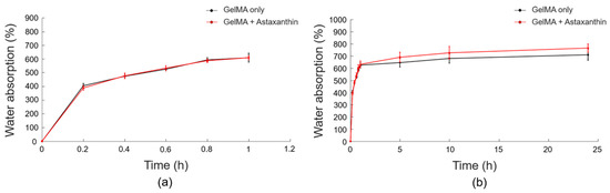
Recently, astaxanthin, a red lipophilic pigment belonging to the xanthophyllic family of carotenoids, has shown the feasibility of its uses in tissue engineering and regenerative medicine, due to its excellent antioxidant activities and its abilities to enhance the self-renewal potency of stem cells. In this study, we demonstrate the influence of astaxanthin on the proliferation of adipose-derived mesenchymal stem cells in tissue-engineered constructs. The tissue engineered scaffolds were fabricated using photopolymerizable gelatin methacryloyl (GelMA) with different concentrations of astaxanthin. The effects of astaxanthin on cellular proliferation in two-dimensional environments were assessed using alamar blue assay and reverse transcription polymerase chain reaction (RT-PCR). Then, rheological properties, chemical structures and the water absorption of the fabricated astaxanthin-incorporated GelMA hydrogels were characterized using NMR analysis, rheological analysis and a swelling ratio test. Finally, the influence in three-dimensional environments of astaxanthin-incorporated GelMA hydrogels on the proliferative potentials of adipose-derived stem cells was assessed using alamar blue assay and the confocal imaging with Live/dead staining. The experimental results of the study indicate that an addition of astaxanthin promises to induce stem cell potency via proliferation, and that it can be a useful tool for a three-dimensional culture system and various tissue engineering applications.
Full article
►▼
Show Figures
Open AccessReview
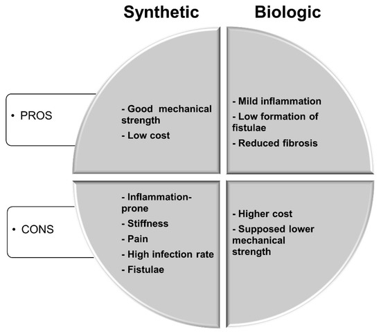
Biological Scaffolds for Abdominal Wall Repair: Future in Clinical Application?
by
Alessandra Costa, Sergio Adamo, Francesco Gossetti, Linda D’Amore, Francesca Ceci, Paolo Negro and Paolo Bruzzone
Cited by 31 | Viewed by 6445
Abstract
Millions of abdominal wall repair procedures are performed each year for primary and incisional hernias both in the European Union and in the United States with extremely high costs. Synthetic meshes approved for augmenting abdominal wall repair provide adequate mechanical support but have
[...] Read more.
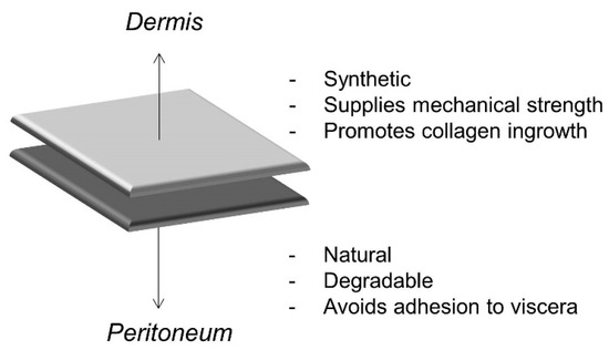
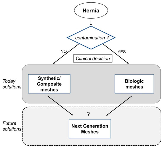
Millions of abdominal wall repair procedures are performed each year for primary and incisional hernias both in the European Union and in the United States with extremely high costs. Synthetic meshes approved for augmenting abdominal wall repair provide adequate mechanical support but have significant drawbacks (seroma formation, adhesion to viscera, stiffness of abdominal wall, and infection). Biologic scaffolds (i.e., derived from naturally occurring materials) represent an alternative to synthetic surgical meshes and are less sensitive to infection. Among biologic scaffolds, extracellular matrix scaffolds promote stem/progenitor cell recruitment in models of tissue remodeling and, in the specific application of abdominal wall repair, have enough mechanical strength to support the repair. However, many concerns remain about the use of these scaffolds in the clinic due to their higher cost of production compared with synthetic meshes, despite having the same recurrence rate. The present review aims to highlight the pros and cons of using biologic scaffolds as surgical devices for abdominal wall repair and present possible improvements to widen their use in clinical practice.
Full article
►▼
Show Figures
Open AccessArticle
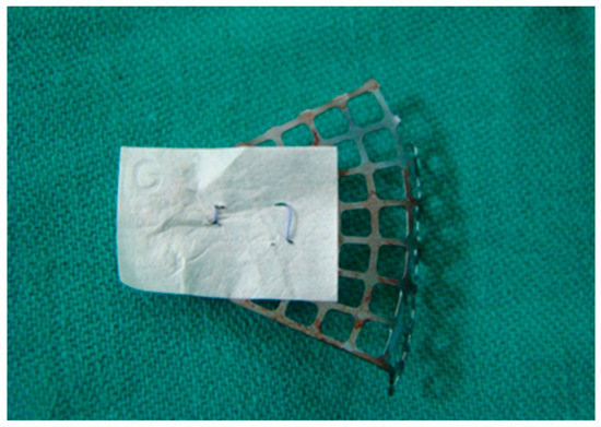
Periorbital Reconstruction by “Periorbital Patch” Technique Using a Pericardium-Based Collagen Membrane and Titanium Mesh
by
Nenad Tanaskovic, Branko Trajkovski, Željka Perić Kačarević, Patrick M. Rider, Alireza Houshmand, Xin Xiong, Ole Jung and Mike Barbeck
Cited by 8 | Viewed by 4178
Abstract
Objective: Titanium mesh is a commonly used material for the reconstruction of orbital floor fractures. However, in some instances, a subsequent inflammatory reaction can occur that causes the adhesion of orbital tissue to the titanium mesh. The adhesion of the orbital soft tissue
[...] Read more.
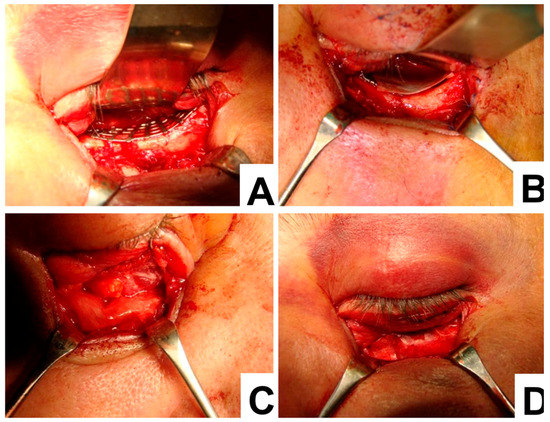
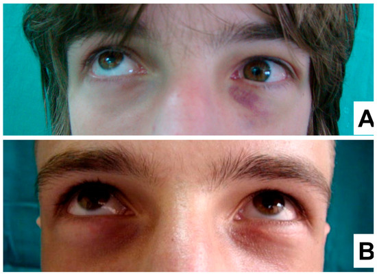
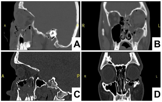
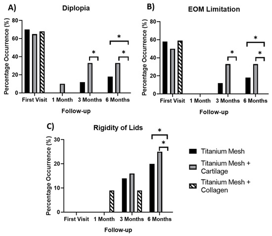
Objective: Titanium mesh is a commonly used material for the reconstruction of orbital floor fractures. However, in some instances, a subsequent inflammatory reaction can occur that causes the adhesion of orbital tissue to the titanium mesh. The adhesion of the orbital soft tissue to the mesh causes diplopia, lid rigidity and extraocular movements restriction. This study was performed to determine if the placement of a collagen membrane over a titanium mesh can prevent the adhesion of orbital soft tissue for an improved clinical outcome. Clinical considerations: A case study was performed investigating 106 patients undergoing a periorbital restoration. Seventy-two patients received a titanium mesh without a barrier membrane, 12 patients received a barrier membrane composed of autologous auricular cartilage to provide a barrier function and 22 patients received a pericardium collagen membrane and titanium mesh. Conclusions: Titanium has been shown to generate an intense inflammatory reaction in host tissues, which can cause fibrosis to adjacent structures. Fibrosis is an essential factor in the repair of fracture sites, however this can lead to adverse effects in the orbital socket. Fibrosis can cause cicatrization and lower eyelid retraction when induced along the lower orbital rim. An improved outcome can be achieved by using a barrier between the titanium mesh and the soft tissue, such as autogenous auricular cartilage, however, only patients treated with a resorbable collagen membrane to act as a soft tissue barricade during site regeneration, prevented the fibrosis reaction and related problems from occurring.
Full article
►▼
Show Figures
Open AccessArticle
Engineering of Chitosan-Hydroxyapatite-Magnetite Hierarchical Scaffolds for Guided Bone Growth
by
Alessandro Pistone, Daniela Iannazzo, Consuelo Celesti, Elpida Piperopoulos, Deepu Ashok, Arianna Cembran, Antonio Tricoli and David Nisbet
Cited by 36 | Viewed by 4257
Abstract
Bioabsorbable materials have received increasing attention as innovative systems for the development of osteoconductive biomaterials for bone tissue engineering. In this paper, chitosan-based composites were synthesized adding hydroxyapatite and/or magnetite in a chitosan matrix by in situ precipitation technique. Composites were characterized by
[...] Read more.
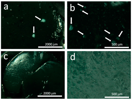
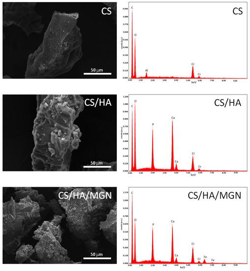
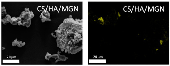
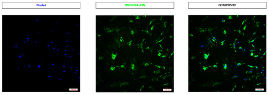
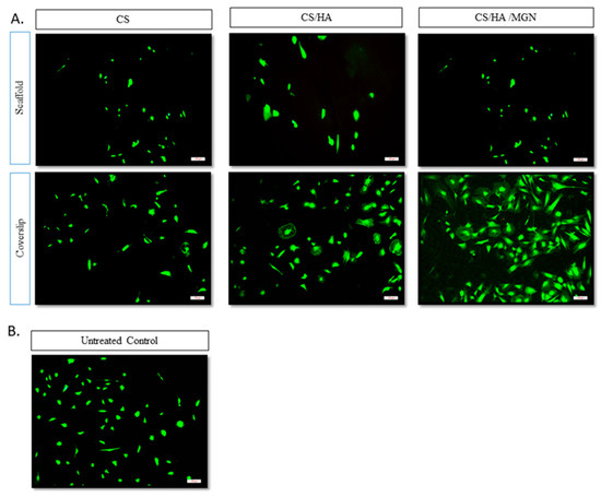
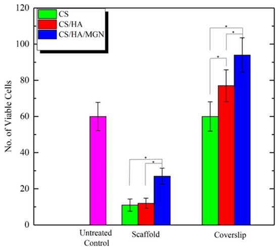
Bioabsorbable materials have received increasing attention as innovative systems for the development of osteoconductive biomaterials for bone tissue engineering. In this paper, chitosan-based composites were synthesized adding hydroxyapatite and/or magnetite in a chitosan matrix by in situ precipitation technique. Composites were characterized by optical and electron microscopy, thermogravimetric analyses (TGA), x-ray diffraction (XRD), and in vitro cell culture studies. Hydroxyapatite and magnetite were found to be homogeneously dispersed in the chitosan matrix and the composites showed superior biocompatibility and the ability to support cell attachment and proliferation; in particular, the chitosan/hydroxyapatite/magnetite composite (CS/HA/MGN) demonstrated superior bioactivity with respect to pure chitosan (CS) and to the chitosan/hydroxyapatite (CS/HA) scaffolds.
Full article
►▼
Show Figures
Open AccessReview
Oral Bone Tissue Engineering: Advanced Biomaterials for Cell Adhesion, Proliferation and Differentiation
by
Alexandra Roi, Lavinia Cosmina Ardelean, Ciprian Ioan Roi, Eugen-Radu Boia, Simina Boia and Laura-Cristina Rusu
Cited by 26 | Viewed by 4172
Abstract
The advancements made in biomaterials have an important impact on oral tissue engineering, especially on the bone regeneration process. Currently known as the gold standard in bone regeneration, grafting procedures can sometimes be successfully replaced by a biomaterial scaffold with proper characteristics. Whether
[...] Read more.
The advancements made in biomaterials have an important impact on oral tissue engineering, especially on the bone regeneration process. Currently known as the gold standard in bone regeneration, grafting procedures can sometimes be successfully replaced by a biomaterial scaffold with proper characteristics. Whether natural or synthetic polymers, biomaterials can serve as potential scaffolds with major influences on cell adhesion, proliferation and differentiation. Continuous research has enabled the development of scaffolds that can be specifically designed to replace the targeted tissue through changes in their surface characteristics and the addition of growth factors and biomolecules. The progress in tissue engineering is incontestable and research shows promising contributions to the further development of this field. The present review aims to outline the progress in oral tissue engineering, the advantages of biomaterial scaffolds, their direct implication in the osteogenic process and future research directions.
Full article
►▼
Show Figures
Open AccessArticle
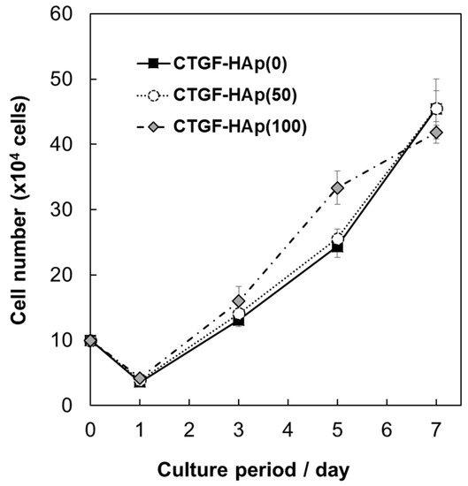
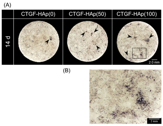
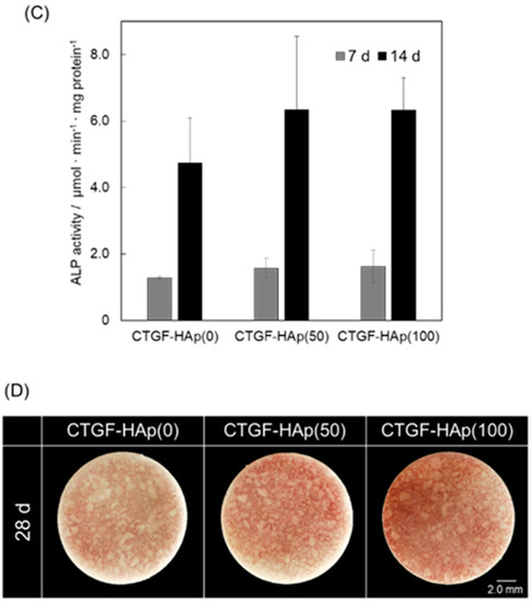
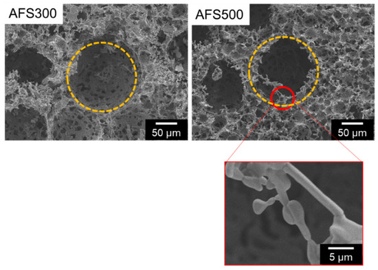
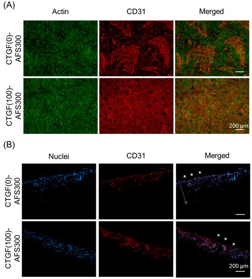
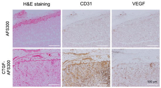
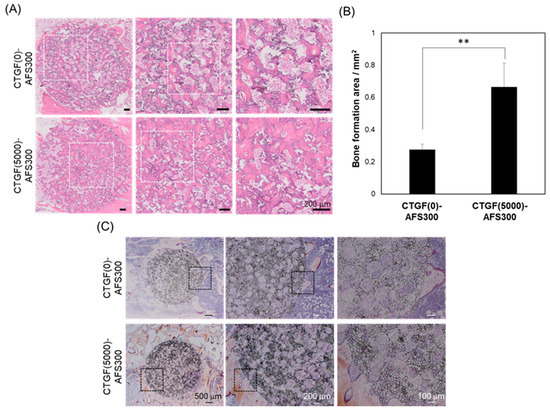
Acceleration of Osteogenesis via Stimulation of Angiogenesis by Combination with Scaffold and Connective Tissue Growth Factor
by
Michiyo Honda, Ryo Hariya, Morio Matsumoto and Mamoru Aizawa
Cited by 17 | Viewed by 3559
Abstract
In bone regeneration, there are some important cellular biological processes, such as mineralization, cell organization, and differentiation. In particular, vascularization into regenerative tissues is a key step for the survival of cells and tissues. In this study, to fabricate biomimetic-engineered bone, including vascular
[...] Read more.
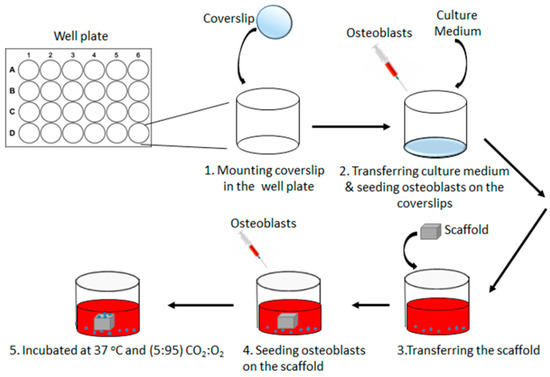
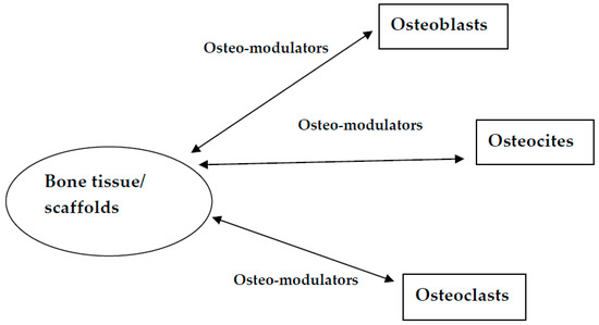
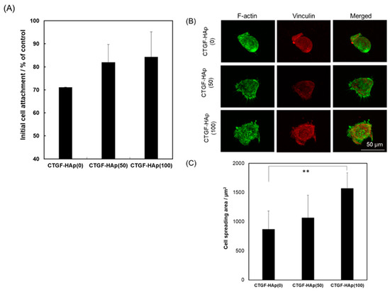
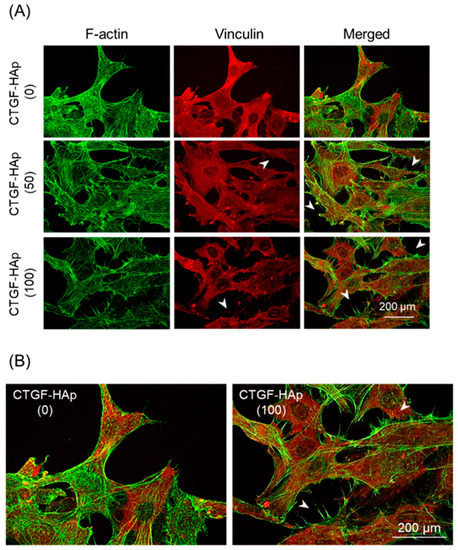
In bone regeneration, there are some important cellular biological processes, such as mineralization, cell organization, and differentiation. In particular, vascularization into regenerative tissues is a key step for the survival of cells and tissues. In this study, to fabricate biomimetic-engineered bone, including vascular networks, we focused on connective tissue growth factor (CTGF), a multifunctional protein which could regulate the extracellular matrix remodeling. By combination with CTGF and hydroxyapatite (HAp) ceramics (2D) or apatite-fiber scaffold (AFS, 3D), we have fabricated bioactive materials. The CTGF-loaded HAp ceramics could enhance the cellular attachment through interaction with integrin and promote actin cytoskeletal reorganization. CTGF-loaded HAp also enhanced the differentiation of osteoblasts by integrin-mediated activation of the signaling pathway. Under co-culture conditions, both osteoblasts and endothelial cells in the CTGF-loaded AFS were stimulated by CTGF, and each cell could penetrate the central region of the scaffold in vitro and in vivo. Direct cell-cell interaction would also improve the functionality of cells in bone formation. These results suggest that coupling between effective optimized scaffold and CTGF with multifunction could provide better mimicking natural bone by stimulation of angiogenesis.
Full article
►▼
Show Figures
Open AccessArticle
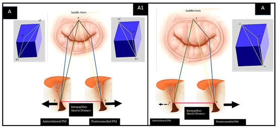
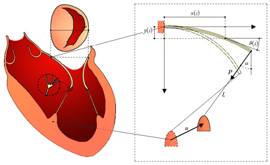
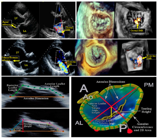
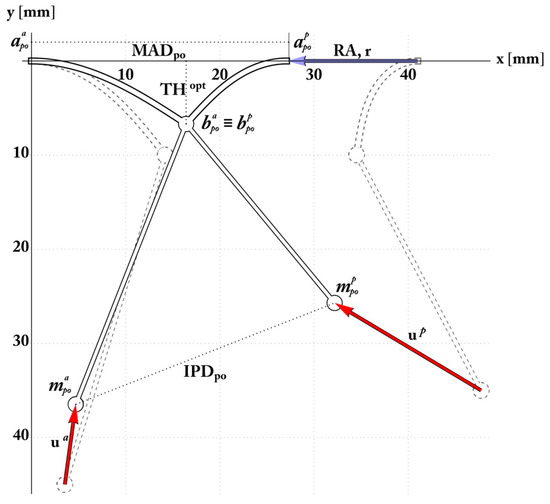
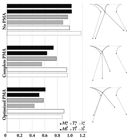
Euler’s Elastica-Based Biomechanics of the Papillary Muscle Approximation in Ischemic Mitral Valve Regurgitation: A Simple 2D Analytical Model
by
Francesco Nappi, Angelo Rosario Carotenuto, Sanjeet Singh Avtaar Singh, Christos Mihos and Massimiliano Fraldi
Cited by 16 | Viewed by 3179
Abstract
Ischemic mitral regurgitation (IMR) occurs as an adverse consequence of left ventricle remodeling post-myocardial infarction. A change in mitral valve configuration with an imbalance between closing and tethering forces underlie this pathological condition. These abnormalities lead to impaired leaflet coaptation and a variable
[...] Read more.
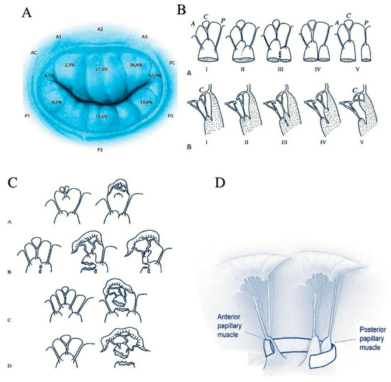
Ischemic mitral regurgitation (IMR) occurs as an adverse consequence of left ventricle remodeling post-myocardial infarction. A change in mitral valve configuration with an imbalance between closing and tethering forces underlie this pathological condition. These abnormalities lead to impaired leaflet coaptation and a variable degree of mitral regurgitation, which can in turn influence the ventricular filling status, the heart rhythm and the afterload regardless of the residual ischemic insult. The IMR correction can be pursued through under-sizing mitral annuloplasty and papillary muscle approximation to restore the mitral valve and left ventricle physiological geometry to, consequently, achieve normalization of the engaged physical forces. Because the structures involved undergo extremely large deformations, a biomechanics model based on the Euler’s Elastica –the mitral leaflet– interlaced with nonlinear chordae tendineae anchored on papillary muscles has been constructed to elucidate the interactions between closing and tethering forces. The model takes into account the actual updated geometrical and mechanical features of the valvular and subvalvular apparatuses in physiological and IMR conditions, as well as in case of papillary muscle approximation, finally furnishing ad hoc geometry-based mathematical relations that could be utilised to support—and optimize—the relevant choices in cardiac surgery.
Full article
►▼
Show Figures
Open AccessReview
Decellularized Extracellular Matrix for Cancer Research
by
Takashi Hoshiba
Cited by 68 | Viewed by 6309
Abstract
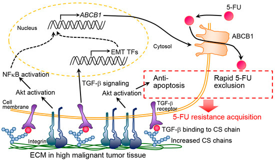
Genetic mutation and alterations of intracellular signaling have been focused on to understand the mechanisms of oncogenesis and cancer progression. Currently, it is pointed out to consider cancer as tissues. The extracellular microenvironment, including the extracellular matrix (ECM), is important for the regulation
[...] Read more.
Genetic mutation and alterations of intracellular signaling have been focused on to understand the mechanisms of oncogenesis and cancer progression. Currently, it is pointed out to consider cancer as tissues. The extracellular microenvironment, including the extracellular matrix (ECM), is important for the regulation of cancer cell behavior. To comprehensively investigate ECM roles in the regulation of cancer cell behavior, decellularized ECM (dECM) is now used as an in vitro ECM model. In this review, I classify dECM with respect to its sources and summarize the preparation and characterization methods for dECM. Additionally, the examples of cancer research using the dECM were introduced. Finally, future perspectives of cancer studies with dECM are described in the conclusions.
Full article
►▼
Show Figures
Open AccessArticle
Temporary Wettability Tuning of PCL/PDMS Micro Pattern Using the Plasma Treatments
by
Wei-Chih Lin and Nur Adila Mohd Razali
Cited by 23 | Viewed by 4450
Abstract
Surface wettability plays an important role in determining the function of a wound dressing. Dressings with hydrophobic surfaces are suitable for bacterial adsorption, however, a hydrophilic surface is needed to improve cell attachment for most anchorage-dependent cell types. Furthermore, the hydrophobicity/hydrophilicity of the
[...] Read more.
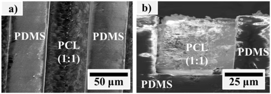
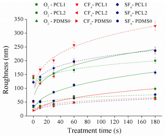
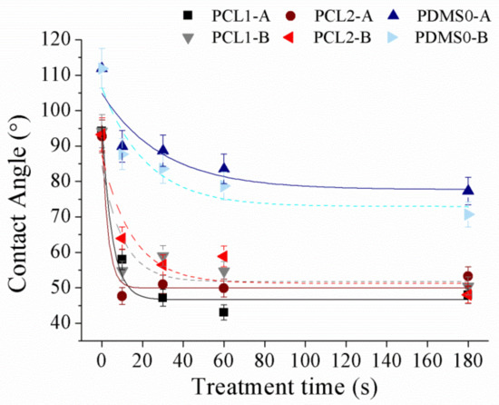
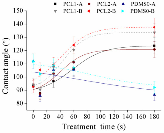
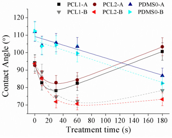
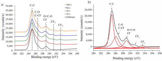
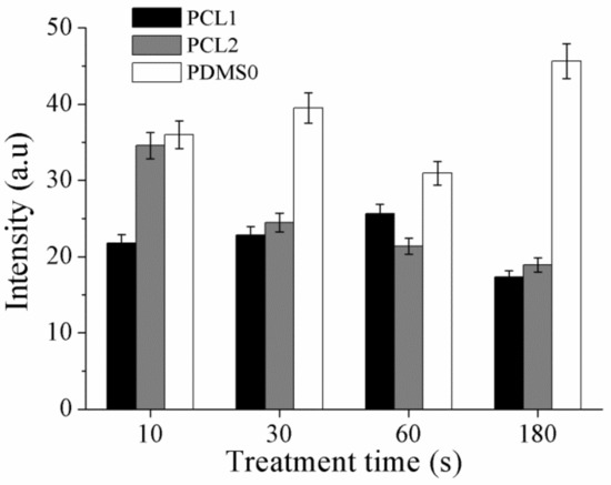
Surface wettability plays an important role in determining the function of a wound dressing. Dressings with hydrophobic surfaces are suitable for bacterial adsorption, however, a hydrophilic surface is needed to improve cell attachment for most anchorage-dependent cell types. Furthermore, the hydrophobicity/hydrophilicity of the surface can be used to direct cellular processes such as cell initial attachment, adhesion, and migration during wound healing. Thus, a surface with an ability to switch their surface wettability improves the practicality of the dressing. In this study, we propose a temporary surface wettability tuning for surface patterning utilizing plasma treatment. Polycaprolactone (PCL) and polydimethylsiloxane (PDMS) surfaces were treated with tetrafluoromethane (CF
4), sulphur hexafluoride (SF
6), and oxygen (O
2) plasma, and the effects on the surface wettability, roughness, and chemical composition were investigated. Based on the contact angle measurement, CF
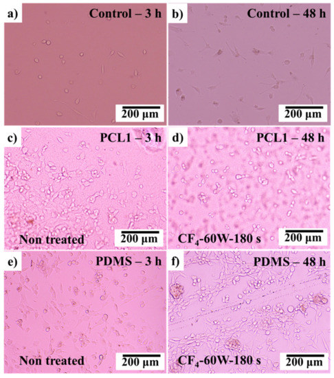
4 plasma altered surface wettability of PCL and PDMS films to hydrophobic and hydrophilic, respectively. After CF
4 treatment, better attachment of primary mouse embryonic fibroblast cell (3T3) was observed on the treated PDMS surface. Embedding PCL into PDMS generated a hydrophobic-hydrophilic pattern mixture surface, which offers great potential in the tissue engineering field such as cell patterning and guidance.
Full article
►▼
Show Figures
Open AccessArticle
Biocompatibility Characteristics of Titanium Coated with Multi Walled Carbon Nanotubes—Hydroxyapatite Nanocomposites
by
Jung-Eun Park, Yong-Seok Jang, Tae-Sung Bae and Min-Ho Lee
Cited by 20 | Viewed by 3439
Abstract
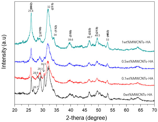
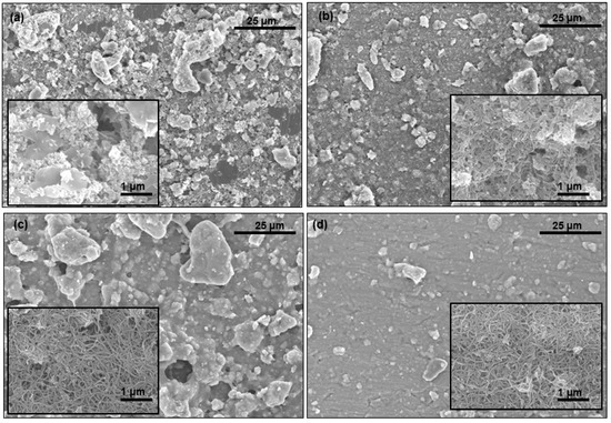
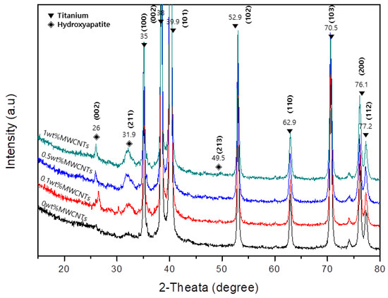
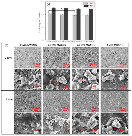
Multi walled carbon nanotubes-hydroxyapatite (MWCNTs-HA) with various contents of MWCNTs was synthesized using the sol-gel method. MWCNTs-HA composites were characterized by X-ray diffraction (XRD) and transmission electron microscopy (TEM). HA particles were generated on the surface of MWCNT. Produced MWCNTs-HA nanocomposites were coated
[...] Read more.
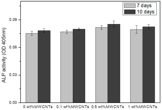
Multi walled carbon nanotubes-hydroxyapatite (MWCNTs-HA) with various contents of MWCNTs was synthesized using the sol-gel method. MWCNTs-HA composites were characterized by X-ray diffraction (XRD) and transmission electron microscopy (TEM). HA particles were generated on the surface of MWCNT. Produced MWCNTs-HA nanocomposites were coated on pure titanium (PT). Characteristic of the titanium coated MWCNTs-HA was evaluated by field-emission scanning electron microscopy (FE-SEM) and XRD. The results show that the titanium surface was covered with MWCNTs-HA nanoparticles and MWCNTs help form the crystalized hydroxyapatite. Furthermore, the MWCNTs-HA coated titanium was investigated for in vitro cellular responses. Cell proliferation and differentiation were improved on the surface of MWCNT-HA coated titanium.
Full article
►▼
Show Figures
Open AccessArticle
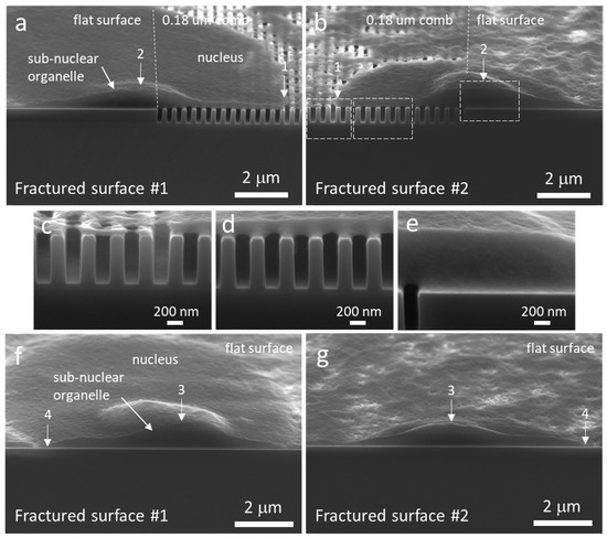
What’s Happening on the Other Side? Revealing Nano-Meter Scale Features of Mammalian Cells on Engineered Textured Tantalum Surfaces
by
Ting Y. Tsui, Megan Logan, Hassan I. Moussa and Marc G. Aucoin
Cited by 3 | Viewed by 4481
Abstract
Advanced engineered surfaces can be used to direct cell behavior. These behaviors are typically characterized using either optical, atomic force, confocal, or electron microscopy; however, most microscopic techniques are generally restricted to observing what’s happening on the “top” side or even the interior
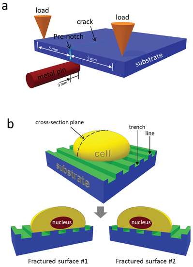
[...] Read more.
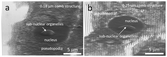
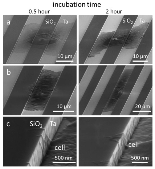
Advanced engineered surfaces can be used to direct cell behavior. These behaviors are typically characterized using either optical, atomic force, confocal, or electron microscopy; however, most microscopic techniques are generally restricted to observing what’s happening on the “top” side or even the interior of the cell. Our group has focused on engineered surfaces typically reserved for microelectronics as potential surfaces to control cell behavior. These devices allow the exploration of novel substrates including titanium, tungsten, and tantalum intermixed with silicon oxide. Furthermore, these devices allow the exploration of the intricate patterning of surface materials and surface geometries i.e., trenches. Here we present two important advancements in our research: (1) the ability to split a fixed cell through the nucleus using an inexpensive three-point bend micro-cleaving technique and image 3D nanometer scale cellular components using high-resolution scanning electron microscopy; and (2) the observation of nanometer projections from the underbelly of a cell as it sits on top of patterned trenches on our devices. This application of a 3-point cleaving technique to visualize the underbelly of the cell is allowing a new understanding of how cells descend into surface cavities and is providing a new insight on cell migration mechanisms.
Full article
►▼
Show Figures
Open AccessArticle
Photothermal Ablation of Cancer Cells by Albumin-Modified Gold Nanorods and Activation of Dendritic Cells
by
Xiuhui Wang, Jingchao Li, Naoki Kawazoe and Guoping Chen
Cited by 24 | Viewed by 4825
Abstract
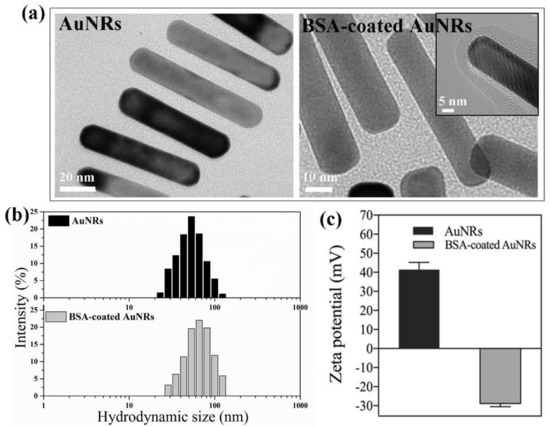
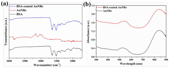
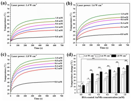
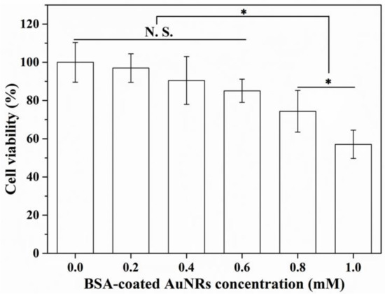
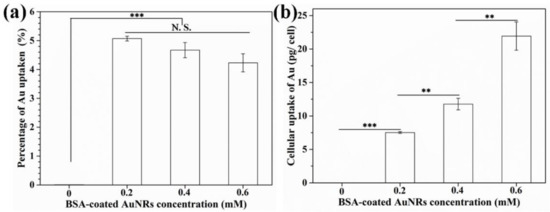
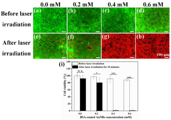
Nanoparticle-mediated photothermal therapy has been widely studied for cancer treatment. It is important to disclose how photothermally ablated tumor cells trigger immune responses. In this study, bovine serum albumin (BSA)-coated gold nanorods (BSA-coated AuNRs) were prepared and used for photothermal ablation of breast
[...] Read more.
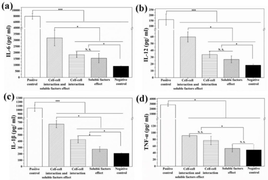
Nanoparticle-mediated photothermal therapy has been widely studied for cancer treatment. It is important to disclose how photothermally ablated tumor cells trigger immune responses. In this study, bovine serum albumin (BSA)-coated gold nanorods (BSA-coated AuNRs) were prepared and used for photothermal ablation of breast tumor cells. The BSA-coated AuNRs showed high photothermal conversion efficiency and good photothermal ablation effect towards tumor cells. The ablated tumor cells were co-cultured with immature dendritic cells (DCs) through a direct cell contacting model and diffusion model to confirm the stimulatory effects of cell–cell interaction and soluble factors released from ablated tumor cells. The results indicated that photothermally ablated tumor cells induced immune-stimulatory responses of DCs through both cell–cell interaction and soluble factors. The results should be useful for synergistic photothermal-immunotherapy of primary and metastatic cancer.
Full article
►▼
Show Figures
Open AccessArticle
Encapsulation of Rat Bone Marrow Derived Mesenchymal Stem Cells in Alginate Dialdehyde/Gelatin Microbeads with and without Nanoscaled Bioactive Glass for In Vivo Bone Tissue Engineering
by
Ulrike Rottensteiner-Brandl, Rainer Detsch, Bapi Sarker, Lara Lingens, Katrin Köhn, Ulrich Kneser, Anja K. Bosserhoff, Raymund E. Horch, Aldo R. Boccaccini and Andreas Arkudas
Cited by 19 | Viewed by 5405
Abstract
Alginate dialdehyde (ADA), gelatin, and nano-scaled bioactive glass (nBG) particles are being currently investigated for their potential use as three-dimensional scaffolding materials for bone tissue engineering. ADA and gelatin provide a three-dimensional scaffold with properties supporting cell adhesion and proliferation. Combined with nanocristalline
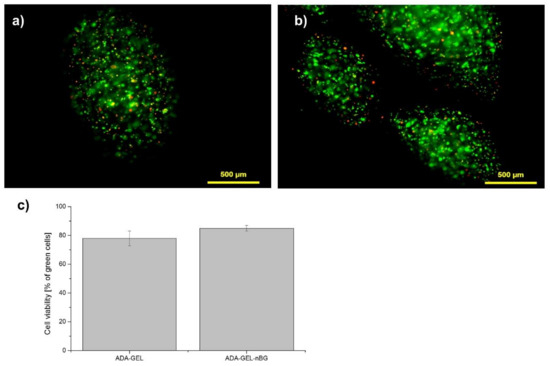
[...] Read more.
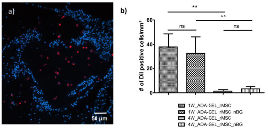
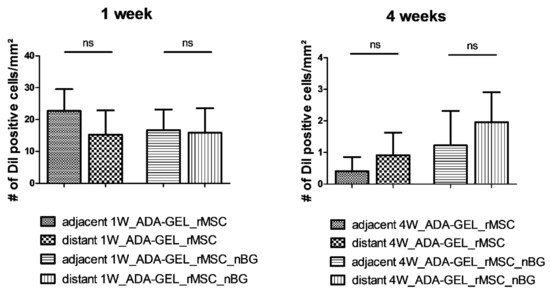
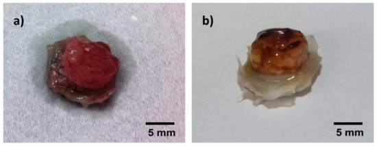
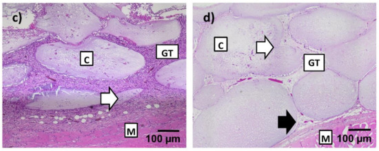
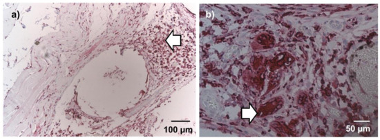
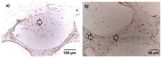
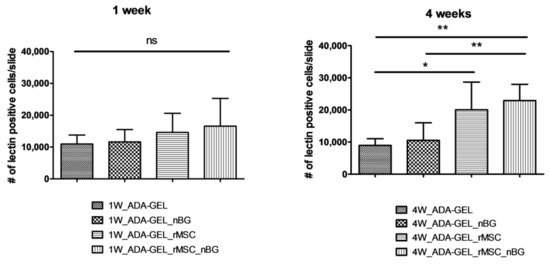
Alginate dialdehyde (ADA), gelatin, and nano-scaled bioactive glass (nBG) particles are being currently investigated for their potential use as three-dimensional scaffolding materials for bone tissue engineering. ADA and gelatin provide a three-dimensional scaffold with properties supporting cell adhesion and proliferation. Combined with nanocristalline BG, this composition closely mimics the mineral phase of bone. In the present study, rat bone marrow derived mesenchymal stem cells (MSCs), commonly used as an osteogenic cell source, were evaluated after encapsulation into ADA-gelatin hydrogel with and without nBG. High cell survival was found in vitro for up to 28 days with or without addition of nBG assessed by calcein staining, proving the cell-friendly encapsulation process. After subcutaneous implantation into rats, survival was assessed by DAPI/TUNEL fluorescence staining. Hematoxylin-eosin staining and immunohistochemical staining for the macrophage marker ED1 (CD68) and the endothelial cell marker lectin were used to evaluate immune reaction and vascularization. After in vivo implantation, high cell survival was found after 1 week, with a notable decrease after 4 weeks. Immune reaction was very mild, proving the biocompatibility of the material. Angiogenesis in implanted constructs was significantly improved by cell encapsulation, compared to cell-free beads, as the implanted MSCs were able to attract endothelial cells. Constructs with nBG showed higher numbers of vital MSCs and lectin positive endothelial cells, thus showing a higher degree of angiogenesis, although this difference was not significant. These results support the use of ADA/gelatin/nBG as a scaffold and of MSCs as a source of osteogenic cells for bone tissue engineering. Future studies should however improve long term cell survival and focus on differentiation potential of encapsulated cells in vivo.
Full article
►▼
Show Figures
Open AccessArticle
Biological Compatibility of a Polylactic Acid Composite Reinforced with Natural Chitosan Obtained from Shrimp Waste
by
Yaret Gabriela Torres-Hernández, Gloria Michel Ortega-Díaz, Lucía Téllez-Jurado, Nayeli Shantal Castrejón-Jiménez, Alejandro Altamirano-Torres, Blanca Estela García-Pérez and Heberto Balmori-Ramírez
Cited by 37 | Viewed by 5114
Abstract
The aim of this work is to evaluate the effect of chitosan content (1, 3 and 5 wt %) dispersed in polylactic acid (PLA) on the structure and properties of composites. Also, the hydrolytic degradation, and the cell viability and adhesion of human
[...] Read more.
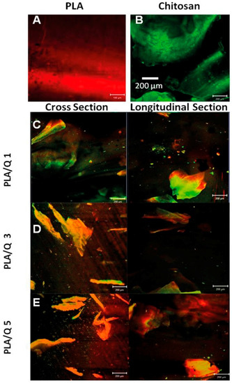
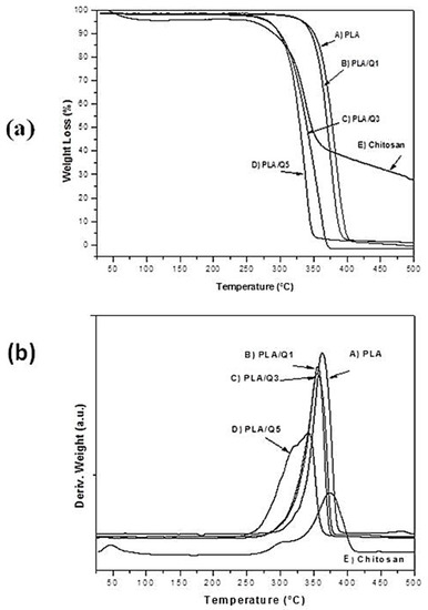
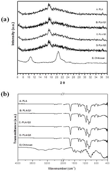
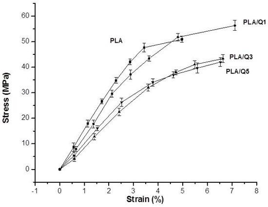
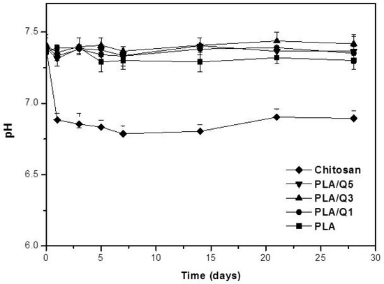
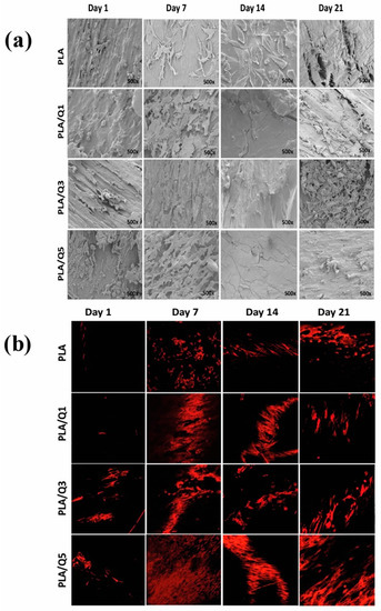
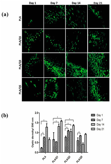
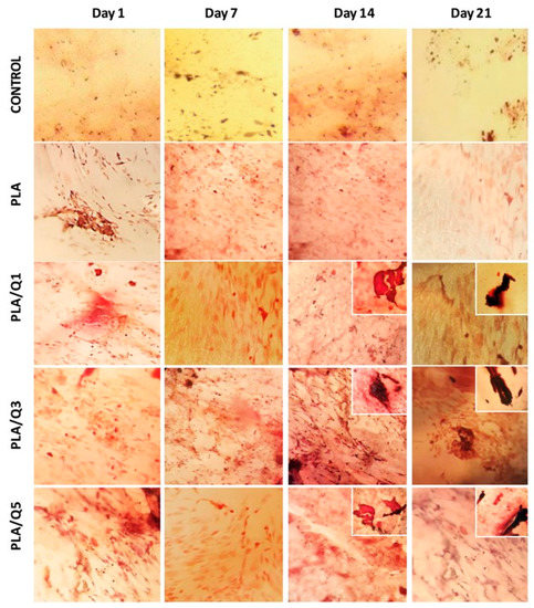
The aim of this work is to evaluate the effect of chitosan content (1, 3 and 5 wt %) dispersed in polylactic acid (PLA) on the structure and properties of composites. Also, the hydrolytic degradation, and the cell viability and adhesion of human MG-63 osteoblasts are analyzed to determine the composites’ suitability for use in tissue engineering. For the manufacture of the materials, natural chitosan was extracted chemically from shrimp exoskeleton. The composites were fabricated by extrusion, because it is a low-cost process, it is reproducible, and it does not compromise the biocompatibility of the materials. FT-IR and XRD show that the chitosan does not change the polymer structure, and interactions between the composite components are discarded. In vitro degradation tests show that the composites do not induce significant pH changes in phosphate buffer solution due to their low susceptibility to hydrolytic degradation. The adhesion and morphological characteristics of the osteoblasts are evaluated using confocal microscopy and scanning electron microscopy. The cell viability is determined by the MTT assay. Osteoblasts adhesion is observed on the surface of PLA and composites. A higher amount of chitosan, higher number of cells with osteoblastic morphology, and mineralized nodules are observed on the composite surface. The highest metabolic activity is evidenced at 21 days. The results suggest that the Polylactic acid/chitosan composites are potentially suitable for use as a biomaterial.
Full article
►▼
Show Figures
Open AccessArticle
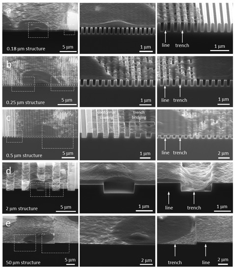
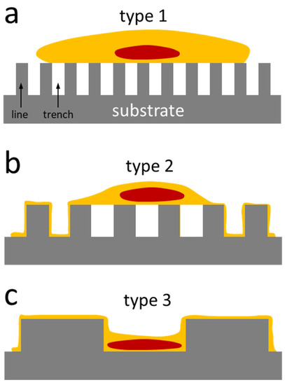
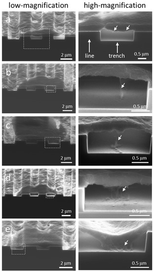
Pattern-Dependent Mammalian Cell (Vero) Morphology on Tantalum/Silicon Oxide 3D Nanocomposites
by
Hassan I. Moussa, Megan Logan, Wing Y. Chan, Kingsley Wong, Zheng Rao, Marc G. Aucoin and Ting Y. Tsui
Cited by 8 | Viewed by 4758
Abstract
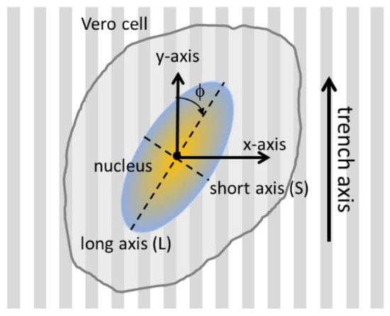
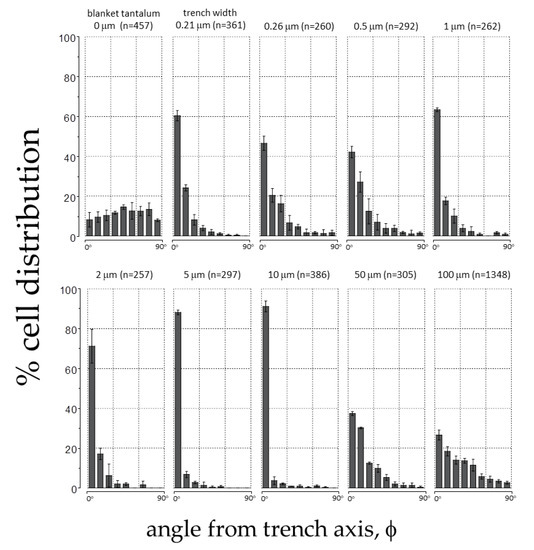
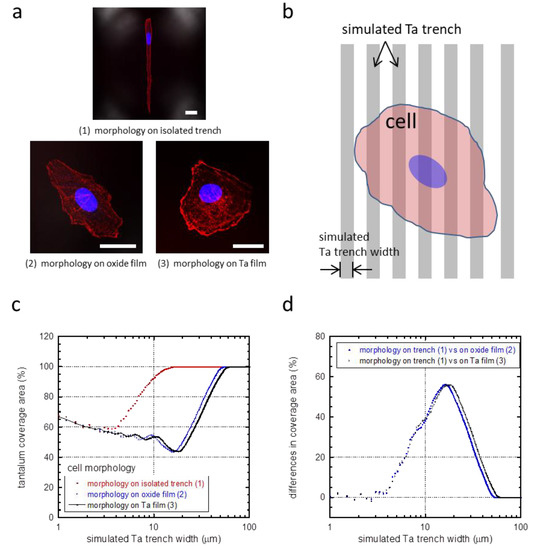
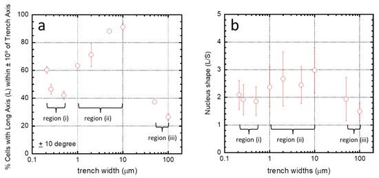
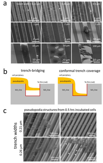
The primary goal of this work was to investigate the resulting morphology of a mammalian cell deposited on three-dimensional nanocomposites constructed of tantalum and silicon oxide. Vero cells were used as a model. The nanocomposite materials contained comb structures with equal-width trenches and
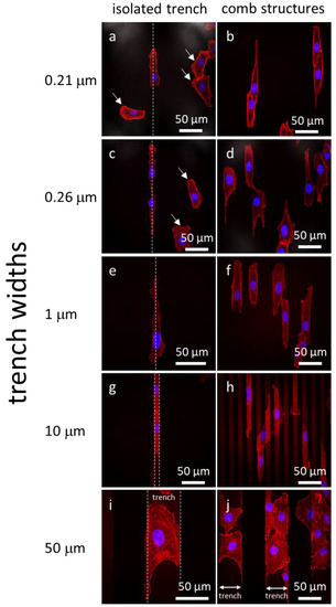
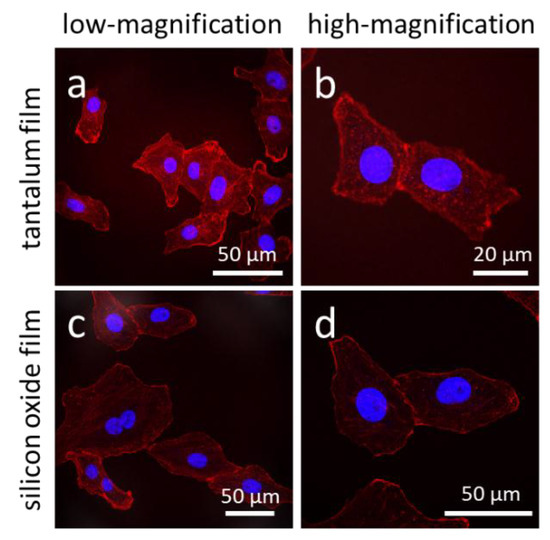
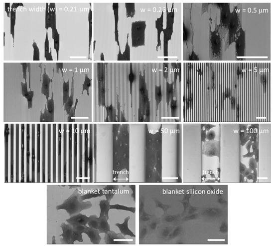
[...] Read more.
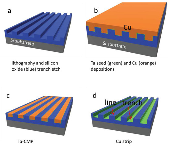
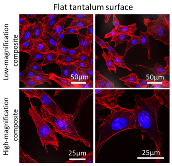
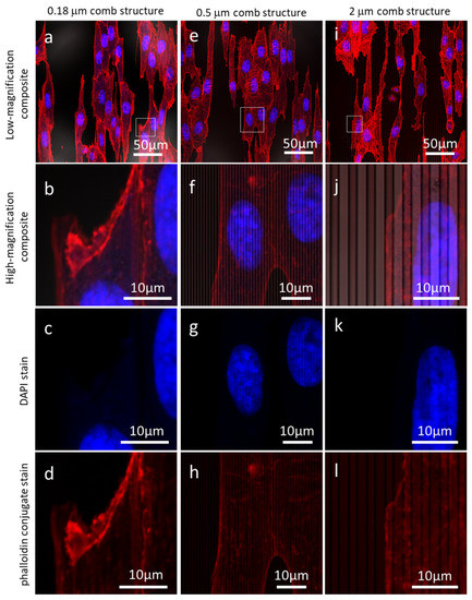
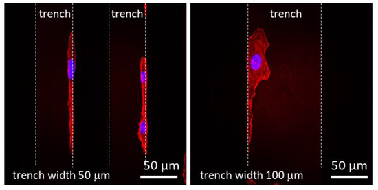
The primary goal of this work was to investigate the resulting morphology of a mammalian cell deposited on three-dimensional nanocomposites constructed of tantalum and silicon oxide. Vero cells were used as a model. The nanocomposite materials contained comb structures with equal-width trenches and lines. High-resolution scanning electron microscopy and fluorescence microscopy were used to image the alignment and elongation of cells. Cells were sensitive to the trench widths, and their observed behavior could be separated into three different regimes corresponding to different spreading mechanism. Cells on fine structures (trench widths of 0.21 to 0.5 μm) formed bridges across trench openings. On larger trenches (from 1 to 10 μm), cells formed a conformal layer matching the surface topographical features. When the trenches were larger than 10 μm, the majority of cells spread like those on blanket tantalum films; however, a significant proportion adhered to the trench sidewalls or bottom corner junctions. Pseudopodia extending from the bulk of the cell were readily observed in this work and a minimum effective diameter of ~50 nm was determined for stable adhesion to a tantalum surface. This sized structure is consistent with the ability of pseudopodia to accommodate ~4–6 integrin molecules.
Full article
►▼
Show Figures
Open AccessArticle
Porous Silk Fibroin Microspheres Sustainably Releasing Bioactive Basic Fibroblast Growth Factor
by
Jing Qu, Lu Wang, Longxing Niu, Jiaming Lin, Qian Huang, Xuefeng Jiang and Mingzhong Li
Cited by 21 | Viewed by 4612
Abstract
Basic fibroblast growth factor (bFGF) plays a significant role in stimulating cell proliferation. It remains a challenge in the field of biomaterials to develop a carrier with the capacity of continuously releasing bioactive bFGF. In this study, porous bFGF-loaded silk fibroin (SF) microspheres,
[...] Read more.
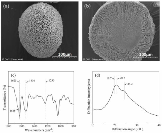
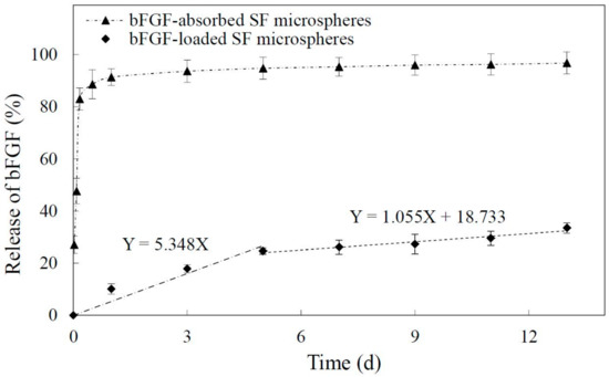
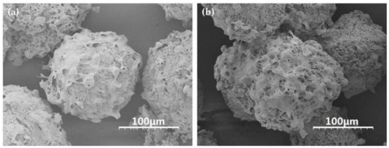
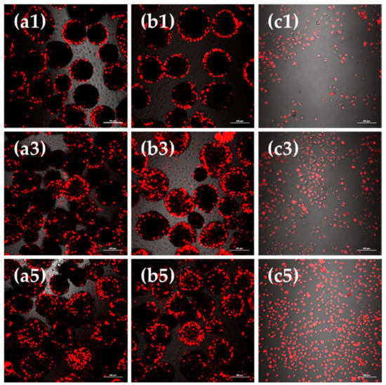
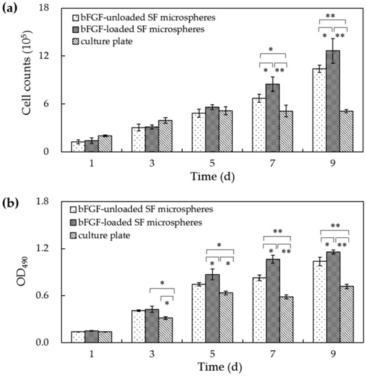
Basic fibroblast growth factor (bFGF) plays a significant role in stimulating cell proliferation. It remains a challenge in the field of biomaterials to develop a carrier with the capacity of continuously releasing bioactive bFGF. In this study, porous bFGF-loaded silk fibroin (SF) microspheres, with inside-out channels, were fabricated by high-voltage electrostatic differentiation, and followed by lyophilization. The embedded bFGF exhibited a slow release mode for over 13 days without suffering burst release. SEM observations showed that incubated L929 cells could fully spread and produce collagen-like fibrous matrix on the surface of SF microspheres. CLSM observations and the results of cell viability assay indicated that bFGF-loaded microspheres could significantly promote cell proliferation during five to nine days of culture, compared to bFGF-unloaded microspheres. This reveals that the bFGF released from SF microspheres retained obvious bioactivity to stimulate cell growth. Such microspheres sustainably releasing bioactive bFGF might be applied to massive cell culture and tissue engineering as a matrix directly, or after being combined with three-dimensional scaffolds.
Full article
►▼
Show Figures
Open AccessArticle
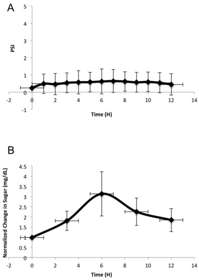
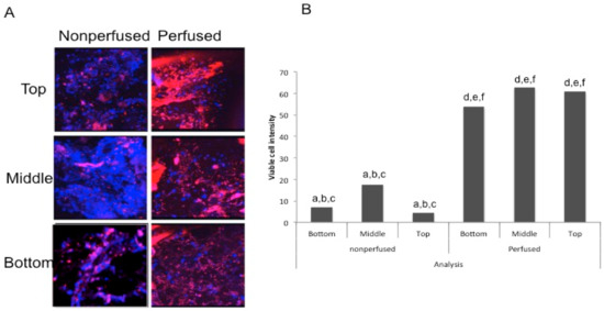
The Preservation of Bone Cell Viability in a Human Femoral Head through a Perfusion Bioreactor
by
Aparna Swarup, Hilary Weidner, Randall Duncan and Anja Nohe
Cited by 6 | Viewed by 3181
Abstract
Current methods for drug development and discovery involve pre-clinical analyses that are extremely expensive and time consuming. Animal models are not the best precedent to use, when comparing to human models as they are not synonymous with the human response, thus, alternative methods
[...] Read more.
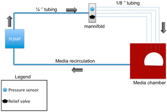
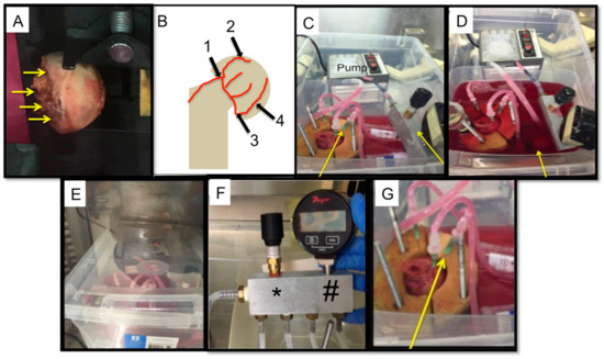
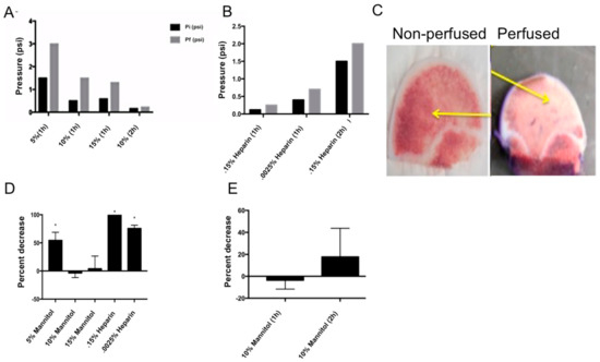
Current methods for drug development and discovery involve pre-clinical analyses that are extremely expensive and time consuming. Animal models are not the best precedent to use, when comparing to human models as they are not synonymous with the human response, thus, alternative methods for drug development are needed. One of which could be the use of an ex vivo human organ where drugs could be tested and the effects of those drugs could be observed. Finding a viable human organ to use in these preliminary ex vivo studies is difficult due to the availability, cost, and viability. Bone tissue and marrow contain a plethora of both bone and stem cells, however, these cells need constant perfusion to be viable over a longer time range. Here we maintain bone cell sustainability in an ex vivo model, through the use of human femoral heads in a novel bioreactor. This bioreactor was designed to directly perfuse cell culture media (DMEM) through the vasculature of a femoral head, providing ideal nutrients and conditions required for maintaining organ viability. We show, for the first time, that cells within a femoral head can stay alive up to 12 h. Further development could be used to determine the effects of drugs on a human organ system and could aid in the understanding of the progression of bone diseases and pathologies.
Full article
►▼
Show Figures
Planned Papers
The below list represents only planned manuscripts. Some of these
manuscripts have not been received by the Editorial Office yet. Papers
submitted to MDPI journals are subject to peer-review.
Title: Promotion of osteogenesis by combination of three-dimensional scaffold and connective tissue growth factor
Abstract: In bone tissue engineering, implanted scaffolds should provide the biological cellular requirements, such as mineralization, cell organization and differentiation. Especially, vascularization within engineered tissues is a key process to improve their survival in vitro and in vivo. In the present study, to construct an active blood vessel network, we have investigated the relation between osteoblasts and endothelial cells. In addition, to develop the vascularized bone scaffolds, we focused on connective tissue growth factor (CTGF), a multifunctional growth factor. Combination of apatite-fiber scaffold and CTGF could promote the adhesion, migration, and proliferation of osteoblasts, together with stimulation of angiogenesis.
Author’s name: Michiyo Honda, Ryo Hariya and Mamoru Aizawa
Abstract: In recent years there has been an increasing trend of recall of drugs and most of these recalls have been due to unforeseen liver toxicity. This could be attributed to the fact there are few reliable in vitro liver models that can give reliable results in drug testing that can be translated to the clinic successfully. To meet this need, there are now numerous three dimensionally cultured liver organoids that are being employed for drug testing and disease modeling. In this study, we have tried to improve on such organoids by creating a simple and well-defined extracellular matrix hyaluronic acid and collagen hydrogel biofunctionalized with fibronectin, which improves physiologic and metabolic activities of these 3D liver constructs. First, we provide basic characterization of this hydrogel through standard material science assessments. Second, we verified biocompatibility using a comparison to a commercially available biomaterial with several common cell lines. Lastly, we demonstrate the utility of the biofunctionalized hydrogel in 3D primary human hepatocyte constructs – or liver “organoids”. The liver organoids constructed with biofunctionalized matrix were then tested for toxicity response with recalled hepatotoxic drugs and environmental toxin wherein we observed dose dependent response. We also show that these liver organoids are capable of being rescued with and reviving their function after a drug insult thus demonstrating clinical relevance and translational potential of this platform.


